Embed Size (px)
Citation preview
RANCINAMYCINS, METABOLITES PRODUCED BY
STREPTOMYCES LINCOLNENSIS IN SULFUR-DEPLETED MEDIA
A. D. ARGOUDELIS, T. R. PYKE and R. W. SPRAGUE*
The Upjohn Company, Kalamazoo, Michigan 49001, U.S.A.
(Received for publication May 26, 1976)
Rancinamycins 1, II, III and IV are secondary metabolites produced by Streptomyces lincolnensis in a sulfur-depleted culture medium. Rancinamycins I and II, the main components of the mixture, show broad spectrum antibiotic activity in vitro. Subcutaneously injected or orally administered antibiotic afforded no protection for experimentally infected mice against lethal challenges of Staphylococcus aureus. Radioactive tracer studies failed to demonstrate that the rancinamycins were precursors in the biosynthesis of lincomycin.
Streptomyces lincolnensis was first reported to produce lincomycin (1) by MASON et al.1) in 1962.
Subsequently, a number of other lincomycin-related antibiotics were found to be produced by this cul-
to re2,8,4,b)_
All of the antibiotics reported to be produced by S. lincolnensis are pro-
duced in either complex or defined media containing inorganic sulfate as the
usual source of sulfur. The sulfur of methionine can (also be utilized effi-
cientlye~ .
When S. lincolnensis is grown in a chemically defined medium in the
absence of sulfur-containing organic or inorganic compounds a group of new
metabolites is produced. The isolation, purification, characterization and
biological properties of these compounds, designated rancinamycins I, II, III
and IV, are the subjects of the present communication.
Experimental
Spectroscopic Procedures PMR spectra were recorded on a Varian A-60 spectrometer. All preparations were run in either
D2O or d6-dimethylsulfoxide (de-DMSO) using sodium 2,2-dimethyl-2-silapentane-5-sulfonate (SDSS) or tetra methylsilane (TMS) as internal reference.
Gas Chromatography-Mass Spectroscopy (GC-MS) Gas-Chromatographic separation was done on a GC Varian Aerograph series 2700 chromatograph using a 6 ft., 3.8% UCW-98 on 80100 mesh Diatoport-S column (Hewlett-Packard). The operation was performed isothermally at 185°C. The mass spectra were recorded on a CH7 Massenspectrometer
(Varian Mat., West Germany) operating at 70 electron volts. It was found necessary to prepare the trimethylsilyl ethers of rancinamycins in order to obtain vol-
atile compounds satisfactory for gas chromatographic analysis. This was done by reacting 50 ,ul of a 1 mg/ml solution of a rancinamycin preparation in dimethylformamide with 50 ,ul of Regisil® (Regis Chemical Co.). The reaction mixture was allowed to stand at room temperature for 30 minutes.
R = CH2CH2CH3
* Participant of the Student Individualized Project Program operated by Kalamazoo College and The Upjohn
Company.
778 THE JOURNAL OF ANTIBIOTICS AUG. 1976
Assay Procedures
Antibiotic production was measured by a microbiological disc-plate assay procedure') using Proteus vulgaris (UC-93), Proteus rettgeri (UC-344), Staphylococcus aureus (UC-80), and Sarcina lutea
(UC-130) as the assay organisms.
Paper and Thin-layer Chromatographic Procedures Fermentation, purification and separation studies were followed by paper chromatography using water-saturated 1-butanol as the solvent system. Thin-layer chromatograms were run on Analtech silica gel GF plates (Analtech Inc.) using ethyl acetate saturated with water (system I), methyl ethyl ketone - acetone - water (186: 52:10, v/v) (system II) or methyl ethyl ketone - acetone - water (186: 52: 22, v/v) (system III) as the solvent systems.
The antibiotics, separated either by paper or tic chromatography, were detected by bioautography on P. vulgaris-seeded agar trays.
Fermentation Procedures Seed cultures of S. lincolnensis were prepared in a medium consisting of glucose monohydrate, 25
g/liter and Pharmamedia, 25 g/liter (Trader's Oil Mill Co., Forth Worth, Texas, USA); seed presterili-zation pH 7.2. The cultures were incubated at 28°C for 72 hours on a rotary shaker (250 rpm). A fermentation medium consisting of anhydrous glucose , 30 g/liter; sodium citrate, 1.8 g/liter; dipotassium phosphate, 2.5 g/liter; sodium chloride 0.5 g/liter; ammonium nitrate, 2.0 g/liter; zinc chloride 0.47 mg/ liter; ferrous chloride tetrahydrate 0.72 mg/liter; magnesium citrate , 1.2 g/liter and lard oil 10 ml/liter was adjusted to pH 7.2 and inoculated at a rate of 5 % (v/v) with the 72-hour seed culture. Fermenta-tions were incubated at 28°C on a rotary shaker and beers were harvested after total fermentation time of 7296 hours.
Isolation Procedures
Adsorption on Amberlite XAD-4: The whole beer (10 liters) was filtered using diatomaceous earth (Dicalite, 4200, Grefco Inc.). Part of the clear beer (1.5 liters) was passed through a column
prepared from 100 ml of Amberlite XAD-4* (Rohm and Haas Co.). The spent, collected in one frac-tion, was bioinactive and was discarded. The column was then washed with 500 ml of water; the aqueous wash was also bioinactive and was discarded. The column was eluted with methanol-water
(95: 5, v/v). Fractions containing rancinamycins were concentrated to an aqueous solution and freeze-dried to yield 2.55 g of crude rancinamycin preparation.
Adsorption on Granulated Activated Carbon: Another portion (1.5 liters) of the clear beer, ob-tained as described above, was passed through a column containing 80 g of Cal Carbon (Pittsburgh Activated Carbon). The column was washed with750 ml of water; both spent and the aqueous wash were found bioinactive and were discarded. The column was successively eluted with 750 ml of ace-tone - water (20: 80, v/v), 250 ml of acetone - water (40: 60, v/v) and acetone - water (90: 10, v/v). Fractions collected with acetone-water (90: 10, v/v), containing rancinamycins, were combined, concen-trated to an aqueous solution and freeze-dried to yield 1.22 g of crude rancinamycins.
Purification Procedures Separation of Rancinamycins by Countercurrent Distribution: Three grams of crude rancina-mycins, obtained by the carbon adsorption method, was distributed in an all glass countercurrent distri-bution apparatus (500 tubes, 10 ml/phase) using a solvent system consisting of equal volumes of 1-butanol and water. A total of 467 transfers were completed. The distribution was followed by determination of solids and bioactivity. Results which7are presented in Fig. 2 showed the presence of four active components with K values of 0.52, 1.15, 2.40 and 5.95. Tubes under the peak with a K value of 1.15 were found to contain rancinamycin I, while rancinamycin II was found in the fractions under the peak with a K value of 2.40. Concentration of appropriate fractions yielded rancinamycins
* Amberlite XAD-4 was successively washed with the following before use: 2 bed volumes of 50: 50 acetone-water, 2 bed volumes of acetone, 2 bed volumes of methanol, 2 bed volumes of 50:50 methanol -water, and 20 bed volumes of water.
779VOL. XXIX NO. 8 THE JOURNAL OF ANTIBIOTICS
I and II isolated as viscous oils which crystallized (colorless needles) on standing. The small amounts of the bioactive components with K values of 0.52 (rancinamycin III) and 5.95 (rancinamycin IV) were not isolated by this procedure but by the purification sequence described below.
Separation of Rancinamycins by Amberlite XAD-4 Chromatography, Silica Gel Chromatography and Countercurrent Distribution: Ninety grams of crude rancinamycins obtained from a large scale
fermentation (by the carbon process) were dissolved in one liter of water (resulting pH was 4.5). This solution was passed over a 2-liter Amberlite XAD-4 column. The spent, collected as one fraction, was bioinactive and was discarded. The column was then washed with water (6 liters) collected as one fraction. The aqueous wash was also bioinactive and was discarded. The column was eluted with
methanol-water (50: 50, v/v). Fractions containing rancinamycins were combined, concentrated to an aqueous solution and freeze-dried to give 56 g of material containing all rancinamycins.
Twenty-one grams of this crude rancinamycin was dissolved in 50 ml of water. The solution was mixed with 100 g of silica gel (Merck-Darmstadt, Art 7704). This mixture was dried in vacuo and added on the top of a glass column (7 cm internal diameter) containing 1.8 kg of silica gel packed in a solvent consisting of methyl ethyl ketone - acetone - water (186: 52: 3, v/v). Bioactive fractions obtained by elution of the column with the solvent system were combined and concentrated to dryness in vacuo to
give 7.9 g of slightly colored viscous material containing rancinamycins I to IV. The purified rancinamycin mixture, obtained as described above, was dissolved in 50 ml of each
phase of the solvent system consisting of equal volumes of 1-butanol -water. The solutions were added in the first five tubes of an all-glass countercurrent distribution apparatus (500 tubes, 10 ml/phase). After 500 transfers the distribution was analyzed by thin-layer chromatography using systems I and II and by bioassays. Rancinamycins I, 0.64 g; 11, 0.58 g; III, 0.29 g; and IV, 1.79 g were isolated by concentration of the appropriate fractions. Characterization of these materials is described in Dis-cussion and Results.
Rancinamycin I-2,4-Dinitrophenylhydrazone
One hundred sixty mg of rancinamycin I was dissolved in 10 ml of water. This solution was mixed with 420 ml of a solution prepared by dissolving I g of 2,4-dinitrophenylhydrazine in 1 liter of 2 N aqueous hydrochloric acid. The mixture was allowed to stand at room temperature for 24 hours. The crystalline precipitate formed, was separated by filtration and dried, 130 mg.
Anal. Calcd. for C17H20N409: C, 48.15; H, 4.75; N, 13.22; 0, 33.96; molecular weight, 424. Found: C, 47.65; H, 4.25; N. 12.93; 0, 33.36; equivalent weight 411.
Rancinamycin II-2,4-Dinitrophenylhydrazone One hundred mg of rancinamycin II was dissolved in 5 ml of 95 % ethanol and this solution was
mixed with 300 ml of a solution prepared by dissolving 1 g of 2,4-dinitrophenylhydrazone in 1 liter of 2 N aqueous hydrochloric acid. Crystalline rancinamycin II-2,4-dinitrophenylhydrazone was isolated by filtration and dried; yield 90 mg.
Anal. Calcd. for C18H22N409: C, 49.36; H, 5.06; N, 12.79; 0, 32.88; molecular weight, 438. Found: C, 48.77; H, 4.96; N, 12.79; 0, 31.50; equivalent weight 421. Preparation of the Mixture of 2,4-Dinitrophenylhydrazones of Rancinamycins
Crude rancinamycin, obtained by the carbon adsorption process described earlier, was dissolved in 50 ml of water. This solution was mixed with 2 liters of a solution prepared by dissolving 1 g of 2,4-dinitrophenylhydrazone in I liter of 2 N aqueous hydrochloric acid. Formation of an orange precipi-tate started immediately. The mixture was allowed to stand at room temperature for 24 hours. The
precipitated mixture of the 2,4-dinitrophenylhydrazones of rancinamycins I and II was separated by filtration and dried (2.28 g). Separation of 2,4-Dinitrophenylhydrazones of Rancinamycins I and d II. Countercurrent Distribu-
tion. The mixture of 2,4-dinitrophenylhydrazones of rancinamycins I and II was distributed in an all-
glass CRAIG countercurrent distribution apparatus (500 tubes, 10 ml/phase) using a solvent system consisting of equal volumes of cyclohexane, ethyl acetate, 95 % ethanol and water. After 465 transfers the distribution was analyzed by solid determination. Tubes 180230, containing rancinamycin 1-2,4-
780 THE JOURNAL OF ANTIBIOTICS AUG. 1976
dinitrophenylhydrazone, were concentrated to dryness to give 790 mg of crystalline material. Simi-larly, tubes 243280 gave 400 mg of crystalline rancinamycin II-2,4-dinitrophenylhydrazone. Preparation of Radioactive Rancinamycins The fermentation conditions were identical to those described earlier. Uniformly labeled 14C_
glucose was used as the radioactive precursor. Approximately one per cent of the radioactivity added was incorporated into the crude rancinamycins isolated by the carbon adsorption process discussed ear-lier. This radioactive rancinamycin mixture (specific activity of 6 x 10⁴ cpm/mg) was added to a linco-mycin-producing fermentation of S. lincolnensis as described below.
Addition of !4C-Rancinamycins to Fermentation of S. lincolnensis Producing Lincomycin
The fermentation conditions were identical to those described by ARGOUDELis et ale). Radioactive 14C-rancinamycin obtained as described above was added to the fermentation medium 48 hours after
inoculation. The fermentation was followed by bioactivity determinations using both P. vulgaris
(specific for rancinamycins) and S. lutea (specific for lincomycin) as the assay organisms. Results are discussed in the Results and Discussion section.
Results and Discussion
Production of Rancinamycins
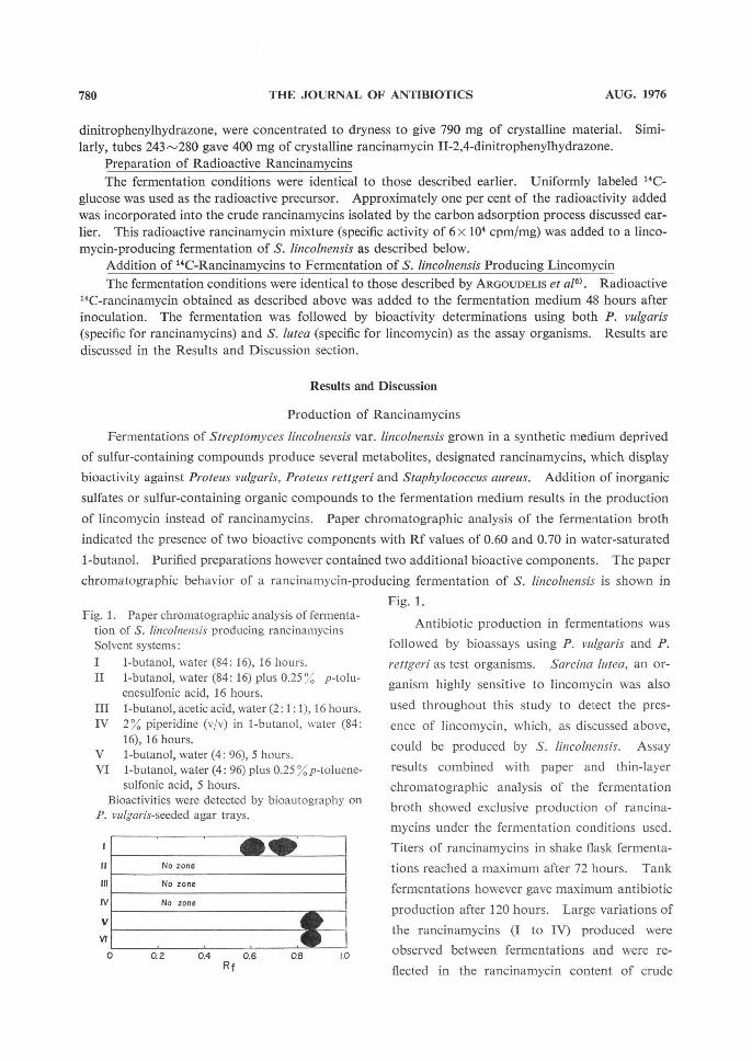
Fermentations of Streptomyces lincolnensis var. lincolnensis grown in a synthetic medium deprived
of sulfur-containing compounds produce several metabolites, designated rancinamycins, which display
bioactivity against Proteus vulgaris, Proteus rettgeri and Staphylococcus aureus. Addition of inorganic
sulfates or sulfur-containing organic compounds to the fermentation medium results in the production
of lincomycin instead of rancinamycins. Paper chromatographic analysis of the fermentation broth
indicated the presence of two bioactive components with Rf values of 0.60 and 0.70 in water-saturated
1-butanol. Purified preparations however contained two additional bioactive components. The paper
chromatographic behavior of a rancinamycin-producing fermentation of S. lincolnensis is shown in
Fig. 1.
Antibiotic production in fermentations was
followed by bioassays using P. vulgaris and P.
rettgeri as test organisms. Sarcina lutea, an or-
ganism highly sensitive to lincomycin was also
used throughout this study to detect the pres-
ence of lincomycin, which, as discussed above,
could be produced by S. lincolnensis. Assay
results combined with paper and thin-layer
chromatographic analysis of the fermentation
broth showed exclusive production of rancina-
mycins under the fermentation conditions used.
Titers of rancinamycins in shake flask fermenta-
tions reached a maximum after 72 hours. Tank
fermentations however gave maximum antibiotic
production after 120 hours. Large variations of
the rancinamycins (I to IV) produced were
observed between fermentations and were re-
flected in the rancinamycin content of crude
Fig. 1. Paper chromatographic analysis of fermenta- tion of S. lincolnensis producing rancinamycins
Solvent systems:
I 1-butanol, water (84: 16), 16 hours. II 1-butanol, water (84: 16) plus 0.25°o p-tolu- enesulfonic acid, 16 hours.
III 1-butanol, acetic acid, water (2:1 :1), 16 hours. IV 2 % piperidine (v/v) in 1-butanol, water (84:
16), 16 hours. V 1-butanol, water (4: 96), 5 hours.
VI 1-butanol, water (4: 96) plus 0.25 %p-toluene- sulfonic acid, 5 hours.
Bioactivities were detected by bioautography on P. vulgaris-seeded agar trays.
No zone
No zone
No zone
Rf
781VOL. XXIX NO. 8 THE JOURNAL OF ANTIBIOTICS
preparations.
Isolation and Purification. Designation of Rancinamycins I, II, III and IV.
The bioactive components were recovered from the fermentation broth by filtration followed by
adsorption on either granulated activated carbon or Amberlite XAD-4. Purification was achieved by
chromatography on Amberlite XAD-4 and silica gel. Separation of rancinamycins was obtained by
countercurrent distribution using 1-butanol - water (1: 1, v/v) as the solvent system (Fig. 2). The
bioactive components in the tubes under the peaks
with K values of 1.15 and 2.40 were defined as ran-
cinamycins I and II, respectively. Rancina-
mycins I and II had Rf values of 0.68 and 0.78 in
the papergram system discussed earlier. Rancina-
mycins III and IV were defined in the bioactive
materials in the tubes under the peaks with K
values of 0.52 and 5.95, respectively.
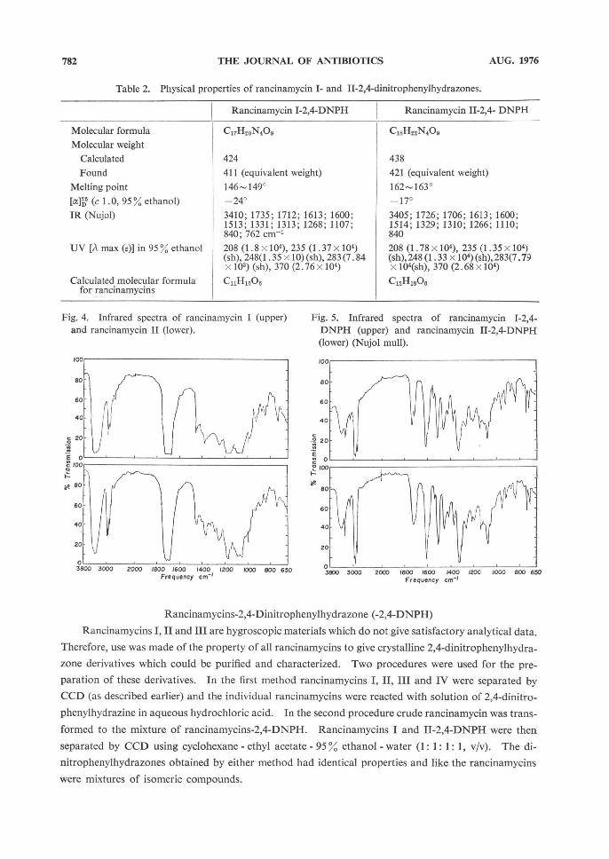
Rancinamycins I, II and III were isolated
as colorless viscous materials which crystallized
on standing (colorless needles). Crystalline
rancinamycins I, II and III behaved as single en-
tities in several paper chromatographic and tlc
systems (Fig. 3). However gas chromatography-
mass spectroscopy (see below) indicated that
each of rancinamycins I, II and III was a mix-
ture of isomeric compounds which could not be
separated by either CCD or the chromatogra-
phic techniques (paper or tic) used. Rancina-mycin IV on the other hand, isolated as color-
less crystalline (needles) solid, was found to con-
tain one material only.
Fig. 2. Countercurrent distribution of rancina-
mycins
Number of transfers
Fig. 3. Thin-layer chromatogram* of rancinamy- cins I, II, III and IV *Silica gel; methyl ethyl ketone - acetone - water
(186: 52: 10, v/v). Antibiotics were detected by bioautography on P. vulgaris-seeded agar trays.
Rf
Table 1. Physical and chemical properties of ran-
cinamycins.
Molecular formula
Molecular weight Found Calculated
Molecular weight of TMS-dderi- vative Found Calculated
Molecular formu- la of TMS-deri-
vative
[a]1 (c 1.0, water) UV [Am_ (s); 95 % ethanol]
Titration IR (Neat)
Rancinamycin I
C11H16Osa'c
244.09191 244.0946
460.21051 460.2138
C11H1306. [Si(CH3)3]31
+96' 220 nm
(9.5x10³) Neutral
3400; 1735; 1700; 1250;1195.1150; 1100-1050 cm-1
Rancinamycin II
C12H1308b, o
258b
474.22821 474.2288
C12H1500. [Si(CH3)3]3a
+69' 220 nm (7.74 x 10³) Neutral
3405; 1726; 1706; 1266; 1110 cm-1
a By high resolution mass spectroscopy . b By mass spectroscopy . By mass spectrometry and data of its 2,4-dinitro- phenylderivative (see Table 2).
d TMS=trimethylsilyl .
782 THE JOURNAL OF ANTIBIOTICS AUG. 1976
Rancinamycins-2,4-Dinitrophenylhydrazone (-2,4-DNPH)
Rancinamycins I, II and III are hygroscopic materials which do not give satisfactory analytical data . Therefore, use was made of the property of all rancinamycins to give crystalline 2,4-dinitrophenylhydra-
zone derivatives which could be purified and characterized. Two procedures were used for the pre-
paration of these derivatives. In the first method rancinamycins I, II, III and IV were separated by
CCD (as described earlier) and the individual rancinamycins were reacted with solution of 2,4-dinitro-
phenylhydrazine in aqueous hydrochloric acid. In the second procedure crude rancinamycin was trans-
formed to the mixture of rancinamycins-2,4-DNPH. Rancinamycins I and II-2,4-DNPH were then
separated by CCD using cyclohexane - ethyl acetate - 95 % ethanol - water (1: 1: 1: 1, v/v). The di-
nitrophenylhydrazones obtained by either method had identical properties and like the rancinamycins
were mixtures of isomeric compounds.
Table 2. Physical properties of rancinamycin I- and II-2,4-dinitrophenylhydrazones.
Molecular formula
Molecular weight
Calculated
Found
Melting point
[a]1 (c 1.0, 95 % ethanol) IR (Nujol)
UV [A max (e)] in 95 % ethanol
Calculated molecular formula for rancinamycins
Rancinamycin I-2,4-DNPH
C11H20N40e
424
411 (equivalent weight)
146149° -24°
3410; 1735; 1712; 1613; 1600; 1513; 1331; 1313; 1268; 1107; 840; 762 cm'
208(1.8 x 10⁴), 235 (1.37 x 10⁴) (sh), 248(1.35 x 10) (sh), 283 (7.84 x 10³) (sh), 370 (2.76 x 10⁴)
C11H1e06
Rancinamycin 11-2,4- DNPH
C,8H22N409
438
421 (equivalent weight)
162-163' -17°
3405; 1726; 1706; 1613; 1600; 1514; 1329; 1310; 1266; 1110; 840
208 (1.78 x 10⁴), 235 (1.35 x 10⁴) (sh), 248 (1.33 x 10⁴) (sh), 283(7.79 X 10⁴(sh), 370 (2.68 x 10⁴)
C12H160e
Fig. 4. Infrared spectra of rancinamycin I (upper) and rancinamycin II (lower).
Frequency cm-'
Fig. 5. Infrared spectra of rancinamycin 1-2,4- DNPH (upper) and rancinamycin II-2,4-DNPH
(lower) (Nujol mull).
Frequency cm-'
783VOL. XXIX NO. 8 THE JOURNAL OF ANTIBIOTICS
Characterization of Rancinamycins
Rancinamycins I, II and III
are hygroscopic materials soluble
in water, lower alcohols, ethyl ace-
tate and acetone. They are less
soluble in chlorinated hydrocarbon
solvents and insoluble in ether and
saturated hydrocarbons. Rancina-
mycin IV has limited solubility in
water but it is soluble in alcohols,
ethyl acetate, acetone, chloroform
and ether.
The properties of rancinamycins
I and II and rancinamycins I-and
II-2,4-dinitrophenylhydrazones are
listed in Tables 1 and 2, respectively.
The data presented indicate that the
two antibiotics are closely related
and that rancinamycin II (C15H18Oe)
Fig. 6. Nuclear magnetic resonance spectra of rancinamycin I-2,4-DNPH and rancinamycin II-2,4-DNPH (60 MHz, in d(,-dimethylsulfoxide).
Offset 225 Cps
Offset 220 cps
Table 3. Gas chromatographic-mass spectroscopic analysis*.
1. Rancina-
mycin I-
TMS**
2. Rancina- mycin II-
TMS
3. Rancina- mycin III-
TMS
Compound
Ia-TMS
Ib-TMS
Ic-TMS
Id-TMS
Ie-TMS
Ila-TMS
IIb-TMS
IIc-TMS
IId-TMS
IIe-TMS
Illa-TMS
IIIb-TMS
IIIc-TMS
IIId-TMS
(Retention time (min.,
19.2 23.5
26.5 28.5 33.3
27.5 31.0 33.3 36.5
40.5
11.5 13 14.5 17
Relative abun-
dance (%)I
36
51
1
3
8
67
7
3
4
19
15
10 15
60
M+
460
460
460
460
460
474
474
474
474
474
462
462
462
462
Number of TMS groups
3
3
3
3
3
3
3
3
3
3
4
4
4
4
* For conditions see Experimental . ** TMS=trimethylsilyl .
784 THE JOURNAL OF ANTIBIOTICS AUG. 1976
is a higher homolog of rancinamycin I (C„H,BO8)
differing from the latter by a -CH.- group. The
IR spectra of rancinamycins I and II (Fig. 4) and
the IR and PMR spectra of rancinamycins I-and
11-2,4-dinitrophenyihydrazones (Figs. 5 and 6)
also indicate the close structural similarity be-
tween these two antibiotics.
Gas chromatographic-mass spectroscopic
(GC-MS) analysis of the trimethylsilyl (TMS)
derivative of rancinamycin I showed that rancina-
mycin I is a mixture of five compounds designat-
ed rancinamycins la, Ib, Ic, Id and Ie in order
of increasing retention time (Table 3) of their
TMS derivatives. All five compounds form
TMS-derivatives of identical molecular weight
(460) and identical fragmentation patterns) in
their mass spectra. Furthermore, all five TMS-
derivatives contain three TMS groups indicating
the presence of three hydroxyl groups in each of
the components. This suggests that the anti-
biotics, themselves, have identical molecular
formulas (C„H,sO8) and isomeric structures.
Rancinamycins la and lb were the main com-
ponents comprising ca. 87% of rancinamycin I.
Rancinamycins Ic, Id and Ie, being present in
small amounts, are not expected to effect the
spectra (CMR, PMR, IR and UV) of rancina-
mycin I which are discussed in detail in a sub-
sequent paper8).
Similarly, rancinamycin II was found to be
a mixture of five components (rancinamycins II-
a, IIb, IIc, lid and IIe) (Table 3). All components
had identical molecular weight (474) and identical fragmentation patterns in the mass spectra of their
TMS-derivativese), indicative of the isomeric character of the rancinamycin II components. The for-
mation of tri-TMS derivatives indicates that rancinamycins II also contains three hydroxyl groups.
Rancinamycin IIa and lie are the main components comprising ca. 86 % of rancinamycin II. The char-
acterization data recorded on Tables 1 and 2 for rancinamycins I, II and their 2,4-dinitrophenylhydra-
zones, with the exception of the specific rotations and the melting points, are not effected by the finding
that each antibiotic is a mixture of isomeric components.
Since small amounts of rancinamycin III were isolated, this material was not completely char-
acterized. High resolution mass spectrum of rancinamycin III-TMS showed molecular formula of
CTHsO5 . [Si(CHB)s]4; (Molecular weight: calcd. 462.21093, found, 462.20788). Therefore the mole-
Table 4. Antibacterial spectra of rancinamycins I,
II, III and IV*.
Kiebsiella pneumoniae (UC-57)
Escherichia coli (UC-51)
Proteus vulgaris (UC-93)
Salmonella schottmuel- leri (UC-126)
Salmonella gallinarum (UC-265)
Pseudomonas aeruginosa (UC-95)
Streptococcus pyogenes (UC-152)
Sarcina lutea (UC-130)
Penicillium oxalicum (UC-1268)
Bacillus subtilis (UC-564)
Mycobacterium avium (UC-159)
Zone size (mm)
I
24
22
33
24
25
0
21
22
0
17
0
II
20
18.'-
30.5
20
21.5
0
20
22
0
16
0
III
16.5
15.5
28.5
16.5
21.5
0
20.5
16.5
0
trace
0
IV
24
25.5
26
24
22.5
16.5
0
18
0
22.5
29
* The concentration of the solutions used was 10
mg/ml; agar diffusion test.
Table 5. Characteristics of a lincomycin-producing
fermentation of S. lincolnensis containing rancina-
mycins as precursors.
Time (hours)
0
12
36
60 84
108
pH
6.1
5.1
7.8
7.9
8.3
8.6
Assay(P vulgaris; bu/ml)
12
8.9
0
0
0 0
Lincomycin (mcg/ml)
4.3
4.3
51.2
128
285 193
Radioacti- vity* (cpm/ml)
61,880
67,890
65,430
63,045
64,940
66,125
* Radioactivity present in the fermentation broth .
785VOL. XXIX NO. 8 THE JOURNAL OF ANTIBIOTICS
cular formula of rancinamycin III is C7H1005. Rancinamycin III, like rancinamycins I and II, was
found to be a mixture of four components (rancinamycins llla, Illb, Tile and IIId) (Table 3). The
molecular weight (462) and fragmentation pattern of the TMS-derivatives of these components was
identical. The formation of tetra-TMS derivative shows the presence of four hydroxyl groups. Ran-
cinamycin III forms a 2,4-dinitrophenylhydrazone which exhibits UV spectrum identical to that of
rancinamycins I and II (Table 2) indicating the presence of the same chromophoric system in rancina-
mycins I, II and III.
Rancinamycin IV, C7H008 (molecular weight: found 138.0307; calcd. 138.0317) was isolated as
colorless crystalline material. Gas chromatographic mass spectroscopic analysis of the trimethylsilyl
derivative of rancinamycin IV indicated the presence of one component with retention time of 4.5
minutes. IR and PMR spectra indicated that rancinamycin IV is 3,4-dihydroxybenzaldehyde8).
Biological Properties of Rancinamycins
The antibacterial spectrum of rancinamycins I, II, III and IV is presented in Table 4. The anti-
bacterial activity of rancinamycins I, TI and III is destroyed by cysteine, by reagents reacting with
carbonyl groups such as hydrogen sulfite and by reduction with sodium borohydride. In addition,
the antibiotics were unstable in acidic or basic solutions. Rancinamycins I and II were inactive both
orally and subcutaneously against Staphylococcus aureus infected mice.
Rancinamycins as Precursors in Lincomycin Biosynthesis
The possibility that rancinamycins might be intermediates in the biosynthesis of lincomycin was
suggested by the finding that these compounds are related to shikimic acid8) and that aromatic acids
(phenylalanine, tyrosine) are involved in the biosynthesis of the amino acid moiety of lincomycin0). To
test this hypothesis uniformly labeled 14C-rancinamycin complex was prepared by the use of uniformly
labeled 14C-glucose. About 1.0% of the radioactivity added to the fermentation medium was incor-
porated into rancinamycins. The radioactive rancinamycin complex was then added into a lincomycin
producing fermentation. As shown in Table 6 the rancinamycin bioactivity (P. vulgaris) was rapidly
lost. The radioactivity level in the fermentation supernatant liquid, however, remained constant
throughout the fermentation. Lincomycin, isolated as the crystalline hydrochloride, was radioinactive
indicating that rancinamycins were not utilized for biosynthesis of lincomycin under the conditions
used. The rapid loss of P. vulgaris activity was due, probably, to the instability of the rancinamycins
to the acidic conditions early in the fermentation.
Acknowledgments
The authors wish to express their appreciation to Mr. K. J. GEIPEL for technical assistance, to the Fermen-tation and Development Unit of The Upjohn Company for large scale extraction, and to the Physical and Ana-lytical Chemistry Unit of The Upjohn Company for analytical and spectral data.
References
1) MASON, D. J.; A. DIETZ & C. DEBOER: Lincomycin, a new antibiotic. 1. Discovery and biological properties. Antimicr. Agents & Chemoth. -1962: 554-559, 1963
2) ARGOUDELIS, A. D.; J. A. Fox & T. E. EBLE: U-21,699: A new lincomycin-related antibiotic. Bio- chemistry 4:698-703, 1965
3) ARGOUDELIS, A. D. & D J. MASON: Studies on the biosynthesis of lincomycin. I. Antibiotic U-11,921, an S-ethyl homolog of lincomycin. Biochemistry 4: 704709, 1965 4) ARGOUDELIS, A. D.; J. A. Fox & D. J. MASON: Studies on the biosynthesis of lincomycin. II. Antibiotic
U-11,973, N-demethyl-lincomycin. Biochemistry 4:710713, 1965
786 THE JOURNAL OF ANTIBIOTICS AUG. 1976
5) ARGOUDELIS, A. D.; T. E. EBLE & D. J. MASON: Studies on the biosynthesis of lincomycin. V. Effect of ethionine on fermentations of S. lincolnensis. J. Antibiotics 23:1- 8, 1970
6) ARGOUDELIS, A. D.; T. E. EBLE, J. A. Fox & D. J. MASON: Studies on the biosynthesis of lincomycin. IV. The origin of methyl groups. Biochemistry 8:34083411, 1969
7) HANKA, L. J.; M. R. BURCH & W. T. SOKOLSKI: Psicofuranine. VI. Microbiological assay. Antibiot. & Chemoth. 9:432-435, 1959
8) ARGOUDELIS, A. D.; R. W. SPRAGUE & S. A. MIZSAK: Rancinamycins I, II, III and IV. Structural studies. J. Antibiotics 29: 787-796,1976
9) Wrrz, D. F.; E. J. HESSLER & T. L. MILLER: Bioconversion of tyrosine into the propylhygric acid moiety of lincomycin. Biochemistry 10:11281132, 1971


![Enhancinghepodcion ofcephalopoinChoghmodlaing ......thesis [17, 18]ike most of secondary metabolites, ceph - alosporin C is produced at the anaphase of exponential growth and stationary](https://img.pdfslide.us/doc/110x75/5e54ac8e0af37e17e962cae2/enhancinghepodcion-ofcephalopoinchoghmodlaing-thesis-17-18ike-most-of.jpg)