Embed Size (px)
Citation preview
Raman Analysis of Sperm Nuclear DNA Integrity
AbstractRaman Spectroscopy was evaluated as a non-invasive method of analysis of sperm DNA and the influence of UV irradiation on the sperm. The results show that Raman Spectroscopy, combined with multivariate analysis provide the reproducible and accurate information on DNA of sperm and the effect and location of damage.
Key wordsDNA damage, sperm, human reproduction, Raman spectroscopy, Raman imaging, DuoScan
Introduction
Infertility has been and still it is a highly studied topic1,2,3. It is known that sperm nuclear DNA (nDNA) integrity is a crucial factor for the success of male reproductive function. However, the status of sperm nDNA cannot be determined by the usual microscopic means which comprise routine semen analysis. Commercially available tests exist which assess the extent of sperm DNA fragmentation; however as they all result in the death of the sample they are of limited use to embryologists and ART (Assisted Reproductive Technology) clinics. The aim of our study, published in detail in “Human Reproduction”4, was to systematically appraise the utility of Raman microspectroscopy in analysing sperm nDNA status and can be compared to the results with established nDNA tests.
Raman map on native sperm
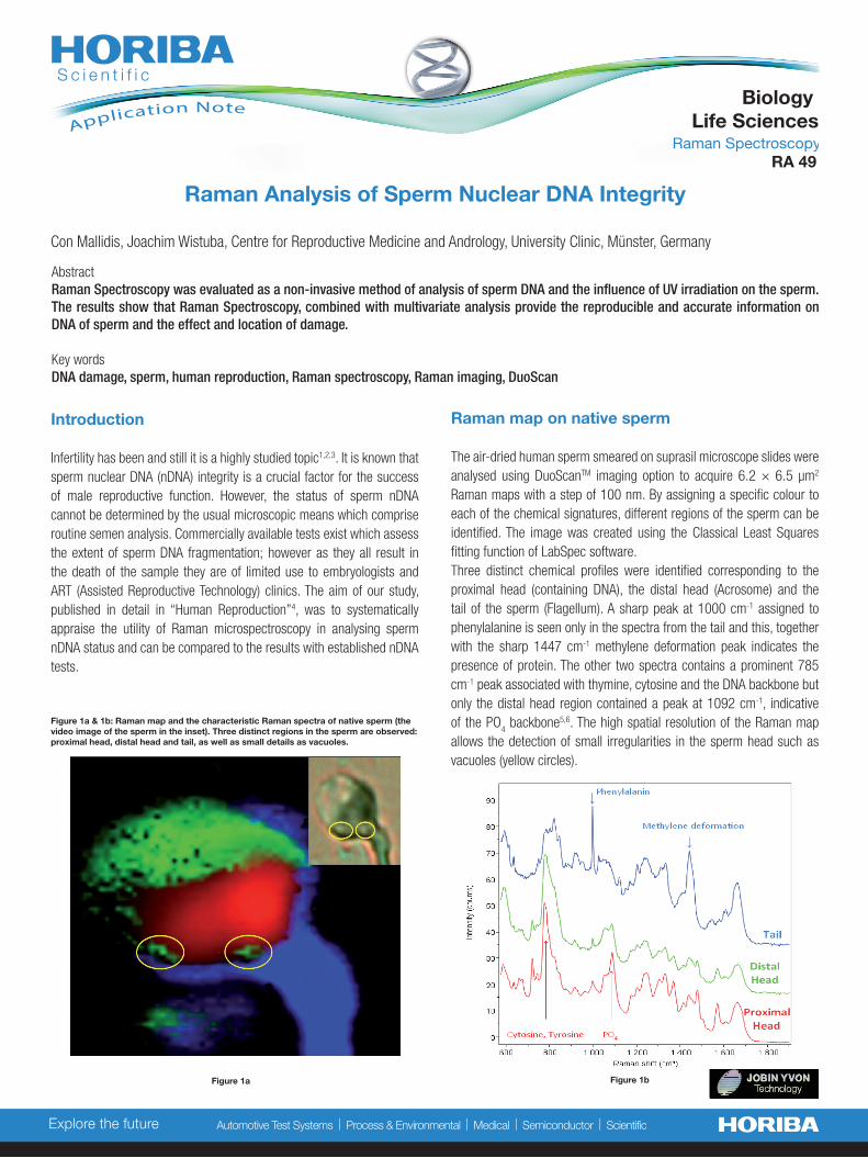
The air-dried human sperm smeared on suprasil microscope slides were analysed using DuoScanTM imaging option to acquire 6.2 × 6.5 µm2 Raman maps with a step of 100 nm. By assigning a specific colour to each of the chemical signatures, different regions of the sperm can be identified. The image was created using the Classical Least Squares fitting function of LabSpec software. Three distinct chemical profiles were identified corresponding to the proximal head (containing DNA), the distal head (Acrosome) and the tail of the sperm (Flagellum). A sharp peak at 1000 cm-1 assigned to phenylalanine is seen only in the spectra from the tail and this, together with the sharp 1447 cm-1 methylene deformation peak indicates the presence of protein. The other two spectra contains a prominent 785 cm-1 peak associated with thymine, cytosine and the DNA backbone but only the distal head region contained a peak at 1092 cm-1, indicative of the PO
4 backbone5,6. The high spatial resolution of the Raman map
allows the detection of small irregularities in the sperm head such as vacuoles (yellow circles).
Explore the future Automotive Test Systems | Process & Environmental | Medical | Semiconductor | Scientific
Raman Spectroscopy
Biology Life SciencesApplication Note
Con Mallidis, Joachim Wistuba, Centre for Reproductive Medicine and Andrology, University Clinic, Münster, Germany
RA 49
Figure 1a & 1b: Raman map and the characteristic Raman spectra of native sperm (the video image of the sperm in the inset). Three distinct regions in the sperm are observed: proximal head, distal head and tail, as well as small details as vacuoles.
Figure 1a Figure 1b
2
Explore the future Automotive Test Systems | Process & Environmental | Medical | Semiconductor | Scientific
Differentiation between native and UV irradiated sperm: chemometrics analysis
Raman measurements were carried out on native and UV irradiated (i.e. DNA damaged) sperm. Standard principal component analysis (PCA) was performed on three datasets (native sperm/observer W, native sperm/observer M and damaged sperm) to analyse a statistically meaningful number of measurements (200 sperm per sample). The results show that no difference is observed between the different observers’ measu-rements on native sperm however they were clearly distinguishable from those obtained after UVB treatment (Figure 2).
In further analysis using the local spectral angle classification, the peak related to DNA PO
4 backbone showed a clear shift towards 1042 cm-1
and changes in its intensity after treatment (Figure 3). This difference indicates the dimerizations of nucleotide bases caused by UVB. Closer examination using wavelet decomposition confirmed the 1042 cm-1 shift and identified a possible second affected region (1400–1600 cm-1) corresponding to protein–DNA interactions and thus susceptible to UVB damage.
[email protected]/scientific T
his
docu
men
t is
not c
ontr
actu
ally
bin
ding
und
er a
ny c
ircum
stan
ces
- P
rinte
d in
Fra
nce
- ©
HO
RIB
A J
obin
Yvo
n 07
/201
3
Conclusions and perspectives
Once optimized, Raman microspectroscopy is a reproducible and reliable technique for sperm nDNA analysis with the acquisition of spectra being quick and easy. Even if the interpretation and analysis of the spectra can be complex, it can be overcome by the development of user friendly mathematical algorithms which would help to identify the presence of damaged DNA; and combined with mapping can give an indication of its distribution within the sperm head. As no studies, ours included, have assessed living sperm, it is prema-ture to advocate the use of Raman microspectroscopy for ART purposes. However if, as in the other cell types that have already been tested, the technique is found to provide a detailed chemical fingerprint whilst not damaging the integrity of living sperm then its application opens oppor-tunities for basic and clinical andrology thus far beyond our technical capabilities. Studies could be undertaken on the nature, causes, impact and importance of sperm DNA fragmentation which in turn would lay the foundations for truly objective sperm selection.
Experimental parameters
A LabRAM Aramis Raman system was used in confocal mode with the HeNe laser (632.8 nm). The spectral resolution was 6.7 cm-1. Peak assignments were based on the attributions in previous publications on Raman assessment of sperm5,6.
References
1 Deutsches IVF Register, J Reproduktionsmed Endokrinol, Suppl 1: 1037, 2010
2Nyboe et al: Hum Reprod 24:1267-87, 2009. 3Agbaje et al Reprod BioMed Online, 16(3):401-, 2008 4Mallidis et al.: Hum Reprod 26:1641-9, 20115Huser et al. .J Biophotonics ;2:322–332, 20096Meister et al. Analyst;135:1370–1374, 20107Sanchez et al Fertil & Steril, 98(5):1124-9, 2012
Figure 3: Averaged spectra showing the distinct shift at 1042 cm-1(arrow) in spectra of irradiated (red) compared with that of the untreated human sperm (green).
Figure 2: Principal component analysis: native sperm (red and blue, for two observers) and UVB-irradiated sperm (green).
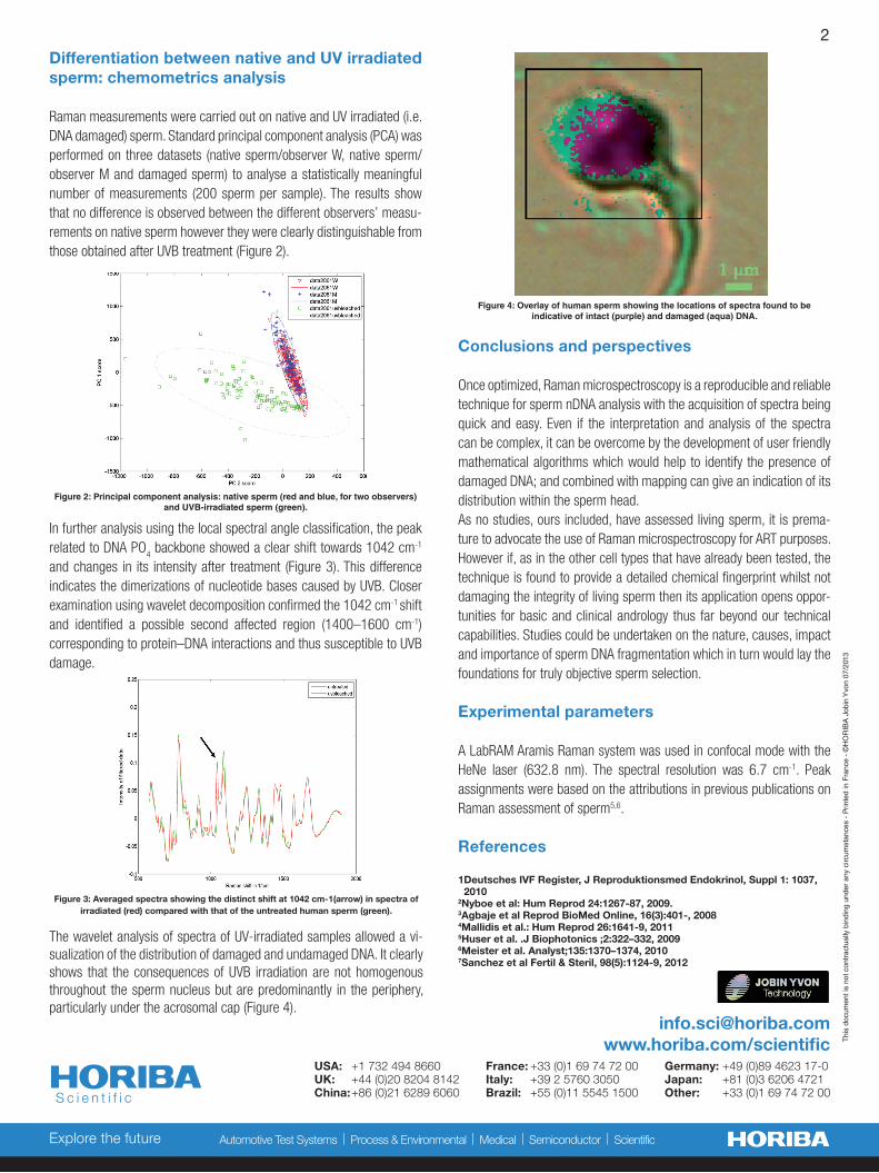
Figure 4: Overlay of human sperm showing the locations of spectra found to be indicative of intact (purple) and damaged (aqua) DNA.
The wavelet analysis of spectra of UV-irradiated samples allowed a vi-sualization of the distribution of damaged and undamaged DNA. It clearly shows that the consequences of UVB irradiation are not homogenous throughout the sperm nucleus but are predominantly in the periphery, particularly under the acrosomal cap (Figure 4).
USA: +1 732 494 8660 France: +33 (0)1 69 74 72 00 Germany: +49 (0)89 4623 17-0UK: +44 (0)20 8204 8142 Italy: +39 2 5760 3050 Japan: +81 (0)3 6206 4721China: +86 (0)21 6289 6060 Brazil: +55 (0)11 5545 1500 Other: +33 (0)1 69 74 72 00