Embed Size (px)
Citation preview
1
12th International Conference & Workshop
“Medical Physics in the Baltic States”, 2015
RAMAN SPECTROSCOPY OF
POLYMERIZATION PROCESSES
IN nPAG AND nMAG DOSE
GELS
Neringa VAIČIŪNAITĖ1, Rimas ŠEPERYS2
1 Physics Department, Kaunas University of Technology
2SME “Šeši partneriai”

2
INTRODUCTION
• Polymer gels have been proven to be a valuable tool for
determination of beam dose characteristics and 3D radiation
dose measurements for medical purposes [1-3].
• A typical gel dosemeter consists of water, gelatin agent,
monomers and a cross-linker comonomer [4,5].
• Many chemical compositions have been proposed for
dosimetric purposes, but only a few have been carefully tested
[4,5].
• To evaluate the interaction between monomer consumption
and polymer production is very important [6].

AIM OF THE STUDY
The aim of this study was to identify the radiation interaction
mechanisms on photon irradiated nPAG and nMAG polymer
gels in accordance with analysis of characteristic vibrational
modes of dose gel components

4
POLYMER GELS PREPARATION
AND IRRADIATION
nPAG nMAG
89% Highly purified water 86% Highly purified water
5% Gelatine 8% Gelatine
3% Acrylaimde 6% Methacrylic acid
3% N; N- methylene-
bisacrylamide
-
10 mmol/l Hydroxymethyl
phosphonium chloride
10 mmol/l Hydroxymethyl
phosphonium chloride
nPAG and nMAG polymer gels were irradiated by
Co60 photon source of teletherapy unit (ROKUS M)
by 2 Gy dose. Dose rate of 0.03 Gy/ min,
SSD – 100 cm, field opening – 10×10 cm. Fig.1. Preparation of the
dose gel.

POLYMER GELS PREPARATION
AND IRRADIATION
Fig. 2. Micro Raman
inVia Renishaw
a)
b)
Micro Raman
(inVia Renishaw)
vibrational
spectroscopy of
the irradiated
copolymerized
nPAG and nMAG
samples in
cuvettes was
performed on the
7th post-irradiation
day.
Fig. 3. a) Non irradiated nPAG
gel, b) Non irradiated nMAG
gel, c) nPAG irradiated by 2
Gy Co-60, d) nMAG irradiated
by 2 Gy Co-60.
a) b) c) d)
6
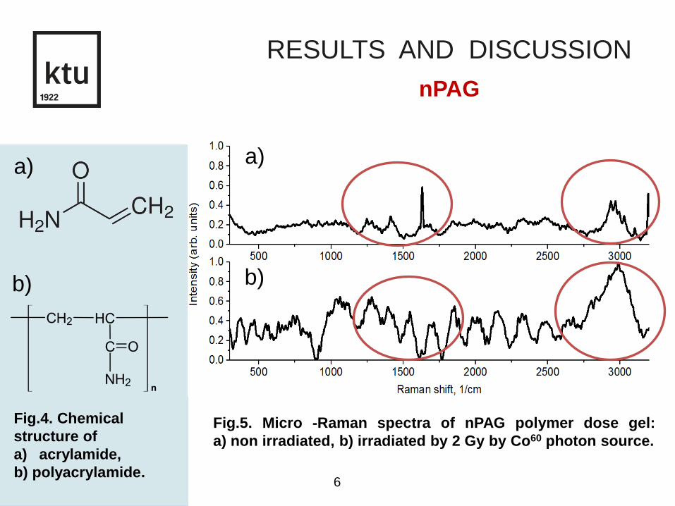
RESULTS AND DISCUSSION
Fig.4. Chemical
structure of
a) acrylamide,
b) polyacrylamide.
a)
b)
Fig.5. Micro -Raman spectra of nPAG polymer dose gel:
a) non irradiated, b) irradiated by 2 Gy by Co60 photon source.
nPAG
a)
b)

RESULTS AND DISCUSSION
Fig.6. Polymer
development observed
in Raman spectra of
normoxic
polyacrylamide (nPAG)
gel in the range of
900-1400 cm-1 and
2800-3200 cm-1.
a), c) Non irradiated
nPAG gel;
b), d) irradiated nPAG
gel.
nPAG
a)
c)
b)
d)
1256
1285
1256
1285
FWHM = 310,9 FWHM = 1229,1
2880
2936
FWHM = 121,7 FWHM = 213,6
2880
2936

8
RESULTS AND DISCUSSION
Fig.7. Chemical
structure of a)
methacrylic acid,
b) polymetchacrylic
acid.
a)
b)
a)
b)
Fig. 8. Raman spectra of nMAG polymer dose gel: a) non
irradiated, b) irradiated by 2 Gy by Co60 photon source.
nMAG

RESULTS AND DISCUSSION
Fig. 9. Polymer
development
observed in Raman
spectra of normoxic
methacrylic acid
(nMAG) gel at the
range of 450-600cm-1
and 2000-3200 cm-1.
a), c) Non irradiated
nMAG gel;
b), d) irradiated nMAG
gel.
539
539
2955
3114
2955
3114
a)
b)
c)
d)
FWHM = 111,4 FWHM = 1198,8
FWHM = 102,8 FWHM = 290,7
nMAG

10
CONCLUSIONS
• Photon irradiation of Co60 was applied to normoxic nPAG and
nMAG polymer dose gels to perform a polymerization process.
• The Micro-Raman spectra and full-width half-maximum
calculations, showed the incensement of polymer representing
vibrational bands and sharp reduction of monomer fraction.
• The reduction of BIS proved the theory of cross-linker
consumption and its importance in network structure formation.
• The results agreed with the studies of other researchers,
however more tests are needed to evaluate the consumption rate
of monomers and cross-linker.

11
Thank You for your attention

12
REFERENCES
1. Papoutsaki M., Maris T.G., Pappas E. Dosimetric characteristics of a new polymer gel
and their dependence on post-preparation and post-irradiation time: Effect on X-ray beam
profile measurements. Physica Medica 9, 2013. p. 453-460.
2. McAuley K.B. Fundamentals of Polymer Gel Dosimeters. Journal of Physics:
Conference Series 56, 2006. p. 35–44, doi:10.1088/1742-6596/56/1/004.
3. De Deene Y. Essential characteristics of polymer gel dosimeters. „Third International
Conference on Radiotherapy Gel Dosimetry“, Journal of Physics: Conference Series 3,
2004. p. 34–57, doi:10.1088/1742-6596/3/1/006.
4. Baldock C., De Deene Y.,Doran S. Topical Review: Polymer gel dosimetry. Phys Med
Biol, 2010. doi:10.1088/0031-9155/55/5/R01.
5. Bong J., Choi K., Yu S.C. Raman Spectroscopy of Irradiated Normoxic
Polymethacrylic Acid Gel Dosimeter. Bull. Korean Chem. Soc., Vol. 32, No. 2, 2011. p.
625 – 629. doi 10.5012/bkcs.2011.32.2.625.
6. Maryanski M. J., Zastavker Y. Z., and Gore J. C. Radiation dose distributions in three
dimensions fromtomographic optical density scanning of polymer gels: II. Optical
properties of the BANG polymer gel. Phys. Med. Biol. 41, 1996. p. 2705–2717.