Embed Size (px)
Citation preview
Accepted Manuscript
Raman-IR vibrational and XRD characterization of ancient and modern mineralogyfrom volcanic eruption in Tenerife Island: Implication for Mars
E.A. Lalla, G. López-Reyes, A. Sansano, A. Sanz-Arranz, J. Martinez-Frías, J.Medina, F. Rull-Pérez
PII: S1674-9871(15)00088-2
DOI: 10.1016/j.gsf.2015.07.009
Reference: GSF 377
To appear in: Geoscience Frontiers
Received Date: 18 November 2014
Revised Date: 28 July 2015
Accepted Date: 31 July 2015
Please cite this article as: Lalla, E.A., López-Reyes, G., Sansano, A., Sanz-Arranz, A, Martinez-Frías,J., Medina, J., Rull-Pérez, F., Raman-IR vibrational and XRD characterization of ancient and modernmineralogy from volcanic eruption in Tenerife Island: Implication for Mars, Geoscience Frontiers (2015),doi: 10.1016/j.gsf.2015.07.009.
This is a PDF file of an unedited manuscript that has been accepted for publication. As a service toour customers we are providing this early version of the manuscript. The manuscript will undergocopyediting, typesetting, and review of the resulting proof before it is published in its final form. Pleasenote that during the production process errors may be discovered which could affect the content, and alllegal disclaimers that apply to the journal pertain.
MANUSCRIP
T
ACCEPTED
ACCEPTED MANUSCRIPT
MANUSCRIP
T
ACCEPTED
ACCEPTED MANUSCRIPTRaman-IR vibrational and XRD characterization of ancient and modern
mineralogy from volcanic eruption in Tenerife Island: Implication for Mars
E. A. Lallaa,b,*,, G. López-Reyesb, A. Sansanob, A.Sanz-Arranzb, J. Martinez-Fríasc, J.
Medinab, F. Rull-Pérezb.
aDepartment of Physics, Faculty of Science, University of La Laguna, San Cristobal de
La Laguna, Santa Cruz de Tenerife, CP 38206, Spain.
b Unidad Asociada al Centro de Astrobiología CSIC-INTA associated to the NASA
Astrobiology Center – Instituto Nacional de Técnica Aeroespacial, Torrejón de Ardoz,
CP 28850, Madrid, Spain.
cDepartment of Earth Dynamics and Observation , Instituto de Geociencias IGEO-
(CSIC-UCM), Facultad de Ciencias Geológicas, Ciudad Universitaria, CP 28040,
Madrid, Spain
*Corresponding author. E-mail: [email protected], [email protected]
MANUSCRIP
T
ACCEPTED
ACCEPTED MANUSCRIPT
ABSTRACT
A detailed vibrational Raman-IR spectroscopic and diffractional analyses have been
performed on basalts from two locations from Tenerife Island: (1) the Arenas Negras
volcano which belongs to the historical eruption not showing visible alteration and (2)
Pillow Lavas zone from Anaga Massif which shows a clearly fluid-rock interaction
caused by submarine alteration. These places have been extensively studied due to its
similarity with the surface of Mars. The analysis is based on the mineral detection of
selected samples by a Micro-Raman study of the materials. The complementary
techniques have confirmed the mineralogy detected by the Raman measurement. The
results show a volcanic environment behavior with primary phases like olivine,
pyroxene, and feldspar/plagioclase. Moreover, the presence of accessory minerals or
secondary mineralization like phosphate, iron oxides, zeolite or carbonates shows the
alteration processes on each outcrop. Moreover, the variation in the crystallinity and
amorphous phases is related to fluid-rock interaction caused by hydrothermal episodes
and external weathering processes, which shows several analogies with the ancient
volcanic activity from Mars.
Keywords: Mars, Volcanoes, Terrestrial Analog, Raman Spectroscopy, Tenerife Island,
mineralogy.

MANUSCRIP
T
ACCEPTED
ACCEPTED MANUSCRIPT
1. INTRODUCTION
Nowadays, Raman spectroscopy is considered one of the next generation techniques for
planetary exploration due to its multiple advantages. Among others, Raman
spectroscopy is a non-destructive analytical tool, capable of obtaining vibrational,
rotational and other low-frequency modes information of the selected target (Rull-Pez
and Martinez-Frias, 2006; Courres-Lacoste et al., 2007). One of the cutting edge
research field applications for the Raman technique is the planetary science, where
several instruments are being developed such as the Scanning Habitable Environments
with Raman & Luminescence for Organics and Chemicals Instrument on NASA
(SHERLOC) or SuperCam on NASA-France consortium for the NASA mission in 2020
( Grossman, 2013; Campbell et al., 2015). On the other hand, the next European mission
to Mars, the ESA-ExoMars rover mission on 2018, will be equipped with a Raman
Laser Spectrometer (RLS) as part of the analytical suite in the body of the vehicle
( Rull-Pez and Martinez-Frias, 2006; Courres-Lacoste et al., 2007; Bost et al., 2015).
The main applications of this instrument are focused on the exobiological and
geochemical targets. Thus, the RLS is capable to detect different mineral phases,
establishing the main mineralogical sequences and following secondary ones such as
carbonates, sulfates and hydrated minerals formation ( Edwards et al., 2004; Rull-Pez
and Martinez-Frias, 2006; Lalla et al., 2010; Bost et al., 2015). The research and
technical progress behind the Raman instrument is of fundamental importance for the
future success. In this regard, it is important to highlight the appropriate preparation of
the prototypes, the calibration, the realistic measurement conditions, the spectroscopic
MANUSCRIP
T
ACCEPTED
ACCEPTED MANUSCRIPTcharacterizations of natural samples and mineral species. Thus, it is necessary to
analyze different Martian analogue materials, terrestrial analogues and different
outcrops that will allow scientists to obtain ground accurate information of past and
present of Mars (Bost et al., 2013, 2015).
The views of young volcanos and their volcanic features provided, such as Hawaii or
Galapagos Island, show that the diversity of volcanic landscapes, size of lava flow
fields, the dimensions of volcanic channels and the mode of emplacement of deposits
may relate similarities with volcanos on the red planet. In this regards, the Viking
Orbiter, Apollo, Voyager, and Magellan images can be inferred and confirm it (Xiao et
al., 2012; Graham et al., 2015). From the geochemical point of view, the results
enforces these considerations when the data received from NASA missions (MER and
Curiosity-MSL) is compared (Kuhn, 2015). Apart from the Hawaiian Islands, the
Canaries Island are one of the most interesting oceanic islands in the world for carried
volcanic and geophysical research considering the geological complexity and
heterogeneity in the Archipelago formation (Carracedo, 1999; Parthasarathy et al., 2003;
Galindo et al., 2005). Furthermore, special attention has been done on the Tenerife
Island, where multiple types of extrusive structure, petrological and geochemical
variability are also present. In this regards, 20 million years of volcanism can be
recognized on the Tenerife Island due to the volcanoes remains emergent until mass-
wasting is completed. These features added to the gravitational general collapses and
different erosion processes increase the interest for deeper studies (Carracedo, 1999;
Acocella, 2007) in the fields of analogy with the Martian volcanology (Lalla et al., 2010;
Lalla, 2014). For this research the two zones chosen present different primary and
secondary mineralogy where alteration processes and rock/fluid interactions are present
(marine, hydrothermal and meteoritic water). Thus, Tenerife geology presents all major

MANUSCRIP
T
ACCEPTED
ACCEPTED MANUSCRIPTconditions for becoming a Martian volcanic analogue, due to the geological complexity
and heterogeneity of the volcanic surface are similar to Mars. In this regard, the geo-
diversity of the two terrestrial analogues is useful for the evaluation of analytical
conditions, the pre-selection of appropriate target sites, sampling and site
characterization strategies for overall mission operations (Bishop et al., 2004; Farr,
2004; Leill? 2009; West and Clarke, 2010; Prinsloo et al., 2011). The main motivation
of this research is to obtain a complete spectral mineralogical analysis of two volcanic
outcrops (the Arenas Negras volcano and the Pillow Lavas zones) by Raman
spectroscopy and presenting its mineralogical analogies with the ancient volcanic
activity of the Martian planet. In this regard, a complete spectral mineralogy analysis
from potential models could be used for the understanding of original unaltered material
on Mars. In addition, the combination of Raman spectroscopy with X-Ray diffraction,
IR- spectroscopy and SEM Microscopy help us to confirm the Raman information and
provide complementary information for the characterization of the mineralogy detected.
2. MATERIALS AND METHODS
2.1 Geological target description and sampling
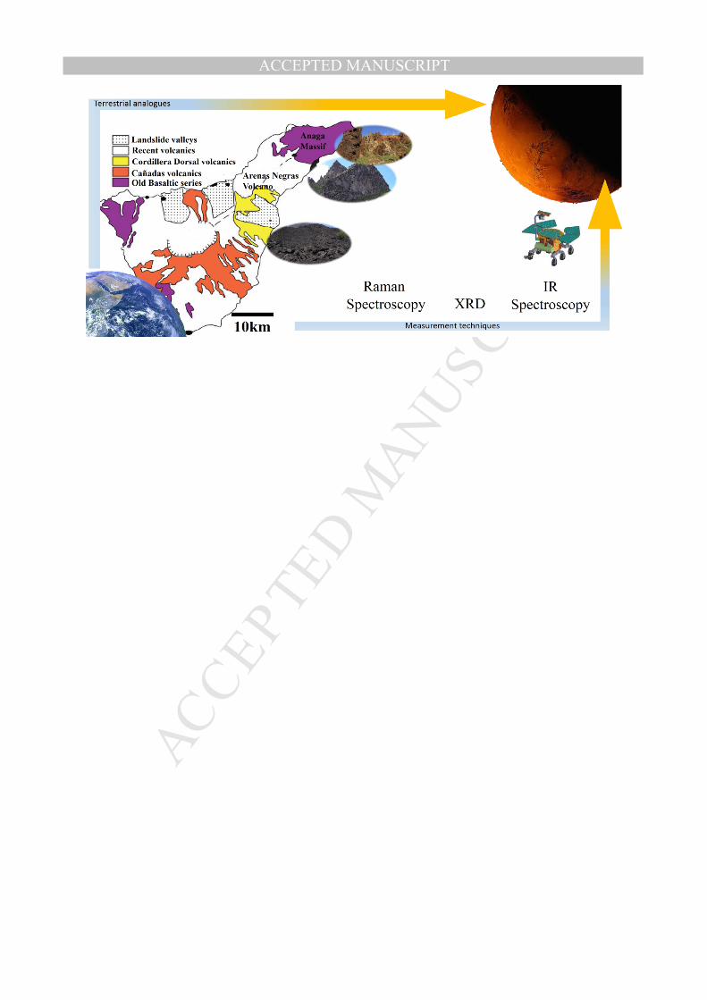
The selected places for the research have been considered especially by the geological
eruptional period on the Tenerife Island. The first zone is placed on the Anaga massif
(Fig. 1). It has been selected due to the submarine hydrothermal and weathering
processes occurred on the outcrop. Moreover, the Pillow Lava formation is an
unequivocal sign of volcanism on aqueous environment and, likely, the most abundant
structural type on Earth (Ancochea et al., 1990) being the base of the island underneath
the sea and the first part of the island formation. In this formation time, the volcanic
activity was more violent due to the combination of water, gas eruptions and magma
MANUSCRIP
T
ACCEPTED
ACCEPTED MANUSCRIPTsurrounding activities, giving the possibility to create peculiar geological fragments
considering the geochemistry and mineralogy. The studies of these geological outcrops
indicate an igneous material with the explained origin. They were formed primary by a
basinitic composition from “8 Ma”, alkali basalt around from “5.8 Ma” and basinitic
activity from “4.2 Ma”, with the existence of fossil hydrothermal systems and the
existence of Fe-rich silica amorphous phase alteration to a groundmass of celadonite
and opaline mixture rich in Fe-(hydro) oxides (Bustillo and Martez-Frs, 2003; Thirlwall
et al., 2000). The selection of this outcrop for planetary implications is due to similar
possible lava deposits have been reported on Mars, which have similar geomorphology
to the terrestrial Tuyas (Leverington, 2011; Martez-Alonso et al., 2011), where exist the
possibility of Pillow Lavas formed by the past water activity on Mars. The samples
were collected from the Igueste ravine at an altitude of eighty six meters, located at
(28º32’06”N, 16º09’24”W) near the southern coast of Anaga massif in 2009.
The second area, named as the Arenas Negras volcano (Fig. 1), dates back to one of
latest eruptions from Tenerife (ca. 400 years ago), which belongs to the triple eruption
of 1704‒1705 (“Siete Fuentes”, “Fasnia” and “Arenas Negras”). It is worth to note that
the samples are minimally affected by the meteoritic water and other external
hydrothermal process (Solana, 1996; Lalla, 2014). The volcanic material belongs to AA
lava type, composed by pyroxene-olivine with idiomorphic phenocrystals (millimeter
size). The olivine is forsterite and the clino-pyroxene is augite (Solana, 1996). The
matrix is composed by micro-crystals of plagioclase, augite, olivine and idiomorphic
opaque of different sizes (Hernandez et al., 1993; Lalla et al., 2010). However, the
secondary alteration could be caused either by the water vapors trapped within the pores
of the glass particles creating alteration material under closed-system conditions or by
weathering process (Hernandez et al., 1993; Villasante-Marcos et al., 2014). The sample
MANUSCRIP
T
ACCEPTED
ACCEPTED MANUSCRIPTset was collected by picking out samples on the studied areas during several expeditions
in 2010, being all of them catalogued and photographed (Fig. 1). In the case of the
Arenas Negras volcano, thirteen samples were collected from three different zones: 3
from the first zone TNFG01 (28º19’53”N, 16º22’34”W); six from the second zone
TNFG02 (28º19’44”N, 16º25’39”W) and two from the third zone TNFG03 (28º19’44
”N, 16º25’39”W). The selection of the zone has been done taking in consideration the
volcanic crater (TNFG03), the middle zone (TNFG02) and the end of lava eruption
(TNFG01) and eruption time. In this regard, the magma accumulation at the crater has a
8‒10 m thick being the last days of the eruption (TNFG03). On the other hand, the
TNFG03 zone corresponds to the beginning of the eruption where the distal part has a
thickness of 2‒3 m. The eruption distance has 9 km along the Güimar Valley where the
water alteration by weathering is increased with the distance.
2.2 Experimental setup and Conditions:
The micro-Raman mineralogical characterization of the samples was performed with a
microscope Nikon Eclipse E600 coupled to a spectrometer KOSI Holospec f/1.8i with
best resolution of 5 cm‒1, illuminated by a laser REO LSRP-3501 He-Ne 632.8 nm. The
detection was performed by a CCD Andor DV420A-OE-130. The laser power used was
14 mW with a spot diameter of 15 µm and the Raman mapping of the bulk surface of
the sample was done by the Micro-Raman Prior Proscan II motorized stage in automatic
mode in order to detect the different mineralization. However, the optimum recording
conditions were obtained by varying the laser power, microscope objective and the
confocal spot size (XY instrument) as required for the different samples. The spectra
were directly acquired on the sample material without any sample preparation. The FT-
Raman analysis was performed with a Fourier Transform Raman (FT-Raman) Bruker
MANUSCRIP
T
ACCEPTED
ACCEPTED MANUSCRIPTRSF 100/S, which allows the analysis of high fluorescence samples, making it is more
suitable for some samples than the visible Raman equipment. The FT-Raman
spectrometer is composed by a Nd:YAG Laser at 1064 nm, spectrograph with spectral
range 851‒1695 nm (NIR) and best spectral resolution of 2 cm‒1. The detector is a
Bruker CCD model D418T cooled by liquid N2. The working conditions achieved a
spectral resolution of 4 cm‒1 and a diameter spot of 100 µm approximately.
The X-ray diffraction analyses were carried out with a XRD diffractometer Philips
PW1710 equipped with automatic divergent slit graphite monochromator and Cu-anode.
Experimental conditions: CuKα radiation, λ = 0.154nm, a niquel filter, an aluminum
sample-holder, 40 kV generator voltage, generator current 30 mA with a relation
intensity of 0.5 (α1/α2) and angle range (2θº) from 5 to 70º. The Terra XRD
diffractometer instrument based on the MSL-CheMin concept with a detector (1024 X
256 pixels) 2D peltier cooled CCD camera for XRD with a source cobalt X-ray tube of
30Kv-300uA was also used. For the XRD analysis a preparation was necessary,
consisting on the powdering of a minimum part of the samples (2‒4 mg) and sieved
with a granulometry lower than 150 µm.
The complementary analysis with scanning electron microscopy (SEM) was performed
with an ESEM-Quanta 200F Microscope. For this analysis, no sample preparation was
necessary and the measurements were directly performed on the bulk samples.
The infrared spectroscopic data were obtained by means of a Fourier Transform Infrared
spectrometer with an attenuated total reflectance accessory (FTIR-ATR). The ATR-
FTIR Pelkin Elmer Spectra 100 spectrometer system was equipped with a diamond
ATR universal system, and the spectral range spans from 650 to 4000 cm‒1 with 4 cm‒1
spectral resolution. The samples used on these analyses were the same as the ones
prepared for the XRD analyses.
MANUSCRIP
T
ACCEPTED
ACCEPTED MANUSCRIPT3. RESULTS AND DISCUSSION
3.1 Raman spectroscopy results
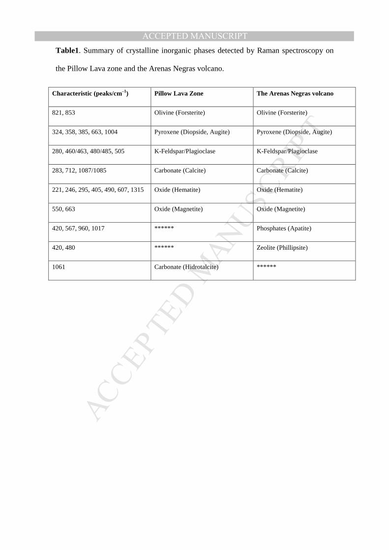
Table 1 and Figs. 2 and 3 compile and summarize the mineral species and phases
identified by Raman Spectroscopy on the two studied zones.
The olivine species detected on both sites correspond to a forsterite, clearly visible by
the typical doublet band at 820 and 850 cm‒1 in accordance with the literature (Chopelas,
1991). Also, the detected mineral species show other vibrational modes which are: (1)
the lattice mode of the olivine, both “translational and rotational” SiO4 movements and
the translational motions of the cations (Mg2+, Fe2+) in the crystal lattice at the zone
below 400 cm‒1; (2) the internal bending vibrational modes of the SiO4 ionic group at
the region 400‒700 cm‒1; and (3) the internal stretching vibrational modes of the SiO4
ionic group at the 700‒1000 cm‒1 region (Kuebler et al., 2006). On the basis of
theoretical modeling, it has been demonstrated that the high frequency peaks of the
olivine are originated from a mixing of symmetric and anti-symmetric stretching modes
of SiO4 units (Piriou and McMillan, 1983; Lam et al., 1990). In this regard, the doublet
at 820‒850 cm‒1 are the most intense peaks on the olivine Raman spectra. These peaks
are used to identify the olivine in the multi-phase spectrum due to the fact that the
relative peak height of the doublet is function of crystal orientation. In other words, it
serves as a calibration method for chemical, compositional and structural
characterization of this kind of mineral species (Kuebler et al., 2006; Mouri and Enami,
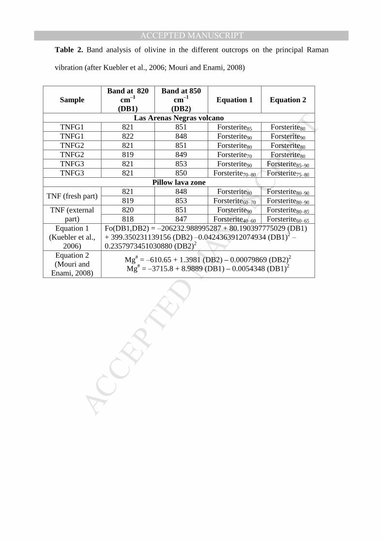
2008; Yasuzuka et al., 2009). The calibration method proposed by several author
(Kuebler et al., 2006; Yasuzuka et al., 2009) has been applied on the samples collection
and the results point to an forsterite-olivine behaviour detailed on Table 2. Nevertheless,
compared to the Arenas Negras volcano, the Pillow Lava zone presents weak/poor
MANUSCRIP
T
ACCEPTED
ACCEPTED MANUSCRIPTresults caused by an insignificant detection of olivine, which have been probably altered
by the hydrothermal process on the submarine pillow lava formation. Thus, the
Mg#=Mg/(Mg+Fe) estimation method is very sensitive to the pressure, the composition
and the alteration process (Kuebler et al., 2006; Mouri and Enami, 2008).
Concerning to the pyroxene mineral species, the Raman technique shows the existence
of diopside and augite. Moreover, the analyses of the spectral pattern, developed by
other authors (Wang et al., 2001), taking into account the empirical rules and spectral
convention enforces the previous results. Special attention has been paid to the
following regions: (1) the 1100‒800 cm‒1 region (where the strong asymmetric peak at
1000 cm‒1 approximately and some wide and weak peaks on its two wings caused by
the Si-Onbr stretching are found); (2) the 800‒600 cm‒1 region (with a strong doublet or
an asymmetric single peak near to 660 cm‒1 due to the Si-Obr stretching); (3) the
600‒450 cm‒1 region with a group of peaks which overlap each other, being of middle
intensity and which correspond to the O-Si-O bending; (4) the 450‒300 cm‒1 region
formed by overlapped strong peaks caused by a M-O stretching and M-O bending; and
(5) some peaks of moderate intensity found below 300 cm‒1 (Wang et al., 2001) . A
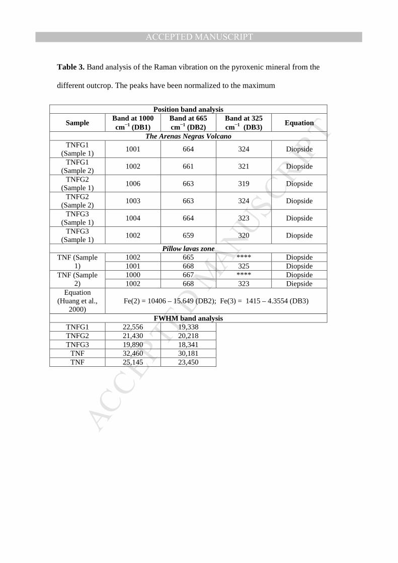
detailed comparison of the intensity or the FWHM (Full Bandwidth at Half Maximum)
of the principal bands has been performed for the different spectral regions detailed on
Table 3. This comparison reveals that Pillows Lavas zone samples have a more
amorphous behavior and possibly a more disordered structure than the Arenas Negras
volcano samples provoked by the submarine hydrothermal activity and the possible
overlapping of other mineral species (Huang et al., 2000; Wang et al., 2001; Prinsloo et
al., 2011). On the Marion Island, the results shows a similar behavior of glassy phase
systems (Prinsloo et al., 2011) and the main cause is the rapid cooling of the lava
extrudes from a volcano, without a correct crystal growing. In the case of submarine
MANUSCRIP
T
ACCEPTED
ACCEPTED MANUSCRIPTalteration, the melting point of the crystal formation is decreased, increasing the
obsidian glass formation. On the other hand, the calibration method developed by
other authors (Huang et al., 2000; Wang et al., 2001), considering Mg/(Mg+Fe+Ca)
relative concentration, where a relative change in the position of the peaks at 327 cm‒1
and at 665 cm‒1 has been applied (Table 3). For the Arenas Negras samples, the
Mg/(Mg+Fe+Ca) value is approximately “0.4 ± 0.1” while it is “0.5 ± 1” for the Pillow
Lava zone. This corresponds to a diopside-augitie behavior (Huang et al., 2000; Wang
et al., 2001) .
The tectosilicate groups detected on the target outcrop are a plagioclase series between
labradorite and andesine in the Pillow Lava zone and a plagioclase series (albite-
oligoclase)- K-feldspar (albite-anorthoclase) on the Arenas Negras volcano. However
this depends upon the sample under analysis. The detailed identification on the Raman
spectral pattern was achieved by the spectral region method, where the strongest
vibrational bands are produced by the structure of SiO4 of tectosilicate group and
located below 600 cm‒1 (Freeman et al., 2008). Thus, special consideration has been
taken for the characteristic triplet or doublet bands, located on the 450‒515 cm‒1 region
where the strongest peak is at 505‒515 cm‒1. Moreover, other vibrational regions have
been considered for the correct mineral identification such as the 200‒400 cm‒1 zone
which corresponds to the rotational-translational modes; the 600‒800 cm‒1 spectral
zone, where the Raman modes are produced by the deformation modes of the
tetrahedral; and the 900‒1200 cm‒1 region, where the vibrations are assigned to the
vibrational stretching mode of the tetrahedral structure (Freeman et al., 2008).
Hematite has been detected both on Pillow Lava zone and the Arenas Negras volcano
(Figs. 2a and 3a). Hematite is the most common Fe-oxide in nature, and it presents a
polymorph structure caused by the high thermodynamics stability. The most important
MANUSCRIP
T
ACCEPTED
ACCEPTED MANUSCRIPTbands are: the vibrations at 221 and 490 cm‒1 caused by an Ag mode; the vibrations at
245, 295, 305, 410 and 607 cm‒1 being assigned to an Eg mode; and the broad peaks at
1323 cm‒1 is caused by the magnons effects (Jubb and Allen, 2010). On the other
hand, magnetite (Fe2O3) has also been detected on both zones, where the most intense
peak of this oxide structure is found at 660 cm‒1 and it can be assigned to the A1g
symmetry mode, while the other two peaks detected correspond to the F2g mode at 550
and 504 cm‒1 (Rull et al., 2007; Jubb and Allen, 2010). The analyses of the spectral
patterns show that the Arenas Negras volcano presents a more crystalline behavior than
the Pillow Lava zone. Furthermore, the concentration of hematite/magnetite is bigger on
the Arenas Negras volcano than in the Pillow Lava zone which could be explained by
the Fe2+ and the Fe3+ cations incorporation in other crystalline solution/mineral species
caused by the hydrothermal alteration on the submarine processes.
The carbonate detected on the samples is calcite, very commonly found as a secondary
product from hydrothermal alteration in the modern Earth system. The Raman
vibrations in the carbonates can be obtained from the vibration of the (CO3)2‒ internal
modes, the vibration of hydroxyl molecules and the vibration modes M-O from the
interactions between cations and O of either (CO3)2‒, external or lattice modes. The
most intense vibrations are the symmetric stretching of CO3 at 1086 cm‒1; the
symmetric bending of CO3 at 715 cm‒1; and the external vibration of CO3‒ at 285 cm‒1
(Rull et al., 2004; Rividi et al., 2010). It is necessary to quote that the signal was very
low and sloped in comparison with other mineral species such as the pyroxene and
olivine on the Pillow Lava zone. On the other hand, in the Arenas Negras volcano, the
calcite was detected in conjunction with zeolite (Urmos et al., 1991; Hernandez et al.,
1993). Moreover, a method, developed by Rull et al (Rull-Perez and Martinez-Frias,
2003), has been applied for obtaining the paragenesis of the mineral species. The
MANUSCRIP
T
ACCEPTED
ACCEPTED MANUSCRIPTanalysis of measured parameters such as intensity and FWHM of the main peaks at
1086, 715 and 280 cm‒1 converge to a magmatic formation in both sites (Rull-Perez
and Martinez-Frias, 2003). The structure of hydrotalcite states that there is a broad
range of compositions depending on cations. However, on the Anaga Pillow Lavas
zone, the measurements show only a Raman vibration at 1060 cm‒1, which is caused by
the combination of symmetric and antisymmetric stretching of the CO3‒. Nevertheless,
more peaks are necessary to perform an identification of the specific hydrotalcite
species (Moroz et al., 2001; Frost et al., 2005). Concerning to the paragenesis, the
rock/fluid interaction with submarine water is clearly the main cause of formation.
The calcium phosphate detected on the Arenas Negras volcano corresponds to an apatite
structure, from which several Raman and IR active modes like the PO3-4, OH- and CO2-
3
active modes have been measured (Mooney et al., 1968; Antonakos et al., 2007; ). The
Td symmetry of the PO3-4 vibrational mode creates the stretching mode A1 at 960 cm‒1
zone, the doubly degenerated bending E mode at 420 cm-1, the triply degenerated anti-
symmetric stretching mode F2 at 1017 cm‒1 and the triply degenerated bending F2 at 567
cm‒1 (Ohea et al., 1974; Cooney et al., 1999; Antonakos et al., 2007; Hill and Jha, 2007;
Zattin et al., 2007).
The zeolites from the Arenas Negras volcano samples are abundant in the vesicular tops
and bottoms of the basalt flows and flow breccias. The detailed classification of the
zeolite performed by the Raman based classification technique determinates that it is a
phillipsite. The spectra have been compared with different Raman spectral data from
zeolite obtained from Raman online database such as RRUFF. Moreover, the vibrations
at 420 and 480 cm‒1 have been compared with the reference (Pechar, 1981), being in
agreement with other authors (Gatta et al., 2010).
3.2 X-Ray diffraction analysis
MANUSCRIP
T
ACCEPTED
ACCEPTED MANUSCRIPTThe qualitative mineralogical analysis was performed based on the peak positions and
its comparison with other standard patterns on the Fig. 4. For the analysis of different
phases in multiphase specimens, the commercial software Phillips PW-1876 Pc-identify
was used, which compares peak intensities attributed to the identified phases. These
analyses reveal the existence of tectosilicates (plagioclase and K-feldspar), pyroxene
(diopside or augite possibly) and olivine (forsterite) lines. Moreover, it is important to
mention the existence of hematite and magnetite in a lower proportion for both analyzed
zones. Nonetheless, the results are in agreement with the Raman analyses verifying the
principal mineral phases.
3.3 ATR-FTIR analysis
The ATR-FTIR complementary analysis only shows the most intense vibration of Al-
silicates at 1630 cm‒1 and silicates at 1000 cm‒1 in the mixture (Fig. 5). The main
problem is that the minor mineral phases are masked by the host matrix mineral phases
when crushed and mixed, and these cannot be detected in the spectra due to a relatively
high detection threshold inherent to this kind of technique. However, IR-Spectroscopy
is a very sensitive technique to the hydrogen bonding of the OH‒ at 3300 cm‒1 and the
water vibration at 3600 cm‒1 (Nakamoto, 1978; Rull et al., 2004). This feature plays an
important role in determining the aqueous mineral identification in natural samples,
which could be consequence of the rock-water interaction. As a result, the presence of
Al-OH species, OH‒ bands and H2O vibrations can be confirmed (Nakamoto, 1978).
The results could point to the hydration of the groundmass, caused either by the action
of percolating water in the subaerial environment (the Arenas Negras volcano) or the
action of the sea water in the case of the submarine environment (Pillow Lava zone).
3.4 SEM analysis
MANUSCRIP
T
ACCEPTED
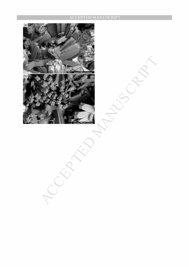
ACCEPTED MANUSCRIPTThe results of SEM show the ubiquitous occurrence of the zeolites (phillipsite), which
were only found in the many cavities of the rock, whose abundance and size are
variable, forming radiating clusters with minor quantities of calcite (Fig. 6). The results
are in agreement with the literature, the phillipsite has been identified by direct
comparison to other SEM photograph, where similar alteration processes have occurred
(Hernandez et al., 1993; Gatta et al., 2010; ).
3.5 Discussions of the results
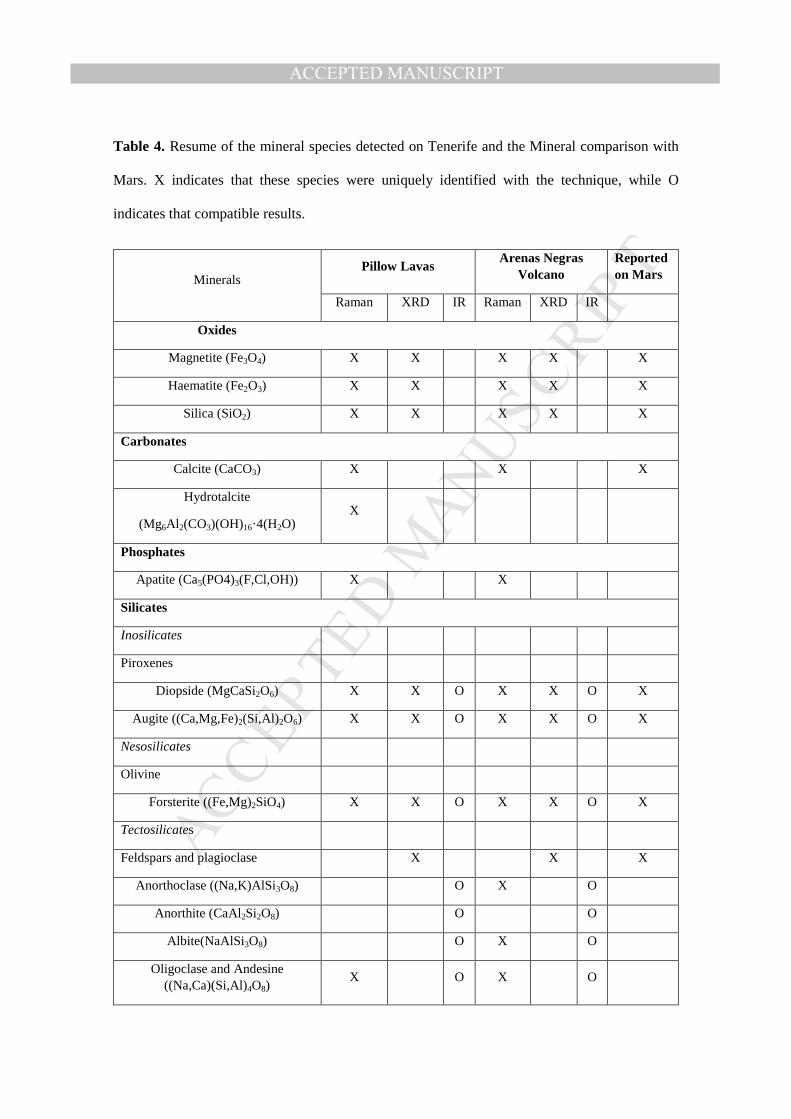
The mineralogy detected by the different techniques is summarized in Table 4. On the
different outcrop, the mineral species detected correspond to a primary and secondary
mineralization. The Pillow Lava zone has been submitted from the first processes of the
island formation compared to the Arenas Negras volcano. The secondary minerals
detected correspond to different variety of origins such as hydrothermal process or
submarine processes. However, the mineralogy on the Arenas Negras volcano presents
several similarities, despite the spectral differences on the Raman analysis.
The olivine is forsterite on the different zones, being in agreement with the references.
On the other hand, the pyroxene detected are diopside-augite and considering the
Raman detailed analysis, the spectral differences correspond to the broadening band
which could be related to the submarine alteration or the overlapping on the mineral
detection. The feldspar and plagioclase on the host matrix was detected by Raman
spectroscopy and confirmed afterward through the two complementary technique (X-
Ray diffraction and Infrared spectroscopy). Their composition varies depending on the
samples and zones and this is indicative that the different cation content is conditioned
by the local environment (temperature of formation and cooling rates). Furthermore, it
causes a discontinuous Bowen series reaction in some cases (Haldar and Tijar, 2014). In
the case of the oxides, they were detectable only by Raman spectroscopy and XRD, the
MANUSCRIP
T
ACCEPTED
ACCEPTED MANUSCRIPTPillow Lava outcrop presents less quantity detection compared to the Arenas Negras
volcano. In this regard, the Fe is incorporated in other solid crystalline solution and
glass by the submarine alteration.
The calcite presents a hydrothermal volcanic origin taking into account the Raman
analysis of the spectra. The apatite corresponds to a volcanic accessory mineral,
however it could be attributed to a contamination from submarine sediment (Pillow
Lavas) or biological contamination (the Arenas Negras volcano) (Prinsloo et al., 2011).
The zeolite was detected only by Raman spectroscopy and confirmed by SEM. The
origin of this mineral specie is clearly hydrothermal. The lava in the historical eruptions
extruded with a big amount of water vapors at the south of Tenerife and it probably
reacts fast with volcanic glasses at high temperatures in a closed-system (Hernandez et
al., 1993; Rodruez et al., 2015). Therefore, the detected zeolite crystals has been
originated by a similar hydrothermal process such as the Deccan Trap outcrop in India,
where the glass is hydrated and dissoluted with the subsequent nucleation and growth of
the zeolite (Hernandez et al., 1993; Parthasarathy et al., 2003; Parthasarathy and Sarkar,
2014). Also, several authors have obtained this zeolite phase by hydrothermal processes
of synthetic water free glasses of some compositions by P-T-t (Pressure-Temperature
close system) close system using distilled water at 200/250ºC and 4‒5 weeks being in
agreement with the natural result found on the outcrop (Ghobarkar et al., 2003;
Parthasarathy et al., 2003; Parthasarathy and Sarkar, 2014; ).
On the Table 4 is also presented a mineral comparison with the mineral detected on
Mars considering the references (Bishop et al., 2004; Chevrier and Math, 2007; Bish et
al., 2013; Wang et al., 2015). The mineral comparison have been considered the results
from the analysis from Martian meteorites and Rover analysis. In this regard, the
analysis from the Tenerife selected outcrops present similar mineral detection such as
MANUSCRIP
T
ACCEPTED
ACCEPTED MANUSCRIPT
the Crater Gusev’s mineralogy (Rice et al., 2010; Bish et al., 2013 ). Moreover, the
Gale crater shows signatures of feldspar, pyroxene, magnetite and olivine. Also the data
shows indication of phyllosilicate in the presence of basanites and a minor amount of
sulfates (Bish et al., 2013). Concerning to the alteration minerals such as zeolite, they
have been detected on the dust by orbiters (Ruff, 2004; Wray et al., 2009). As it can be
observed, the mineralization presented on the outcrop and the compared with the
Martian shows that the analogues present similar types of mineral origin: evaporitic
process, water alteration, hydrothermalism and weathering (Chevrier and Math, 2007)).
Moreover, future field-testing of the portable instrumentation and analysis of resulting
multispectral with other analytical techniques (XRD and IR) have shown to be valuable,
including the identification of spectral and synergy working of the Raman
instrumentation with other techniques for planetary research. Also, the Raman data can
reveal overall outcrop mineralogy and mineral structure, being a crucial factor in the
selection of drill targets and interpretation of the local geology.
4. CONCLUSSIONS
Different samples from Pillow Lava zone from Anaga zone and the Arenas Volcano
were characterized and studied by Raman spectroscopic techniques and several
laboratory complementary techniques for the very first time, through a complete
analysis of the mineralogy from the selected materials. Crystalline primary phases such
as olivine, pyroxene, oxide, feldspar; and secondary minerals like carbonate, zeolite and
phosphate have been confirmed by Raman spectroscopy and in some case confirmed by
complementary methods detailed along this paper. Also, other amorphous/glassy
materials, resulting from the hydrothermal alteration and weathering, were detected.
MANUSCRIP
T
ACCEPTED
ACCEPTED MANUSCRIPTThe crystalline phases of mineral species described along the paper are similar to those
reported on materials of other volcanic terrestrial analogues and Mars observations.
However, the samples present some differences on the secondary mineral species, such
as the zeolite. The possibility to distinguish the zeolite by Raman spectroscopy helps us
to deduce the rock-process formation or rock-process alteration. Thus, the enlargement
of knowledge on terrestrial analogues helps on the planetary research with
astrogeological implications, specially focused on the development of future Martian
missions.
It has been shown that Raman spectroscopy on the altered igneous rocks is capable of
detecting minor mineral phases which can be used to correlate the spectra with the
cooling rate and temperature formation of the rocks, becoming a very useful technique
for in-situ planetary exploration. Thus, the results reveal that Raman techniques are
powerful and robust systems for the detection of aqueous processes and support the
continued endeavors to use the Raman spectroscopy for Mars exploration. The
combination of complementary (IR, XRD and SEM) techniques with Raman allows us
to obtain comprehensive information about the mineralogy and to interpret the future
information to be received from the solid solution structure on the Martian surface.
ACKNOWLEDGEMENTS
The work was supported by the MICINN with the Project AYA-2008-04529 for the
development of the Raman-LIBS combined spectrometer for the ESA-ExoMars
Mission. E. Lalla wish to thank MICINN for the FPI grants (BES-2009-024992).
REFERENCES
MANUSCRIP
T
ACCEPTED
ACCEPTED MANUSCRIPTAcocella, V., 2007. Understanding caldera structure and development: An overview of
analogue models compared to natural calderas. Earth-Science Rev. 85, 125–160.
doi:http://dx.doi.org/10.1016/j.earscirev.2007.08.004
Ancochea, E., Fuster, J., Ibarrola, E., Cendrero, A., Coello, J., Hernan, F., Cantagrel,
J.M., Jamond, C., 1990. Volcanic evolution of the island of Tenerife (Canary
Islands) in the light of new K-Ar data. J. Volcanol. Geotherm. Res.
doi:10.1016/0377-0273(90)90019-C
Antonakos, A., Liarokapis, E., Leventouri, T., 2007. Micro-Raman and (FTIR) studies
of synthetic and natural apatites. Biomaterials 28, 3043–3054.
doi:http://dx.doi.org/10.1016/j.biomaterials.2007.02.028
Bish, D.L., Blake, D.F., Vaniman, D.T., Chipera, S.J., Morris, R. V, Ming, D.W.,
Treiman, A.H., Sarrazin, P., Morrison, S.M., Downs, R.T., Achilles, C.N., Yen,
A.S., Bristow, T.F., Crisp, J.A., Morookian, J.M., Farmer, J.D., Rampe, E.B.,
Stolper, E.M., Spanovich, N., Team, M.S.L.S., 2013. X-ray Diffraction Results
from Mars Science Laboratory: Mineralogy of Rocknest at Gale Crater. Sci. 341 .
doi:10.1126/science.1238932
Bishop, J.L., Murad, E., Lane, M.D., Mancinelli, R.L., 2004. Multiple techniques for
mineral identification on Mars: a study of hydrothermal rocks as potential
analogues for astrobiology sites on Mars. Icarus 169, 311–323.
doi:http://dx.doi.org/10.1016/j.icarus.2003.12.025
Bost, N., Ramboz, C., LeBreton, N., Foucher, F., Lopez-Reyes, G., De Angelis, S.,
Josset, M., Venegas, G., Sanz-Arranz, A., Rull, F., Medina, J., Josset, J.-L.,
Souchon, A., Ammannito, E., De Sanctis, M.C., Di Iorio, T., Carli, C., Vago, J.L.,
MANUSCRIP
T
ACCEPTED
ACCEPTED MANUSCRIPTWestall, F., 2015. Testing the ability of the ExoMars 2018 payload to document
geological context and potential habitability on Mars. Planet. Space Sci. 108, 87–
97. doi:10.1016/j.pss.2015.01.006
Bost, N., Westall, F., Ramboz, C., Foucher, F., Pullan, D., Meunier, A., Petit, S.,
Fleischer, I., Klingelhöfer, G., Vago, J.L., 2013. Missions to Mars:
Characterisation of Mars analogue rocks for the International Space Analogue
Rockstore (ISAR). Planet. Space Sci. 82–83, 113–127.
doi:http://dx.doi.org/10.1016/j.pss.2013.04.006
Bustillo, M.A., Martínez-Frías, J., 2003. Green opals in hydrothermalized basalts
(Tenerife Island, Spain): alteration and aging of silica pseudoglass. J. Non. Cryst.
Solids 323, 27–33. doi:http://dx.doi.org/10.1016/S0022-3093(03)00288-6
Campbell, K.A., Guido, D.M., Gautret, P., Foucher, F., Ramboz, C., Westall, F., 2015.
Geyserite in hot-spring siliceous sinter: Window on Earth’s hottest terrestrial
(paleo)environment and its extreme life. Earth-Science Rev. 148, 44–64.
doi:10.1016/j.earscirev.2015.05.009
Carracedo, J.C., 1999. Growth, structure, instability and collapse of Canarian volcanoes
and comparisons with Hawaiian volcanoes. J. Volcanol. Geotherm. Res. 94, 1–19.
doi:http://dx.doi.org/10.1016/S0377-0273(99)00095-5
Chevrier, V., Mathé, P.E., 2007. Mineralogy and evolution of the surface of Mars: A
review. Planet. Space Sci. 55, 289–314.
doi:http://dx.doi.org/10.1016/j.pss.2006.05.039
MANUSCRIP
T
ACCEPTED
ACCEPTED MANUSCRIPTChopelas, A., 1991. Single crystal Raman spectra of forsterite, fayalite, and
monticellite. Am. Mineral. 76, 1101–1109.
Cooney, T.F., Scott, E.R.D., Krot, A.N., Sharma, S.K., Yamaguchi, A., 1999.
Vibrational spectroscopic study of minerals in the Martian meteorite ALH84001.
Am. Mineral. 84, 1569–1576. doi:10.1016/0003-004X-99-0010-1569
Courrèges-Lacoste, G.B., Ahlers, B., Rull Perez, F., 2007. Combined Raman
spectrometer/laser-induced breakdown spectrometer for the next ESA mission to
Mars. Spectrochim. Acta Part A Mol. Biomol. Spectrosc. 68, 1023–1028.
doi:http://dx.doi.org/10.1016/j.saa.2007.03.026
Edwards, H.G.M., Moody, C.A., Jorge Villar, S.E., Mancinelli, R., 2004. Raman
spectroscopy of desert varnishes and their rock substrata. J. Raman Spectrosc. 35,
475–479. doi:10.1002/jrs.1170
Farr, T.G., 2004. Terrestrial analogs to Mars: The NRC community decadal report.
Planet. Space Sci. 52, 3–10. doi:http://dx.doi.org/10.1016/j.pss.2003.08.004
Freeman, J.J., Wang, A., Kuebler, K.E., Jolliff, B.L., Haskin, L.A., 2008.
Characterization of natural feldspars by raman spectroscopy for future planetary
exploration. Can. Mineral. 46, 1477–1500. doi:10.3749/canmin.46.6.1477
Frost, R.L., Adebajo, M.O., Erickson, K.L., 2005. Raman spectroscopy of synthetic and
natural iowaite. Spectrochim. Acta Part A Mol. Biomol. Spectrosc. 61, 613–620.
doi:http://dx.doi.org/10.1016/j.saa.2004.05.015
Galindo, I., Soriano, C., Martí, J., Pérez, N., 2005. Graben structure in the Las Cañadas
edifice (Tenerife, Canary Islands): Implications for active degassing and insights
MANUSCRIP
T
ACCEPTED
ACCEPTED MANUSCRIPTon the caldera formation. J. Volcanol. Geotherm. Res. 144, 73–87.
doi:10.1016/j.jvolgeores.2004.11.017
Gatta, G.D., Cappelletti, P., Langella, A., 2010. Crystal-chemistry of phillipsites from
the Neapolitan Yellow Tuff. Eur. J. Mineral. doi:10.1127/0935-1221/2010/0022-
2027
Ghobarkar, H., Schäf, O., Massiani, Y., Knauth, P., 2003. The Reconstruction of
Natural Zeolites: A New Approach to Announce Old Materials by their Synthesis,
Kluwer Aca. ed. Springer-Verlag New York Inc. doi:10.1007/978-1-4419-9142-3
Graham, L., Graff, T.G., Aileen Yingst, R., ten Kate, I.L., Russell, P., 2015. 2012 Moon
Mars Analog Mission Activities on Mauna Kea, Hawaii. Adv. Sp. Res. 55, 2405–
2413. doi:10.1016/j.asr.2015.01.024
Grossman, L., 2013. NASA urged to seek live Martians with 2020 rover. New Sci. 219,
9. doi:10.1016/S0262-4079(13)61775-3
Haldar, S.K., Tišljar, J., 2014. Chapter 4 - Igneous Rocks, in: Tišljar, S.K.H. (Ed.),
Introduction to Mineralogy and Petrology. Elsevier, Oxford, pp. 93–120.
doi:http://dx.doi.org/10.1016/B978-0-12-408133-8.00004-3
Hernandez, J.E.G., del Pino, J.S.N., Martin, M.M.G., Reguera, F.H., Losada, J.A.R.,
1993. Zeolites in pyroclastic deposits in southeastern tenerife (Canary Islands).
Clays Clay Miner. 41, 521–526. doi:10.1346/CCMN.1993.0410501
Hill, C.J., Jha, A., 2007. Development of novel ternary tellurite glasses for high
temperature fiber optic mid-IR chemical sensing. J. Non. Cryst. Solids 353, 1372–
1376. doi:http://dx.doi.org/10.1016/j.jnoncrysol.2006.10.061
MANUSCRIP
T
ACCEPTED
ACCEPTED MANUSCRIPTHuang, E., Chen, C.H., Huang, T., Lin, E.H., Xu, J.A., 2000. Raman spectroscopic
characteristics of Mg-Fe-Ca pyroxenes. Am. Mineral. 85, 473–479.
Jubb, A.M., Allen, H.C., 2010. Vibrational Spectroscopic Characterization of Hematite,
Maghemite, and Magnetite Thin Films Produced by Vapor Deposition. ACS Appl.
Mater. Interfaces 2, 2804–2812. doi:10.1021/am1004943
Kuebler, K.E., Jolliff, B.L., Wang, A., Haskin, L.A., 2006. Extracting olivine (Fo–Fa)
compositions from Raman spectral peak positions. Geochim. Cosmochim. Acta 70,
6201–6222. doi:http://dx.doi.org/10.1016/j.gca.2006.07.035
Kuhn, N., 2015. Chapter 2 - Overview of Mars, in: Kuhn, N.B.T.-E. in R.G. (Ed.),
Experiments in Reduced Gravity. Elsevier, Oxford, pp. 17–26.
doi:http://dx.doi.org/10.1016/B978-0-12-799965-4.00002-9
Lalla, E., Caramazana Sansano, A., Sanz-Arranz, A., Alonso Alonso, P., Medina
García, J., Martinez-frías, J., Rull-Perez, F., 2010. Espectroscopía Raman de
Basaltos Correspondientes al Volcán de Las Arenas , Tenerife. MACLA - Soc.
Española Mineral. 13, 129–130.
Lalla, E.A., 2014. Tenerife como análogo de Marte: Caracterización multianalítica
(Raman, DRX, ATR-FTIR, SEM y MossBaeur) de muestras de interés planetario y
astrobiológico. Dep. Física la Mater. Condens. Cristalogr. y Minerealogía - Univ.
Valladolid. University of Valladolid.
Lam, P.K., Yu, R., Lee, M.W., Sharma, S.K., 1990. Structural distortions and
vibrational modes in Mg2SiO4. Am. Mineral.
MANUSCRIP
T
ACCEPTED
ACCEPTED MANUSCRIPTLéveillé, R., 2009. Validation of astrobiology technologies and instrument operations in
terrestrial analogue environments. Comptes Rendus Palevol 8, 637–648.
doi:http://dx.doi.org/10.1016/j.crpv.2009.03.005
Leverington, D.W., 2011. A volcanic origin for the outflow channels of Mars: Key
evidence and major implications. Geomorphology 132, 51–75.
doi:http://dx.doi.org/10.1016/j.geomorph.2011.05.022
Martínez-Alonso, S., Mellon, M.T., Banks, M.E., Keszthelyi, L.P., McEwen, A.S.,
2011. Evidence of volcanic and glacial activity in Chryse and Acidalia Planitiae,
Mars. Icarus 212, 597–621. doi:http://dx.doi.org/10.1016/j.icarus.2011.01.004
Mooney, R.W., Toma, S.Z., Goldsmith, R.L., Butler, K.H., 1968. Normal vibrations of
the PO43− ion, site symmetry C3v, IN Sr3(PO4)2 and Ba3(PO4)2. J. Inorg. Nucl.
Chem. doi:10.1016/0022-1902(68)80337-9
Moroz, T., Razvorotneva, L., Grigorieva, T., Mazurov, M., Arkhipenko, D., Prugov, V.,
2001. Formation of spinel from hydrotalcite-like minerals and destruction of
chromite implanted by inorganic salts. Appl. Clay Sci. 18, 29–36.
Mouri, T., Enami, M., 2008. Raman spectroscopic study of olivine-group minerals. J.
Mineral. Petrol. Sci. 103, 100–104. doi:10.2465/jmps.071015
Nakamoto, K., 1978. Infrared and Raman Spectra of Inorganic and Coordination
Compounds, Part A: Theory and Applications in Inorganic Chemistry, Sixth Edit.
ed. John Wiley & Sons, Ltd.
MANUSCRIP
T
ACCEPTED
ACCEPTED MANUSCRIPTO’Shea, D.C., Bartlett, M.L., Young, R.A., 1974. Compositional analysis of apatites
with laser-raman spectroscopy:(oh,f,cl)apatites. Arch. Oral Biol. 19, 995–906.
doi:http://dx.doi.org/10.1016/0003-9969(74)90086-7
Parthasarathy, G., Choudary, B.M., Sreedhar, B., Kunwar, A.C., Srinivasan, R., 2003.
Ferrous saponite from the Deccan Trap , India , and its application in adsorption
and reduction of hexavalent chromium. Am. Mineral. 88, 1983–1988.
Parthasarathy, G., Sarkar, P.K., 2014. High pressure temperature studies of
phyllosilicates from the Deccan Trap: Implications to Martian Mineralogy and
Near Subsurface Processes, in: Lunar and Planetary Science Conference. Houston,
Texas, p. 1326. doi:10.1002/jgre.20161.
Pechar, F., 1981. Study of the Raman Polarization Spectra of the Single Crystal
Phillipsite. Krist. und Tech. 16, 917–920. doi:10.1002/crat.19810160810
Piriou, B., McMillan, P., 1983. The high-frequency vibrational spectra of vitreous and
crystalline orthosilicates. Am. Mineral.
Prinsloo, L.C., Colomban, P., Brink, J.D., Meiklejohn, I., 2011. A Raman spectroscopic
study of the igneous rocks on Marion Island: A possible terrestrial analogue for the
geology on Mars. J. Raman Spectrosc. 42, 626–632. doi:10.1002/jrs.2756
Rice, M.S., Bell, J.F., Cloutis, E.A., Wang, A., Ruff, S.W., Craig, M.A., Bailey, D.T.,
Johnson, J.R., de Souza, P.A., Farrand, W.H., 2010. Silica-rich deposits and
hydrated minerals at Gusev Crater, Mars: Vis-NIR spectral characterization and
regional mapping. Icarus 205, 375–395. doi:10.1016/j.icarus.2009.03.035
MANUSCRIP
T
ACCEPTED
ACCEPTED MANUSCRIPTRividi, N., van Zuilen, M., Philippot, P., Ménez, B., Godard, G., Poidatz, E., 2010.
Calibration of Carbonate Composition Using Micro-Raman Analysis: Application
to Planetary Surface Exploration. Astrobiology 10, 293–309.
doi:10.1089/ast.2009.0388
Rodríguez, F., Pérez, N.M., Padrón, E., Melián, G., Piña-Varas, P., Dionis, S.,
Barrancos, J., Padilla, G.D., Hernández, P.A., Marrero, R., Ledo, J.J., Bellmunt, F.,
Queralt, P., Marcuello, A., Hidalgo, R., 2015. Surface geochemical and
geophysical studies for geothermal exploration at the southern volcanic rift zone of
Tenerife, Canary Islands, Spain. Geothermics 55, 195–206.
doi:10.1016/j.geothermics.2015.02.007
Ruff, S.W., 2004. Spectral evidence for zeolite in the dust on Mars. Icarus 168, 131–
143. doi:10.1016/j.icarus.2003.11.003
Rull, F., Martinez-Frias, J., Rodríguez-Losada, J.A., 2007. Micro-Raman spectroscopic
study of El Gasco pumice, western Spain. J. Raman Spectrosc. 38, 239–244.
doi:10.1002/jrs.1628
Rull, F., Martinez-Frias, J., Sansano, A., Medina, J., Edwards, H.G.M., 2004.
Comparative micro-Raman study of the Nakhla and Vaca Muerta meteorites. J.
Raman Spectrosc. 35, 497–503. doi:10.1002/jrs.1177
Rull-Perez, F., Martinez-Frias, J., 2003. Identification of calcite grains in the Vaca
Muerta mesosiderite by Raman spectroscopy. J. Raman Spectrosc. 34, 367–370.
doi:10.1002/jrs.1003
MANUSCRIP
T
ACCEPTED
ACCEPTED MANUSCRIPTRull-Pérez, F., Martinez-Frias, J., 2006. Raman spectroscopy goes to Mars. Spectrosc.
Eur. 18, 18–21.
Solana, M.C., 1996. La erupción de 1704-1705 en Tenerife, Islas Canarias.
Reconstrucción, peligros asociados y su mitigación. Geogaceta 20, 540–542.
Thirlwall, M.F., Singer, B.S., Marriner, G.F., 2000. 39Ar–40Ar ages and geochemistry
of the basaltic shield stage of Tenerife, Canary Islands, Spain. J. Volcanol.
Geotherm. Res. 103, 247–297. doi:http://dx.doi.org/10.1016/S0377-
0273(00)00227-4
Urmos, J., Sharma, S.K., Mackenzie, F.T., 1991. Characterization of some biogenic
carbonates with Raman spectroscopy. Am. Mineral.
Villasante-Marcos, V., Finizola, A., Abella, R., Barde-Cabusson, S., Blanco, M.J.,
Brenes, B., Cabrera, V., Casas, B., De Agustín, P., Di Gangi, F., Domínguez, I.,
García, O., Gomis, A., Guzmán, J., Iribarren, I., Levieux, G., López, C., Luengo-
Oroz, N., Martín, I., Moreno, M., Meletlidis, S., Morin, J., Moure, D., Pereda, J.,
Ricci, T., Romero, E., Schütze, C., Suski-Ricci, B., Torres, P., Trigo, P., 2014.
Hydrothermal system of Central Tenerife Volcanic Complex, Canary Islands
(Spain), inferred from self-potential measurements. J. Volcanol. Geotherm. Res.
272, 59–77. doi:10.1016/j.jvolgeores.2013.12.007
Wang, A., Jolliff, B.L., Haskin, L.A., Kuebler, K.E., Viskupic, K.M., 2001.
Characterization and comparison of structural and compositional features of
planetary quadrilateral pyroxenes by Raman spectroscopy. Am. Mineral. 86, 790–
806.
MANUSCRIP
T
ACCEPTED
ACCEPTED MANUSCRIPTWang, A., Korotev, R.L., Jolliff, B.L., Ling, Z., 2015. Raman imaging of extraterrestrial
materials. Planet. Space Sci. 112, 23–34. doi:10.1016/j.pss.2014.10.005
West, M.D., Clarke, J.D.A., 2010. Potential martian mineral resources: Mechanisms and
terrestrial analogues. Planet. Space Sci. 58, 574–582.
doi:http://dx.doi.org/10.1016/j.pss.2009.06.007
Wray, J.J., Murchie, S.L., Squyres, S.W., Seelos, F.P., Tornabene, L.L., 2009. Diverse
aqueous environments on ancient Mars revealed in the southern highlands. Geol.
37 , 1043–1046. doi:10.1130/G30331A.1
Xiao, L., Huang, J., Christensen, P.R., Greeley, R., Williams, D.A., Zhao, J., He, Q.,
2012. Ancient volcanism and its implication for thermal evolution of Mars. Earth
Planet. Sci. Lett. 323-324, 9–18. doi:10.1016/j.epsl.2012.01.027
Yasuzuka, T., Ishibashi, H., Arakawa, M., Yamamoto, J., Kagi, H., 2009. Simultaneous
determination of Mg# and residual pressure in olivine using micro-Raman
spectroscopy. J. Mineral. Petrol. Sci. 104, 395–400. doi:10.2465/jmps.090615
Zattin, M., Bersani, D., Carter, A., 2007. Raman microspectroscopy: A non-destructive
tool for routine calibration of apatite crystallographic structure for fission-track
analyses. Chem. Geol. 240, 197–204. doi:10.1016/j.chemgeo.2007.02.007
MANUSCRIP
T
ACCEPTED
ACCEPTED MANUSCRIPT
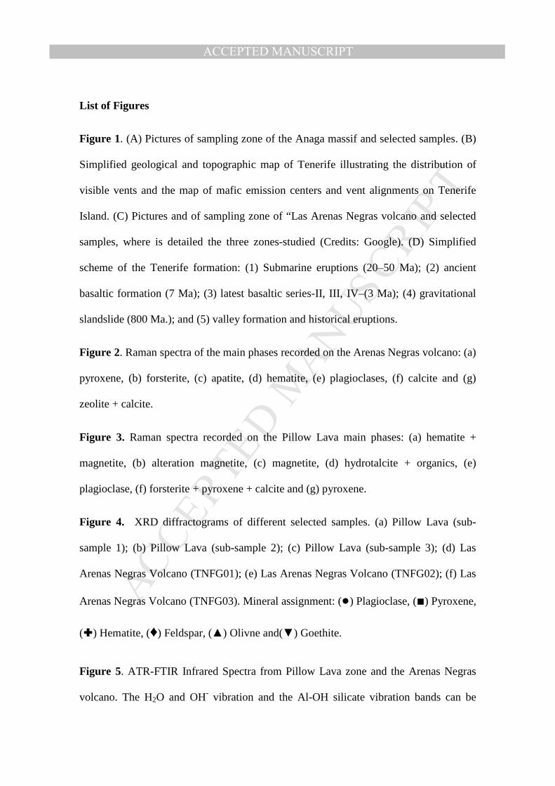
List of Figures
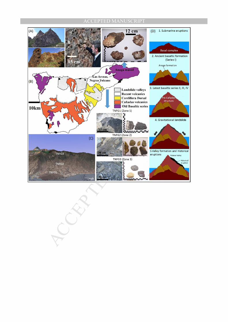
Figure 1. (A) Pictures of sampling zone of the Anaga massif and selected samples. (B)
Simplified geological and topographic map of Tenerife illustrating the distribution of
visible vents and the map of mafic emission centers and vent alignments on Tenerife
Island. (C) Pictures and of sampling zone of “Las Arenas Negras volcano and selected
samples, where is detailed the three zones-studied (Credits: Google). (D) Simplified
scheme of the Tenerife formation: (1) Submarine eruptions (20‒50 Ma); (2) ancient
basaltic formation (7 Ma); (3) latest basaltic series-II, III, IV–(3 Ma); (4) gravitational
slandslide (800 Ma.); and (5) valley formation and historical eruptions.
Figure 2. Raman spectra of the main phases recorded on the Arenas Negras volcano: (a)
pyroxene, (b) forsterite, (c) apatite, (d) hematite, (e) plagioclases, (f) calcite and (g)
zeolite + calcite.
Figure 3. Raman spectra recorded on the Pillow Lava main phases: (a) hematite +
magnetite, (b) alteration magnetite, (c) magnetite, (d) hydrotalcite + organics, (e)
plagioclase, (f) forsterite + pyroxene + calcite and (g) pyroxene.
Figure 4. XRD diffractograms of different selected samples. (a) Pillow Lava (sub-
sample 1); (b) Pillow Lava (sub-sample 2); (c) Pillow Lava (sub-sample 3); (d) Las
Arenas Negras Volcano (TNFG01); (e) Las Arenas Negras Volcano (TNFG02); (f) Las
Arenas Negras Volcano (TNFG03). Mineral assignment: (●) Plagioclase, (■) Pyroxene,
(�) Hematite, (♦) Feldspar, (▲) Olivne and(▼) Goethite.
Figure 5. ATR-FTIR Infrared Spectra from Pillow Lava zone and the Arenas Negras
volcano. The H2O and OH- vibration and the Al-OH silicate vibration bands can be

MANUSCRIP
T
ACCEPTED
ACCEPTED MANUSCRIPTobserved. (a-c) Pillow Lava (selected samples spectra) (d) TNFG01 sample and (e)
TNFG03 sample.
Figure 6. SEM Photograph of the zeolite occurrence. Radiating cluster of the zeolite
crystal inside the cavities from the Arenas Negras volcano (a) and (b).
MANUSCRIP
T
ACCEPTED
ACCEPTED MANUSCRIPTTable1. Summary of crystalline inorganic phases detected by Raman spectroscopy on
the Pillow Lava zone and the Arenas Negras volcano.
Characteristic (peaks/cm‒1) Pillow Lava Zone The Arenas Negras volcano
821, 853 Olivine (Forsterite) Olivine (Forsterite)
324, 358, 385, 663, 1004 Pyroxene (Diopside, Augite) Pyroxene (Diopside, Augite)
280, 460/463, 480/485, 505 K-Feldspar/Plagioclase K-Feldspar/Plagioclase
283, 712, 1087/1085 Carbonate (Calcite) Carbonate (Calcite)
221, 246, 295, 405, 490, 607, 1315 Oxide (Hematite) Oxide (Hematite)
550, 663 Oxide (Magnetite) Oxide (Magnetite)
420, 567, 960, 1017 ****** Phosphates (Apatite)
420, 480 ****** Zeolite (Phillipsite)
1061 Carbonate (Hidrotalcite) ******
MANUSCRIP
T
ACCEPTED
ACCEPTED MANUSCRIPTTable 2. Band analysis of olivine in the different outcrops on the principal Raman
vibration (after Kuebler et al., 2006; Mouri and Enami, 2008)
Sample Band at 820
cm‒1 (DB1)
Band at 850 cm‒1
(DB2) Equation 1 Equation 2
Las Arenas Negras volcano TNFG1 821 851 Forsterite85 Forsterite80 TNFG1 822 848 Forsterite90 Forsterite90 TNFG2 821 851 Forsterite80 Forsterite80 TNFG2 819 849 Forsterite70 Forsterite80 TNFG3 821 853 Forsterite90 Forsterite85‒90 TNFG3 821 850 Forsterite70‒80 Forsterite75‒80
Pillow lava zone
TNF (fresh part) 821 848 Forsterite80 Forsterite80‒90 819 853 Forsterite60‒70 Forsterite80‒90
TNF (external part)
820 851 Forsterite90 Forsterite80‒85 818 847 Forsterite40‒60 Forsterite60‒65
Equation 1 (Kuebler et al.,
2006)
Fo(DB1,DB2) = ‒206232.988995287 + 80.190397775029 (DB1) + 399.350231139156 (DB2) ‒0.0424363912074934 (DB1)2 ‒ 0.2357973451030880 (DB2)2
Equation 2 (Mouri and
Enami, 2008)
Mg# = ‒610.65 + 1.3981 (DB2) – 0.00079869 (DB2)2
Mg# = ‒3715.8 + 8.9889 (DB1) – 0.0054348 (DB1)2
MANUSCRIP
T
ACCEPTED
ACCEPTED MANUSCRIPT
Table 3. Band analysis of the Raman vibration on the pyroxenic mineral from the
different outcrop. The peaks have been normalized to the maximum
Position band analysis
Sample Band at 1000 cm‒1 (DB1)
Band at 665 cm‒1 (DB2)
Band at 325 cm‒1 (DB3)
Equation
The Arenas Negras Volcano TNFG1
(Sample 1) 1001 664 324 Diopside
TNFG1 (Sample 2)
1002 661 321 Diopside
TNFG2 (Sample 1)
1006 663 319 Diopside
TNFG2 (Sample 2)
1003 663 324 Diopside
TNFG3 (Sample 1)
1004 664 323 Diopside
TNFG3 (Sample 1)
1002 659 320 Diopside
Pillow lavas zone TNF (Sample
1) 1002 665 **** Diopside 1001 668 325 Diopside
TNF (Sample 2)
1000 667 **** Diopside 1002 668 323 Diepside
Equation (Huang et al.,
2000) Fe(2) = 10406 – 15.649 (DB2); Fe(3) = 1415 – 4.3554 (DB3)
FWHM band analysis TNFG1 22,556 19,338 TNFG2 21,430 20,218 TNFG3 19,890 18,341
TNF 32,460 30,181 TNF 25,145 23,450
MANUSCRIP
T
ACCEPTED
ACCEPTED MANUSCRIPT
Table 4. Resume of the mineral species detected on Tenerife and the Mineral comparison with
Mars. X indicates that these species were uniquely identified with the technique, while O
indicates that compatible results.
Minerals Pillow Lavas
Arenas Negras Volcano
Reported on Mars
Raman XRD IR Raman XRD IR
Oxides
Magnetite (Fe3O4) X X X X X
Haematite (Fe2O3) X X X X X
Silica (SiO2) X X X X X
Carbonates
Calcite (CaCO3) X X X
Hydrotalcite
(Mg6Al 2(CO3)(OH)16·4(H2O) X
Phosphates
Apatite (Ca5(PO4)3(F,Cl,OH)) X X
Silicates
Inosilicates
Piroxenes
Diopside (MgCaSi2O6) X X O X X O X
Augite ((Ca,Mg,Fe)2(Si,Al)2O6) X X O X X O X
Nesosilicates
Olivine
Forsterite ((Fe,Mg)2SiO4) X X O X X O X
Tectosilicates
Feldspars and plagioclase X X X
Anorthoclase ((Na,K)AlSi3O8) O X O
Anorthite (CaAl2Si2O8) O O
Albite(NaAlSi3O8) O X O
Oligoclase and Andesine ((Na,Ca)(Si,Al)4O8)
X O X O

MANUSCRIP
T
ACCEPTED
ACCEPTED MANUSCRIPTLabradorite ((Ca,Na)(Si,Al)4O8) X O O
Zeolites X
Phillipsite ((Ca,K,Na)6(Si10Al 6)O3212H2O)
X
MANUSCRIP
T
ACCEPTED
ACCEPTED MANUSCRIPT

MANUSCRIP
T
ACCEPTED
ACCEPTED MANUSCRIPT

MANUSCRIP
T
ACCEPTED
ACCEPTED MANUSCRIPT

MANUSCRIP
T
ACCEPTED
ACCEPTED MANUSCRIPT

MANUSCRIP
T
ACCEPTED
ACCEPTED MANUSCRIPT
MANUSCRIP
T
ACCEPTED
ACCEPTED MANUSCRIPT

MANUSCRIP
T
ACCEPTED
ACCEPTED MANUSCRIPTHIGHLIGHTS:
Tenerife eruptions as analogue for ancient Mars Micro-Raman characterization of mineral phases Zeolite mineral phase identified in the Arenas Negras volcano