Embed Size (px)
Citation preview
RAISON D’ETRE OF THE IMMUNE SYSTEM:
To Distinguish Self from Non-Self Thereby Protecting Us From Our Hostile
Environment.
Innate Immunity Acquired Immunity
Innate immunity:
mechanisms that are used by
several hours of encountering
(Antigen nonspecific) defense
the host immediately or within
antigen.
Cellular Components of the Innate Immune Response
NK cells
Granulocytes
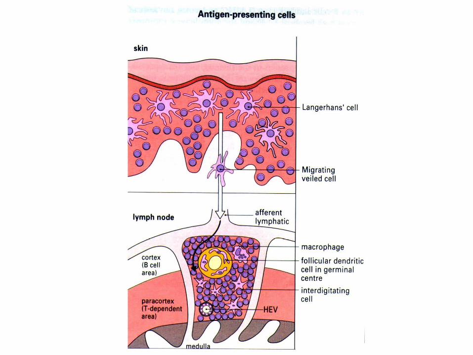
Antigen Presenting Cells:Dendritic cellsMacrophages
Antigen Presenting Cells
These specialized cells internalize antigen byphagocytosis or endocytosis and then express parts of the antigen on the cell surface. These cells are distinguished by two properties:
1. Express class II MHC molecules
2. Provide co-stimulatory signals necessary foractivation of T-cells.
Acquired Immunity
Is adaptive and displays four characteristic attributes:
•Antigen specific•Diversity•Immunologic Memory•Self/non-self recognition
Acquired Immunity
Involves two major types of cells:
Lymphocytes:
a. B-cells: Originate in the bone-marrow
b. T-cells: Originate in the thymus
• All lymphocytes have an antigen receptor, a surface protein that engages with a portion of an invading pathogen
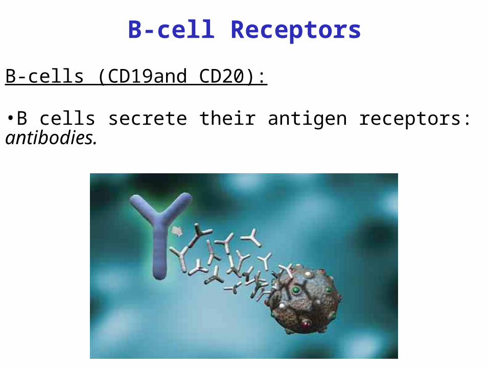
B-cell Receptors
B-cells (CD19and CD20):
•B cells secrete their antigen receptors: antibodies.
Antibodies cont.•Antibodies can help elicit clearance of an antigen, or can prevent proper functioning of the antigen: neutralization.
•Antibodies are effective against extracellular pathogens, such as bacteria, or virus that has budded from the cell.
•Antibodies can work at distal sites. Are in interstitial fluids, blood and lymph fluids.
•Can bind soluble antigen
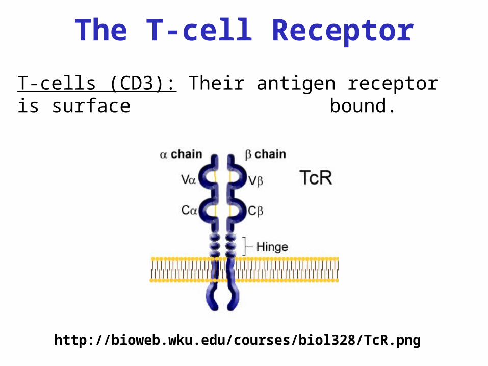
The T-cell Receptor
http://bioweb.wku.edu/courses/biol328/TcR.png
T-cells (CD3): Their antigen receptor is surface bound.
1. Cytotoxic T cells (CTL) kill infected cells.
• Are identified by the surface marker CD8(CD8+ T-cells)
• Control intracellular pathogens such as viruses and bacteria
• Require cell to cell contact to bind antigen • Bind only antigen presented on the surface of
cells
T-cell Subsets and Functions
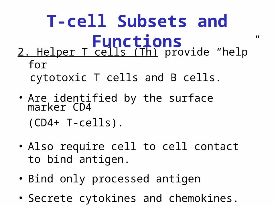
2. Helper T cells (Th) provide “help” forcytotoxic T cells and B cells.
• Are identified by the surface marker CD4
(CD4+ T-cells).
• Also require cell to cell contact to bind antigen.
• Bind only processed antigen
• Secrete cytokines and chemokines.
T-cell Subsets and Functions
Cell to Cell Communication
• Cytokine: Small molecules secreted during an immune response that help to signal and activate responding cells.
• Chemokines: Also small molecules secreted during an immune response, these often signal cells to migrate to areas of inflammation.
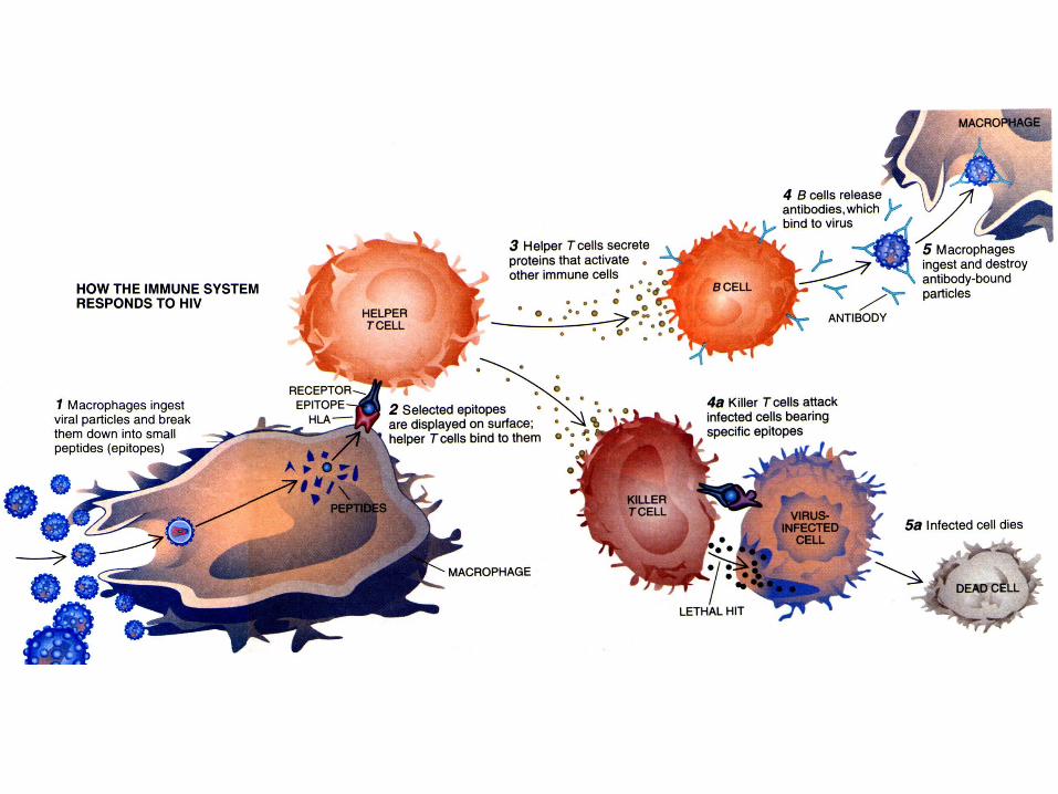
Antigen Processing
• Antigen presenting cells pick up, or endocytose, antigens and degrade them within endosomes via acid-dependent proteases
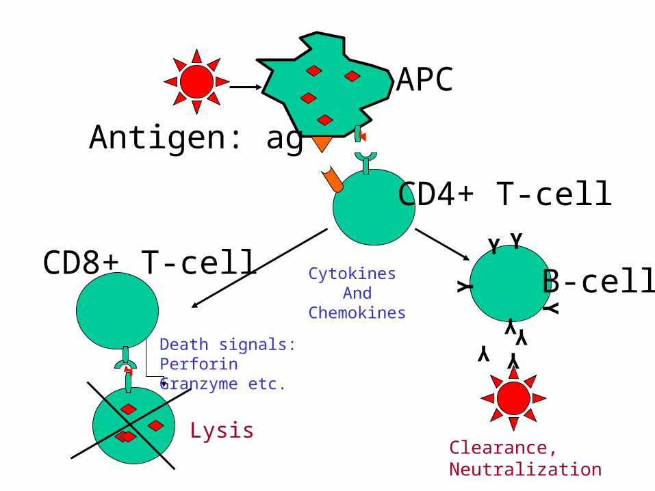
Antigen: ag
APC
CD4+ T-cell
CD8+ T-cell
Death signals:PerforinGranzyme etc.
Cytokines And
Chemokines
B-cell
Y
YY
Y Y
YY Y
LysisClearance,Neutralization
Antigen Specificity:
Is determined by interactions between cellular
receptors (T-cell receptor and B-cell receptor
complex), antigen, and human leukocyte antigens
(HLA).
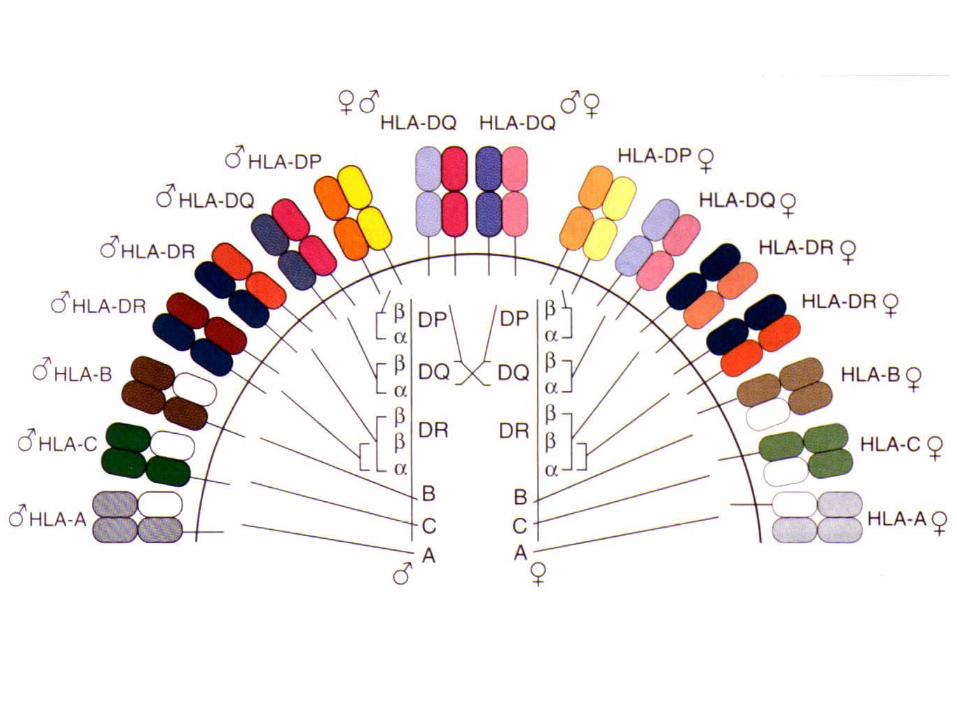
Human Leukocyte Antigens (HLA):
Are a group of proteins encoded within the major histocompatibility complex (MHC) on chromosome6 in humans.
Are the proteins that the body uses to identify self.
“Present” antigens for recognition by B- and T-cells.
Variation between individuals and between ethnicgroups is extensive.
Class I antigens are found on all nucleated cells.= A,B,C
Present endogenous antigens.
CD8+ T cells recognize antigen when presented by HLA Class I molecules
Human Leukocyte Antigens (HLA):
Class II antigens are primarily on antigen presentingcells (macrophages, dendritic cells and B cells).
= DR, DP, DQ
Present exogenous antigens
CD4+ T cells recognize antigen plus MHC Class II
Human Leukocyte Antigens (HLA):
Human Leukocyte Antigens (HLA):
Each HLA allele encodes a surface protein that hasmolecule has its own distinct requirements for peptide binding.
For example, HLA-A*0201 prefers to bind to leucine and valine, while HLA-A*0301 prefers to bind to leucine and lysine.
Therefore, a person’s constellation of HLA moleculeswill determine which portions of a pathogen will bepresented to the immune system.
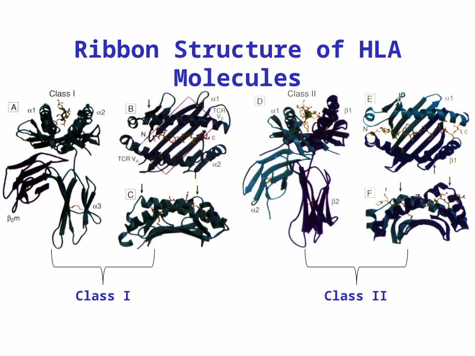
Ribbon Structure of HLA Molecules
Class I Class II
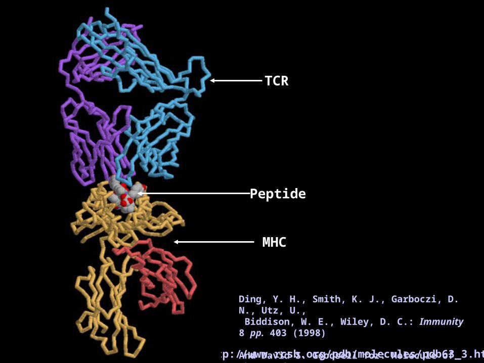
http://www.rcsb.org/pdb/molecules/pdb63_3.html
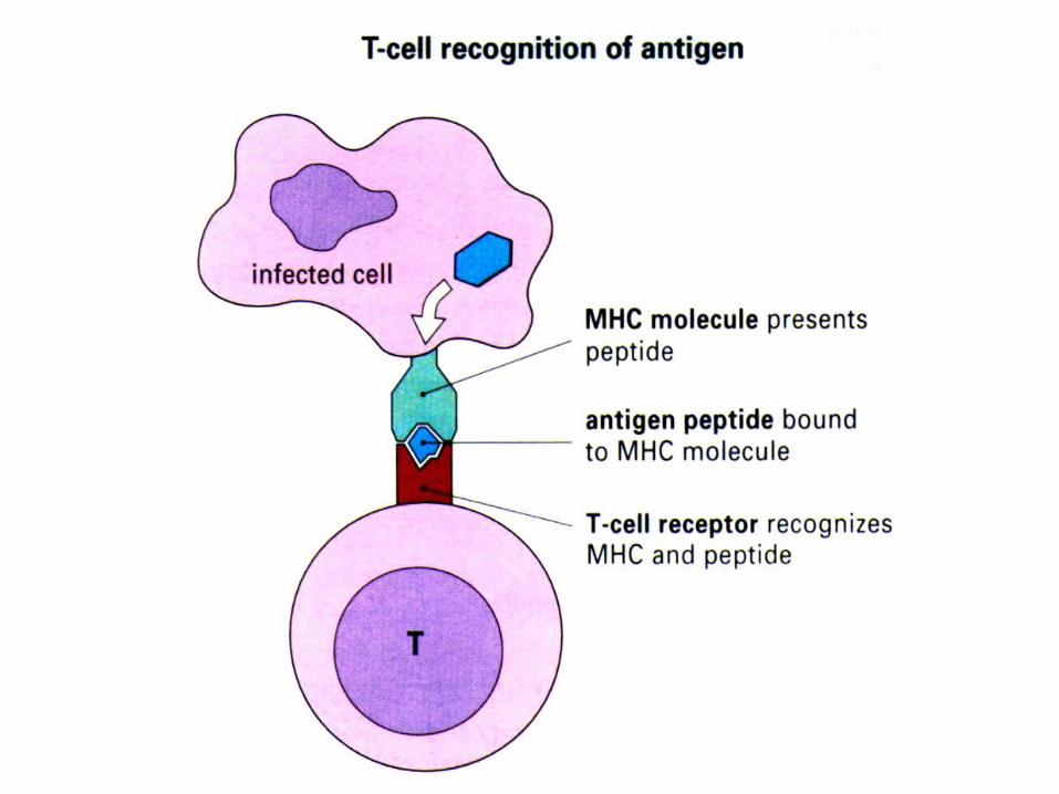
Peptide
TCR
MHC
Ding, Y. H., Smith, K. J., Garboczi, D. N., Utz, U., Biddison, W. E., Wiley, D. C.: Immunity 8 pp. 403 (1998)
And David S. Goodsell for: Molecule of the Month
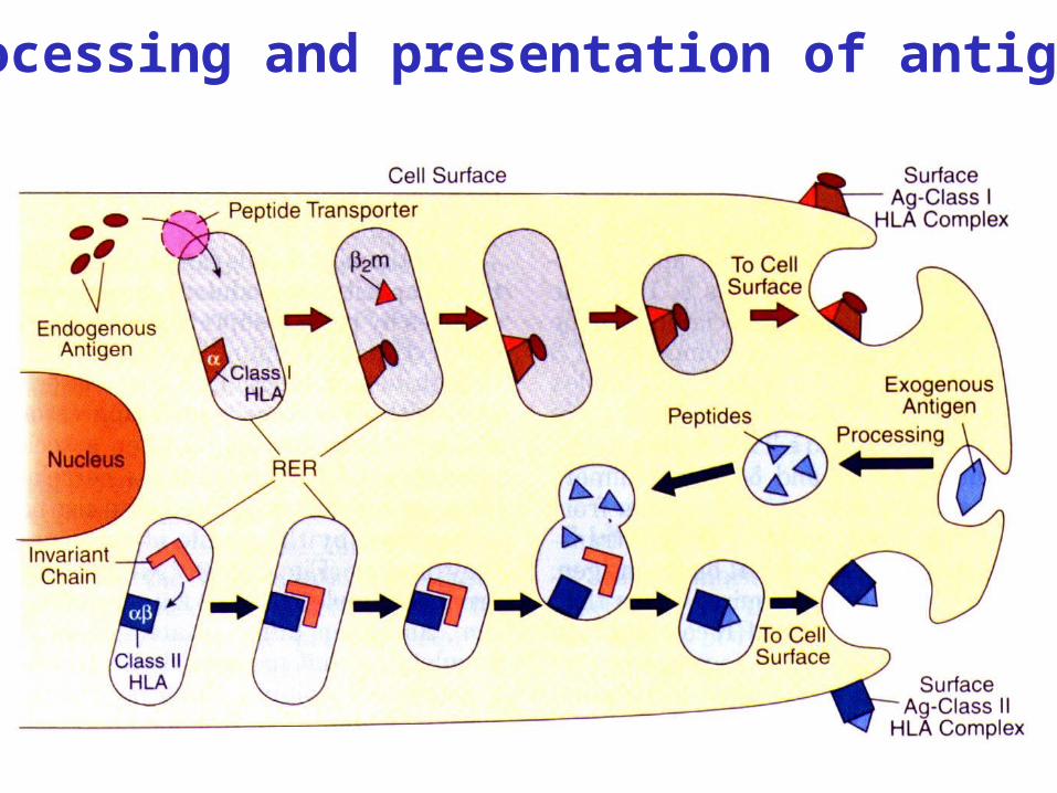
Processing and presentation of antigens
Diversity
Diversity of the adaptive immune response is due to the diversity of the T-cell and B-cell receptor complexes.
Comes at the level of the T-cell and B-cell population. The receptors expressed on each cellare specific for only one antigen, but vary from cell to cell.
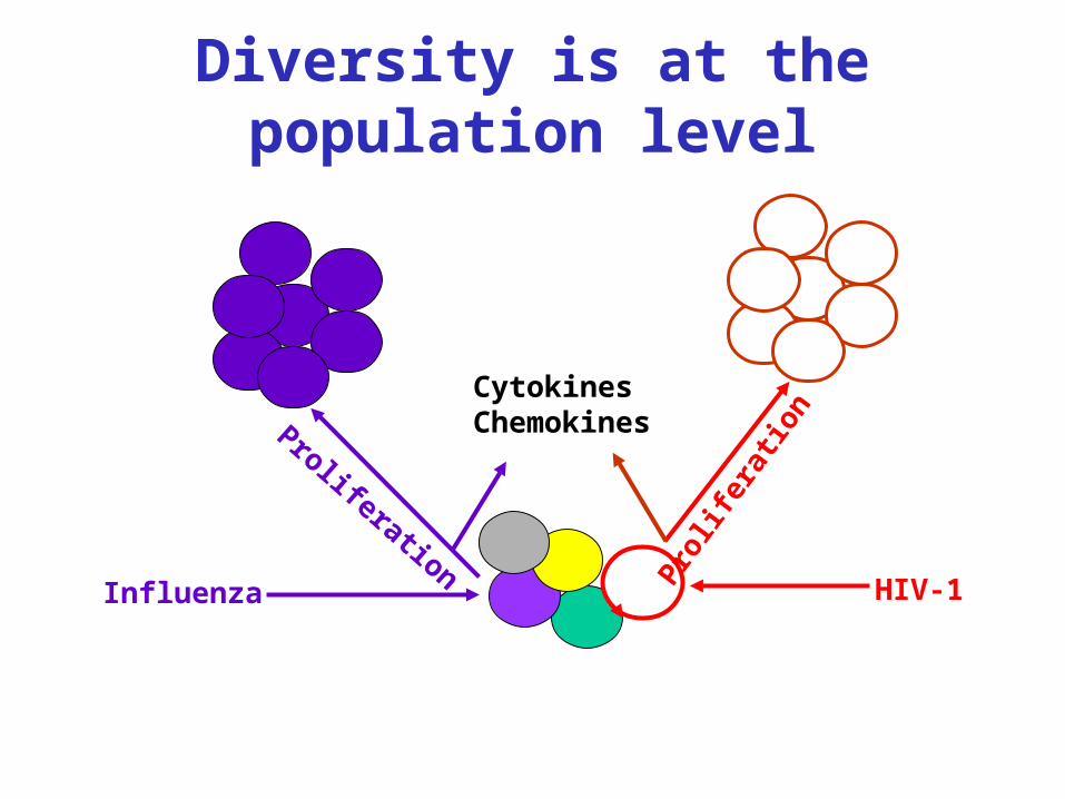
Diversity is at the population level
Prol
ifera
tion
HIV-1Influenza
Proliferation
CytokinesChemokines
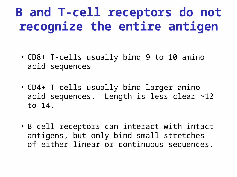
B and T-cell receptors do not recognize the entire antigen
• CD8+ T-cells usually bind 9 to 10 amino acid sequences
• CD4+ T-cells usually bind larger amino acid sequences. Length is less clear ~12 to 14.
• B-cell receptors can interact with intact antigens, but only bind small stretches of either linear or continuous sequences.
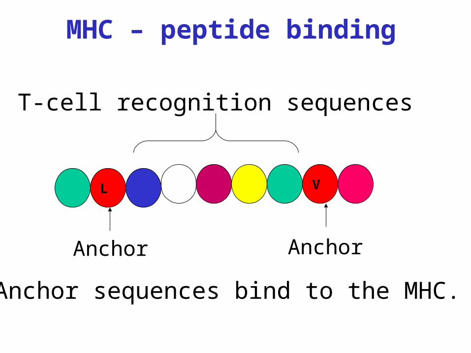
L V
AnchorAnchor
T-cell recognition sequences
Anchor sequences bind to the MHC.
MHC – peptide binding
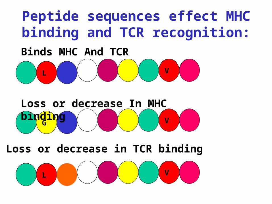
L V
G V
L V
Peptide sequences effect MHC binding and TCR recognition:
Binds MHC And TCR
Loss or decrease In MHC binding
Loss or decrease in TCR binding
Y
Antibody – Antigen Recognition
Antibodies recognize either linear epitopes orepitopes in secondary structures. A changeis the amino acid sequence or secondary structure can eliminate or diminish the antibodybinding. Y
No binding
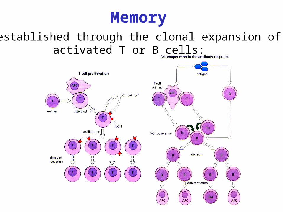
MemoryIs established through the clonal expansion of
activated T or B cells:
Self/Non-self Recognition:
Is achieved through the interaction of antigenreceptors, HLA, and antigen.
Responses to this complex are controlled throughA process of “education”.
Tolerance:The inability to react with self.
Autoimmunity:The state in which tolerance toself is lost.
Immune responses are most efficient in tissue parenchyma.
Lymph nodes and the spleen providearchitectural support for cell-to-cellinteractions, and serve as “filters” forfluids draining other tissues.
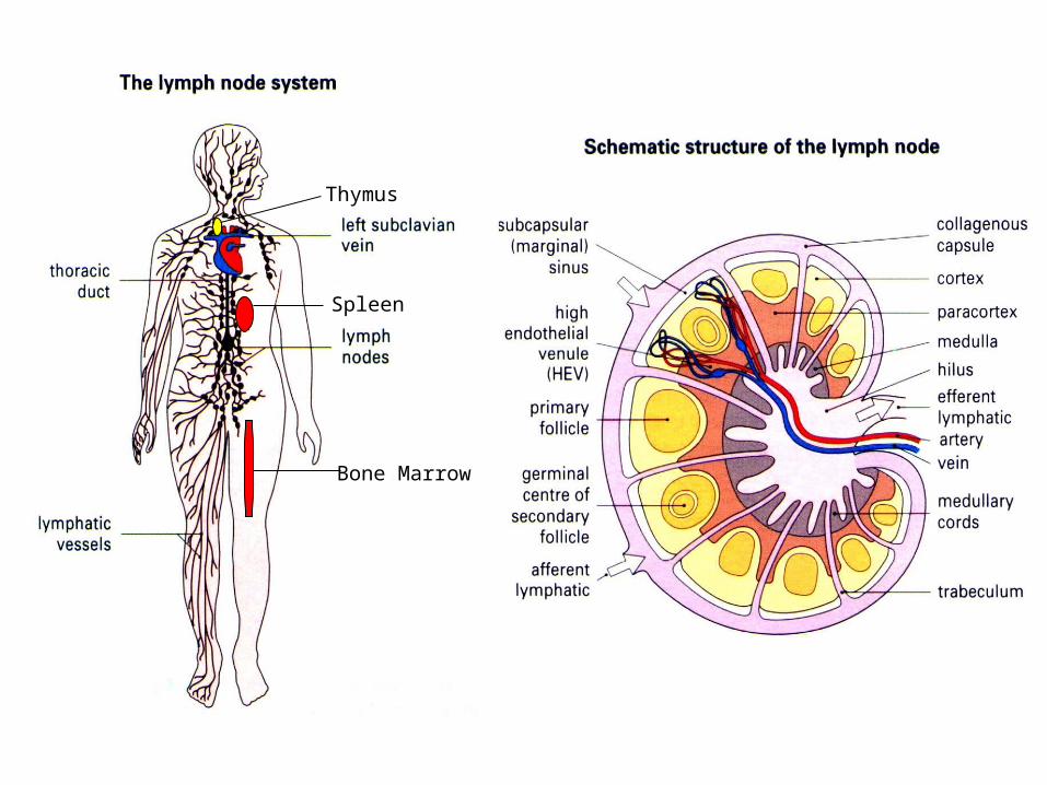
Spleen
Thymus
Bone Marrow
Immune Response To HIV
Infection
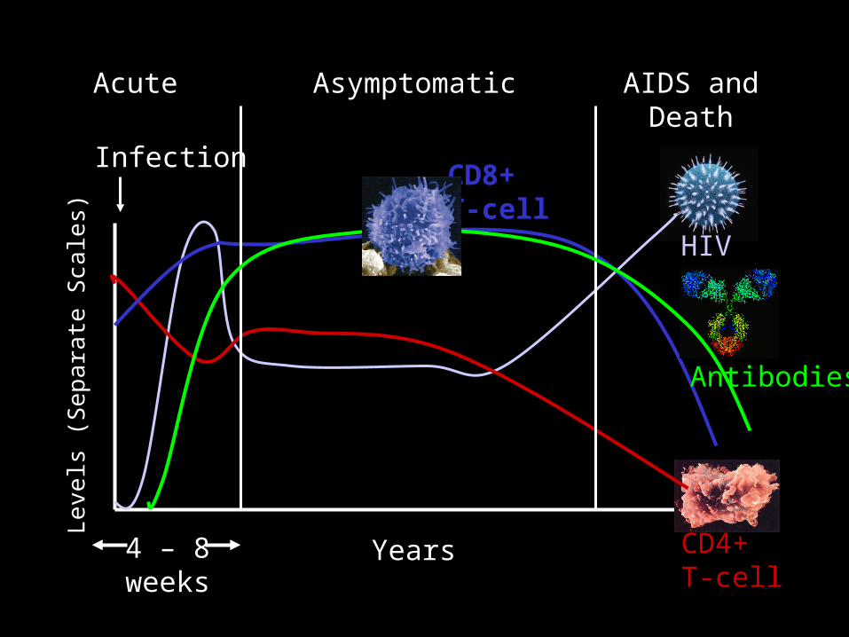
Infection
Levels
(S
ep
ara
te S
cale
s)
CD4+T-cell
HIV
CD8+T-cell
Antibodies
Years
AIDS and Death
Acute Asymptomatic
4 – 8 weeks
Immune Response to HIV
• CD4: Helper T-cell responses
• CTL: Cytotoxic T-cell responses
• B-cell: Antibody responses
• APC: Antigen Presenting Cells
CD4 Responses To HIV
CD4+ T-cell responses to antigens are usually indirectly measured by proliferation (cell division).• 3H-Thy uptake• CFSE
•Cytokine production is another measure of activation•Eliza•ELISpot
CD4+ T-cell responses are predictive of disease progression.
In most individuals, the following pattern is observed:
CD4+ T-cell responses decline at various stages: response to HIV and recall antigens (early) response to alloantigens (mid) response to mitogens (late) expression of IL-2 receptor (CD25)
In addition, there is aberrant cytokine production production of IFN-g, IL-2 production of IL-4, IL-10
CD4+ T-cell Response To HIV cont.
Mandell & Mildvan I AIDS
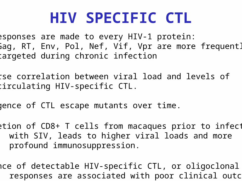
HIV SPECIFIC CTLCTL responses are made to every HIV-1 protein:
Gag, RT, Env, Pol, Nef, Vif, Vpr are more frequently targeted during chronic infection
•Inverse correlation between viral load and levels of circulating HIV-specific CTL.
•Emergence of CTL escape mutants over time.
•Depletion of CD8+ T cells from macaques prior to infectionwith SIV, leads to higher viral loads and more profound immunosuppression.
•Absence of detectable HIV-specific CTL, or oligoclonal CTL responses are associated with poor clinical outcome.
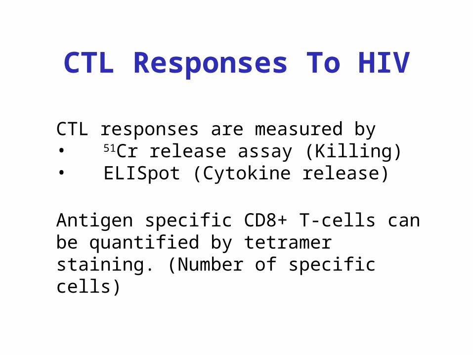
CTL Responses To HIV
CTL responses are measured by• 51Cr release assay (Killing)• ELISpot (Cytokine release)
Antigen specific CD8+ T-cells can be quantified by tetramer staining. (Number of specific cells)
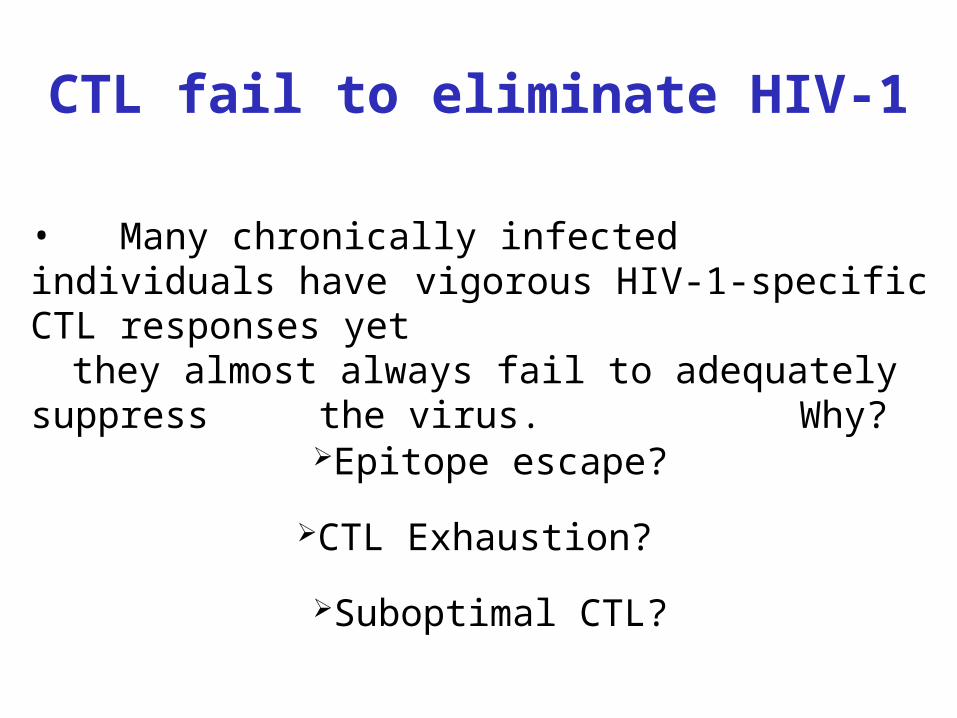
CTL fail to eliminate HIV-1
• Many chronically infected individuals have vigorous HIV-1-specific CTL responses yet
they almost always fail to adequately suppress the virus. Why?
Epitope escape?
CTL Exhaustion?
Suboptimal CTL?
0
0.5
1
1.5
2
01/85 01/87 01/89 01/91 01/93 01/95
Date
% P
osi
tive
CD
8 T
cel
ls
0
0.5
1
Pro
po
rtio
n o
f S
LY
NT
VA
TL
GAG tetramer
Gamma-INF
SLYNTVATL
Donor A: CD8 response to SL9
Antibody Responses
General Properties of Anti-viral Antibodies
•Can be generated to any accessible portion of the virus.
•Effective in blocking entry (neutralizing) if directed to viral receptors such as gp120 of HIV.
•Can block fusion(neutralizing) if antibody (Ab) binds to fusion protein such as gp41 of HIV.
•Can effect clearance of virus if it binds the virus and then binds Fc receptors on monocytes and macrophages.
•Can also bind complement and kill enveloped viruses.
•Most effective if they are present at the site of viral entry.
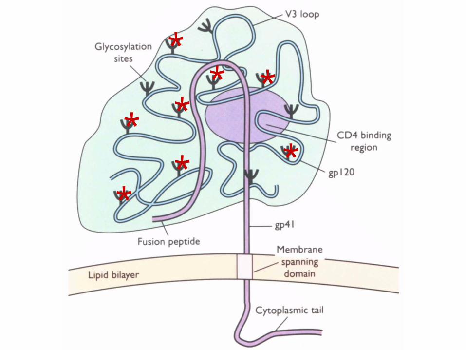
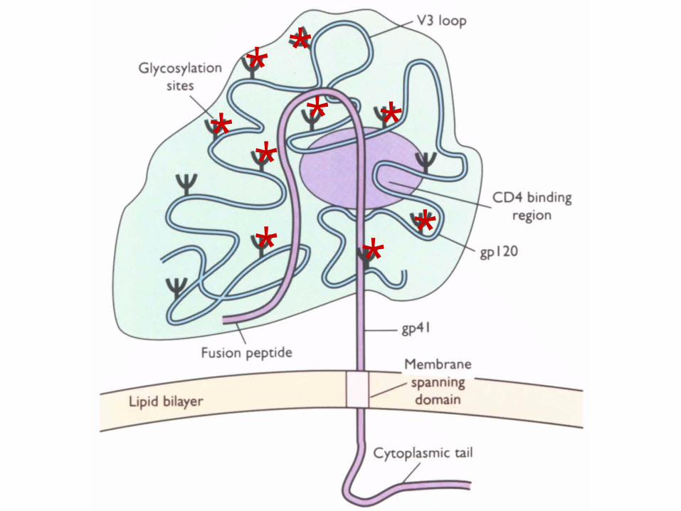
Gp120 and Gp41-mediated fusion
CD4 binding site is devoid of glycosylation and relatively conserved between isolates but is masked by V1V2 loops and is in a depression which is too small for good antibody binding.
Gp120 is highly glycosylated, meaning it has sugar molecules over much of its surface. Because many human proteins are glycosylated, humans rarely make antibody responses to glycoslyated portions of proteins.
Gp120 is presented as a trimer which protects some of the potential antibody binding sites.
Neutralizing antibody responses to HIV are difficult to generate
because:
CD4bs
V1V2 V3
V4
V5Gp41C NInner Outer
CD4bs
Bridging Sheet
Glycosylation:Silent face
Non-nuetralizingface
Trimerization
Neutralizing face
2G12
Co-R bs
Coreceptor bs
CD4bs
Bridging Sheet
C N
Changes in gp120 glycosylation allow HIV escape from Nab responses
Richman et al. PNAS 2003 vol. 100:4149
**
*
**
**
*
*
**
*
* **
*
*
HIV and APC’s
•APC’s may exhibit altered:chemotaxis IL-1 productionantigen presentationoxidative burst responseantimycobacterial activity
•Antigen presenting cells can act astrojan horses.
Dendritic Cells and DC-SIGN
DC-Specific, ICAM-3 Grabbing, Nonintegrin.
Interaction of DC-SIGN with ICAM-3 establishes the initial contact of the DC with a resting T-cell.
This is important because of the low number (100-1000copies/cell) of MHC-peptide ligands on the DC. This enhanced binding allows the T-cell to scan the surface of the DC.
DC-SIGN also binds the glycan-rich HIV-1 envelope in theabsence of CD4.
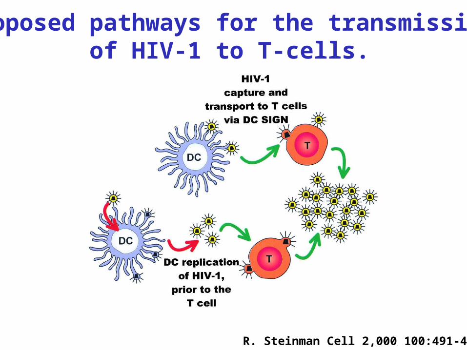
R. Steinman Cell 2,000 100:491-494
Proposed pathways for the transmissionof HIV-1 to T-cells.
Why does the immune response fail
to clear HIV?•HIV integrates into the host genome.
Therefore, to eliminate HIV, infected cells must be killed.
•Host factors can paradoxically enhanceHIV replication. Therefore, by responding to HIV, CD4+ T-cells can be destroyed.
Why does the immune response fail
to clear HIV?
•HIV can mutate and escape immune mediated opposition.
•Suboptimal CTL responses can be elicited.
• Sugar coating (glycosylation) and folding of gp120 protects against Ab recognition.
• Critical binding sites on gp41 arerevealed for only a short period of time.
Why does the immune response fail
to clear HIV?
Why does the immune response fail
to clear HIV?•APC’s may exhibit altered functionsdiminishing their ability to elicit immune responses.
•Antigen presenting cells can act astrojan horses, spreading HIV to CD4+T-cells as they begin to respond to antigen.
Why does the immune response fail to clear HIV?
Role of viral genes:
Tat: Extracellular Tat stimulates CD4+ andCD8+ T-cells.
Nef: Intracellular Nef appears to activate cells to promote viral replication. Affecton cellular function?
Intracellular Nef downregulates CD4 and MHC class I molecules. In vivo significance?