Embed Size (px)
Citation preview

Cavalier-Smith (1987) created the phylum Radiozoa toinclude the marine zooplankton Acantharia, Phaeodariaand Radiolaria, united by the presence of a central capsule. Only the Radiolaria including the siliceousPolycystina (which includes the orders Spumellariaand Nassellaria) and the mixed silica–organic matterPhaeodaria are preserved in the fossil record. TheAcantharia have a skeleton of strontium sulphate (i.e. celestine SrSO4). The radiolarians range from theCambrian and have a virtually global, geographicaldistribution and a depth range from the photic zonedown to the abyssal plains. Radiolarians are most usefulfor biostratigraphy of Mesozoic and Cenozoic deep seasediments and as palaeo-oceanographical indicators.
Heliozoa are free-floating protists with roughlyspherical shells and thread-like pseudopodia thatextend radially over a delicate silica endoskeleton.Fossil heliozoans occur as scales or spines, less than500 µm in size. They are found in marine or freshwaterhabitats from the Pleistocene to Recent.
Phylum Radiozoa
The living radiolarian
The individual single-celled radiolarians averagebetween 50 and 200 µm in diameter, with colonialassociations extending to metres in length. The cyto-plasm of each cell is divided into an outer ectoplasm(extracapsulum) and an inner endoplasm (intracap-sulum), separated by a perforate organic membranecalled the central capsule (Fig. 16.1a), a feature that is unique to the Radiolaria. The nucleus or nuclei inmultinucleate species are found within the endoplasm.
Radiating outwards from the central capsule are thepseudopodia, either as thread-like filopodia or asaxopodia, which have a central rod of fibres for rigid-ity. The ectoplasm typically contains a zone of frothy,gelatinous bubbles, collectively termed the calymmaand a swarm of yellow symbiotic algae called zooxan-thellae. The calymma in some spumellarian Radiolariacan be so extensive as to obscure the skeleton.
A mineralized skeleton is usually present within thecell and comprises, in the simplest forms, either radialor tangential elements, or both. The radial elementsconsist of loose spicules, external spines or internalbars. They may be hollow or solid and serve mainly tosupport the axopodia. The tangential elements, wherepresent, generally form a porous lattice shell of veryvariable morphology, such as spheres, spindles andcones (Fig. 16.1b,c). Often there is an arrangement ofconcentric or overlapping lattice shells.
Skeletal composition differs within the Radiozoa,being of strontium sulphate (i.e. celestine, SrSO4) in the class Acantharia, opaline silica in the classPolycystina (orders Spumellaria and Nassellaria) andorganic with up to 20% opaline silica in the orderPhaeodaria. Radiolarians are able to repair broken elements and grow by adding to their skeletons. Theabsence of gradational forms between adults and juven-iles in plankton samples indicates this process is not asimple addition of material alone.
Radiolarians reproduce by fission and possibly sex-ually by the release of flagellated cells, called swarmers.In the family Collosphaeridae (Spumellaria), the cellsremain attached to form colonies. Individual radio-larians are thought to live no longer than 1 month. Asmarine zooplankton, radiolarians occupy a wide rangeof trophic types including bacterivores, detritivores,
CHAPTER 16
Radiozoa (Acantharia, Phaeodaria andRadiolaria) and Heliozoa
188
MICC16 26/09/2005 12:21 PM Page 188

Chapter 16: Radiozoa (Acantharia, Phaeodaria and Radiolaria) and Heliozoa 189
omnivores and osmotrophs (Casey 1993, in Lipps1993, pp. 249–285). With increasing size there is atrend from herbivory to omnivory (Anderson 1996).Many species use their sticky radiating axopodia totrap and paralyse passing organisms (e.g. phytoplank-ton and bacteria). Food particles are digested in vac-uoles within the calymma and nutrients are passedthrough the perforate central capsule to the endo-plasm. Those living in the photic zone may also con-tain zooxanthellae and can survive by symbiosis.
Buoyancy is maintained in several ways. The specificgravity is lowered by the accumulation of fat globulesor gas-filled vacuoles. Frictional resistance is increasedby the development of long rigid axopods borne onskeletal spines. Holes in the skeleton allow the cyto-plasm to pass through and also reduce weight. Thespherical and discoidal skeletal shapes are furtherdevices to reduce sinking, as in foraminifera, coccol-ithophores and diatoms. The turret- and bell-likeskeletons of the Nassellaria appear to be adaptions forareas of ascending water currents, the mouth beingheld downwards and the axis held vertically, much asin silicoflagellates.
Radiolarian distribution and ecology
Living radiolarians prefer oceanic conditions, espe-cially just seaward of the continental slope, in regionswhere divergent surface currents bring up nutrientsfrom the depths and planktonic food is plentiful.Although most diverse and abundant at equatorial latitudes, where they may reach numbers of up to82,000 m−3 water, they also thrive with diatoms in thesubpolar seas (Fig. 16.2). Radiolarians tend to bloomseasonally in response to changes in food and silicacontent, currents and water masses.
Different trophic types live in different parts of theocean; herbivores are restricted to the upper 200 m ofthe ocean whereas symbiotrophs are found to dom-inate the subtropical gyres and warm shelf areas.Detritivores and bacterivores dominate high latitudeshallow subsurface waters. Different species may alsooccur in vertically stratified assemblages, each approx-imately corresponding to discrete water masses withcertain physical and chemical characteristics (Fig. 16.3).Assemblage boundaries at 50, 200, 400, 1000 and 4000 m
Fig. 16.1 (a) Cross-section through a naked radiolarian cell(Thalassicola). (b) Cross-section through a spumellarian showing the relationship of the nucleus, endoplasm andectoplasm to three concentric lattice shells and radial spines. (c) SEM photomicrograph of a Neogene spumellarianradiolarian. ((b) After Westphal 1976.)
MICC16 26/09/2005 12:21 PM Page 189

190 Part 4: Inorganic-walled microfossils
are reported, though these depths vary with latitude.Acantharia and Spumellaria generally dominate thephotic zone (<200 m) and Nassellaria and Phaeodariadominate in depths below 2000 m. Some radiolarianspecies occupy a wide depth range, with juveniles andsmall adults thriving at the shallower end of the rangeand the larger adults living in the deeper waters.
Radiolaria zoogeography is directly related to oceancirculation and water mass distribution patterns. Theboundaries of radiolarian provinces thus correspondto major current convergences in the subtropical andtropical regions and have been used to plot the chang-
ing history of currents and water masses through theCenozoic (see Casey et al. 1983; Casey 1989) and henceas a proxy for palaeotemperature. Gradients of tem-perature, silica and other macronutrient concentra-tions probably influence the latitudinal abundance ofliving Radiolaria (Abelmann & Gowing 1996). In themodern oceans eight shallow-water and seven deep-water provinces have been defined (Casey et al. 1982;Casey 1989). Of these the Subtropical Anticyclonic GyreProvince has the highest radiolarian diversity, speci-men density and species endemism, probably reflect-ing to the presence of algal symbionts in the majorityof taxa inhabiting this province. Deep-water provincesappear to be related to water masses found at depth. Aswith the Foraminiferida, some cold-water species thatlive near the surface in subpolar waters occur at greaterdepths near the Equator (Fig. 16.3).
Radiolarians and sedimentology
Both the SrSO4 skeletons of the Acantharia and theweakly silicified tubular skeletons of the Phaeodariaare very prone to dissolution in the water column afterdeath and on the deep sea floor, and they are thereforerare as fossils. Conversely, the solid opaline skeletonsof the Spumellaria and Nassellaria tend to be more
Fig. 16.2 Abundance of Radiolaria insurface sediments of the South Atlantic.(Modified from Goll & Bjørklund 1974.)
Fig. 16.3 Latitudinal and vertical assemblages of polycystineradiolarians in the pacific along 170° W transect. (Modifiedfrom Casey, in Funnell & Reidel 1971, figure 7.1.)
MICC16 26/09/2005 12:21 PM Page 190

Chapter 16: Radiozoa (Acantharia, Phaeodaria and Radiolaria) and Heliozoa 191
resistant even than in silicoflagellates and diatoms,although all are susceptible to dissolution because seawater is very undersaturated relative to silica. Belowthe calcium carbonate compensation depth (usually3000–5000 m) nearly all CaCO3 enters into solution sothat siliceous radiolarian or diatomaceous oozes tendto accumulate. Radiolarian oozes are mostly found inthe equatorial Pacific below zones of high productivityat 3000–4000 m depth and can contain as many as100,000 skeletons per gram of sediment, but they mayalso occur abundantly in marine diatomaceous oozesor in Globigerina and coccolith oozes.
With increasing depth and dissolution, the abund-ance of Radiolaria in deep sea sediments decreases,through a progressive loss of the more delicate skele-tons (Fig. 16.4). If the settling or sedimentation rate is slow, the chances for solution of skeletons will alsoincrease, eventually lending a bias to the compositionof fossil assemblages. Consequently, red muds of theabyssal plains mainly consist of volcanic and meteoriticdebris barren of all but the most resistant parts of radio-larian skeletons and fish debris. The best preserved ofradiolarians are those that have sunk rapidly to theocean floor, usually within the faecal pellets of copepodcrustaceans (Casey 1977, in Swain 1977, p. 542).
Fossil radiolarians are frequently found in cherthorizons. Nodular cherts found interbedded with cal-careous pelagic sediments of Mesozoic and Cenozoicage are probably deep-water deposits formed belowbelts of upwelling plankton-rich waters, as at the present day (see Casey 1989). The massive and ribbon-bedded cherts (radiolarites) found in Palaeozoic suc-cessions are interbedded with black shales and basicvolcanic rocks in settings that have been interpreted as ancient oceanic crust. Ancient radiolarite depositshave been compared to those in the modern Owen andSomalia basins, both narrow, partially restricted basinswith active monsoonal upwelling. However, the bestradiolarian assemblages of Palaeozoic age come fromcontinental shelf facies (Holdsworth 1977, in Swain1977, pp. 167–184) and Bogdanov & Vishnevskaya(1992) proposed a shift in radiolarian habitats fromthe shallow, carbonate shelves of the Palaeozoic to theexclusively oceanic realm today.
Like other microfossils, Radiolaria are very prone toexhumation and reburial in younger sediments. These
Fig. 16.4 The vertical distribution of living and deadradiolarians through the water column at a station in the central Pacific. (After Petrushevskaya, in Funnell & Reidel 1971,figure 21.4.)
MICC16 26/09/2005 12:21 PM Page 191

192 Part 4: Inorganic-walled microfossils
and other aspects of radiolarians in sediments arereviewed more fully in Anderson (1983), Sanfilippo et al. (in Bolli et al. 1985) and Casey (1993, in Lipps1993, pp. 249–285).
Classification of radiolarians
The classification of the radiolarians is in a state of flux.Living radiolarians are subdivided on morphology ofthe unmineralized (and therefore unfossilized) centralcapsule as well as on the composition and geometry of the skeleton. Fossil Radiolaria are classified usingskeletal morphology. Separate schemes have beendevised for the taxa present in the different eras and to date little attempt has been made to rationalize themany schemes. The scheme followed herein (Box 16.1)follows that proposed by Hart & Williams (1993 inBenton 1993, pp. 66–69) with modifications as recom-mended by Cavalier-Smith (1993). Suprageneric cat-egories are probably best regarded as informal.
Kingdom PROTOZA
Parvkingdom ACTINOPODA
Phylum RADIOZOA
Subphylum RADIOLARIA
Class POLYCYSTINEA
Polycystine Radiolaria are generally spherical. ThePalaeozoic spherical Radiolaria (order Archaeo-spicularia) may not be closely related to the youngerSpumellaria and comprise several but as yet little-studied groups (see Holdsworth 1977, in Swain 1977,pp. 167–184). For example, Entactinosphaera (U.Dev.-Carb., Fig. 16.5a) has a six-rayed internal spiculesupporting two or more concentric lattice shells.
Order Spumellaria comprises skeletons in the formof a spherical or discoidal lattice, with several concen-tric shells bearing radial spines and supporting bars. InThalassicola (Rec., Fig. 16.1a) a skeleton is either lack-ing or consists merely of isolated spicules. Actinomma(Rec., Fig. 16.5c) has three concentric, spherical latticeshells with large and small radial spines and bars.Dictyastrum (Jur.-Rec., Fig. 16.5d) has a flattenedskeleton with three concentric chambers leading tothree radiating chambered arms. Related genera alsohave radial beams and subdivide the chambers intochamberlets. Albaillella (Carb., Fig. 16.5b) belongs to a
group of radiolarians with bilaterally symmetrical, triangulate skeletons that flourished in Silurian toCarboniferous times. Their systematic position is un-certain, with Holdsworth (1977, in Swain 1977, p. 168)placing them in a separate suborder Albaillellaria; butthey have also been compared with the later Nassellaria.
Order Nassellaria have skeletons usually com-prising a primary spicule, a ring or a lattice shell. Theprimary spicule comprises three, four, six or more rays that may be simple, branched or anastomosing. In Campylacantha (Rec., Fig. 16.6a), for example, the skeleton comprises a three-rayed spicule, each raybearing similar but smaller branches. Evolutionarymodifications of these rays led in certain stocks to asagittal ring that may bear spines, sometimes in theform of tripod-like basal feet. In Acanthocircus (Cret.,Fig. 16.6b) the ring bears three simple spines, two ofthem projecting inwards.
The phylogeny of taxa with more elaborate latticeshells can be traced from the study of the form of theprimary spicule or ring elements (see Campbell 1954).The lattice may be spherical, discoidal, ellipsoidal orfusiform and constructed of successive chambers (seg-ments) that partially enclose earlier ones. The skele-tons differ from those of Acantharia and Spumellariain having a wide aperture (basal shell mouth) at theterminal pole. This may be open or closed by a lattice.The initial chamber (cephalis) is closed and containsthe primary spicule elements referred to above. Thecephalis may also bear diagnostic features such as anapical horn. The second chamber is called the thoraxand the third the abdomen with, sometimes, manymore post-abdominal segments, each separated by a ‘joint’ or constriction. Bathropyramis (Cret.-Rec.,Fig. 16.6c) has a conical lattice with rectangular poresand about nine radial spines around the open basalshell mouth. Podocyrtis (Cret.-Rec., Fig. 16.6d) has a conical, segmented skeleton with an apical horn anda tripod of three radial spines around the open mouth.Successive chambers of the fusiform Cyrtocapsa (Jur.-Rec., Fig. 16.6e) form prominent segments and themouth is closed by a lattice.
Class Phaeodaria have skeletons that comprise 95%organic and 5% opaline silica constructed in the formof a lattice of hollow or solid elements, often with complex dendritic spines called styles. The central
MICC16 26/09/2005 12:21 PM Page 192

Box 16.1 The classification of Radiolaria with diagrammatic representatives of a typicalform (after Casey, in Lipps 1993, figure 13.5)
AAccttiinnoommmmiiddaaee Spongy
cylindrical forms.
Cosmopolitan. ?Trias.-Rec.
CCooccccooddiisscciiddaaee Lens shaped with
a latticed centre and spongy
chambered girdle or arms.
Meso.-Eoc.
CCoolllloosspphhaaeerriiddaaee Single spheres,
usually more interpore area than
pore area; weakly developed
external projections. Commonly
colonial and possessing
symbionts. Warm water in
oligotrophic anticyclonic gyres,
Mioc.-Rec.
EEnnttaaccttiinniiiiddaaee Spherical or
ellipsoidal; latticed wall structure,
bars running to the centre of the
skeleton. L. Sil.-Carb.
HHaaggiiaassttrriiiiddaaee Spongy
‘rectangular’ mesh and two
to four large radial arms
Palaeo.-Meso./Rec.
LLiitthheelliiiiddaaee Coiled and latticed
forms; tightly coiled morphotypes
are cold water and loosely coiled
morphotypes warm water.
Cosmopolitan Carb.-Rec.
OOrroosspphhaaeerriiddaaee Spherical or
cup shapes with coarse
polygonal lattice. Usually large
specimens. Eoc.-Rec.
Actinommidium
Lithocyclia
Collospaera
PPhhaaccooddiisscciiddaaee Lens or biconvex
disc-shaped forms. Warm water,
?Palaeo./Meso.-Rec.
PPsseeuuddooaauulloopphhaacciiddaaee Lens-like
commonly triangular, usually
with a few marginal spines.
Cret. (Val.-Mass.)
PPyylloonniiiiddaaee Skeleton comprises
an ellipsoid of girdles and holes
(gates). Warm water, Eoc.-Rec.
SSppoonnggooddiisscciiddaaee Polyphyletic
grouping of discoid, spongy
forms. Dev.-Rec.
SSppoonngguurriiddaaee A polyphyletic group
containing many subgroups of
discoidal and ‘spongy’ forms.
Cosmopolitan. Meso.-Rec.
TThhoolloonniiiiddaaee Outer shell elliptical
with bulb-like extensions. Deep
cold water. Mioc.-Rec.
Class POLYCYSTINA
Order ARCHAEOSPICULARIA: Includes Lower Palaeozoic Radiolaria previously included in the Spumullaria and Collodaria
characterized by spherical forms with a globular shell of several spicules. Members of this order are some of the earliest radi-
olaria and may have provided the ancestors to the Spumullaria and the Albiaillellidae.
Order SPUMELLARIA
MICC16 26/09/2005 12:21 PM Page 193
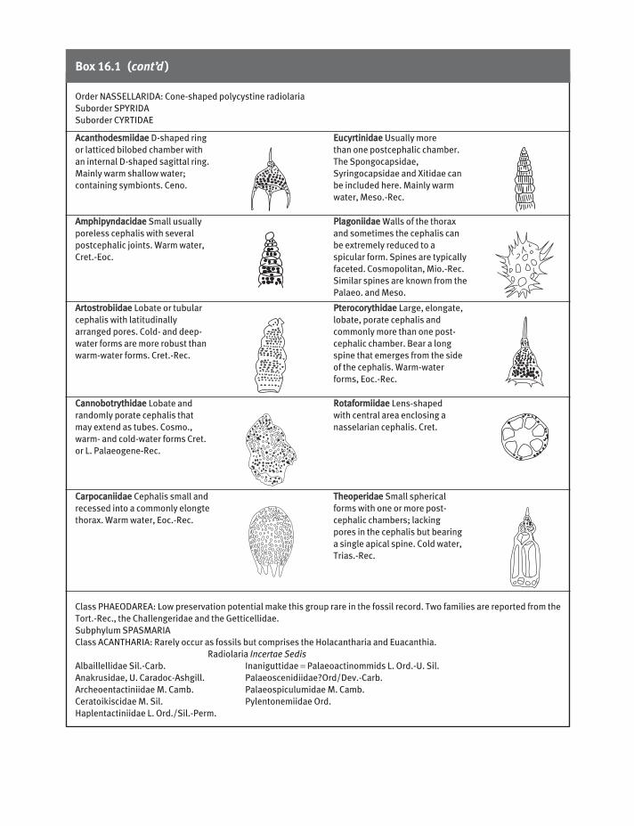
Box 16.1 (cont’d)
AAccaanntthhooddeessmmiiiiddaaee D-shaped ring
or latticed bilobed chamber with
an internal D-shaped sagittal ring.
Mainly warm shallow water;
containing symbionts. Ceno.
AAmmpphhiippyynnddaacciiddaaee Small usually
poreless cephalis with several
postcephalic joints. Warm water,
Cret.-Eoc.
AArrttoossttrroobbiiiiddaaee Lobate or tubular
cephalis with latitudinally
arranged pores. Cold- and deep-
water forms are more robust than
warm-water forms. Cret.-Rec.
CCaannnnoobboottrryytthhiiddaaee Lobate and
randomly porate cephalis that
may extend as tubes. Cosmo.,
warm- and cold-water forms Cret.
or L. Palaeogene-Rec.
CCaarrppooccaanniiiiddaaee Cephalis small and
recessed into a commonly elongte
thorax. Warm water, Eoc.-Rec.
EEuuccyyrrttiinniiddaaee Usually more
than one postcephalic chamber.
The Spongocapsidae,
Syringocapsidae and Xitidae can
be included here. Mainly warm
water, Meso.-Rec.
PPllaaggoonniiiiddaaee Walls of the thorax
and sometimes the cephalis can
be extremely reduced to a
spicular form. Spines are typically
faceted. Cosmopolitan, Mio.-Rec.
Similar spines are known from the
Palaeo. and Meso.
PPtteerrooccoorryytthhiiddaaee Large, elongate,
lobate, porate cephalis and
commonly more than one post-
cephalic chamber. Bear a long
spine that emerges from the side
of the cephalis. Warm-water
forms, Eoc.-Rec.
RRoottaaffoorrmmiiiiddaaee Lens-shaped
with central area enclosing a
nasselarian cephalis. Cret.
TThheeooppeerriiddaaee Small spherical
forms with one or more post-
cephalic chambers; lacking
pores in the cephalis but bearing
a single apical spine. Cold water,
Trias.-Rec.
Order NASSELLARIDA: Cone-shaped polycystine radiolaria
Suborder SPYRIDA
Suborder CYRTIDAE
Class PHAEODAREA: Low preservation potential make this group rare in the fossil record. Two families are reported from the
Tort.-Rec., the Challengeridae and the Getticellidae.
Subphylum SPASMARIA
Class ACANTHARIA: Rarely occur as fossils but comprises the Holacantharia and Euacanthia.
Radiolaria Incertae Sedis
Albaillellidae Sil.-Carb. Inaniguttidae = Palaeoactinommids L. Ord.-U. Sil.
Anakrusidae, U. Caradoc-Ashgill. Palaeoscenidiidae?Ord/Dev.-Carb.
Archeoentactiniidae M. Camb. Palaeospiculumidae M. Camb.
Ceratoikiscidae M. Sil. Pylentonemiidae Ord.
Haplentactiniidae L. Ord./Sil.-Perm.
MICC16 26/09/2005 12:21 PM Page 194

Fig. 16.5 Polycystine Radiolaria. (a) Entactinosphaera ×195. (b) Albaillella (scale unknown). (c) Actinomma (scale unknown). (d) Ditryastrum × 66. ((a) After Foreman 1963; (b) after Holdsworth 1969; (c) after Campbell 1954; (d) after Campbell 1954 fromHaeckel ((c), (d) from the Treatise on Invertebrate Paleontology, courtesy of and © 1954, Part C, The Geological Society of America andThe University of Kansas).)
Fig. 16.6 Nasellarian and phaeodarian Radiolaria. (a) Campylacantha ×200. (b) Acanthocircus ×40. (c) Bathropyramis ×133. (d) Podocyrtis ×100. (e) Cyrtocapsa ×200. (f) Challengerianum ×187. ((a) After Campbell 1954 from Jorgensen; (b) after Campbell 1954from Squinabol; (c), (d), (e) after Campbell 1954 from Haeckel; (f) redrawn after Reshetnjak, in Funnell & Riedel 1971, figure 24.19b.((a), (c)–(e) from the Treatise on Invertebrate Paleontology, courtesy of and © 1954, Part C, The Geological Society of America and The University of Kansas).)
MICC16 26/09/2005 12:21 PM Page 195

196 Part 4: Inorganic-walled microfossils
Acanthometra (Rec., Fig. 16.7b) has thin radial spinesembedded in cytoplasm that invariably disarticulateafter death. Belonaspis (Rec., Fig. 16.7c) has an ellip-soidal lattice formed by fused spine branches (orapophyses) with 20 projecting radial spines.
General history of radiolarians
Radiolaria first appeared in the Cambrian and wereone of the first groups to change from a benthic to free-floating mode of life (Knoll & Lipps 1993, in Lipps1993, pp. 19–29). The earliest well-preserved examplesare spicules, cones and the closed spheres of sphericalArcheoentactiniidae and spicules of the Palaeospicul-umidae from the Middle Cambrian of the GeorginaBasin, Australia (Won & Below 1999) and the UpperCambrian and Lower Ordovician of Kazakhstan(Nazarov 1975). Cold- and warm-water types can bedistinguished in the Cambrian and a deep-water radi-olarian fauna was present by the Silurian. A variety ofdistinct spumellarians flourished in the Palaeozoic,joined by the first deep, cold-water albaillellarians inthe Late Devonian to Early Permian (Holdsworth1977, in Swain 1977, pp. 167–184).
The dramatic reduction of cold- and warm-waterspecies during the Permian and Triassic periods(Tappan & Loeblich 1973; Kozur 1998) has been attri-buted to the tectonic closure of some Late Palaeozoicocean basins, the reorganization and reduction in thenumber of surface currents and eutrophication due toLate Permian glaciation (Hallam & Wignall 1997;Martin 1998). The first unequivocal nassellariansappeared in the Triassic. About half of the extantgroups of Radiolaria appeared in the Mesozoic. Theearliest unequivocal Phaeodaria are of Cretaceous age, with equivocal records from the Permian or evenolder.
From the Cretaceous Radiolaria had to share theirniches with the rapidly radiating planktonic forami-nifera (Anderson 1996). The fossil record suggests that,unlike diatoms and silicoflagellates, the Radiolaria didnot flourish in the cooler Cenozoic Era (Fig. 16.8), asthe equatorial belt in which they achieve their highestdiversity contracted steadily during this time. Throughthe Cenozoic, radiolarians also show a progressive
capsule also has a double wall rather than the singlewall found in the former groups, and a basal shellmouth as in the Nassellaria. Only the more robustshells are known as fossils, such as Challengerianum(Mioc.-Rec., Fig. 16.6f). This has an ovate shell with an apical horn, a marginal keel, an open basal shellmouth surrounded by oral teeth and a skeleton wallwith a fine hexagonal, diatom-like mesh.
Subphylum SPASMARIA
Class Acantharia
These have skeletons generated at the cell centre ratherthan peripherally as is usual in the other groups. Thisskeleton generally comprises 20 spines of SrSO4 joinedat one end (in the endoplasm) and arranged like the fourspokes of five wheels in different planes and of varyingdiameters (e.g. Zygacantha, ?Mioc.-Rec., Fig. 16.7a).
Fig. 16.7 Acantharian radiolarians. (a) Zygacantha skeleton×160. (b) Acanthometra cell with spicules ×71. (c) Belonaspisskeleton ×100. ((a) After Campbell 1954 from Popofsky; (b) redrawn after Westphal 1976; (c) after Campbell 1954 fromHaeckel ((a), (c) from the Treatise on Invertebrate Paleontology,courtesy of and © 1964, Part C, The Geological Society ofAmerica and The University of Kansas).)
MICC16 26/09/2005 12:21 PM Page 196

Chapter 16: Radiozoa (Acantharia, Phaeodaria and Radiolaria) and Heliozoa 197
diversity has been interpreted as an increase in com-petitive pressure for dissolved silica by the diatoms andsilicoflagellates, a pattern also seen during the coolersperiods of the Cenozoic (Harper & Knoll 1975). Hencemany living Nassellaria and Phaeodaria are delicatelyconstructed and do not occur as fossils. The apparentdrop in diversity may be misleading, with the datamerely recording a decrease in the preservation poten-tial of radiolarians, of oceanic environments, or both.
Fossil Acantharia have been reported fromPaleocene and younger strata. The last major radio-larian radiation occurred at the Paleocene–Neogeneboundary in response to the development of newintermediate and circumpolar water masses and theoligotrophic subtropical gyres.
Applications of radiolarians
Most of the studies of fossil Radiolaria emphasize theirvalue to biostratigraphical correlation of oceanic sedi-ments, particularly where the calcareous microfossilshave suffered dissolution. Sanfilippo et al. (in Bolli et al.1985) provides a review of Mesozoic and Cenozoicradiolarian biozonations, with tropical Cenozoic biozonations the best developed. The Late Paleoceneto Recent has been divided into 29 biozones that can berecognized around the world and have been relateddirectly to the well-dated magnetostratigraphy. Thecomplex nature of the radiolarian skeleton and analmost complete Mesozoic to Recent geological recordmakes this group ideal for charting microevolutionarychanges (see Moore 1972; Foreman 1975; Knoll &Johnson 1975).
Radiolaria have an increasing value as depth,palaeoclimate and palaeotemperature indicators andchanges in radiolarian provinciality through theCenozoic are highlighted (Casey et al. 1990). Theyhave also been used to indicate palaeogeographic andtectonic changes in ocean basins. For example, radio-larian stratigraphy gave early support to the hypothesisof sea-floor spreading (Riedel 1967), and the closure of the Panama isthmus about 3.5 Myr is reflected in changing radiolarian assemblages in the Atlantic(Casey & McMillen in Swain 1977, pp. 521–524). Theresistant nature of radiolarian chert to tectonism and
decrease in the amount of silica used to build the skele-ton, particularly in silica-depleted, warm surfacewaters (Casey et al. 1983).
The Eocene–Oligocene boundary is marked bygreatly enhanced siliceous ooze accumulation,Horizon Ac, comprising radiolarian-diatom depositsthat occur in a broad belt across the northern Atlantic,equatorial Pacific and Mediterranean region. Thisevent has been correlated with a large hiatus associatedwith volcanogenic deposits and vigorous deep-watercurrents and upwelling. Berger (1991) in his chert-climate hypothesis noted the Eocene ‘opal revolution’corresponded with a declining volcanic silica source,increased oceanic mixing and progressive oxygena-tion, colonization of upwelling zones by diatoms andthe more effective recirculation of biogenic silica.
A substantial change in the marine siliceous plank-ton occurred in the Early Oligocene, with a markeddecline in many thickly silicified radiolarians (e.g.Conley et al. 1994; Khokhlova 2000). This decline in
Fig. 16.8 Apparent changes in the species diversity ofpolycystine Radiolaria through time. (Based on Tappan &Loeblich 1973 with modifications after Vishnevskaya 1997.)
MICC16 26/09/2005 12:21 PM Page 197

198 Part 4: Inorganic-walled microfossils
diagenesis means Radiolaria are often the only commonfossils preserved in orogenic belts and within accretedterranes (e.g. Murchey 1984; De Wever et al. 1994;Nokleberg et al. 1994; Cordey 1998).
Phylum Heliozoa
The Heliozoa closely resemble the Radiolaria but theylack the distinctive central capsule membrane betweenectoplasm and endoplasm. Their skeletons may com-prise a spherical lattice of chitinous matter weaklyimpregnated with silica, or isolated siliceous spiculesand plates embedded in the mucilage near the outerectoplasm. A few can agglutinate a skeleton of sandgrains or diatom frustules or even survive without askeleton at all. These delicate structures tend to fallapart after death, thereby obscuring their heliozoanorigin. Heliozoans are, none the less, known as fossilsfrom a few Pleistocene lake sediments (Moore 1954).
Further reading
Anderson’s book on Radiolaria (1983) provides anexcellent review of the biology of living radiolariansplus other aspects on the research into Radiolaria up tothat time. Casey (in Lipps 1993, pp. 249–285) providesa good general review with sections on oceanographicapplications and biostratigraphy. Sanfilippo et al. (inBolli et al. 1985) provides a detailed review of radio-larian biostratigraphy plus many illustrations. Case studies of the application of Radiolaria in orogenicbelts can be found in a special issue of Palaeogeogra-phy, Palaeoclimatology, Palaeoecology 1996, 96, 1–161).Identification of specimens should be assisted by refer-ence to Foreman & Riedel (1972 to date). Racki &Cordey (2000) review radiolarian palaeoecology in thecontext of the evolution of the marine silica cycle.
Hints for collection and study
Fossil Radiolaria can be extracted from mudstones,shales and marls using methods A to E (see Appendix),from limestones using method F and from cherts using
method F using HF. The residues should then bewashed over a 125- and 68-µm sieve, dried and con-centrated with CCl4 (methods I and J) and viewed withreflected light (method O) or with well-condensedtransmitted light, as with diatoms (q.v.). Radiolariancherts can be studied in relatively thick petrographicthin sections, viewed with transmitted light at about400× or higher. Further information on preparatorytechniques is given by Riedel & Sanfilippo (in Ramsay1977, pp. 852–858).
REFERENCES
Abelmann, A. & Gowing, M.M. 1996. Horizontal and verticaldistribution pattern of living radiolarians along a tran-sect from the Southern Ocean to the South AtlanticSubtropical Region. Deep Sea Research, Part I 43, 361–382.
Anderson, O.R. 1983. Radiolaria. Springer Verlag. New York.Anderson, O.R. 1996. The physiological ecology of plank-
tonic sarcodines with application to palaeoecology: pat-terns in space and time. Journal of Eukaryotic Microbiology43, 261–274.
Benton, M.J. (ed.) 1993. The Fossil Record 2. Chapman &Hall, London.
Berger, W.H. 1991. Produktivität des Ozeans aus geolo-gischer Sicht: Denkmodelle und Beispiele. Zeitschrift.Deutsche Geologische gesellschaft 42, 149–178.
Bogdanov, N.A. & Vishnevskaya, V.S. 1992. Influence of evo-lutionary changes in Radiolaria on sedimentary processes.Doklady Akademii Nauk SSSR 324, 162–166 [in Russian].
Bolli, H.M., Saunders, J.B. & Perch-Nielsen, K. 1985. PlanktonStratigraphy. Cambridge University Press, Cambridge.
Campbell, A.S. 1954. Radiolaria. In: Moore, R.C. (ed.)Treatise on Invertebrate Paleontology. Part D, Protista 3:Protozoa (chiefly Radiolaria and Tintinnina). GeologicalSociety of America and University of Kansas Press, Lawrence, Kansas, pp. 11–163.
Casey, R.E. 1989. Model of modern polycystine radiolarianshallow-water zoogeography. Palaeogeography, Palaeo-climatology, Palaeoecology 74, 15–22.
Casey, R.E., Spaw, J.M. & Kunze, F.R. 1982. Polycystine radiolarian distribution and enhancements related tooceanographic conditions in a hypothetical ocean. Bulletin.American Association of Stratigraphic Palynologists, 66,1426.
Casey, R.E., Wigley, C.R. & Perez-Guzmann, A.M. 1983.Biogeographic and ecologic perspective on polycystineradiolarian evolution. Paleobiology 9, 363–376.
MICC16 26/09/2005 12:21 PM Page 198

Chapter 16: Radiozoa (Acantharia, Phaeodaria and Radiolaria) and Heliozoa 199
Casey, R.E., Weinheimer, A.L. & Nelson, C.O. 1990.Cenozoic radiolarian evolution and zoogeography of thePacific. Bulletin. Marine Science 47, 221–232.
Cavalier-Smith, T. 1987. The origin of eukaryote andarchaeobacterial cells. Annals of the New York Academy ofSciences 503, 17–54.
Cavalier-Smith, T. 1993. Kingdom Protozoa and its 18 phyla.Microbiological Reviews 57, 953–994.
Conley, D.J., Zimba, P.V. & Theriot, E. 1994. Silica content of freshwater and marine benthic diatoms. In: Kociolek,J.P. (ed.) Proceedings of the 11th International Diatom Sym-posium, San Francisco, 1990. Memoir. Californian Academyof Science 17, 95–101.
Cordey, F. 1998. Radiolaires des complexes d’accretion de laCordillere Canadienne (Columbie-Britannique). Bulletin.Geoogical Survey of Canada 207, 1–209.
De Wever, P., Azéma, J. & Fourcade, E. 1994. Radiolaires etradiolarites: production primaire, diagenése et paléo-géographie. Buletin. Centres des Recherches Exploration-Production ELF-Aquitaine 18, 315–379.
Foreman, H.P. 1963. Upper Devonian Radiolaria from theHuron member of the Ohio Shale. Micropalaeontology 9,267–304.
Foreman, H.P. 1975. Radiolaria from the North Pacific, DeepSea Drilling Project, Leg 32. Initial Reports of the Deep SeaDrilling Project 32, 579–673.
Foreman, H.P. & Riedel, W.R. 1972 to date. Catalogue ofPolycystine Radiolaria. Micropaleontology Press, AmericanMuseum of Natural History, New York.
Funnell, B.M. & Riedel, W.R. (eds) 1971. The Micropalaeonto-logy of Oceans. Cambridge University Press, Cambridge.
Goll, R.M. & Bjørklund, K.R. 1974. Radiolaria in the surfacesediments of the South Atlantic. Micropalaeontology 20,38–75.
Hallam, A. & Wignall, P. 1997. Mass Extinctions and theirAftermath. Oxford University Press, Oxford.
Harper, H.E. & Knoll, A.H. 1975. Silica, diatoms andCenozoic radiolarian evolution. Geology 3, 175–177.
Holdsworth, B.K. 1969. The relationship between the genusAlbaillella Deflandre and the ceratoikiscid Radiolaria.Micropalaeontology 15, 230–236.
Khokhlova, I.E. 2000. Changes in generic composition ofCenozoic radiolarians in tropical realm of the World ocean:correlation with abiotic events. Byulletin’ MoskovskogoObshchestva Ispytatelei Prirody Otdel Geologicheskii 75,34–40 [in Russian].
Knoll, A.H. & Johnson, D.A. 1975. Late Pleistocene evolutionof the collosphaerid radiolarian Buccinosphaera invagi-nata. Micropalaeontology 21, 60–68.
Kozur, H.W. 1998. Some aspects of the Permian–Triassicboundary (PTB) and the possible causes for the biotic crisis around this boundary. Palaeogeography, Palaeo-climatology, Palaeoecology 143, 227–272.
Lipps, J.H. (ed.) 1993. Fossil Prokaryotes and Protists.Blackwell, Boston.
Martin, R.E. 1998. Catastrophic fluctuations in nutrient levels as an agent of mass extinction: upward scaling ofecological processes? In: McKinney, M.L. & Drake, J.A.(eds) Biodiversity Dynamics. Turnover and populations,taxa and communities. Columbia University Press, NewYork, pp. 405–429.
Moore, R.C. 1954. Heliozoa. In: Moore, R.C. (ed.) Treatise onInvertebrate Paleontology. Part D, Protista 3: Protozoa(chiefly Radiolaria and Tintinnina). Geological Society ofAmerica and University of Kansas Press, Lawrence,Kansas.
Moore Jr, T.C. 1972. Mid-Tertiary evolution of the radio-larian genus Calocycletta. Micropalaeontology 18, 144–152.
Murchey, B. 1984. Biostratigraphy and lithostratigraphy of chert in Franciscan Complex, Marin headlands,California. In: Blake, M.C. (ed.) Franciscan Geology ofNorthern California. SEPM Pacific Section 43, 51–70.
Nazarov, B.B. 1975. Lower and Middle Paleozoic radio-larians of Kazakhstan. Trudy Instituta Geologiceskih NaukSSSR 275, 202pp. [in Russian].
Nokleberg, W.J., Parfenov, L.M. & Monger, J.W.H. 1994.Circum-North Pacific Tectono-Stratigraphic TerraneMap. US Geological Survey Open-File 94.
Racki, G. & Cordey, F. 2000. Radiolarian palaeoecology andradiolarites: is the present the key to the past? Earth-Science Reviews 52, 83–120.
Ramsay, A.T.S. (ed.) 1977. Oceanic Micropalaeontology, 2vols. Academic Press, London.
Riedel, W.R. 1967. Radiolarian evidence consistent withspreading of the Pacific floor. Science 157, 540–542.
Swain, F.M. (ed.) 1977. Stratigraphic Micropaleontology ofAtlantic Basin and Borderlands. Elsevier, Amsterdam.
Tappan, H. & Loeblich Jr, A.R. 1973. Evolution of the oceanplankton. Earth Science Reviews 9, 207–240.
Vishnevskaya, V.S. 1997. Development of Palaeozoic-Mesozoic Radiolaria in the Northwestern Pacific Rim.Marine Micropalaeontology 30, 79–95.
Westphal, A. 1976. Protozoa. Blackie, Glasgow.Won, M.Z. & Below, R. 1999. Cambrian Radiolaria from the
Georgina Basin, Queensland, Australia. Micropalaeontology45, 325–363.
MICC16 26/09/2005 12:21 PM Page 199

Diatoms are unicellular algae with golden-brown photosynthetic pigments that differ from other chryso-phytes in lacking flagella. Their cell wall is silicified to form a frustule, comprising two valves, one over-lapping the other like the lid of a box. Diatoms live in almost all kinds of aquatic and semi-aquatic envir-onments that are exposed to light, and their remainsmay accumulate in enormous numbers in diatomite.Diatoms are the dominant marine primary producers(see Nelson et al. 1995 for a review) and play a particu-larly important role in the carbon, silica and nutrientbudgets of the modern ocean. Over a century of carefulbotanical research into living forms has resulted in arelatively clear-cut taxonomy and considerable know-ledge about their biology and ecology. Living speciesare extremely sensitive to physical and chemical con-ditions, so that they provide a valuable tool for studiesof modern water quality and for the reconstruction of past environments. Diatoms are also important asbiostratigraphical zone fossils in marine deposits fromhigh latitudes or at great water depth, both of whichtend to lack calcareous microfossils.
The living diatom
The diatom cell ranges in size from 1 to 2000 µm inlength, although most species encountered are in thesize range 10 to 100 µm. The cell may be single or colo-nial, the latter bound together by mucous filaments orby bands into long chains. Each cell possesses two ormore yellow, olive or golden-brown photosyntheticchloroplasts, a central vacuole and a large centraldiploid nucleus, although it lacks flagella and pseudo-podia. Pennate diatoms (Fig. 17.1) can glide over
the substrate by the production of a stream of mucusbetween the frustule and the sediment, but the plank-tonic centric diatoms (Fig. 17.2) are non-motile. Toavoid sinking below the photic zone, the latter aretherefore provided with low-density fat droplets oroccasionally with spines and they may also constructlong colonial chains of frustules.
Reproduction is primarily asexual, by mitotic divisionof the parent cell into two. This binary fission can takeplace from one to eight times per day. Because eachdaughter cell takes one of the parent valves for its ownand adds a new valve, there is a gradual diminution inthe average size of the diatom stock with each generation.This trend is eventually reversed by sexual reproduction.
The frustule
About 95% of the cell wall in diatoms is impregnatedwith opaline silica. The region of overlap between theepivalve and hypovalve is called the girdle, and a studyof the valve and the girdle view aids identification (Fig.17.1a). Frustules are usually either circular (centric) orelliptical (pennate) in valve view, these kinds alsocomprising the two orders of diatoms (Centrales andPennales). From 10 to 30% of the valve surface area iscovered in tiny pores called punctae, the arrangementof which is also significant for classification, perforatesurface. The punctae, which allow connection betweenthe cytoplasm and the external environment, can eitherbe simple holes or are occluded by thin transverse plateswith minute pores, referred to as sieve membranes(Fig. 17.1d). Arrangement of the punctae in lines givesrise to striae, usually separated by imperforate ridgescalled costae.
CHAPTER 17
Diatoms
200
MICC17 26/09/2005 12:21 PM Page 200

Chapter 17: Diatoms 201
Members of the order Pennales, or pennate diatoms,have frustules that are elliptical or rectangular in valveview, with sculpture that is bilaterally symmetricalabout a central line. In many diatoms, this central lineis a longitudinal unsilicified groove down the middleof each valve face called a raphe, which has rows ofpunctae arranged at right angles on either side (Fig.17.1a). The raphe facilitates a flow of mucus that leadsto a creeping motion. Some do not have a groove butmerely a similar, silicified area clear of punctae, called apseudoraphe (Fig. 17.1b,c). A central nodule in themid-point of the valve face divides the raphe into two,and similar polar nodules may occur at the extremities(Fig. 17.1a). The raphe or pseudoraphe can occur onone or both valves. Such features are used for furthertaxonomic subdivision of pennate diatoms. Membersof the suborder Araphidineae only have a pseudorapheand generally occur attached by mucilage pads at the
apex of the cell. For example, Fragilaria (Fig. 17.1b) is abenthic, freshwater genus with a very narrow frustule,rectangular in girdle view and commonly united onthe valve faces into long chains. The punctae arearranged in striae without intervening costae. In thesuborder Monoraphidineae, a raphe is present on the hypovalve and a pseudoraphe on the epivalve.Achnanthes (Fig. 17.1c), for example, is solitary orunited in chains and has boat-shaped (naviculoid)valves with punctae arranged in striae. The example inFig. 17.1c is a brackish-water species but freshwaterand marine species occur. The Biraphidineae have atrue raphe on both valves, such as in the commonfreshwater genus Pinnularia (Fig. 17.1a).
The order Centrales (syn. Coscinodiscophyceae) ischaracterized by members that have a structural centreformed by a point. Centric diatoms have frustuleswhich are circular, triangular or quadrate in valve view
Fig. 17.1 Pennate diatoms. (a) Pinnularia, oblique view with raphe ×320. (b) Fragilaria, valve view with pseudoraphe (left) and girdleview of colony (right, about ×545). (c) Achnanthes, hypovalve with raphe (left), epivalve view with pseudoraphe (centre) and girdleview (right, all about ×545). (d) Detail of diatom punctae. Scale bar = 10 µm ((a) After Scagel et al. 1965; (b) and (c) after van der Werff& Huls 1957–1963; (d) after Chapman & Chapman 1973 from Fott.)
MICC17 26/09/2005 12:21 PM Page 201

202 Part 4: Inorganic-walled microfossils
and rectangular or ovate in girdle view. Being mostlyplanktonic and non-motile, they lack the raphe andpseudoraphe. Melosira (Fig. 17.2a) thrives in fresh-water and brackish-water habitats, its pillbox-like frus-tules united into long filaments. The punctae are smalland arranged in numerous fine striae radiating from a central region of fewer punctae. In Coscinodiscus(Fig. 17.2b) the frustule is also discoidal but with verylarge radiating punctae. This genus is typical of manyinshore and outer shelf planktonic assemblages. Actinop-tychus (Fig. 17.2c) has the valve face divided into compartments, alternately elevated and depressed,with punctae of different size and shape. It thrives inthe nearshore plankton. Thalassiosira (Fig. 17.2d) is an open-ocean planktonic form with radial punctaeand small submarginal spines, the frustules united inchains by a delicate mucus filament.
Many planktonic diatoms living in shelf seas pro-duce a thick-walled siliceous resting cyst or statosporewhen temperature and nutrients fall below a criticallevel. The statospore may sink down to the sea flooruntil favourable conditions return as, for example,
during seasonal upwelling. Unlike normal frustules,the sculpture of the epivalve and hypovalve of thestatospore is different, and it also lacks a girdle.
Diatom distribution and ecology
Diatoms are autotrophic and form the basis of foodchains in many aquatic ecosystems. Different speciesoccupy benthic and planktonic niches in ponds, lakes,rivers, salt marshes, lagoons, seas and oceanic waters,while some thrive in the soil, in ice, or attached to treesand rocks.
Pennate diatoms dominate the freshwater, soil andepiphytic niches although they also thrive in benthicmarine habitats. Centric diatoms thrive as plankton inmarine waters, especially at subpolar and temperatelatitudes. Distinct planktonic assemblages are knownto dwell in nearshore, neritic and oceanic environments.They can also occur as plankton in freshwater bodies.
Diatoms require light and are therefore limited tothe photic zone (<200 m) during life. Each species
Fig. 17.2 Centric diatoms. (a) Melosira, valve view (left) and girdle view of colony (right, about ×342). (b) Coscinodiscus, valve view,about ×535. (c) Actinoptychus, valve view (left, about ×277) and girdle view (right, about ×340). (d) Thalassiosira, valve view (above)and girdle view of colony (below, both ×670). Scale bar = 10 µm. (After van der Werff & Huls, 1957–1963.)
MICC17 26/09/2005 12:21 PM Page 202

Chapter 17: Diatoms 203
tends to have a preference for a particular water mass, with distinctive ranges of temperature, salinity,acidity, oxygen and mineral concentrations. Seasonalfluxes in these factors at high latitudes may lead tospring and late summer blooms, particularly amongthe plankton where diatoms may number as much as 1000 million cells per m3 of water. Diatoms are especially abundant in regions of upwelling caused bycurrent divergences, as in those of the Antarctic diver-gence (Fig. 17.3) and off the coast of Peru. Thesewaters are favoured because of their high silica, phosphate, nitrate and iron content. Diatoms living attimes of high nutrient availability often face an acuteshortage of dissolved silica, which they overcome bythe production of weakly silicified frustules (Conley et al. 1994; Baron & Baldauf 1995). After death, thethin and highly porous skeleton dissolves rapidly,making more silica available for the next generation ofdiatoms. Where the concentrations of biolimitingnutrients are low, as in the centres of oceanic gyres,then diatoms tend to be rare.
Diatomaceous sediments
Diatom productivity is high where nutrient levels arehigh. These conditions can lead to the accumulation ofdiatomites on the deep sea floor. Diatomites are form-ing in three main areas at the present time: beneathsub-Arctic waters of the northern hemisphere;beneath sub-Antarctic waters of the southern hemi-sphere; and in an equatorial belt around the Indianand Pacific Oceans, related to a belt of equatorialupwelling (Fig. 17.3). These equatorial deposits aresome 4–6 m thick and may contain over 400 millionvalves per gram (largely of Ethmodiscus sp.). Such vastaccumulations of diatoms also require conditions oflow terrigenous influx (e.g. away from coastlines) andhigh CaCO3 solubility (e.g. at abyssal depths).
Modern sea water is undersaturated with respect tosilica, largely because of the huge amount of silicaremoved from solution by diatom biomineralization.This means that diatom frustules are prone to dissolu-tion by pressure at depth or under alkaline conditions;
Fig. 17.3 The distribution of diatomfrustules in surface sediments of theIndian and Pacific oceans, in millions per gram of sediment. (Based on Lisitzin,in Funnell & Riedel 1971, figure 10.11.)
MICC17 26/09/2005 12:21 PM Page 203

204 Part 4: Inorganic-walled microfossils
The classification of diatoms has been traditionallybased on frustule form and sculpture. Hustedt (1930)gave the group the status of a division, Bacillariophyta,but Hendey (1964) and many others regard diatoms as a class within the Chrysophyta, which also includesthe coccolithophores. Cavalier-Smith (1993) placed thediatoms (along with the coccolithophores) in the king-dom Chromista based on the location of the chloro-plasts in the lumen of the endoplasmic reticulum. Thisorganelle arrangement also distinguishes the diatomsfrom other photosynthetic Protozoa including the dino-flagellates. Two orders are widely recognized, namelythe Pennales and the Centrales (Table 17.1).
Evolutionary history
The fossil record of marine diatoms is still incom-pletely known due to dissolution and taphonomiceffects (e.g. Hesse 1989; De Wever et al. 1994; Martin1995; Schieber et al. 2000). The ancestor of the diatomsmay have been a spherical chrysophyte provided withthin siliceous scales, such as are known from Pro-terozoic cherts. The earliest unequivocal recordeddiatom frustules are centric forms from the EarlyJurassic although very few remains are known beforethe Campanian (Late Cretaceous).
Diatoms were only moderately affected by events atthe Cretaceous–Tertiary boundary (c. 23% extinc-tion). A major radiation took place among centricdiatoms in the Paleocene when the first pennate typesalso appeared (Fig. 17.5), expanding their numbersgradually through time.
Periods of turnover in diatom species have coincidedwith steps in global cooling leading to the increasing oflatitudinal thermal gradients through the Cenozoic.High- and low-latitude diatom assemblages began todifferentiate in the Late Eocene to Oligocene, and pro-vincialism increased again in the latest Miocene. With-in the Pleistocene, diatom assemblages closely resemblemodern ones but they show a marked increase in abun-dance during glacial maxima owing to increased surfacewater circulation, upwelling and raised nutrient levels.
Prior to the Oligocene, diatom assemblages aredominated by robust genera such as Hemiaulus (Jur.-Oligo., Fig. 17.5). From the Oligocene onward theserobust forms are progressively replaced by more finely
especially the less robust or weakly silicified forms (Fig. 17.4). This selective dissolution in marine environ-ments and the non-preservation of many freshwaterforms leads to fossil assemblages rarely being repre-sentative of the living assemblage. Less than 5% of theliving assemblage at the ocean surface reaches the seafloor to form a death assemblage. The latter mainlycomprises robust frustules and statospores, plus moredelicate forms that have reached the bottom throughincorporation into zooplankton faecal pellets. Further-more, planktonic diatoms may travel far before com-ing to rest on the sea bed; even freshwater diatoms arenot uncommon in deep sea sediments. The latter aremainly blown off the land by strong winds.
Classification
Kingdom CHROMISTA
Subkingdom EUCHROMISTA
Infrakingdom DIATOMEA
Fig. 17.4 Changes in the silicoflagellate and diatom flora withdepth, mainly through dissolution. Dictyocha and Distephanusare silicoflagellates; the rest are diatoms. (Based on Lisitzin, inFunnell & Riedel 1971, figure 10.8.)
MICC17 26/09/2005 12:21 PM Page 204

Chapter 17: Diatoms 205
silicified genera such as Coscinodiscus (Eoc.-Rec., Figs17.2b, 17.5), Thalassiosira (?Eoc.-Rec., Figs 17.2d,17.5) and Thalassionema (Oligo.-Rec., Fig. 17.5). Bythe Late Miocene, very finely silicified forms such asNitschia (Mioc.-Rec., Fig. 17.5) and Denticulopsis(Mioc.-Rec., Fig. 17.5) are abundant. Very delicate,small and elongate forms such as Chaetoceras andSkeletonema dominate blooms in modern coastalupwelling zones. The high number of living species(Fig. 17.6) reflects the contribution made by suchsmall, weakly silicified forms with low preservationpotential. This trend towards more weakly silicifiedforms through the Cenozoic has accompanied globalcooling, more vigorous circulation and raised nutrientlevels. It therefore seems that diatoms, along with theRadiolaria, have adapted to the increasing competitionfor scarce silica in surface waters by reducing theirdemand for silica in the skeleton.
The fossil record of freshwater diatoms is also veryincomplete owing to dissolution of their delicate frus-tules. Pennate diatoms had certainly colonized fresh-water habitats by the Paleocene, while a radiation ofboth centric and pennate diatoms during the MiddleMiocene may have been enhanced by added suppliesof silica from widespread volcanism.
Applications of diatoms
Few microfossil groups can rival diatoms for the breadthof their potential applications and these have been re-viewed by Stoermer & Smol (1999). Planktonic diatomsprovide the primary means of correlating high-latitude,and deep-water deposits where calcareous microfossilstend to be sparse and of low diversity. Their value as bio-zonal indices for the Cretaceous and Tertiary successions
Table 17.1 Suprageneric classification of the diatoms. (Redrawn after Barron in Lipps 1993, after Simonsen 1979.)
Order
Centrales
Central point formed
by a point, auxospore
formation by oogamy
Pennales
Structural centre normally
formed by a line, auxospore
formation not by oogamy
Suborder
Coscinodiscineae
Valves with a ring of marginal pores, symmetry
primarily without development of polarities,
e.g. Coscinodiscus
Rhizosoleniineae
Valves primarily unipolar, strongly elongated in
the direction perpendicular to the plane at which
the two valves are joined in the frustule, e.g. Pyxilla
Biddulphineae
Valves primarily bipolar, secondarily tri- to
multipolar to cicular, e.g. Triceratium
Araphidineae – valves without a raphe,
e.g. Thalassiothrix
Raphidineae – valves with a raphe, e.g. Nitzchia
Family
Thalassiosiraceae
Melosiraceae
Coscinodiscaceae
Hemidiscaceae
Asterolampraceae
Heliopeltaceae
Pyxillaceae
Rhizosoleniaceae
Biddullphiaceae
Chaetoceraceae
Lithodemiaceae
Eupodiscaceae
Diatomaceae
Protoraphidaceae
Eunotiaceae
Achanthaceae
Naviculaceae
Auriculaceae
Epithemiaceae
Nitzsciaceae
Surirellaceae
MICC17 26/09/2005 12:21 PM Page 205

206 Part 4: Inorganic-walled microfossils
Fig. 17.5 Stratigraphical ranges of important genera of planktonic marine diatoms. The width of the bar indicates the relativeabundance of the genus during its range. (Reproduced from Barron in Lipps 1993, figure 10.11.)
is outlined in Barron (in Lipps 1993, pp. 155–169) anddetailed by Fenner (in Bolli et al. 1985) and Barron (in Bolli et al. 1985). After the Eocene, both high- andlow-latitude biozonations are needed and correlationbetween these can be problematic. Although the north-
ern and southern high-latitude assemblages may sharespecies, few of these show synchronous appearancesand extinctions. Before the Miocene, the retrieval ofbiostratigraphical information is also hampered by theadverse effect of burial upon frustule preservation.
MICC17 26/09/2005 12:21 PM Page 206

Chapter 17: Diatoms 207
(e.g. Shennan et al. 1994). Freshwater diatoms havebeen used to study the history of lakes since the lastglaciation, revealing the effects of changing pH and climate (e.g. Battarbee 1984; Battarbee & Charles 1987;Mackay et al. 1998; Leng et al. 2001; Marshall et al.2002) and the effects of human pollution (e.g. Jones et al. 1989; Stewart et al. 1999; Joux-Arab et al. 2000; Ek & Renberg 2001).
The ratios between the oxygen isotopes 18O and 16Oin the silica of fossil diatom frustules can also be usedto indicate absolute temperatures in Quaternary de-posits (e.g. Mikkelsen et al. 1978; Shemesh et al. 1992,2002), though vital effects cannot be fully discounted(Schmidt et al. 1997, 2001). Carbon isotopic recordsfrom diatoms have been used to model whether theSouthern Ocean was a source or sink for carbon diox-ide during the last glacial (Rosenthal et al. 2000).
Mention should be made here of the economic valueof diatomites, a porous and lightweight sedimentaryrock resulting from the accumulation of diatom frus-tules. Deposits in California are marine ranging in agefrom Late Cretaceous to Late Pliocene. In the Miocenediatomites occur in units up to 1000 m thick with oversix million frustules per cubic centimetre. Diatomiteshave been deposited in freshwater environments sinceat least the Eocene. Although rarely greater than 1 mthick, they are still of economic interest. The silica isgraded and used for filtering, sugar refining tooth-paste, insulation, abrasive polish, paint and lightweightbricks. The association of many oil fields with diatom-bearing shale indicates the high lipid-oil content indiatoms is a likely source of petroleum (Harwood1997, in Stoermer & Smol 1999, pp. 436–447).
Further reading
The biology and ecology of living diatoms are outlinedby Round et al. (1990) and Barron (in Lipps 1993, pp. 155–169) and more detailed aspects and taxonomycan be found in Hendey (1964) and Simonsen (1979).Sieburth (1975) provides many beautiful photographsof living forms in their natural habitat. The distribu-tion and significance of diatoms in oceanic sedimentsare also reviewed in Funnell & Riedel (1971), whilstStoermer & Smol (1999) clearly sets forth the geologicalvalue of diatoms and contains a useful bibliography.
The palaeoecological value of diatoms is well estab-lished, particularly for evidence of climatic cooling and changing sedimentation rates in the Arctic andAntarctic oceans (e.g. Retallack et al. 2001; Bianchi & Gersonde 2002; Shemesh et al. 2002; Wilson et al.2002; Whittington et al. 2003) and to provide sea surface temperature estimates (e.g. Birks & Koc 2002).Gardner & Burckle (1975) have shown that theEthmodiscus oozes of the equatorial Atlantic weredeposited during glacial maxima.
Quaternary diatoms are useful indicators of localhabitat changes from terrestrial to deep marine envir-onments and provide insight into relative lake leveland sea-level changes and water chemistry. Diatomassemblages have been classified – the halobian system(Hustedt 1957) – according to their salinity prefer-ences. Mapping these assemblages through cores fromsalt marsh, lake and estuaries allows the constructionof diatom diagrams and plotting of sea-level change
Fig. 17.6 Changes in species diversity of diatoms through theCenozoic Era. (Based on Tappan & Loeblich 1973.)
MICC17 26/09/2005 12:21 PM Page 207

208 Part 4: Inorganic-walled microfossils
Recent and fossil genera and species can be identifiedwith the aid of the catalogue by van Landingham (1967to date). Hartley (1996) provides a useful guide toBritish living diatoms. Reviews of the importance ofterrestrial diatoms can be found in Clarke (2003) andConley (2002).
Hints for collection and study
Recent diatoms can be easily collected by scraping up the green scum from the floors of ponds or from the surfaces of mud, pebbles, shells and vegetation inshallow marine waters. Temporary mounts in distilledwater can be prepared on glass slides and viewed withwell-condensed transmitted light at about 400× mag-nification or higher.
Fossil diatoms are readily studied in freshwater ormarine diatomites. Any reputable aquarium shop or petshop will sell the ‘diatom powder’ used for aquariumfilters. These diatomites usually require little in theway of disaggregation or concentration, but diatoms inshales and limestones will need to be treated withmethods B, C, D or E to release them and method J toconcentrate them (see Appendix). Treat the samplewith formic (or even concentrated hydrochloric) acidif calcareous shells are not wanted (see method F).Temporary mounts can be prepared with distilled waterand strewn on glass slides. For permanent mounts drythe residue on a glass slide, add a blob of CanadaBalsam to the cover slip and place this over the residue.When dry, examine with transmitted light. Some moresophisticated techniques are given by Setty (1966).
REFERENCES
Baron, J.A. & Baldauf, J.G. 1995. Cenozoic marine diatombiostratigraphy and applications to paleoclimatology andpaleoceanography. Siliceous Microfossils. PaleontologicalSociety Short Course, Paleontology 8, 107–118.
Battarbee, R.W. 1984. Diatom analysis and the acidificationof lakes. Philosophical Transactions of the Royal Society ofLondon B305, 451–477.
Battarbee, R.W. & Charles, D.F. 1987. The use of diatomassemblages in lake sediments as a means of assessing thetiming, trends and causes of lake acidification. Progress inPhysical Geography 11, 552–580.
Bianchi, C. & Gersonde, R. 2002. The Southern Ocean sur-face between Marine Isotope Stages 6 and 5d: shape andtiming of climate changes. Palaeogeography, Palaeoecology,Palaeoclimatology 187, 151–177.
Birks, C.J.A. & Koc, N. 2002. A high-resolution diatomrecord of late-Quaternary sea-surface temperatures andoceanographic conditions from the eastern NorwegianSea. Boreas 31, 323–344.
Bolli, H.M., Saunders, J.B. & Perch-Nielsen, K. 1985. PlanktonStratigraphy. Cambridge University Press, Cambridge.
Cavalier-Smith, T. 1993. Kingdom Protozoa and its 18 phyla.Microbiological Reviews 57, 953–994.
Chapman, V.J. & Chapman, D.J. 1973. The Algae.Macmillan, London.
Clarke, J. 2003. The occurrence and significance of biogenicopal in the regolith. Earth Science Reviews 60, 175–194.
Conley, D.J. 2002. Terrestrial ecosystems and the global bio-geochemical silica cycle. Global Biogeochemical Cycles 16,article no. 1121.
Conley, D.J., Zimba, P.V. & Theriot, E. 1994. Silica content offreshwater and marine benthic diatoms. In: Kociolek, J.P.(ed.) Proceedings of the 11th International Diatom Sym-posium, San Francisco, 1990. Memoir. Californian Academyof Science 17, 95–101.
De Wever, P., Azéma, J. & Fourcade, E. 1994. Radiolaires et radiolarites: production primaire, diagenése et paléo-géographie. Buletin. Centres des Recherches Exploration-Production ELF-Aquitaine 18, 315–379.
Ek, A.S. & Renberg, I. 2001. Heavy metal pollution and lakeacidity changes caused by one thousand years of coppermining at Falun, central Sweden. Journal of Paleolimnology26, 89–107.
Funnell, B.M. & Riedel, W.R. (eds) 1971. The Micropalaeon-tology of Oceans. Cambridge University Press, Cambridge.
Gardner, J.V. & Burckle, L.H. 1975. Upper PleistoceneEthmodiscus rex oozes from the eastern equatorial Atlantic.Micropalaeontology 21, 236–242.
Hartley, B. 1996 (ed.). An Atlas of British Diatoms. BiopressLtd, Bristol.
Hendey, N.L. 1964. An Introductory Account of the Smaller Algaeof British Coastal Waters, Part v: Bacillariophyceae (Diatoms).Fisheries Investigations Series, IV. HMSO, London.
Hesse, R. 1989. Origin of chert diagenesis of biogenicsiliceous sediments. Geoscience Canada 15, 171–192.
Hustedt, F. 1930. Bacillariophyta (Diatoms). In: Pascher A.(ed.) Die Sussltwasserflora Mitteleuropas. G. Fischer, Jena.
Hustedt, F. 1957. Die Diatomeenflora des Fluss-systems derWeser im Gebiet der Hansenstadt Bremen. Abhandlungenherausgegeben vom naturwissen schaflichen Verein zu Bremen34, 181–440.
Jones, V.J., Stevenson, A.C. & Batterbee, R.W. 1989.
MICC17 26/09/2005 12:21 PM Page 208

Chapter 17: Diatoms 209
Acidification of lakes in Galloway, south-west Scotland: adiatom and pollen study of the post-glacial history of theRound Loch of Glenhead. Journal of Ecology 77, 1–23.
Joux-Arab, L., Berthet, B. & Robert, J.M. 2000. Do toxicityand accumulation of copper change during size reductionin the marine pennate diatom Haslea ostrearia? MarineBiology 136, 323–330.
van Landingham, S.L. 1967 to date. Catalogue of the Fossil andRecent Genera and Species of Diatoms and their Synonyms.(A revision of F.W. Millsa, An index to the genera and speciesof the Diatomaceae and their synonyms.) J. Cramer Verlag,Germany.
Leng, M., Barker, P., Greenwood, P. et al. 2001. Oxygen isotopeanalysis of diatom silica and authigenic calcite from LakePinarbasi, Turkey. Journal of Palaeolimnology 25, 343–349.
Lipps, J. (ed.). 1993 Fossil Prokaryotes and Protists. BlackwellScientific Publications, Oxford.
Mackay, A.W., Flower, R.J., Kuzmina, A.E. et al. 1998.Diatom succession trends in recent sediments from LakeBaikal and their relation to atmospheric pollution and toclimate change. Philosophical Transactions of the RoyalSociety of London B353, 1011–1055.
Marshall, J.D., Jones, R.T., Crowley, S.F. et al. 2002. A highresolution Late-Glacial isotopic record from Hawes Water,Northwest England Climatic oscillations: calibration andcomparison of palaeotemperature proxies. Palaeogeogra-phy, Palaeoecology, Palaeoclimatology 185, 25–40.
Martin, R.E. 1995. Catastrophic fluctuations in nutrient levelsas an agent of mass extinction: upward scaling of ecologicalprocesses? In: McKinney, M.L. & Drake, J.A. (eds) Bio-diversity Dynamics. Turnover and populations, taxa andcommunities. Columbia University Press, New York, pp.405–429.
Mikkelsen, N., Labeyris Jr, L. & Berger, W.H. 1978. Silicaoxygen isotopes in diatoms, a 20000 yr record in deep seasediments. Nature 271, 536–538.
Nelson, D.M., Tréguer, P., Brzezinski, M.A., Leynaert, A. &Quéguiner, B. 1995. Production and dissolution of biogenicsilica in the ocean-revised global estimates, comparisonwith regional data and relationship to biogenic sedimenta-tion. Global Biogeochemical Cycles 9, 359–372.
Retallack, G.J., Krull, E.S. & Bockheim, J.G. 2001. New groundsfor reassessing palaeoclimate of the Sirius Group, Antarctica.Journal of the Geological Society, London 158, 923–935.
Rosenthal, Y., Dahan, M. and Shemesh, A. 2000. SouthernOcean contributions to glacial-interglacial changes ofatmospheric PCO2: an assessment of carbon isotoperecords in diatoms. Paleoceanography 15, 65–75.
Round, F.E., Crawford, R.M. & Mann, D.G. 1990. TheDiatoms: biology and morphology of the genera. CambridgeUniversity Press, Cambridge.
Scagel, R.F.R.J., Bandoni, G.E., Rouse, W.E. et al. 1965. AnEvolutionary Survey of the Plant Kingdom. Blackie, London.
Schieber, J., Krinsley, D. & Riciputi, L. 2000. Diagenetic ori-gin of quartz silt in mudstones and implication for silicacycling. Nature 406, 981–985.
Schmidt, M., Botz, R., Stoffers, P., Anders, T. & Bohrmann, G.1997. Oxygen isotopes in marine diatoms: a comparativestudy of analytical techniques and new results on the isotope composition of recent marine diatoms. Geochimicaet Cosmochimica Acta 61, 2275–2280.
Schmidt, M., Botz, R., Rickert, D., Bohrmann, G., Hall, S.R.& Mann, S. 2001. Oxygen isotopes of marine diatoms andrelations to opal-A maturation. Geochimica et Cosmochim-ica Acta 65, 201–211.
Setty, M.G.A.P. 1966. Preparation and method of study offossil diatoms. Micropalaeontology 12, 511–514.
Shemesh, A., Charles, C.D. & Fairbanks, R.G. 1992. Oxygenisotopes in biogenic silica-global changes in ocean temper-ature and isotopic composition. Science 256, 1434–1436.
Shemesh, A., Hodell, D., Crosta, X. et al. 2002. Sequence ofevents during the last deglaciation in Southern Ocean sediments and Antarctic ice cores. Palaeoceanography 17,article no. 1056.
Shennan, I., Innes, J., Long, A.J. & Zong, Y. 1994. LateDevensian and Holocene sea-level at Loch nan Eala nearArisaig, northwest Scotland. Journal of Quaternary Science9, 261–284.
Sieburth, J.M. 1975. Microbial Seascapes. A pictorial essay onmarine microorganisms and their environments. UniversityPark Press, Baltimore.
Simonsen, R. 1979. The diatom system: ideas on phylogeny.In: Bacillaria, vol. 2. J. Cramer, Brauschweig, pp. 9–71.
Stewart, P.M., Butcher, J.T. & Gerovac, P.J. 1999. Diatom(Bacillariophyta) community response to water qualityand land use. Natural Areas Journal 19, 155–165.
Stoermer, E.F. & Smol, J.P. (eds) 1999. The Diatoms: applica-tions for the environmental and earth sciences. CambridgeUniversity Press, Cambridge.
Tappan, H. & Loeblich Jr, A.R. 1973. Evolution of the oceanplankton. Earth Science Reviews 9, 207–240.
van der Werff, A. & Huls, H. 1957–1963. Diatomeeerifloravan Nederland (in 7 parts). Published privately.
Whittington, G., Buckland, P., Edwards, K.J. et al. 2003.Multiproxy Devensian Late-glacial and Recent environ-mental records at an Atlantic coastal site in Shetland.Journal of Quaternary Science 18, 151–168.
Wilson, G.S., Barron, J.A., Ashworth, A.C. et al. 2002. TheMount Feather Diamicton of the Sirius Group: an accumulation of indicators of Neogene Antarctic glacialand climatic history. Palaeogeography, Palaeoecology,Palaeoclimatology 182, 117–131.
MICC17 26/09/2005 12:21 PM Page 209

Silicoflagellates have been traditionally referred to the Chrysophyceae, the golden algae, due to the colour imparted by their photosynthetic pigments(chlorophylls a and c, β-carotene, fucoxanthin andcarotenoids) and the Phytomastigophora. However,Cavalier-Smith (1993) placed them in the kingdomChromista based on their chloroplast structure and18sRNA phylogenetics. He considered them a separateclass based on their silica skeleton. Silicoflagellateshave been minor components of marine phytoplank-ton since Early Cretaceous times. They are only wellpreserved in siliceous rocks such as diatomites andhave been little used except in deep oceanic stratawhere they are now widely employed both for correla-tion and for estimation of palaeoclimatic conditions.
The living silicoflagellate
The unicellular organism is usually from 20 to 100 µmin diameter and contains golden-brown photosyn-thetic pigments, a nucleus, several pseudopodia and a single flagellum at the anterior end of the cell (Fig. 18.1a). The cytoplasm is supported internally by askeleton of hollow rods, composed of opaline silica.Reproduction appears to be predominantly asexual,beginning with the secretion of a daughter skeletonand followed by simple cell division. Silicoflagellatesare photoautotrophic; that is, they feed both by photo-synthesis and the function of the tentacles is unknown(Moestrup, in Sandgren et al. 1995, pp. 75–93). Theyare restricted to the shallow photic zone of the ocean(0–300 m), thriving in the silica-enriched waters associated with current upwelling in equatorial andhigh-latitude waters along the western margins of
continents. In these cooler waters they may also bloomseasonally. For these reasons the silicoflagellates arecommonest as fossils in biogenic silica deposits formedduring cool periods with marked seasonality or withstrong upwelling. They are unknown in freshwaterhabitats.
The silicoflagellate skeleton
The basic skeleton is built upon a basal ring which maybe elliptical, circular or pentagonal with from two toseven spines on the corners of the outer margins (Fig.18.1b). This basal ring is usually traversed by one or,rarely, more apical bars arched upward in an apicaldirection and connected to the basal ring by shorterlateral bars. In some genera these features are elabor-ated into a complex hemispherical lattice (Fig. 18.1).The skeleton is thought to function as a mechanism forimprovement of buoyancy, spreading out the cyto-plasm and increasing resistance to downward sinking.To reduce weight further, the skeleton is also hollow.In life, the domed apical portion is orientated upwardstowards the light.
Classification
Botanists generally consider silicoflagellates membersof the Chrysophyceae whilst protozoologists considerthem to be an order of the Phytomastigophora.Palaeontologists emphasize skeletal morphology as theprimary taxonomic criterion and many form specieshave been described. Biologists prefer a broad speciesconcept. Six basic morphological groups (placed in the
CHAPTER 18
Silicoflagellates and chrysophytes
210
MICC18 26/09/2005 12:22 PM Page 210

Chapter 18: Silicoflagellates and chrysophytes 211
family Dictyochaceae) are recognized in the Cenozoicwith an additional four groups in the Cretaceous.
Corbisema group
This group has a basal ring with three sides and threestruts. In Corbisema (Cret.-Rec., Fig. 18.1e) the sym-metry is trigonal. Most species are roughly equilateralbut some have one side shorter that the others whichgives the impression of bilateral symmetry. Rare two-and four-sided variants have been reported. Thisgroup dominates the Cretaceous but declines inimportance from the Oligocene.
Dictyocha group
Silicoflagellates with a four-sided basal ring bearing a spine at each corner. The struts generally meet to
form an apical bridge but this is absent in D. medusaand forms of Neogene age. In Dictyocha (Cret.-Rec.,Fig. 18.1d) the quadrate basal ring has corner spinesand a diagonal apical bar with bifid ends (lateral bars).This group is common throughout the Cenozoic.
Distephanus group
This group contains species with three to eight sides,the apical and basal rings are typically the same size.
Cannopilus group
This group contains multiwindowed forms thatresemble Radiolaria in appearance. Cannopilus (Olig.-Rec., Fig. 18.1g) resembles a radiolarian but has ahemispherical lattice with spines both on the basal ringand on the lattice.
Fig. 18.1 Silicoflagellates. (a) Living cell and skeleton of Distephanus ×267. (b) Side view of Distephanus skeleton ×267. (c) Mesocena×533. (d) Dictyocha ×400. (e) Corbisema ×533. (f) Vallacerta ×446. (g) Cannopilus ×500. (h) Naviculopsis ×375. ((a) Modified fromMarshall 1934.)
MICC18 26/09/2005 12:22 PM Page 211

212 Part 4: Inorganic-walled microfossils
Vallacerta group
A unique group of silicoflagellates with basal rings that have apical domes, lacking windows and portals.Vallacerta (Cret., Fig. 18.1f ) has a pentagonal basalring with corner spines and a convex, sculptured disc(apical plate) of silica. This group is known from theCretaceous.
Lyramula Group
This group includes Y-shaped forms and some work-ers question a silicoflagellate affinity. The skeleton typically has two relatively long limbs and rarely ashorter third limb (Fig. 18.2b). This group can beextremely common in siliceous sediments of LateCretaceous age.
Cornua group
This group is characterized by three radiating skeletalelements that bifurcate distally (Fig. 18.2c). Some consider these are aberrant corbisemids. Others havesuggested Cornua is an evolutionary intermediatebetween Corbisema and the more primitive Variramus.Cornua is only found in shallow-water sediments.
Variramus Group
Silicoflagellates with branching skeletons and lack-ing basal rings are included in this group (Fig. 18.2d).The skeleton incorporates spines and spikes and isextremely variable in morphology.
Geological history of silicoflagellates
Lyramula and Vallacerta are the earliest silicoflagel-lates, found in the lower Cretaceous of the southernhemisphere high latitudes. Corbisema and Dictyochasurvived the Mesozoic and are ancestoral to theCenozoic lineages. Distephanus (Eoc.-Rec.), Dictyocha(U. Cret.-Rec.) and Octactis (Pleisto.-Rec.) are the onlyliving genera. Silicoflagellates have been both numer-ous and diverse during periods of climatic cooling, i.e. the Late Cretaceous, Late Eocene, Miocene and
Bachmannocena group
Three- to many-sided forms that only develop a basalring comprise this group (Fig. 18.2a). Some workersconsider these ecophenotypic variants of the Corbi-sema and Dictyocha groups.
Naviculopsis group
These are elongate forms, lacking struts and withmajor axis spines. Naviculopsis (Palaeoc.-Mioc., Fig.18.1h) has a long and narrow ring with an arched crossbar and a spine at each end. An apical bridge can varyin width and appears to be a useful taxonomic discrim-inator and is biostratigraphically significant. Membersof this group are locally abundant from the Eocene butdeclined through the Oligocene to become extinct inthe Miocene.
Fig. 18.2 Diagrammatic representation of silicoflagellate genera from the Cretaceous and Cenozoic, all about ×500. (a) Bachmannocea (L. Plio.). (b) Lyramula (U. Cret.). (c) Cornua (Cret.). (d) Variramus (L. Cret.). (Redrawn fromillustrations in Lipps & McCartney, in Lipps 1993.)
MICC18 26/09/2005 12:22 PM Page 212

Chapter 18: Silicoflagellates and chrysophytes 213
Quaternary (Fig. 18.3). At these times oceanic currentcirculation is thought to have been more rapid leadingto more vigorous upwelling of mineral-rich waters andblooms of siliceous phytoplankton (see Lipps, 1970;McCartney, in Lipps 1993, pp. 143–155).
Applications of silicoflagellates
Silicoflagellate biostratigraphy is particularly welldeveloped for tropical and subtropical settings and hasbeen reviewed in SEPM Special Publication 32 (1981)and Perch-Nielsen (in Bolli et al. 1985, pp. 811–847).Because silicoflagellates have evolved slowly, silico-flagellate biozones are comparatively few and longranging, with reliance on the relative abundance ofspecies (or assemblages of species). The high degree ofecophenotypic variation means biozonations are onlyof local use and at higher latitudes where other groupsare rare. Emphasis has also been placed on their value
as palaeoclimatic indicators, especially from ratios of the warm-water Dictyocha to the cool-waterDistephanus in sediments (Fig. 18.4). Warm- and cool-water species of Dictyocha have been used by Cornell(1974) to indicate fluctuations in the Miocene climateof California. Aspects of palaeoclimatology are reviewedby Louse (pp. 407–421) and Muhina (pp. 423–431) inFunnell & Riedel (1971). Although only a minor frac-tion of biogenic silicates, their role in sedimentologyhas also been outlined in the above volume by Kozlova(in Funnell & Riedel 1977, pp. 271–275).
Chrysophyte cysts
Chrysophyte cysts are commonly abundant in marine,freshwater and damp terrestrial habitats. The siliceouscyst encloses a granular cytoplasm. For the most partthey are unicellular, non-marine and phytoplanktonic,others can be colonial with a coccoid or filamentous
Fig. 18.3 Species diversity of described silicoflagellates throughtime. (Based on Tappan & Loeblich 1973.)
Fig. 18.4 Distribution of Recent Dictyocha and Distephanus inthe South Atlantic waters. (Based on Lipps 1970.)
MICC18 26/09/2005 12:22 PM Page 213

214 Part 4: Inorganic-walled microfossils
habit. The living Mallomonas and Synura form benthicresting cysts called stomatocycsts, especially afterreproduction (Duff et al. 1995).
Stomatocysts are from 3 to 25 µm across, usuallyspherical and can be distinguished by the presence of a single pore through which the germinating cellemerges (Fig. 18.5a). The pore may be surrounded byan elevated collar (Zeeb & Smol 1993). The outer sur-face sculpture, form of collar and pore and the overallshape may be used to distinguish cyst genera andspecies (e.g. fossil Archaeomonas, Fig. 18.5b).
Fossil chrysophyte cysts are mainly known fromLate Cretaceous and younger diatomites, shales andsilts (Cornell 1970; Tynan 1971), but similar structuresare reported from the Late Precambrian Beck SpringChert, about 1300 Myr (Cloud 1976). It appears thatsome of the more distinctive species may have poten-tial as guide fossils in deep sea strata (Gombos 1977).
Further reading
Tappan (1980), McCartney (in Lipps 1993, pp. 143–155) and Sandgren et al. (1995) provide useful reviewsof biology, ecology and evolution. Perch-Nielsen (inBolli et al. 1985, pp. 811–847) summarizes the biostrai-graphical utility and illustrates many species.
Hints for collection and study
Silicoflagellates and chrysomonads are most readilyobtained from marine diatomites and prepared andstudied in the same way as diatoms. Disaggregatedresidues in water can be smeared on a glass slide and
covered with a cover slip for viewing in transmittedlight. For more permanent mounts allow the residue to dry on the slide, add a drop of Caedax or CanadaBalsam to the cover slip and place over the residue.Allow to dry before examining with transmitted light.
REFERENCES
Bolli, H.M., Saunders, J.B. & Perch-Nielsen, K. 1985. PlanktonStratigraphy. Cambridge University Press, Cambridge.
Cavalier-Smith, T. 1993. Kingdom Protozoa and its 18 phyla.Microbiological Reviews 57, 953–994.
Cloud, P. 1976. Beginnings of biospheric evolution and theirbiochemical consequences. Paleobiology 2, 351–387.
Cornell, W.C. 1970. The chrysomonad cyst-familiesChrysostomataceae and Archaeomonadaceae: their statusin paleontology. Proceedings. North American Paleonto-logical Convention 1969 Part a, 958–994.
Cornell, W.C. 1974. Silicoflagellates as paleoenvironmentalindicators in the Modelo Formation. Journal of Paleonto-logy 48, 1018–1029.
Duff, K.E., Zeeb, B.A. & Smol, J.P. 1995. Atlas of chrysophytecysts. Developments in Hydrobiology 99, 1–189.
Funnell, B.M. & Riedel, W.R. (eds) 1971. The Micropalaeonto-logy of Oceans. Cambridge University Press, Cambridge.
Gombos Jr, A.M. 1977. Archaeomonads as Eocene andOligocene guide fossils in marine sediments. InitialReports of the Deep Sea Drilling Project 36, 689–695.
Lipps, J.H. 1970. Ecology and evolution of silicoflagellates.Proceedings. North American Paleontological Convention1969, Part G, 965–993.
Lipps, J.H. (ed.) 1993. Fossil Prokaryotes and Protists.Blackwell Scientific Publications, Oxford.
Marshall, S.M. 1934. The Silicoflagellata and Titininoinea.British Museum (Natural History) Great Barrier ReefExpedition 1928–1-29, Scientific Reports 4, 623–624.
Sandgren, C.D., Smol, J.P. & Kristiansen, J. 1995. ChrysophyteAlgae: ecology, phylogeny and development. CambridgeUniversity Press, Cambridge.
Tappan, H. 1980. The Palaeobiology of Plant Protists. W.H.Freeman, San Fransisco.
Tappan, H. & Loeblich Jr, A.R. 1973. Evolution of the oceanplankton. Earth Science Reviews 9, 207–240.
Tynan, E.J. 1971. Geologic occurrence of the archaeomo-nads. Proceedings. 2nd International Plankton Conference:Rome 1970. Edizioni Tecnoscienza, Rome, pp. 1225–1230.
Zeeb, B.A. & Smol, J.P. 1993. Chrysophycean cyst recordfrom Elk Lake, Minnesota. Canadian Journal of Botany 71,737–756.
Fig. 18.5 Chrysophyte cysts. (a) Recent statocyst ×2670. (b) Fossil Archaeomonas ×3330.
MICC18 26/09/2005 12:22 PM Page 214


![Pleistocene Radiolaria from Leg 119 Site 738B …The classification of the subclass Radiolaria followed here is that of [54-57]. Remarks on observed morpho- logical features and their](https://img.pdfslide.us/doc/110x75/5e601f1154a4e843ed7a99ae/pleistocene-radiolaria-from-leg-119-site-738b-the-classification-of-the-subclass.jpg)