Embed Size (px)
Citation preview

Case reportA 54-year-old man came to the emergency department
complaining of weakness, dizziness, blurred vision, and gait disturbance of three days' duration. He had also had two episodes of vomiting within the last 2 hours. His arterial blood pressure was 280/110 mm Hg, with a pulse rate of 80/min, suggestive of malignant hypertension. Physical examination revealed an unaffected level of consciousness (15/15 GCS) and mild cerebellar signs (broad-based walk-ing). There was no evidence of focal neurologic deficit, and plantar reflexes were normal. Muscular strength was nor-mal (5/5). Visual field examination was unremarkable, but fundoscopic examination showed grade IV hypertensive retinopathy changes. The rest of the physical examination was unremarkable, as was the patient’s previous medical history. Specifically, there was no frank history of hyperten-sion according to the patient, who had never been on anti-
hypertensive medication. It was presumed, though, that he was unaware of being hypertensive. This was suggested by his low Mini Mental State Examination (MMES) score of 20 (normal >25), indicative of mild cognitive impairment. In addition, ECG in the emergency setting showed evi-dence of left ventricular hypertrophy, consistent with long-standing hypertension. Laboratory values from were unremarkable.
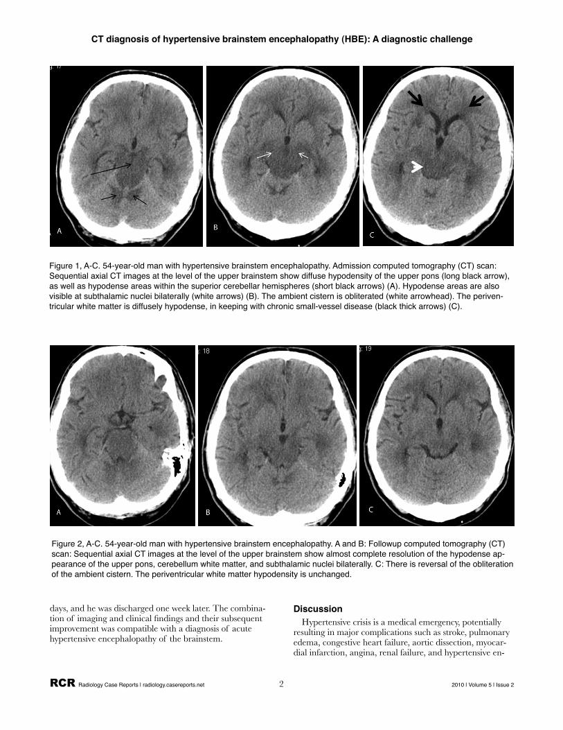
The patient was referred for an emergent brain CT (Fig. 1, A-C). The examination showed that the upper pons and midbrain were markedly and diffusely hypodense and that the ambient cistern was obliterated. Low attenuation ex-tended to the superior cerebellar white matter. There was no evidence of obstructive hydrocephalus. The periven-tricular white matter was mildly hypodense, in keeping with chronic small-vessel disease. The parieto-occipital regions were relatively spared.
The patient was unable to withstand an MRI examina-tion due to claustrophobia. He was subsequently admitted to the nephrology clinic, due to his malignant hypertensive crisis, in order to achieve better blood pressure control. Continuous IV pump infusion of clonidine was adminsi-tered. The blood pressure reached normal levels after 5 days.
Followup CT after blood pressure normalization showed almost complete reversal of the brainstem hypodensity and ambient cistern obliteration (Fig. 2,A-C). Periventricular hypodensity due to presumed chronic small-vessel disease was unchanged. These findings correlated with alleviation of the patient’s symptoms. The patient’s overall clinical status was also markedly improved within the next several
RCR Radiology Case Reports | radiology.casereports.net 1 2010 | Volume 5 | Issue 2
Radiology Case ReportsVolume 5, Issue 2, 2010
CT diagnosis of hypertensive brainstem encephalo-pathy (HBE): A diagnostic challenge in the emergency departmentPantelis Kraniotis, MD; Petros Zampakis, MD, PhD; Christina Kalogeropoulou, MD, PhD; Pantelitsa Kalliakmani, MD; and Theodore Petsas, MD, PhD
Hypertensive encephalopathy usually involves the posterior supratentorium, with uncommon involve-ment of the brainstem. We present a case of acute hypertensive encephalopathy of the brainstem diag-nosed by means of CT. The brainstem was markedly hypodense, with no evidence of typical concomi-tant parieto-occipital involvement. The patient’s symptoms and imaging findings improved after hyper-tension had been controlled.
Citation: Kraniotis P, Zampakis P, Kalogeropoulou C, Kalliakmani P, Petsas T. CT diagnosis of hypertensive brainstem encephalopathy (HBE): A diagnostic challenge in the emergency department. Radiology Case Reports. [Online] 2010;5:385.
Copyright: © 2010 The Authors. This is an open-access article distributed under the terms of the Creative Commons Attribution-NonCommercial-NoDerivs 2.5 License, which permits reproduction and distribution, provided the original work is properly cited. Commercial use and derivative works are not permitted.
Drs. Kraniotis, Zampakis, Kalogeropoulou, and Petsas are in the Department of Radiology, and Dr. Kalliakmani is in the Department of Nephrology, all at University Hospital of Patras, Rion, Greece. Contact Dr. Kraniotis at [email protected].
Competing Interests: The authors have declared that no competing interests exist.
DOI: 10.2484/rcr.v5i2.385
days, and he was discharged one week later. The combina-tion of imaging and clinical findings and their subsequent improvement was compatible with a diagnosis of acute hypertensive encephalopathy of the brainstem.
DiscussionHypertensive crisis is a medical emergency, potentially
resulting in major complications such as stroke, pulmonary edema, congestive heart failure, aortic dissection, myocar-dial infarction, angina, renal failure, and hypertensive en-
CT diagnosis of hypertensive brainstem encephalopathy (HBE): A diagnostic challenge
RCR Radiology Case Reports | radiology.casereports.net 2 2010 | Volume 5 | Issue 2
Figure 2, A-C. 54-year-old man with hypertensive brainstem encephalopathy. A and B: Followup computed tomography (CT) scan: Sequential axial CT images at the level of the upper brainstem show almost complete resolution of the hypodense ap-pearance of the upper pons, cerebellum white matter, and subthalamic nuclei bilaterally. C: There is reversal of the obliteration of the ambient cistern. The periventricular white matter hypodensity is unchanged.
Figure 1, A-C. 54-year-old man with hypertensive brainstem encephalopathy. Admission computed tomography (CT) scan: Sequential axial CT images at the level of the upper brainstem show diffuse hypodensity of the upper pons (long black arrow), as well as hypodense areas within the superior cerebellar hemispheres (short black arrows) (A). Hypodense areas are also visible at subthalamic nuclei bilaterally (white arrows) (B). The ambient cistern is obliterated (white arrowhead). The periven-tricular white matter is diffusely hypodense, in keeping with chronic small-vessel disease (black thick arrows) (C).

cephalopathy (1). Hypertensive encephalopathy (HE) is caused by severe hypertension and has a relatively acute onset (2). This sudden increase in systemic blood pressure often occurs in patients with no history of chronic hyper-tension (3). HE rates are up to 16% in patients presenting with a hypertensive episode (4). Symptoms are nonspecific and include headache, confusion, stupor, visual distur-bances, nausea, vomiting, and seizures (5). Because of the nonspecific nature of the symptoms, the diagnosis is not commonly made by imaging (3). HE is a subset of posterior reversible encephalopathy syndrome (PRES), which also includes conditions such as pre-eclampsia/eclampsia and cyclosporine- and tacrolimus-related encephalopathy (6, 7). This syndrome has also been associated with renal insufficiency (6).
The most characteristic feature of PRES is its predomi-nant involvement of the posterior supratentorial areas. Brainstem involvement is not infrequent but is commonly associated with the more typical supratentorial lesions (7, 8), which were absent in our patient. Isolated involvement of the brainstem and cerebellum is rare, with a few cases in the literature. Although MRI has greatly increased the rec-ognition of hypertensive encephalopathy, the brainstem variant has been rarely reported (1-3, 9-13). This occurs more often in patients less than 40 years of age and is asso-ciated with secondary hypertension (4).
Vasogenic edema caused by failure of cerebral autoregu-lation and endothelial dysfunction is considered to be the underlying mechanism of hypertensive encephalopathy (14). When systemic blood pressure rises over the autoregu-latory threshold of the cerebral vasculature, it results in brain hyperperfusion, due to dilatation of cerebral arteri-oles. This causes blood-brain barrier breakdown, with sub-sequent transudation of fluid and protein material (vaso-genic edema) (6, 8, 15). This pathophysiologic mechanism is supported by the increased ADC values reported in these patients (16). (This of course could not be demonstrated in our case due to the patient’s claustrophobia.) Another pro-posed mechanism is endothelial damage or dysfunction, which may trigger, via increased production of nitric oxide, increased capillary permeability and loss of autoregulation (3). Responsive vasoconstriction-causing ischemia to the affected territory may play a role in some cases (6, 15). This tends not to predominate, though, given the reversible na-ture of the clinical and radiological findings. In cases re-solving after control of the hypertensive episode, the lesions visualized on imaging are most consistent with vasogenic edema.
As mentioned, hypertensive encephalopathy lesions occur mainly posteriorly, which may be due to relatively de-creased sympathetic innervation of the posterior circulation (vertebrobasilar and posterior cerebral arteries) compared to the anterior circulation (2). This accounts for the in-creased susceptibility of the parieto-occipital regions, brain-stem, and cerebellum when autoregulation breakdown oc-curs (17). It has been suggested by Kumai et al that differ-ences in the arterial pressure level are sufficient to cause the development of vasogenic edema in cortical and subcorti-
cal regions and deep structures, such as the basal ganglia and brainstem (18). Cortical and subcortical regions are less tolerant to hypertension compared to deep structures. For this reason, vasogenic edema is thought to involve the deep structures when the systemic blood pressure rises at a highly accelerated rate. Differences in the sympathetic innervation may also exist between the posterior supratentorial and infratentorial circulation, which could explain infratentorial predominance (2).
Newer evidence with diffusion-weighted MRI and ani-sotropy diffusion studies also suggests that MRI signal change is caused by transient vasogenic edema (19). MRI characteristically shows a posterior leukoencephalopathy, affecting predominantly the white matter of the parieto-occipital regions (20). On the other hand, HBE affects pre-dominantly the brainstem and cerebellum, while parieto-occipital regions are spared (12). However, brainstem and deep-white-matter involvement seems to have less reversi-bility than cortical and subcortical areas (19). DWI is re-ported be helpful in HBE cases because lack of restricted diffusion can rule out infarct.
In cases when no typical parieto-occipital lesions coexist, the differential diagnosis for brainstem lesions includes acute infarction, tumor, encephalitis, and vasculitis. Our patient’s clinical status of mild cerebellar syndrome that subsequently resolved was not consistent with acute infarc-tion. Consciousness level was normal, and there was no focal neurologic deficit. Mild symptomatology, like head-ache and confusion, with lack of cranial nerve findings and focal neurologic deficits despite brainstem involvement (re-ferred to as clinical-radiological dissociation) (4) suggests hypertensive encephalopathy (2). The improvement would also not be compatible with tumor. Laboratory tests during his hospitalization were negative, including inflammatory and collagen-vascular-disease markers. These findings, along with the rapid clinical evolution and resolution of both symptoms and brainstem lesions with correction of hypertension, established the clinical diagnosis of hyperten-sive encephalopathy (12).
Recognition of the brainstem variant of hypertensive encephalopathy is important so that prompt treatment can be initiated. Radiologists may be the first to notice this alarming appearance of the brainstem. Along with the im-aging findings, the presence of “clinical-radiological disso-ciation” should alert radiologists to suggest the diagnosis of this rare variant of hypertensive encephalopathy.
References1. Seet RC, Lim EC. Images in cardiovascular medicine.
Hypertensive brainstem encephalopathy. Circulation 2007;115:e310-311 [PubMed]
2. Fujiwara H, Momoshima S, Kuribayashi S, Sasamura H. Hypertensive encephalopathy of brain stem with minimal supratentorial involvement: a rare manifesta-tion of hypertensive encephalopathy. Radiat Med 2005;23:504-507 [PubMed]
CT diagnosis of hypertensive brainstem encephalopathy (HBE): A diagnostic challenge
RCR Radiology Case Reports | radiology.casereports.net 3 2010 | Volume 5 | Issue 2

3. McCarron MO, McKinstry CS. Vanishing brainstem edema. J Stroke Cerebrovasc Dis 2008;17:156-157 [Pub-Med]
4. Cruz-Flores S, de Assis Aquino Gondim F, Leira EC. Brainstem involvement in hypertensive encephalopa-thy: clinical and radiological findings. Neurology 2004;62:1417-1419 [PubMed]
5. Chester EM, Agamanolis DP, Banker BQ, Victor M. Hypertensive encephalopathy: a clinicopathologic study of 20 cases. Neurology 1978;28:928-939 [Pub-Med]
6. Hinchey J, Chaves C, Appignani B, et al. A reversible posterior leukoencephalopathy syndrome. N Engl J Med 1996;334:494-500 [PubMed]
7. Casey SO, Sampaio RC, Michel E, Truwit CL. Poste-rior reversible encephalopathy syndrome: utility of fluid-attenuated inversion recovery MR imaging in the detection of cortical and subcortical lesions. AJNR Am J Neuroradiol 2000;21:1199-1206 [PubMed]
8. Weingarten K, Barbut D, Filippi C, Zimmerman RD. Acute hypertensive encephalopathy: findings on spin-echo and gradient-echo MR imaging. AJR Am J Roent-genol 1994;162:665-670 [PubMed]
9. Kitaguchi H, Tomimoto H, Miki Y, et al. A brainstem variant of reversible posterior leukoencephalopathy syndrome. Neuroradiology 2005;47:652-656 [PubMed]
10. Yasuda Y, Akiguchi I, Imai T, Sonobe M, Kage M. Hypertensive brainstem encephalopathy. Intern Med 2003;42:1131-1134 [PubMed]
11. Fong CS. Hypertensive encephalopathy involving the brainstem and deep structures: a case report. Acta Neu-rol Taiwan 2005;14:191-194 [PubMed]
12. Albini TA, Lakhanpal RR, Foroozan R, Lopez GA, McPherson AR. Retinopathy and choroidopathy as the initial signs of hypertensive brainstem encephalopathy. Arch Ophthalmol 2006;124:1784-1786 [PubMed]
13. Nagata M, Maeda M, Tsukahara H, Maier SE, Takeda K. Brain stem hypertensive encephalopathy evaluated by line scan diffusion-weighted imaging. AJNR Am J Neuroradiol 2004;25:803-806 [PubMed]
14. Lamy C, Oppenheim C, Meder JF, Mas JL. Neuroi-maging in posterior reversible encephalopathy syn-drome. J Neuroimaging 2004;14:89-96 [PubMed]
15. Skinhoj E, Strandgaard S. Pathogenesis of hyperten-sive encephalopathy. Lancet 1973;1:461-462 [PubMed]
16. Schwartz RB, Mulkern RV, Gudbjartsson H, Jolesz F. Diffusion-weighted MR imaging in hypertensive en-cephalopathy: clues to pathogenesis. AJNR Am J Neuro-radiol 1998;19:859-862 [PubMed]
17. de Seze J, Mastain B, Stojkovic T, et al. Unusual MR findings of the brain stem in arterial hypertension. AJNR Am J Neuroradiol 2000;21:391-394 [PubMed]
18. Kumai Y, Toyoda K, Fujii K, Ibayashi S. Hypertensive encephalopathy extending into the whole brainstem and deep structures. Hypertens Res 2002;25:797-800 [PubMed]
19. Pande AR, Ando K, Ishikura R, et al. Clinicoradio-logical factors influencing the reversibility of posterior reversible encephalopathy syndrome: a multicenter study. Radiat Med 2006;24:659-668 [PubMed]
20. Vaughan CJ, Delanty N. Hypertensive emergencies. Lancet 2000;356:411-417 [PubMed]
CT diagnosis of hypertensive brainstem encephalopathy (HBE): A diagnostic challenge
RCR Radiology Case Reports | radiology.casereports.net 4 2010 | Volume 5 | Issue 2

CT diagnosis of hypertensive brainstem encephalopathy (HBE): A diagnostic challenge
RCR Radiology Case Reports | radiology.casereports.net 5 2010 | Volume 5 | Issue 2
Figure 6: 49-year-old woman with meningioma. Axial CT in (A) bone window and (B) soft-tissue window shows subtle hyperos-tosis of the left petrous ridge at the porus acousticus (black arrow) and coarse calcification (white arrows) anteriorly within the mass.