Embed Size (px)
Citation preview
S. Wirth Department of Clinical Radiology; Hospital of the Ludwigs-Maximilians University of Munich
Radiologische Diagnostik
bei akutem Abdomen
Notfalldiagnostik bei nicht-traumatischen Notfällen
Klinik, Management und interventionelle Therapie
S. Wirth
S. Wirth Department of Clinical Radiology; Hospital of the Ludwigs-Maximilians University of Munich
Definition: Akutes Abdomen
„An acute abdomen is, when a good surgeon says, it is an acute abdomen. There is no other test for it - however a CT scan is a good idea.“ Clifton K. Meador, Internal Med.
Akute Manifestation von Erkrankungen im Bauchraum, die einer sofortigen Diagnostik und Therapie bedürfen.
Leitsymptome: - “starker” lokaler oder diffuser Bauchschmerz - Abwehrspannung, Übelkeit, eingeschränkter AZ In ca. 10% Ursache für Aufsuchen einer Notaufnahme
S. Wirth Department of Clinical Radiology; Hospital of the Ludwigs-Maximilians University of Munich
Ätiologie
1. Intraperitoneale Erkrankungen Entzündungen ohne/ mit Perforation:
Appendizitis, Divertikulitis, Pankreatitis, Gastritis, Ulkus, M. Crohn Obstruktion eines Hohlorgans Ileus Vaskuläre Erkrankungen:
Mesenterialinfakt, Darmischämie, Aortenaneurysma, Blutungen Gynäkologisch:
EUG, Torsion, Endometriose
S. Wirth Department of Clinical Radiology; Hospital of the Ludwigs-Maximilians University of Munich
Ätiologie
2. Extraperitoneale Erkrankungen Thorax: MI, Pneumonie, LE, Pneumonie, Pleuritis Retroperitoneum: Nierenkolik Infektionen Systemische Erkrankungen: DM, HPT, CF, … Neurologische Erkrankungen: u.a. Psychosen Intoxikationen 3. NSAP (non specific abdominal pain) Ohne Nachweis einer Ursache Klingen i.d.R. innerhalb 48h ab
S. Wirth Department of Clinical Radiology; Hospital of the Ludwigs-Maximilians University of Munich
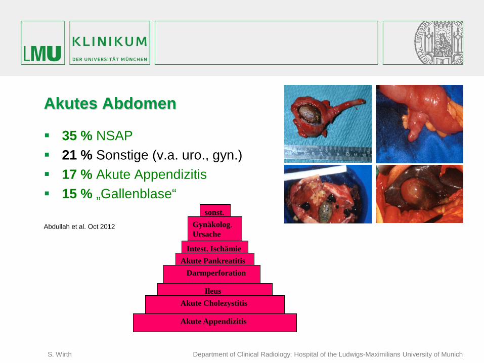
Akutes Abdomen
35 % NSAP 21 % Sonstige (v.a. uro., gyn.) 17 % Akute Appendizitis 15 % „Gallenblase“ Abdullah et al. Oct 2012
Akute Appendizitis
Akute Cholezystitis Ileus
Darmperforation Akute Pankreatitis
Intest. Ischämie
Gynäkolog. Ursache
sonst.
S. Wirth Department of Clinical Radiology; Hospital of the Ludwigs-Maximilians University of Munich
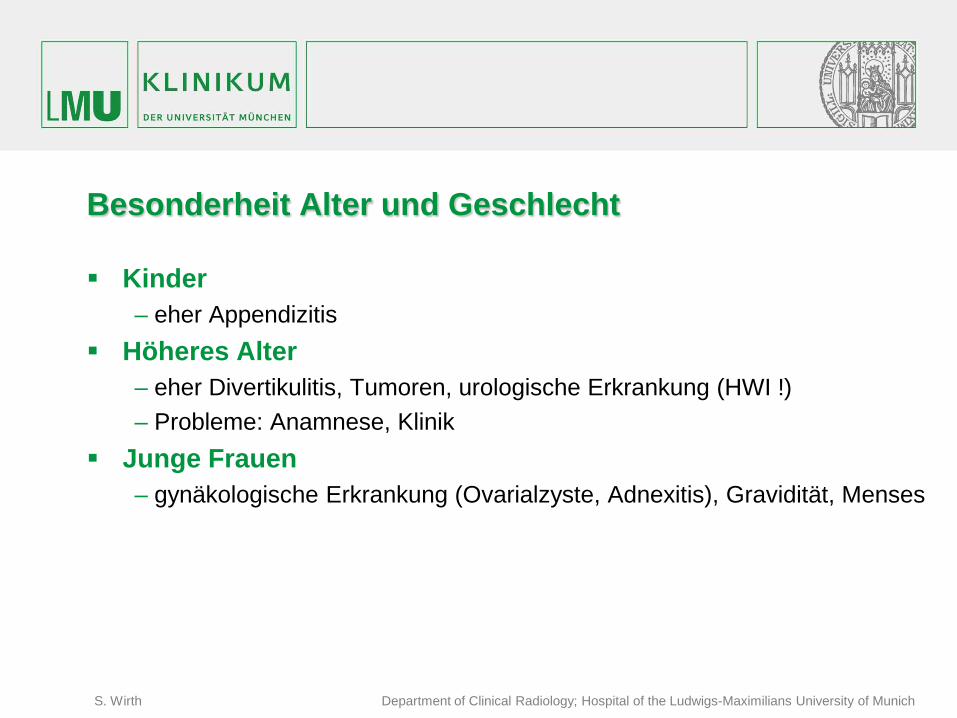
Besonderheit Alter und Geschlecht Kinder
– eher Appendizitis Höheres Alter
– eher Divertikulitis, Tumoren, urologische Erkrankung (HWI !) – Probleme: Anamnese, Klinik
Junge Frauen – gynäkologische Erkrankung (Ovarialzyste, Adnexitis), Gravidität, Menses
S. Wirth Department of Clinical Radiology; Hospital of the Ludwigs-Maximilians University of Munich
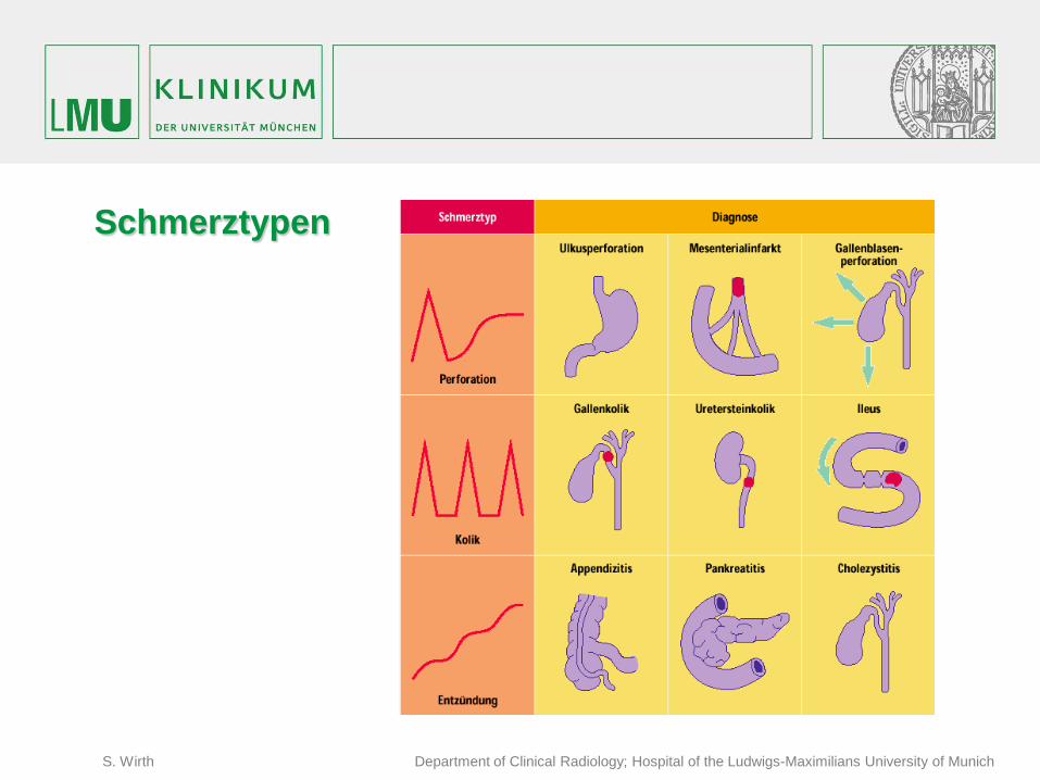
Schmerztypen
S. Wirth Department of Clinical Radiology; Hospital of the Ludwigs-Maximilians University of Munich
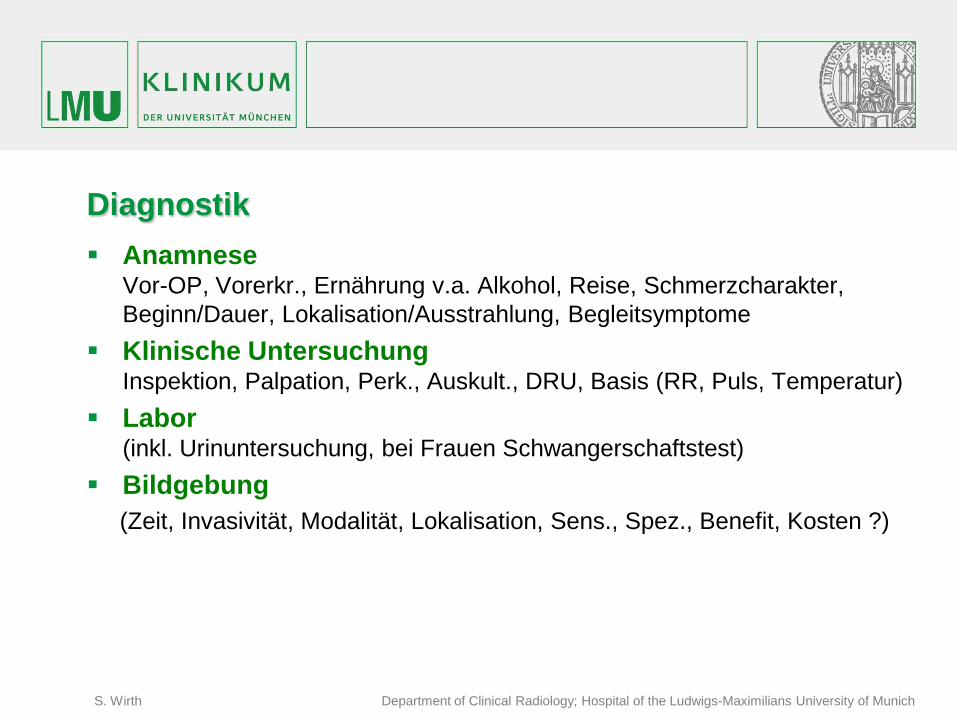
Diagnostik Anamnese
Vor-OP, Vorerkr., Ernährung v.a. Alkohol, Reise, Schmerzcharakter, Beginn/Dauer, Lokalisation/Ausstrahlung, Begleitsymptome
Klinische Untersuchung Inspektion, Palpation, Perk., Auskult., DRU, Basis (RR, Puls, Temperatur)
Labor (inkl. Urinuntersuchung, bei Frauen Schwangerschaftstest)
Bildgebung (Zeit, Invasivität, Modalität, Lokalisation, Sens., Spez., Benefit, Kosten ?)
S. Wirth Department of Clinical Radiology; Hospital of the Ludwigs-Maximilians University of Munich
GCS <13
no
no
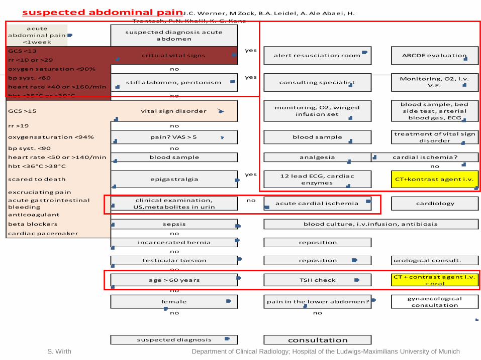
GCS >15 vital sign disordermonitoring, O2, winged
infusion set
blood sample, bed side test, arterial
blood gas, ECG
rr >19 no
pain? VAS > 5 blood sampletreatment of vital sign
disorderno
blood sample analgesia
no
epigastralgiayes 12 lead ECG, cardiac
enzymesCT+kontrast agent i.v.
clinical examination, US,metabolites in urin
noacute cardial ischemia cardiology
sepsis
no
incarcerated hernia reposition
no
testicular torsion reposition urological consult.
no
age > 60 years TSH checkCT + contrast agent i.v.
+ oralno
female pain in the lower abdomen?gynaecological
consultationno no
suspected diagnosis consultation
blood culture, i.v.infusion, antibiosis
cardial ischemia?
critical vital signs alert resusciation roomyes
ABCDE evaluation
stiff abdomen, peritonism consulting specialistMonitoring, O2, i.v.
V.E.
yes
acute abdominal pain
<1week
suspected abdominal painJ.C. Werner, M Zock, B.A. Leidel, A. Ale Abaei, H. Trentsch, P.N. Khalil, K.-G. Kanz
suspected diagnosis acute abdomen
rr <10 or >29
oxygen saturation <90%
bp syst. <80
heart rate <40 or >160/min
hbt <35°C or >39°C
oxygensaturation <94%
acute gastrointestinal bleedinganticoagulant
beta blockers
cardiac pacemaker
bp syst. <90
heart rate <50 or >140/min
hbt <36°C >38°C
scared to death
excruciating pain
S. Wirth Department of Clinical Radiology; Hospital of the Ludwigs-Maximilians University of Munich
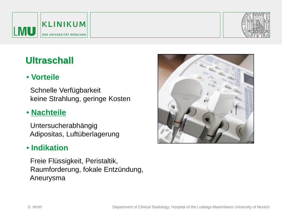
Ultraschall • Vorteile Schnelle Verfügbarkeit keine Strahlung, geringe Kosten
• Nachteile Untersucherabhängig Adipositas, Luftüberlagerung
• Indikation Freie Flüssigkeit, Peristaltik, Raumforderung, fokale Entzündung, Aneurysma
S. Wirth Department of Clinical Radiology; Hospital of the Ludwigs-Maximilians University of Munich
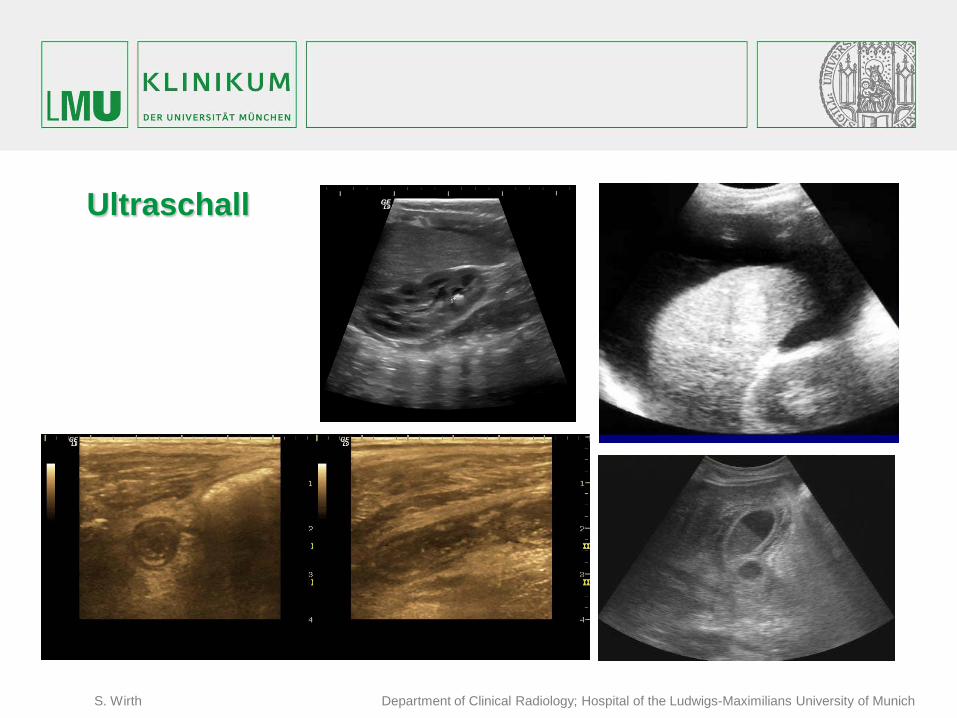
Ultraschall
S. Wirth Department of Clinical Radiology; Hospital of the Ludwigs-Maximilians University of Munich
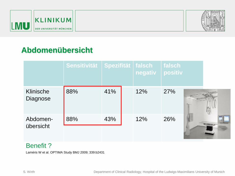
Abdomenübersicht
Sensitivität Spezifität falsch negativ
falsch positiv
Klinische Diagnose
88% 41% 12% 27%
Abdomen- übersicht
88% 43% 12% 26%
Benefit ? Laméris W et al. OPTIMA Study BMJ 2009; 339:b2431
S. Wirth Department of Clinical Radiology; Hospital of the Ludwigs-Maximilians University of Munich
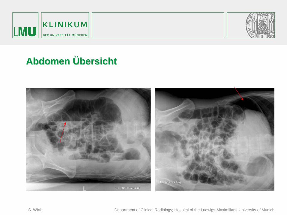
Abdomen Übersicht
S. Wirth Department of Clinical Radiology; Hospital of the Ludwigs-Maximilians University of Munich
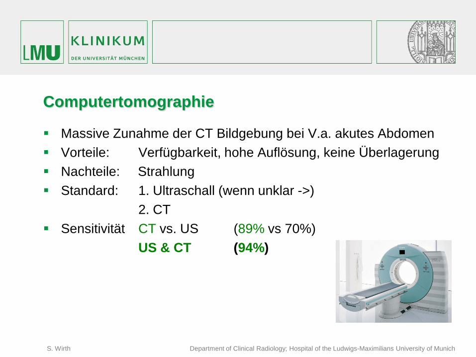
Computertomographie
Massive Zunahme der CT Bildgebung bei V.a. akutes Abdomen Vorteile: Verfügbarkeit, hohe Auflösung, keine Überlagerung Nachteile: Strahlung Standard: 1. Ultraschall (wenn unklar ->) 2. CT Sensitivität CT vs. US (89% vs 70%) US & CT (94%)
S. Wirth Department of Clinical Radiology; Hospital of the Ludwigs-Maximilians University of Munich
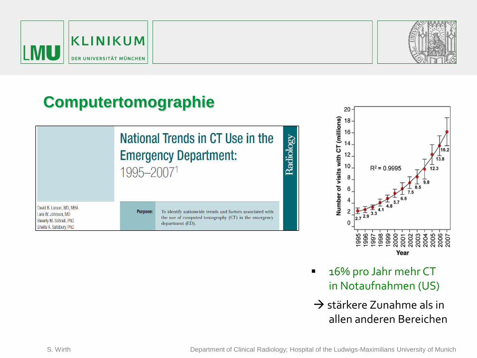
Computertomographie
16% pro Jahr mehr CT in Notaufnahmen (US)
stärkere Zunahme als in allen anderen Bereichen
S. Wirth Department of Clinical Radiology; Hospital of the Ludwigs-Maximilians University of Munich
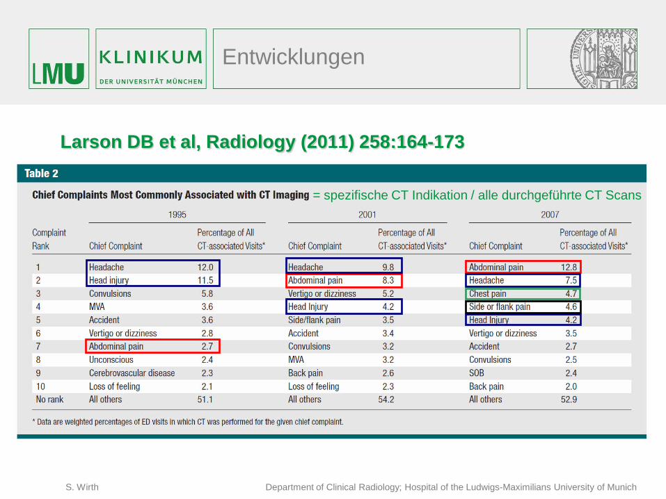
Larson DB et al, Radiology (2011) 258:164-173
= spezifische CT Indikation / alle durchgeführte CT Scans
Entwicklungen
S. Wirth Department of Clinical Radiology; Hospital of the Ludwigs-Maximilians University of Munich
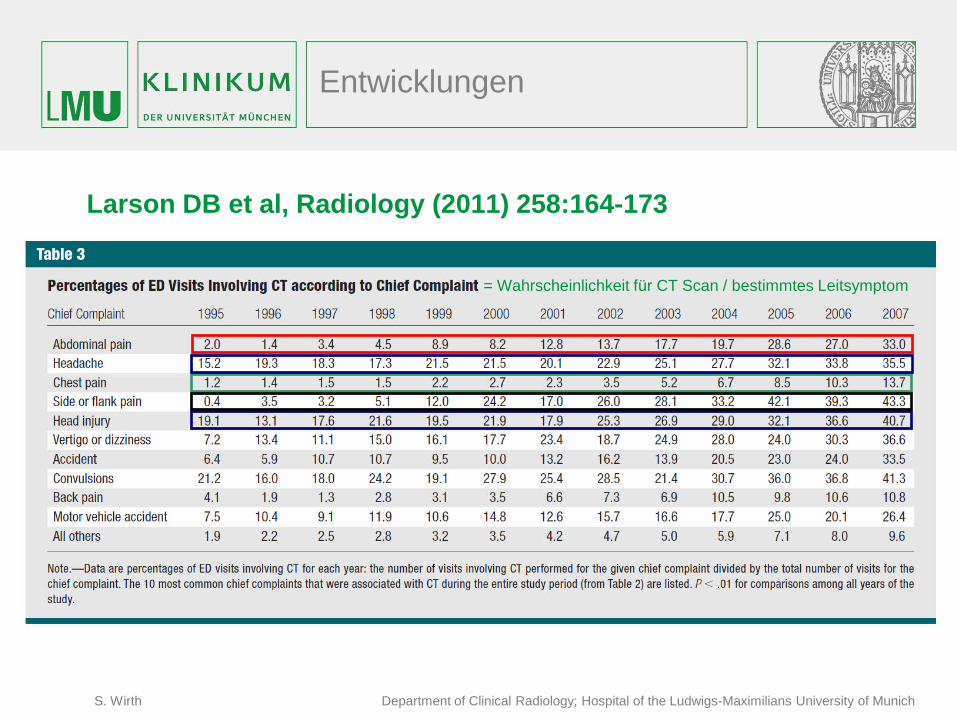
Larson DB et al, Radiology (2011) 258:164-173
= Wahrscheinlichkeit für CT Scan / bestimmtes Leitsymptom
Entwicklungen
S. Wirth Department of Clinical Radiology; Hospital of the Ludwigs-Maximilians University of Munich
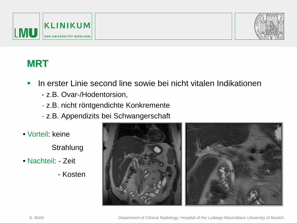
MRT
In erster Linie second line sowie bei nicht vitalen Indikationen - z.B. Ovar-/Hodentorsion, - z.B. nicht röntgendichte Konkremente - z.B. Appendizits bei Schwangerschaft • Vorteil: keine
Strahlung
• Nachteil: - Zeit
- Kosten
S. Wirth Department of Clinical Radiology; Hospital of the Ludwigs-Maximilians University of Munich
Interventionelle Therapie:
Interdisziplinäres Management ! Beispiele
– intraabdominelle Abszesse – Primärversorgung akuter gastrointestinaler Blutungen. – akute Mesenterialischämie – akuter Gallengangsverschluss
S. Wirth Department of Clinical Radiology; Hospital of the Ludwigs-Maximilians University of Munich
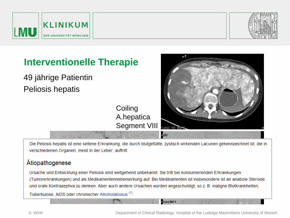
Interventionelle Therapie 49 jährige Patientin Peliosis hepatis
Coiling A.hepatica Segment VIII
S. Wirth Department of Clinical Radiology; Hospital of the Ludwigs-Maximilians University of Munich
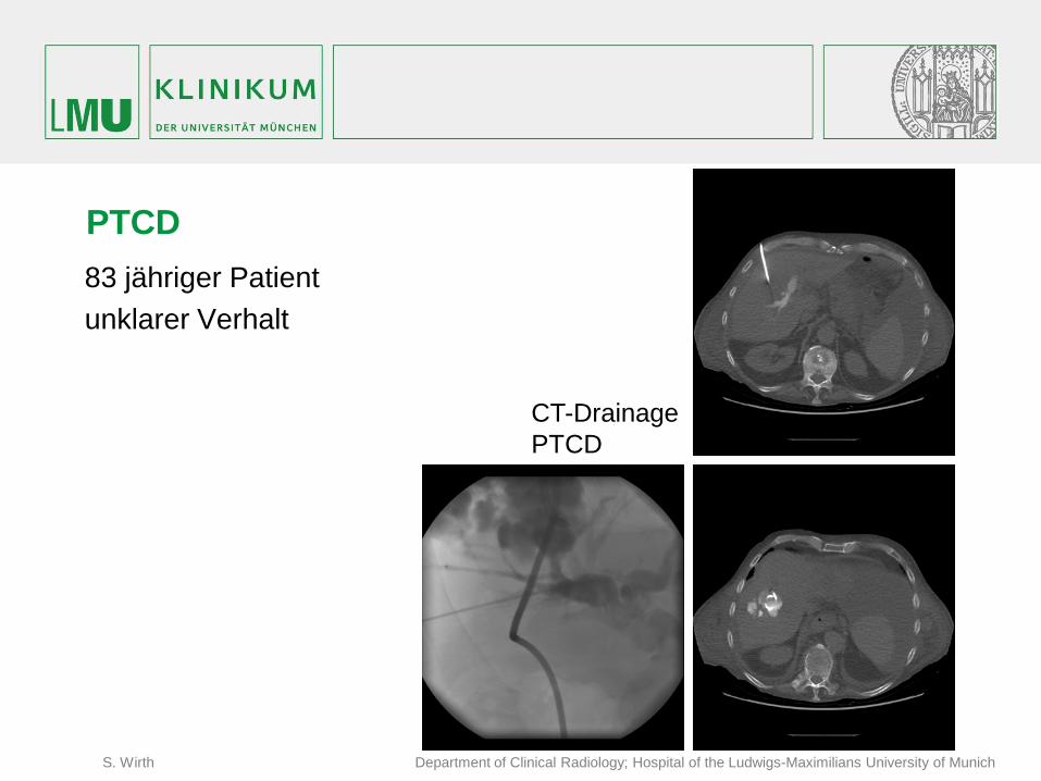
PTCD 83 jähriger Patient unklarer Verhalt
CT-Drainage PTCD
S. Wirth Department of Clinical Radiology; Hospital of the Ludwigs-Maximilians University of Munich
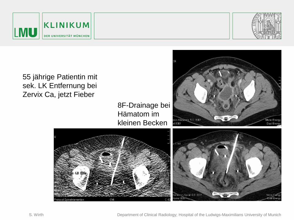
8F-Drainage bei Hämatom im kleinen Becken
55 jährige Patientin mit sek. LK Entfernung bei Zervix Ca, jetzt Fieber
S. Wirth Department of Clinical Radiology; Hospital of the Ludwigs-Maximilians University of Munich
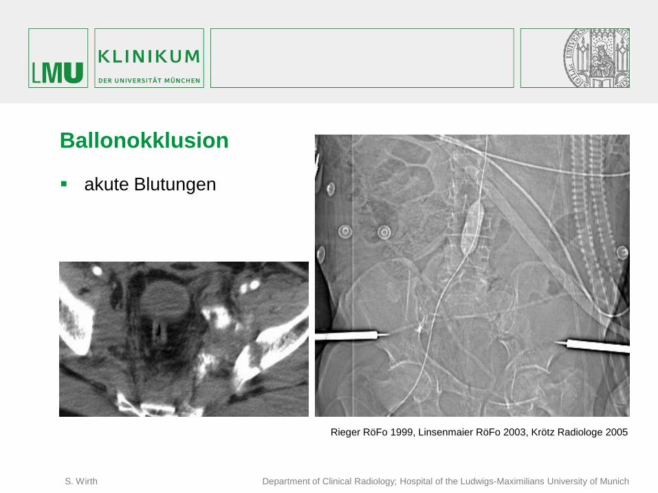
Ballonokklusion
akute Blutungen
Rieger RöFo 1999, Linsenmaier RöFo 2003, Krötz Radiologe 2005
S. Wirth Department of Clinical Radiology; Hospital of the Ludwigs-Maximilians University of Munich
Zusammenfassung
Akutes Abdomen Notfall akuter Handlungsbedarf Abdominelle Schmerzen Anamnese, körperl. Unt., Labor, Bildgebung interdisziplinäre Zusammenarbeit : Chirurgie, Radiologie, (Anästhesie)
– vitalitätsbedrohend Stabilisation, Bildgebung, Intervention – möglich vitalitätsbedrohend:
• Sono (FAST), KM CT (ohne orales KM, Zeit!) – keine dringende vitalitätsbedrohende Indikation
• Gynäkologie ?, MRI ? • Nierenkolik ? CT ohne KM, low dose • KM-CT mit oralem KM
– Intervention: Drainage, PTCD, Ballonblockade (besser Embolisation, Stenting) Wann immer möglich kein CT bei Kindern jungen Patienten Ausnutzung von Dosisreduktion wird weitere Verbreitung der CT fördern
(Diskussion von Risiko, aber bitte auch von Nutzen !!!)
S. Wirth Department of Clinical Radiology; Hospital of the Ludwigs-Maximilians University of Munich
Quellen: Evaluation of acute abdominal pain in adults. Cartwright SL, Knudson MP.Source, Department
of Family and Community Medicine, Wake Forest University School ofMedicine, Winston-Salem, North Carolina 27157, USA.
Imaging patients with acute abdominal pain.Stoker J, van Randen A, Laméris W, Boermeester MA.Source, Radiology. 2009 Oct;253(1):31-46. doi: 10.1148/radiol.2531090302.
Diagnostic approach and management of acute abdominal pain. Abdullah M, Firmansyah MA. National trends in CT use in the emergency department: 1995-2007.Larson DB, Johnson LW,
Schnell BM, Salisbury SR, Forman HP. Radiology. 2011 Jan;258(1):164-73. doi: 10.1148/radiol.10100640. Epub 2010 Nov 29.
S. Wirth Department of Clinical Radiology; Hospital of the Ludwigs-Maximilians University of Munich
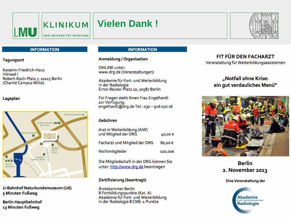
Vielen Dank !
S. Wirth Department of Clinical Radiology; Hospital of the Ludwigs-Maximilians University of Munich
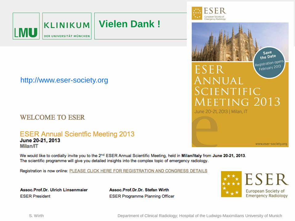
Vielen Dank !
http://www.eser-society.org