Embed Size (px)
Citation preview

Radiography of Patients
with Special Needs
Veronika Stiles, RDH
University of Michigan
June 16, 2009
Introduction
• Various special needs exist
• RDH must be competent to alter
techniques
Patients with gag reflex
• Confident attitude
• Patience, tolerance, &
understanding
• Key: limit amount of time
• Prepare patient & equipment
ahead of time
• Start with anterior exposures
• Then, premolar shot before
molar
• Maxillary molar films must be
exposed last
Patients with Gag Reflex:
Film Placement & Technique
• Each film must be placed and
exposed as quickly as possible
• Avoid the palate
• Demonstrate film placement to
the patient
• Never suggest gagging
• Suggest deep breathing through
the nose
• Try to distract patient
• Reduce tactile stimuli (ice H2O,
salt, topical anesthetic)
Patients with Physical
Disabilities
A. Vision impairment:
communicate using clear verbal
explanations
B. Hearing impairment:
Gestures
Written instructions
Ask caretaker for assistance
C. Mobility impairment:
Assist the patient in transfer
Ask caretaker for assistance
Patients with Developmental
Disabilities
• Examples: autism, cerebral
palsy, epilepsy, mental
retardation
• Coordination problems: mild
sedation
• Comprehension problems: ask
for assistance
• If the patient cannot tolerate IO
films No intraoral films must
be exposed
• Alternative: Panoramic X-Ray
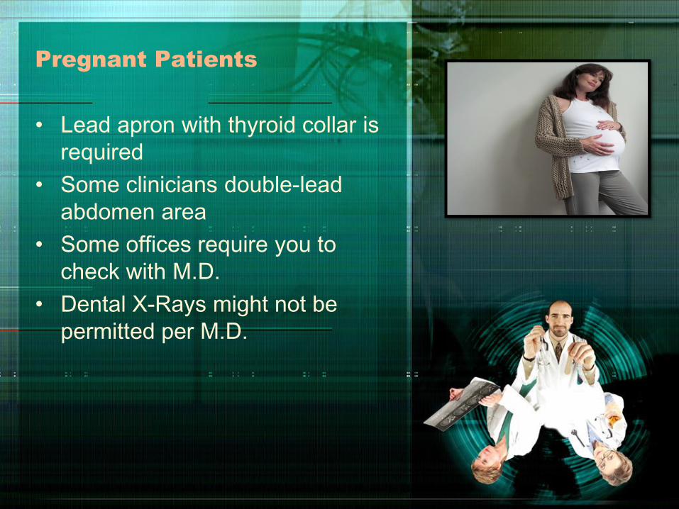
Pregnant Patients
• Lead apron with thyroid collar is
required
• Some clinicians double-lead
abdomen area
• Some offices require you to
check with M.D.
• Dental X-Rays might not be
permitted per M.D.
Pediatric Patients
• Follow KCC prescribing
guidelines
• Explain procedure:
– Tubehead = camera
– Lead apron = coat
– Radiograph = picture
• Exposure factors must be
reduced
• Size 0 = primary dentition;
Size 1 = mixed dentition;
Size 2 = mainly for occlusal
radiographs

Edentulous Patients
• Why radiographs are necessary?
– To detect presence of root
tips, impacted teeth, and
lesions;
– To identify objects embedded
in bone;
– To observe the quantity and
quality of bone that is present
Pediatrics: Helpful Hints
• Show and tell
• Demonstrate behavior
• Request assistance if needed
from a parent
• Postpone examination if child is
too scared
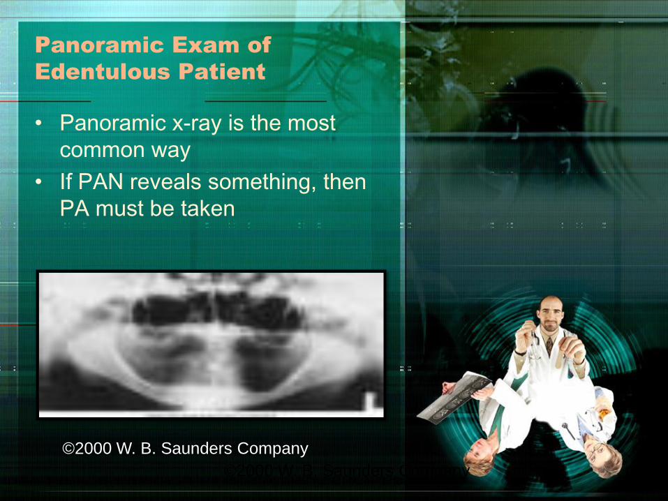
Panoramic Exam of
Edentulous Patient
• Panoramic x-ray is the most
common way
• If PAN reveals something, then
PA must be taken
©2000 W. B. Saunders Company
©2000 W. B. Saunders Company
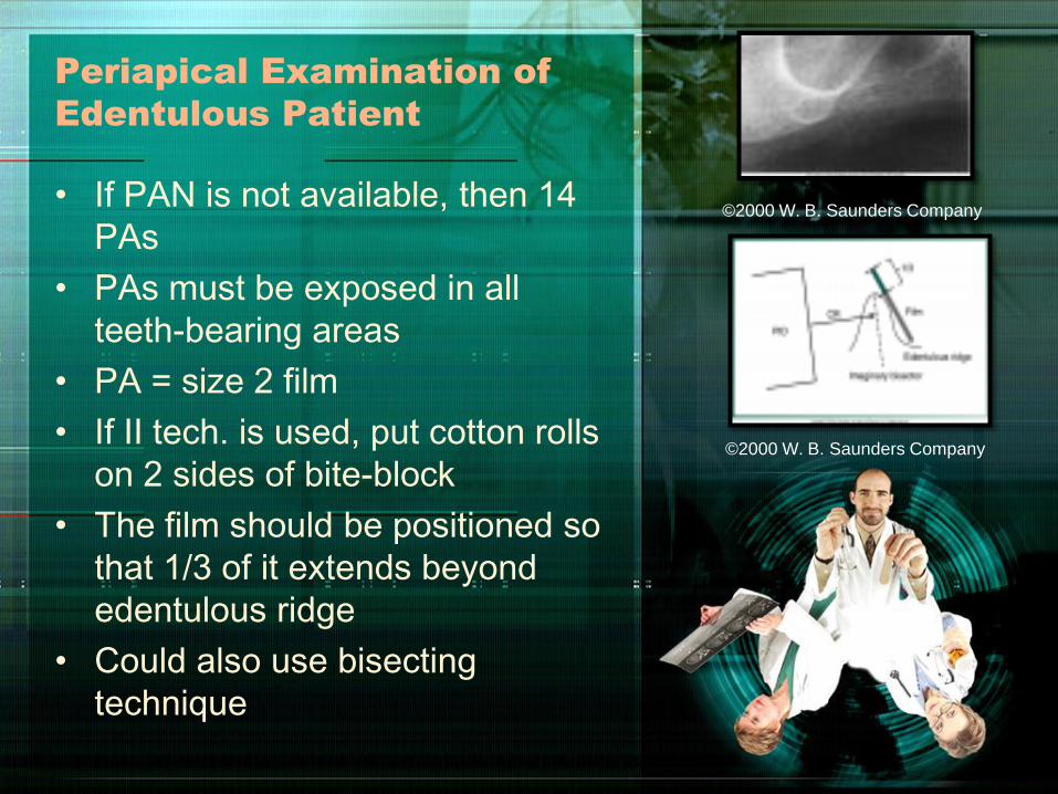
Periapical Examination of
Edentulous Patient
• If PAN is not available, then 14
PAs
• PAs must be exposed in all
teeth-bearing areas
• PA = size 2 film
• If II tech. is used, put cotton rolls
on 2 sides of bite-block
• The film should be positioned so
that 1/3 of it extends beyond
edentulous ridge
• Could also use bisecting
technique
©2000 W. B. Saunders Company
©2000 W. B. Saunders Company
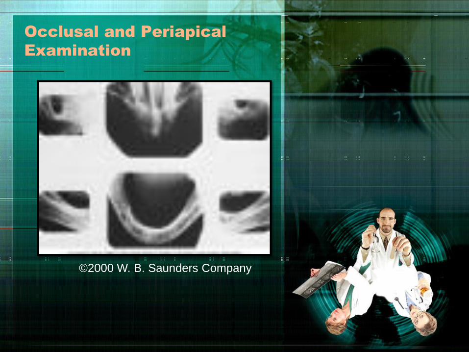
Occlusal and Periapical
Examination
©2000 W. B. Saunders Company
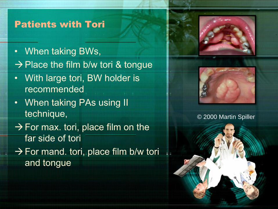
Patients with Tori
• When taking BWs,
Place the film b/w tori & tongue
• With large tori, BW holder is
recommended
• When taking PAs using II
technique,
For max. tori, place film on the
far side of tori
For mand. tori, place film b/w tori
and tongue
© 2000 Martin Spiller
Patients with Missing Teeth
& Shallow Palate
Missing teeth:
• Use cotton rolls to “replace”
missing tooth
• Helps to position the film parallel
to long axis
Shallow palate:
• If the lack of parallelism is > 20,
then
– Use 2 cotton rolls one placed
on each side of bite-block
– Vertical angulation can be
increased by 5-15 degrees
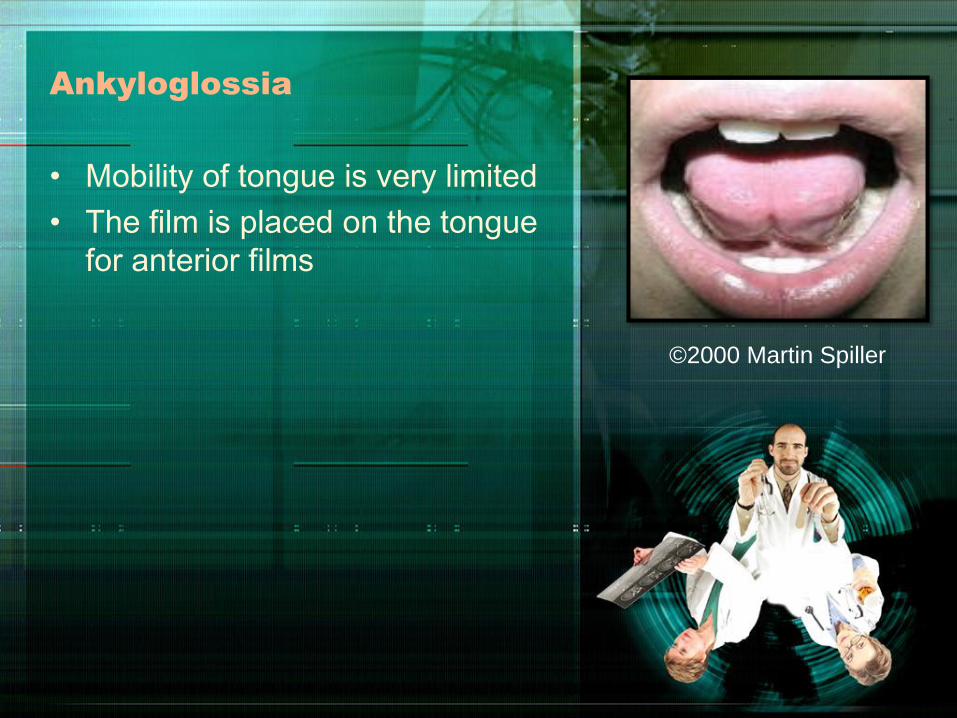
Ankyloglossia
• Mobility of tongue is very limited
• The film is placed on the tongue
for anterior films
©2000 Martin Spiller
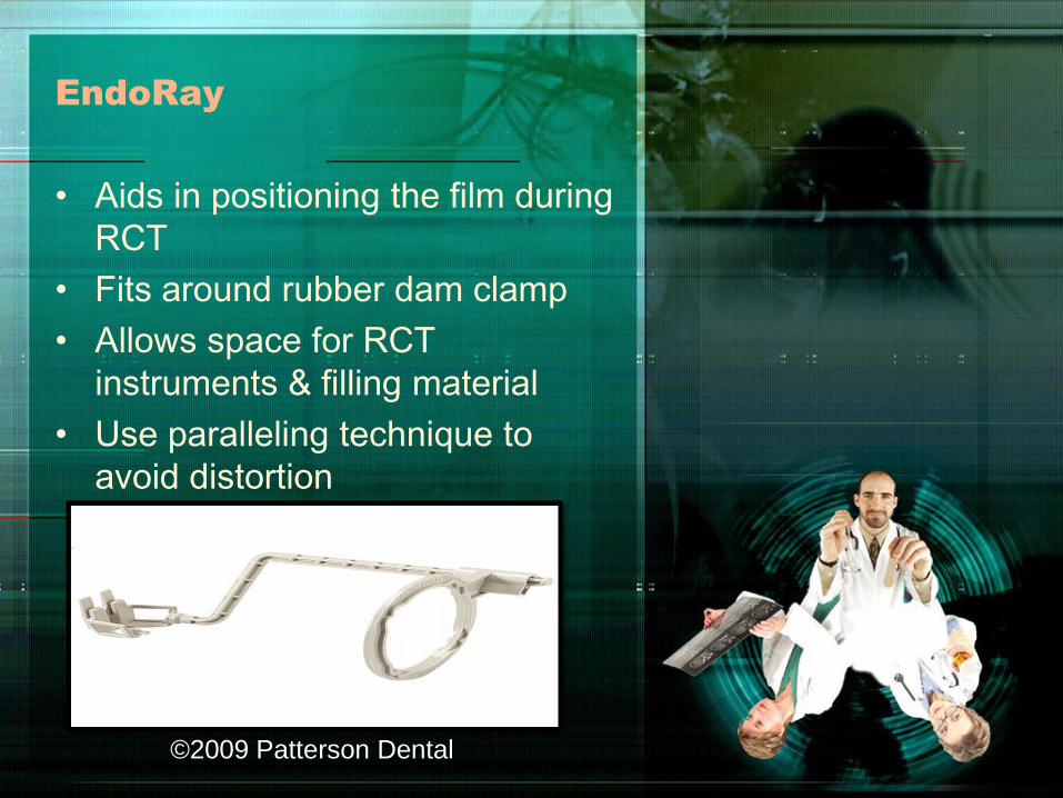
EndoRay
• Aids in positioning the film during
RCT
• Fits around rubber dam clamp
• Allows space for RCT
instruments & filling material
• Use paralleling technique to
avoid distortion
©2009 Patterson Dental

References
1. Haring JI, Jansen L. Dental
Radiography Principles and
Techniques. 2nd ed. Philadelphia:
W. B. Saunders Company; 2000.