Embed Size (px)
Citation preview

1
Radiographic Procedures III (RAD 228)
Urinary System
RADIOGRAPHIC EXAMINATIONS
Urinary System
Antegrade Exam
IVU
Functional test
Hypertensive evaluation as per protocol
Retrograde Exams
Retrograde Urography
Non-functional test (OR Suite)
Retrograde Cystography
Non-functional test
Voiding Cystourethrography
Functional test
Urinary System Kidneys (2)
Ureters (2)
Urinary bladder
Urethra
3
Anterior view

2
Urinary System
Retroperitoneal
structures
Kidneys and ureters
Infraperitoneal
structures
Distal ureters
Urinary bladder
Urethra
4
Lateral view
Kidney Location
Halfway between
xiphoid process and
iliac crest
Between T11-T12
and L3
Nephroptosis
7
IVU Demonstrating Kidneys, Ureters,
and Bladder
10

3
Equipment Preparation for IVU
13
Nonionic Contrast Media
18
Effects of Ionic versus Nonionic
Contrast Media
Ionic Nonionic
Dissociates into separate
ions when injected Does not dissociate
Creates hypertonic
condition
Remains near
isotonic
Increase in blood
osmolality
No significant
increase in
osmolality
19

4
Side Effect versus Reaction
Side effects: expected
outcome of injected
contrast media
Common side effects
Temporary hot flash
Metallic taste in
mouth
Reaction:
An unexpected
outcome of injected
contrast media
20
Technologist Responsibilities
1. Patient history
Clinical complaints?
Food or drug allergies?
Previous contrast media reaction?
Asthma, hay fever, or hives?
21
Patient History
Management of non–insulin-dependent
diabetes: Glucophage (metformin
hydrochloride)
Check chart and/or ask patient the following:
“Are you currently taking glucophage or other
medication for diabetes mellitus?”
To be withheld 48 hours following iodinated
contrast media procedure
Must verify normal kidney function before
resuming medication
22

5
Patient History
Check blood chemistry—normal ranges
Creatinine level (adult)—0.6 -1.5 mg/dL
BUN levels (adult)—8-25 mg/100 mL
23
Premedication Protocol
Common protocol:
Prescribed combination of Benadryl and
prednisone over period of 12 or more hours
before procedure
Patients who have history of hay fever,
asthma, food allergies, or previous contrast
media reaction may be candidates for
premedication procedure
26
Categories of Contrast Media
Reactions
Local
Reactions that affect only a specific region of the
body
Systemic
Reactions that affect the entire body or a specific
organ system
27

6
Local Reactions
Extravasation
Leakage of iodinated contrast media outside
the vessel and into surrounding soft tissues
(also referred to as infiltration)
May be toxic to skin
Notify department nurse and/or physician
Elevate affected extremity above heart
Cold compress followed by warm compresses
first to relieve pain and then to improve
resorption
28
Local Reactions
Phlebitis
Inflammation of a vein
Signs include pain, redness, and possibly swelling
surrounding the venous access site
Discontinue the venous access at this site
Notify department nurse and/or physician
29
Systemic Reactions
Mild Nonallergic reaction does not typically require
drug intervention or medical assistance
Symptoms include the following: Anxiety
Light-headedness
Nausea
Vomiting
Metallic taste (common side effect)
Mild erythema
Warm, flush sensation during injection (common side effect)
Itching
Mild, scattered hives
30
7
Systemic Reactions
Moderate
A true allergic reaction (anaphylactic
reaction)
Symptoms include the following:
Urticaria (moderate to severe hives)
Possible laryngeal swelling
Bronchospasm
Tachycardia (100 beats/min)
Bradycardia (60 beats/min)
Angioedema
Hypotension
31
Systemic Reactions
Severe (Vasovagal)
Life-threatening reaction
Symptoms include the following:
Hypotension (systolic blood pressure 80 mm Hg)
Bradycardia (50 beats/min)
Cardiac arrhythmias
Laryngeal swelling
Possible convulsions
Loss of consciousness
Cardiac arrest
Respiratory arrest
No detectable pulse
32
Excretory Urography—IVU
• Correct term
Intravenous urogram (IVU): Radiographic examination of the urinary system
• Purpose of IVU (twofold)
1. Visualize the collecting portion of the urinary system.
2. Assess the functional ability of the kidneys (a timed procedure).
34

8
Common Clinical Indications—IVU
1. Abdominal or pelvic
mass
2. Renal or urethral
calculi
3. Kidney trauma
4. Flank pain
5. Hematuria
6. Hypertension
7. Renal failure
8. Urinary tract infection
(UTI) (pyelonephritis)
Renal calculi in right kidney
35
Patient Preparation for IVU*
Light evening meal prior to procedure
Bowel-cleansing laxative
NPO after midnight (minimum of 8 hours)
Enema on the morning of examination
Voiding prior to procedure
* Suggested protocol; prep may vary among
departments and clinical needs
37
IVU—Basic Routine
Scout radiograph
Injection Note time at beginning
of injection
Sample imaging routine 1 min nephrogram or
nephrotomography • (Hypertensive)
5 min AP supine
10-15 min AP supine
20 min posterior obliques
Postvoid (prone or erect)
38

9
IVU
Routine
AP scout
Nephrotomography (1 min following injection)
AP
RPO and LPO
AP postvoid (recumbent or erect)
Special
AP ureteric compression
43
IVU—AP Projection
• No rotation
• CR to level of iliac crest (include symphysis pubis)
44
IVU—Posterior Obliques
CR at level of iliac crest
48
30° RPO 30° LPO
10
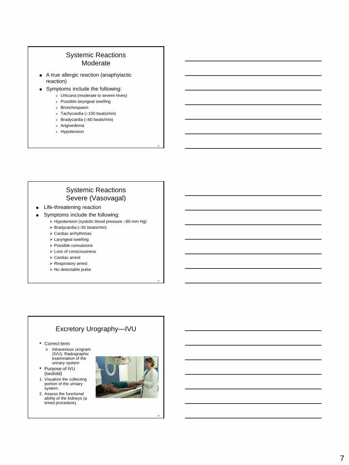
Evaluation Criteria
Posterior Oblique
• Elevated side: Kidney is parallel to plane of IR
• Downside: Ureter is free of superimposition from spine
• Entire urinary system visualized
• No motion
• Appropriate technique employed
• Minute marker visible
RPO
49
Voiding Cystourethrography
(VCUG)
Technical—Positioning Factors IR size: 24 × 30 cm (10 × 12 in), lengthwise
70-80 kV, grid
CR perpendicular to symphysis pubis
52
Cystogram
CR 2 inches (5 cm) superior to symphysis pubis
53
AP: 10°-15° caudad
Posterior oblique: 45°-60°
11
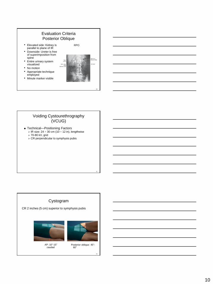
Evaluation Criteria
Cystogram
AP: urinary bladder
not superimposed
by pubic bones
Posterior obliques:
urinary bladder not
superimposed by
lower limbs
Distal ureter,
bladder, proximal
urethra on male to
be included
54
Voiding Cystourethrography
Purpose: Functional study of the bladder and
urethra
Performed after routine cystogram
Catheter removed and imaged while voiding
55
Female—AP Male—30° RPO
Retrograde Urography
Performed in surgery
Contrast media delivered retrograde through
catheter
58
12
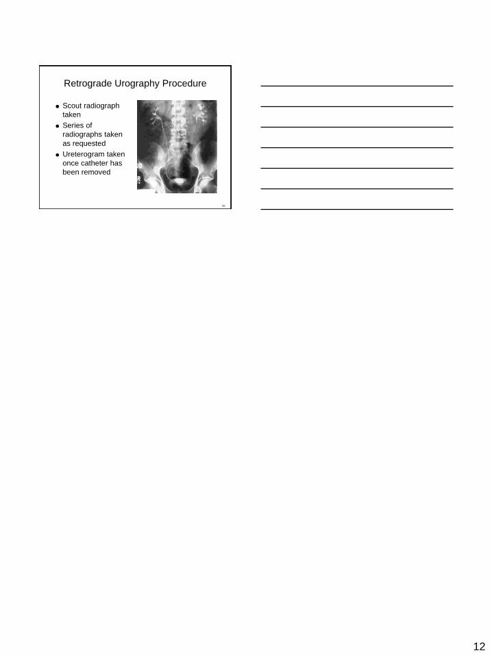
Retrograde Urography Procedure
Scout radiograph
taken
Series of
radiographs taken
as requested
Ureterogram taken
once catheter has
been removed
59