Embed Size (px)
Citation preview
Radiographic examinations of the temporomandibular joint
Dobó-Nagy, CsabaSemmelweis University
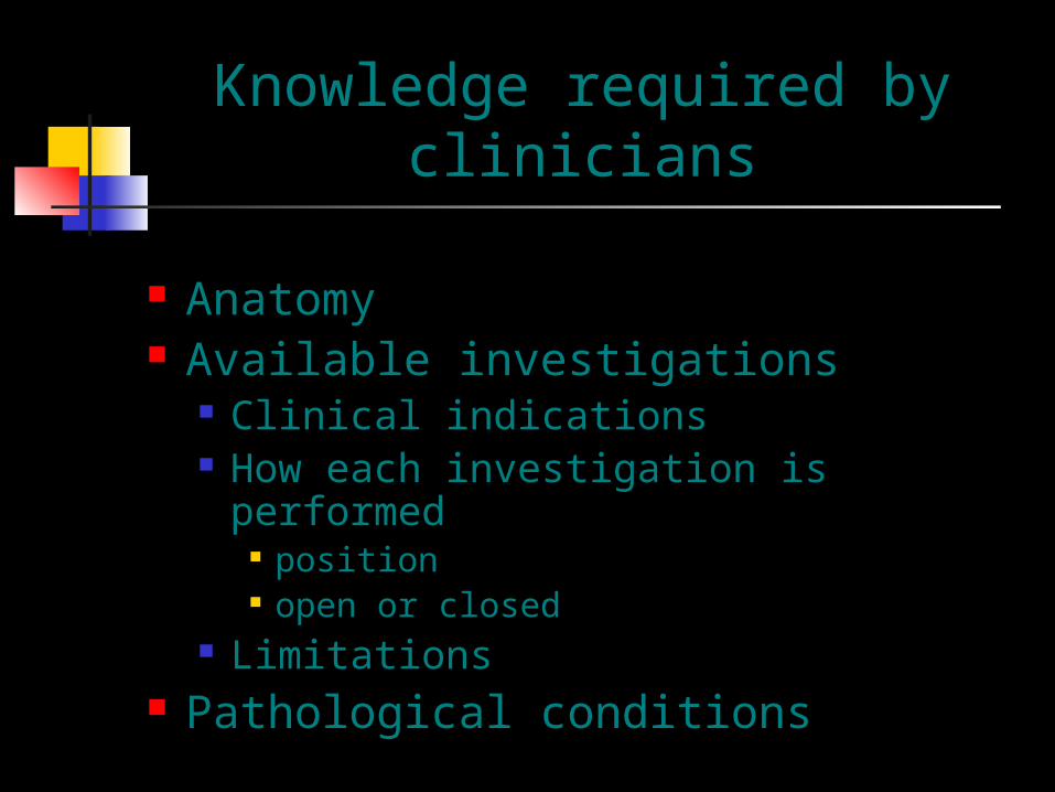
Knowledge required by clinicians
Anatomy Available investigations
Clinical indications How each investigation is performed
position open or closed
Limitations Pathological conditions
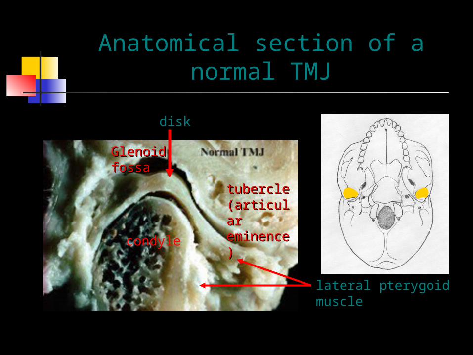
Anatomical section of a normal TMJ
condyle
disk
Glenoid fossaGlenoid fossa
tubercle tubercle (articular (articular eminence)eminence)
lateral pterygoid muscle

Skull base model
AA PP
LL RR

Visualization of the TMJ
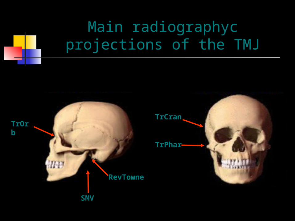
Main radiographyc projections of the TMJ
TrCran
TrPhar
TrOrb
SMV
RevTowne

Transcranial projection
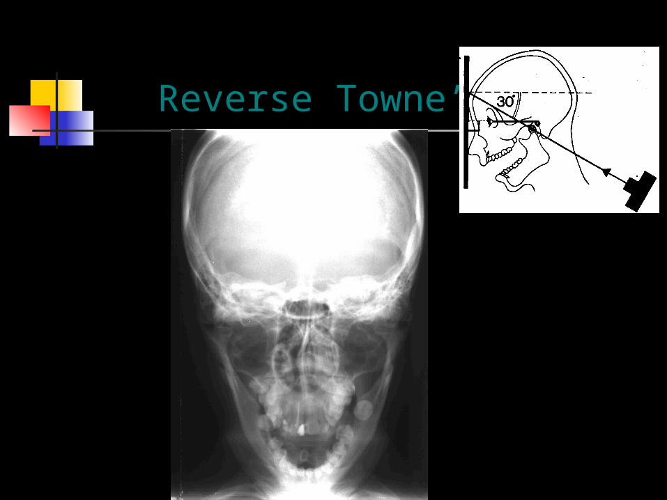
Reverse Towne’s

Transorbital
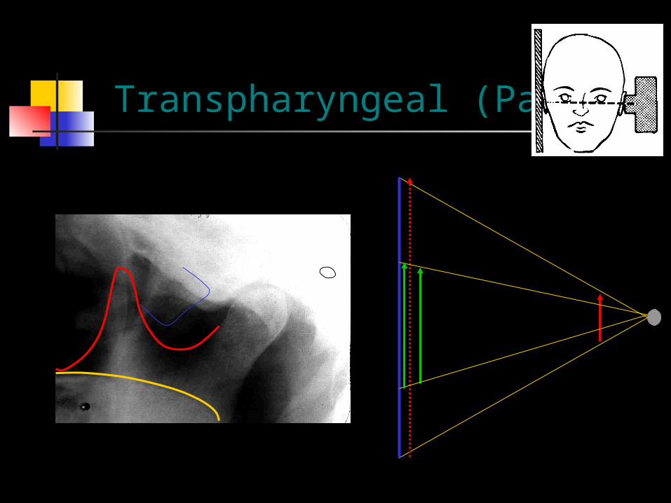
Transpharyngeal (Parma)
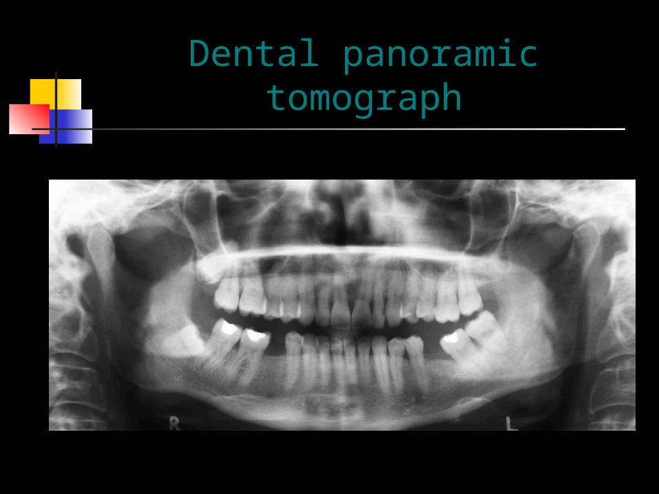
Dental panoramic tomograph
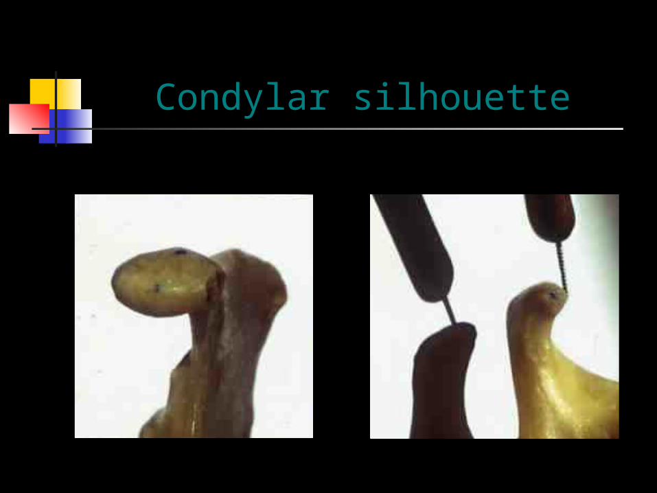
Condylar silhouette

Projection of panoramic tomography

Double lateral view of TMJ
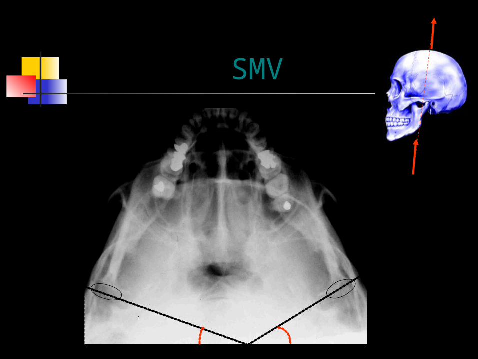
SMV
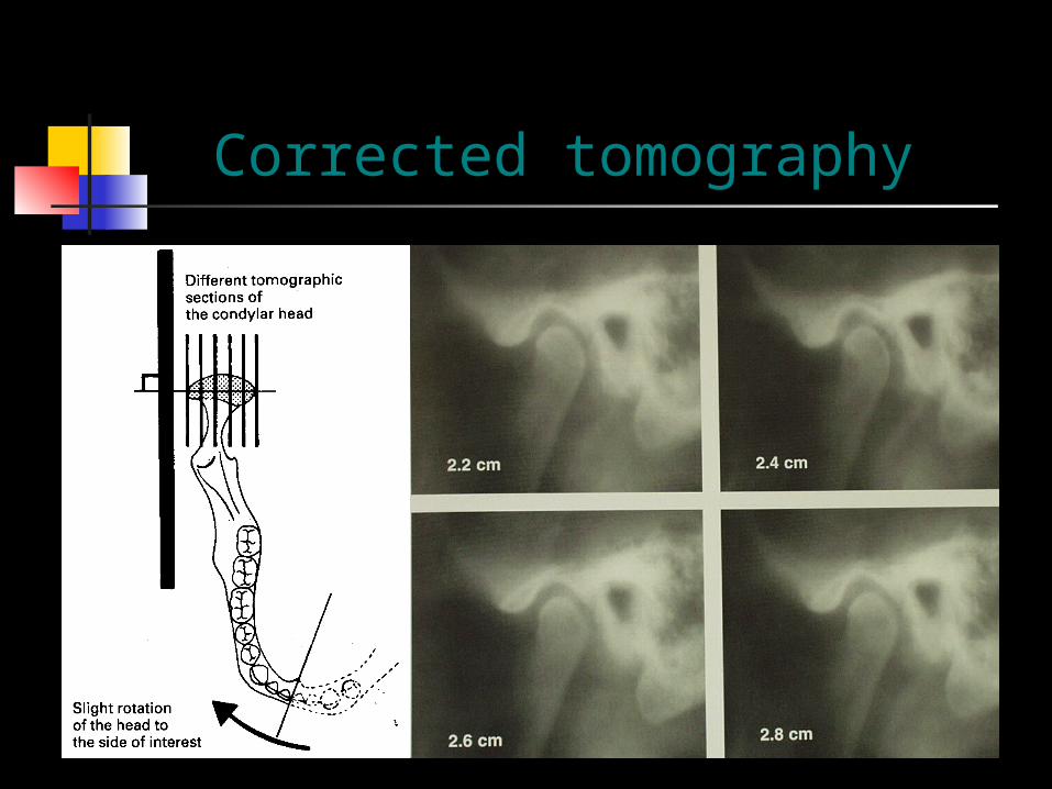
Corrected tomography

Limitations of routin radiographic projections
Soft tissue abnormalities Effusion Small and localised bony defects One aspect of the joint (except for
tomography)
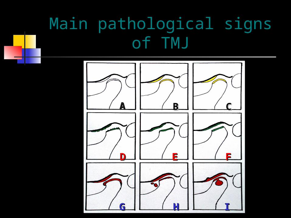
Main pathological signs of TMJ
AA BBBB CCCC
DDDD EEEE FFFF
GGGG HHHH IIII
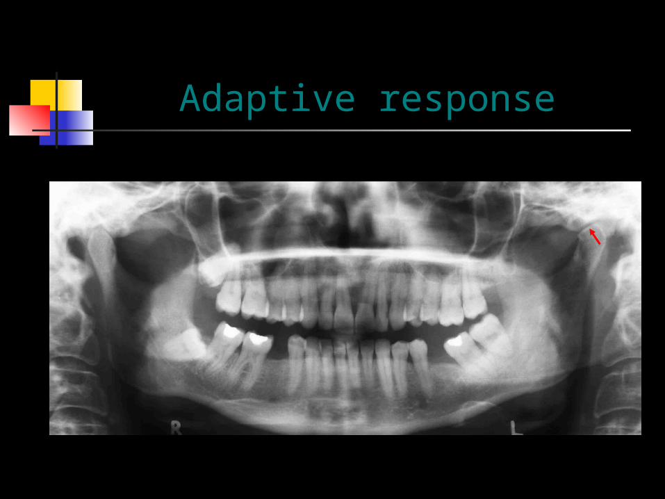
Adaptive response
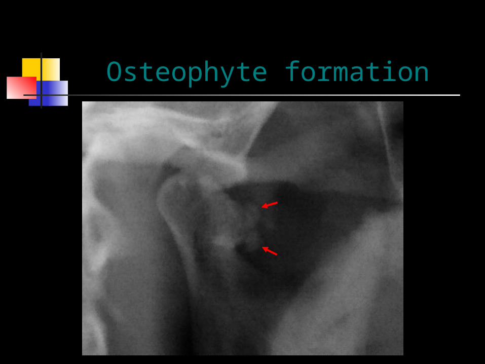
Osteophyte formation

Fracture
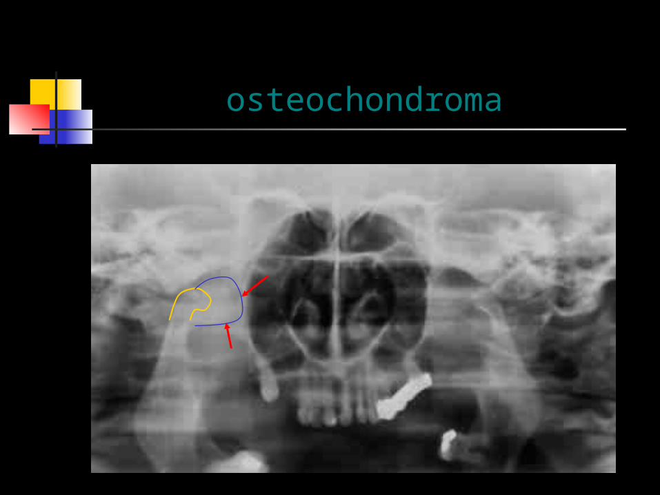
osteochondroma
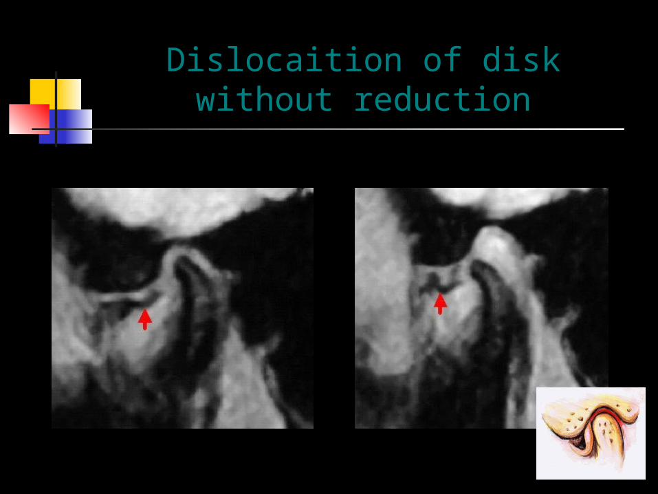
Dislocaition of disk without reduction

Diagnostic informations of MR
Disk displacement The integrity of the disk and its
soft tissue attachments Effusion (T2-weighted image) Integrity of the articular surface Research (gold standard)
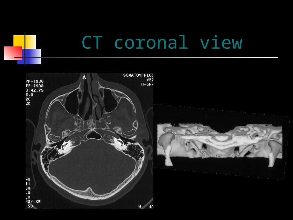
CT coronal view
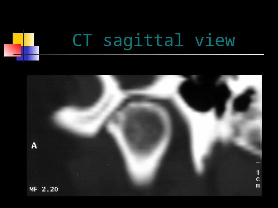
CT sagittal view
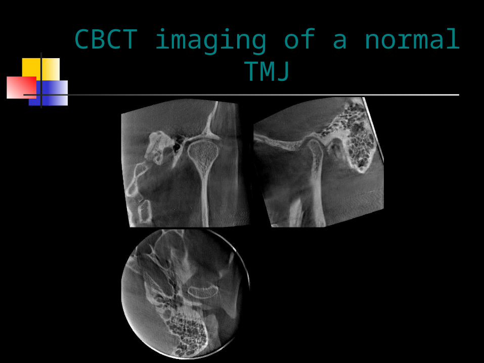
CBCT imaging of a normal TMJ
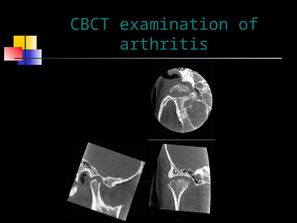
CBCT examination of arthritis
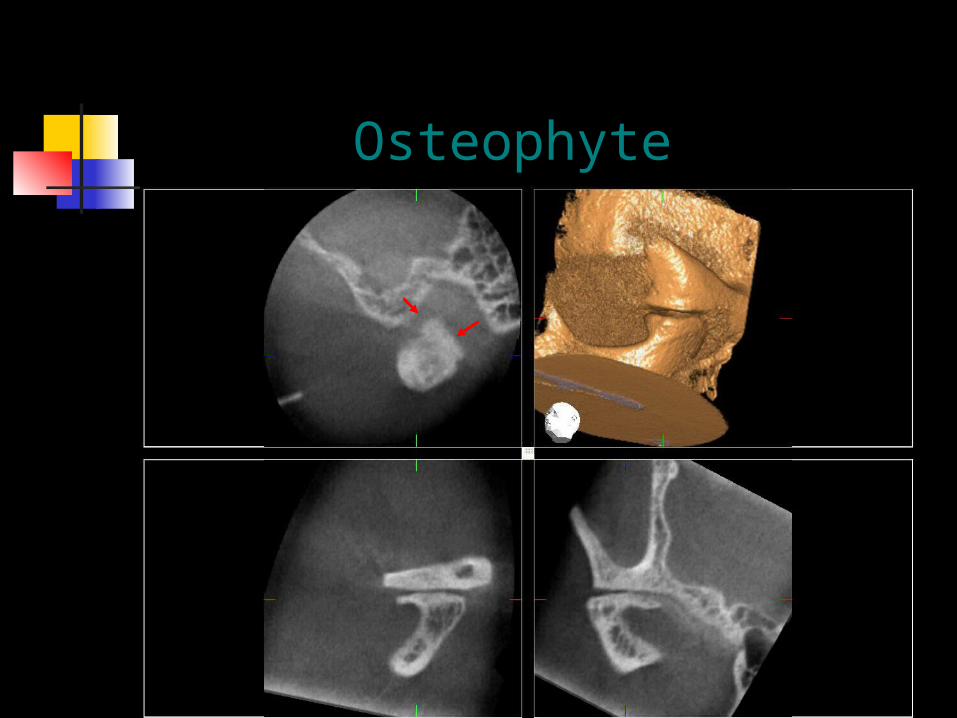
Osteophyte
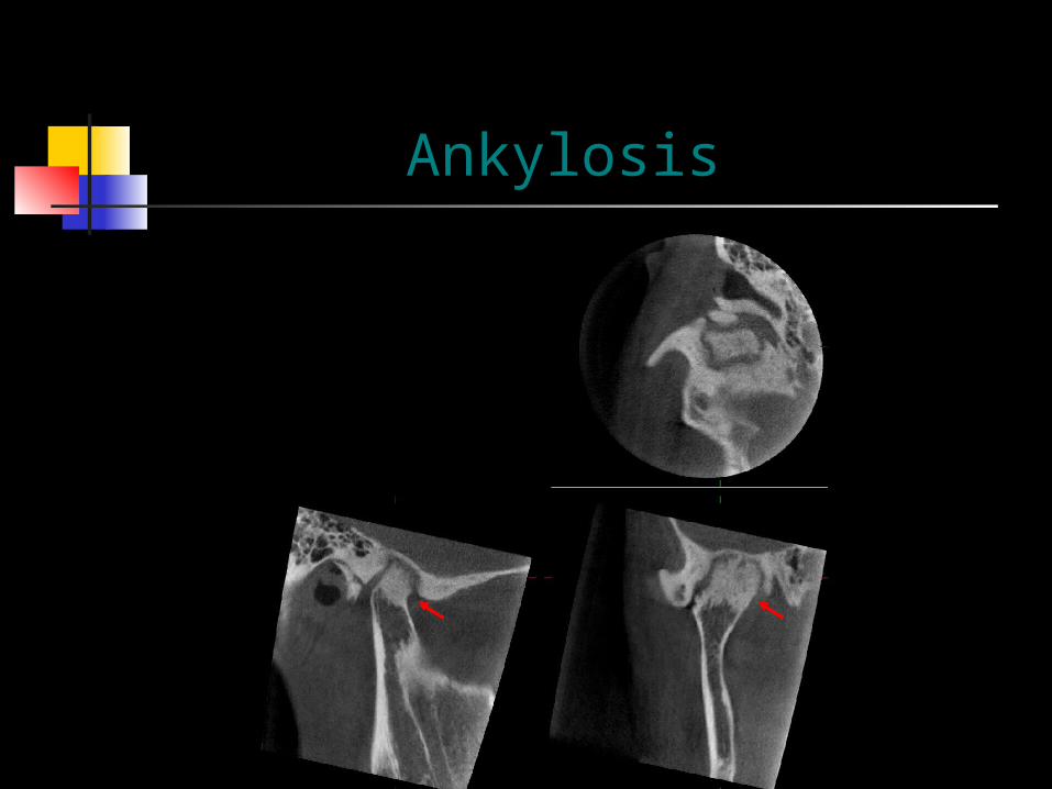
Ankylosis
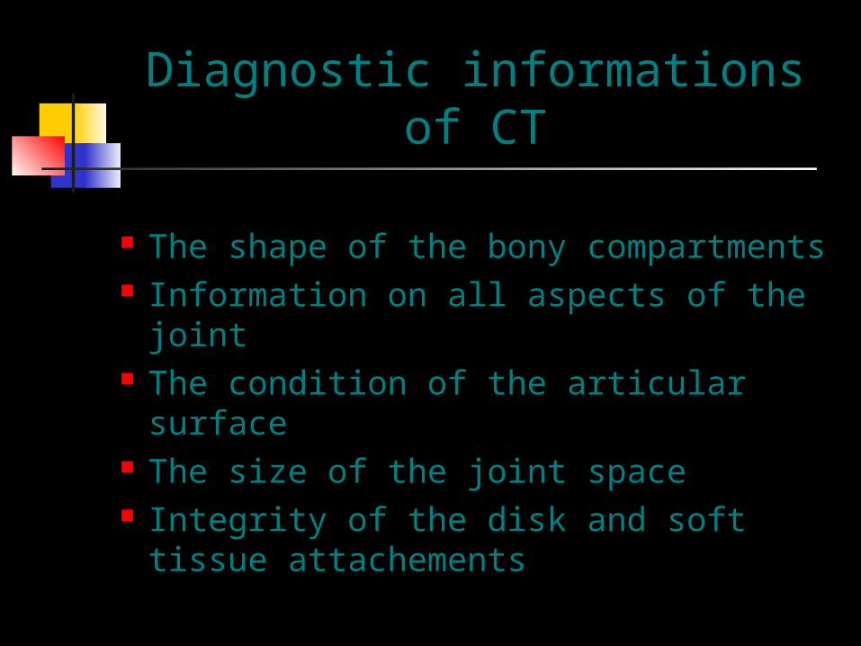
Diagnostic informations of CT
The shape of the bony compartments Information on all aspects of the joint The condition of the articular surface The size of the joint space Integrity of the disk and soft tissue
attachements

Size of TMJ space and soft tissue changes
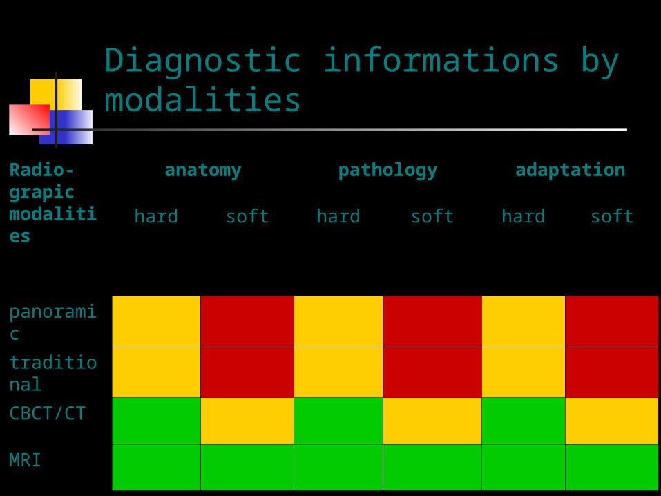
Diagnostic informations by modalities
Radio-grapic modalities
anatomy pathology adaptation
hard soft hard soft hard soft
panoramic
traditional
CBCT/CT
MRI
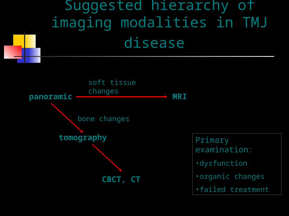
Suggested hierarchy of imaging modalities in TMJ disease
panoramic MRI
tomography
CBCT, CT
soft tissue changes
bone changes
Primary examination:
•dysfunction
•organic changes
•failed treatment

Summary of views of main projections
projection area of joint shown
Transcranial lateral aspect
Transpharyngeal lateral aspect
Dental panoramic lateral aspect
Reverse Towne’s posterior view
Transorbital anterior view