Embed Size (px)
DESCRIPTION
Skeletal System. Radiographic Anatomy. Skull. Educational Objectives. By the end of this lecture you should be able to: Identify the anatomical parts of skull on diagrams and radiographs. Identify the different sutures of the skull - PowerPoint PPT Presentation
Citation preview

Radiographic Anatomy
Skeletal System
Skull
Educational Objectives
By the end of this lecture you should be able to: Identify the anatomical parts of skull on diagrams and
radiographs. Identify the different sutures of the skull State the surface land marks of skull and explain
its importance in radiographic positioning Explain how to hang skull radiographs on the
view box
4
1. Text book of radiographic positioning and related anatomy; by Kenneth L.Bontrager,6th edition. 2. Introduction to Human Anatomy and Physiology: by Eldra Pearl Solomon:W.B.Saunders Company 3. Handbook of Anatomy and physiology for Students of Medical Radiation Technology: Mallett.M:Jaspar
Websiteshttp://www6.district125.k12.il.us/science/anatomy/http://www.innerbody.com/htm/body.html http://www.e-radiography.net/
http://www.getbodysmart.com/index.htm
References
5
Skull
8 Cranial bones (cranium)
+
14 Facial bones
6
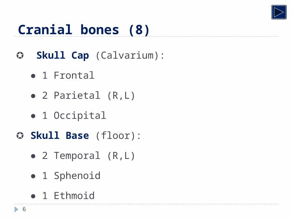
Cranial bones (8)
Skull Cap (Calvarium):
● 1 Frontal
● 2 Parietal (R,L)
● 1 Occipital
Skull Base (floor):
● 2 Temporal (R,L)
● 1 Sphenoid
● 1 Ethmoid
7
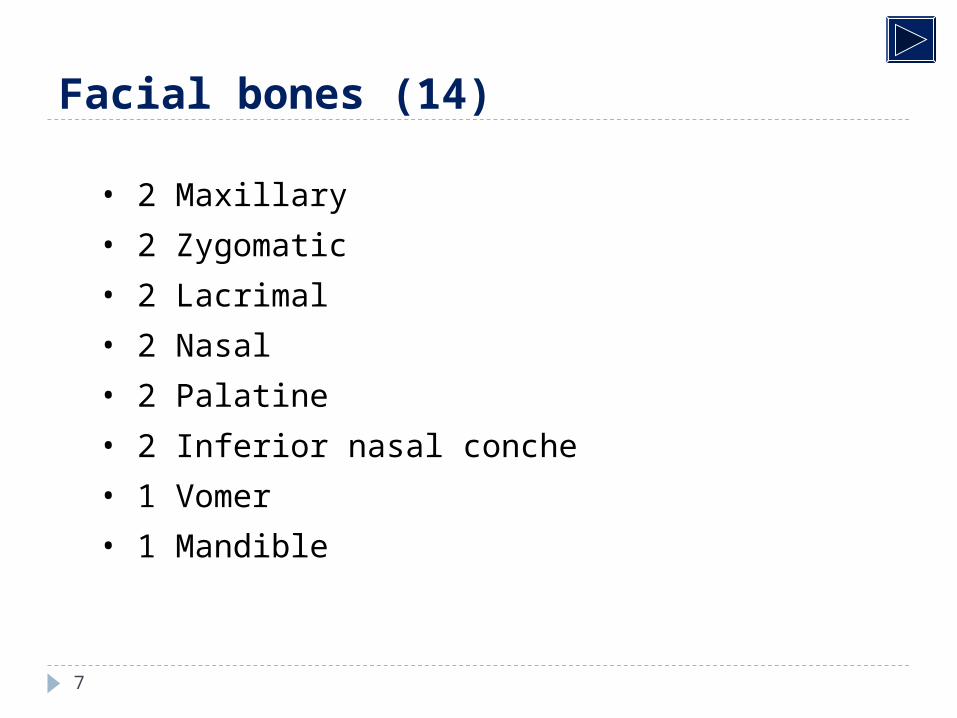
Facial bones (14)
• 2 Maxillary
• 2 Zygomatic
• 2 Lacrimal
• 2 Nasal
• 2 Palatine
• 2 Inferior nasal conche
• 1 Vomer
• 1 Mandible
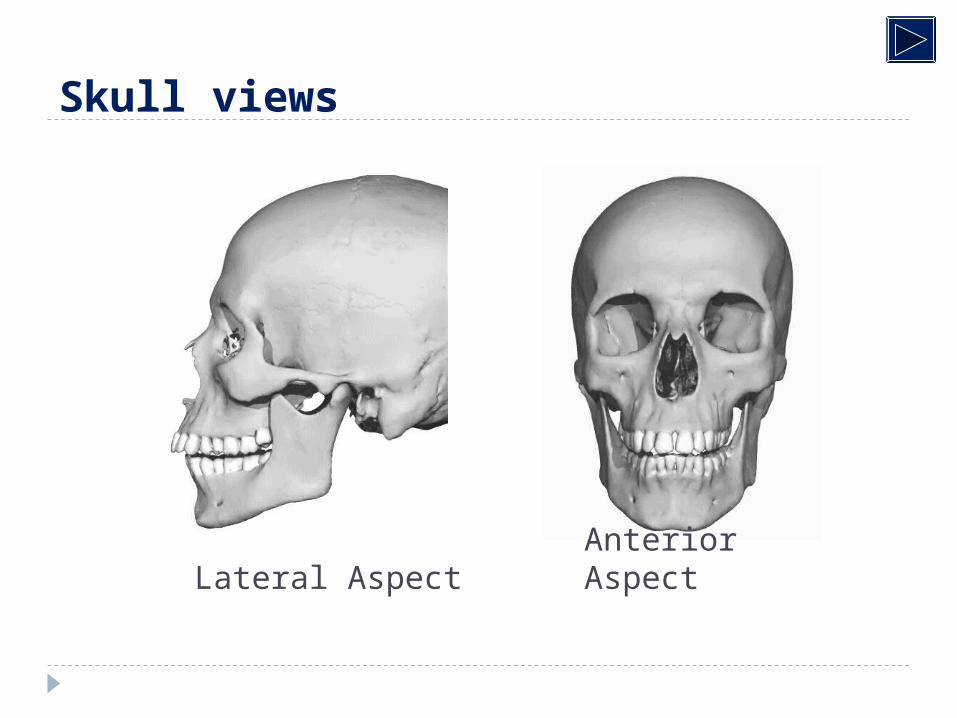
Skull views
Anterior AspectLateral Aspect
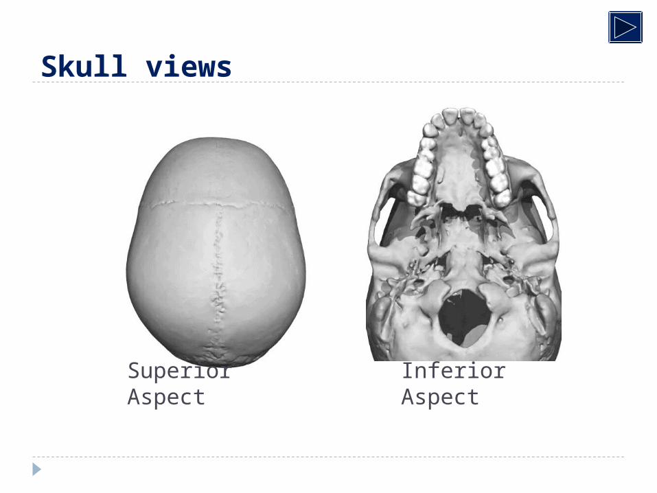
Skull views
Inferior AspectSuperior Aspect
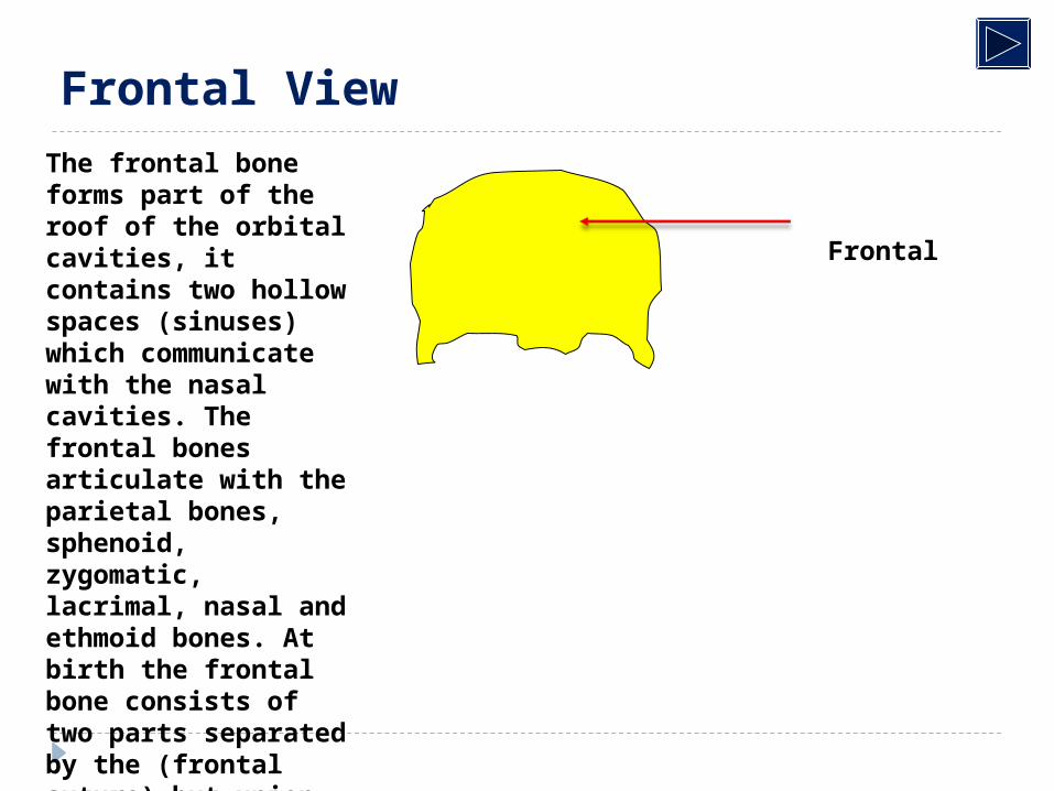
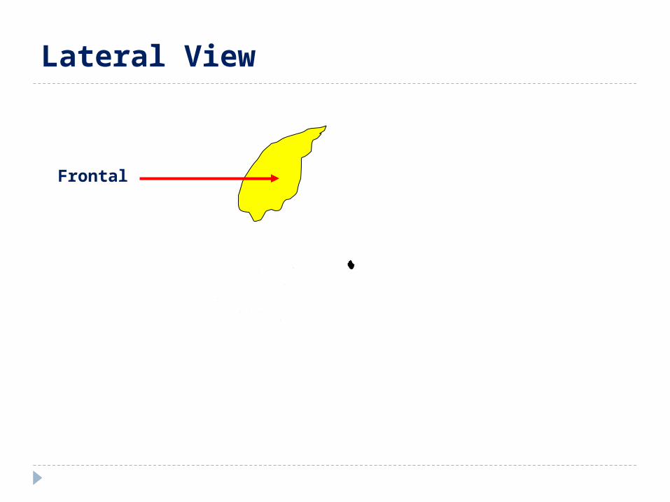
Frontal
Frontal ViewThe frontal bone forms part of the roof of the orbital cavities, it contains two hollow spaces (sinuses) which communicate with the nasal cavities. The frontal bones articulate with the parietal bones, sphenoid, zygomatic, lacrimal, nasal and ethmoid bones. At birth the frontal bone consists of two parts separated by the (frontal suture) but union is usually complete by the 8th year of life.
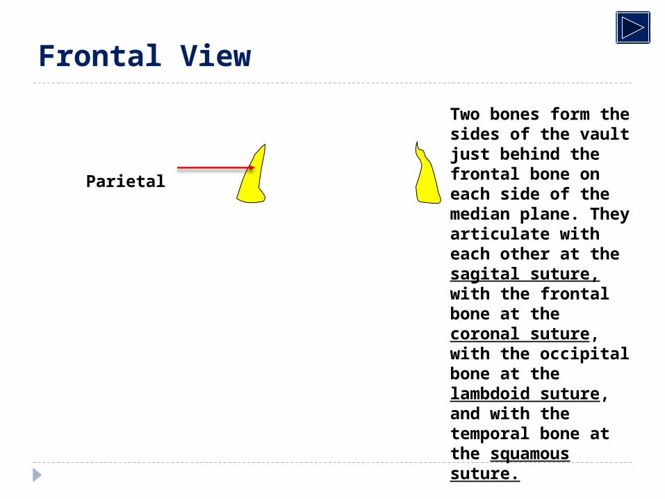
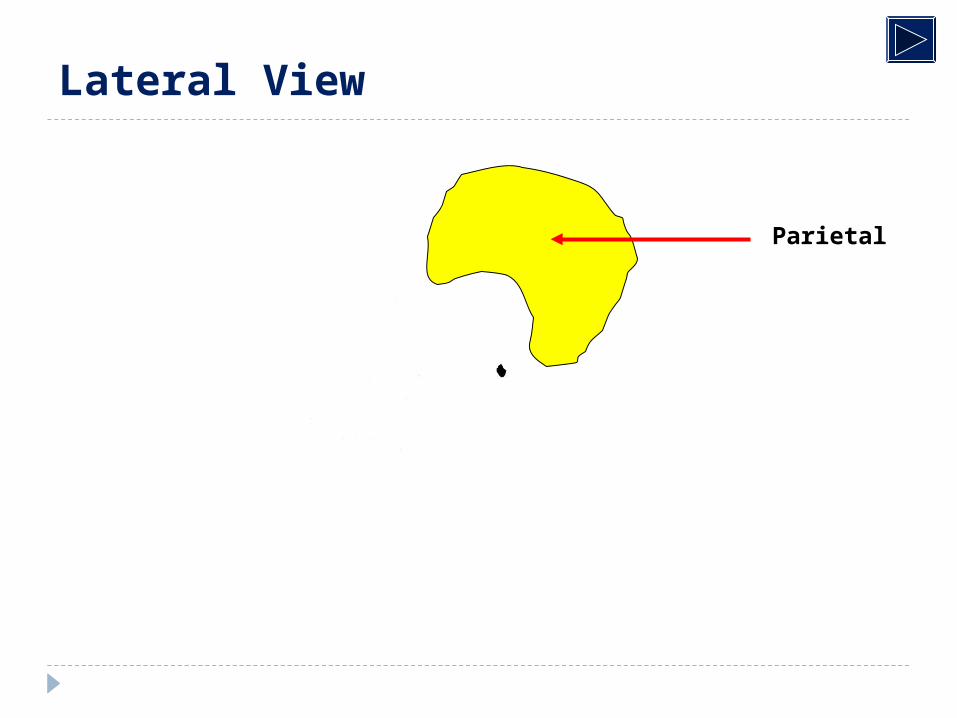
Parietal
Frontal View
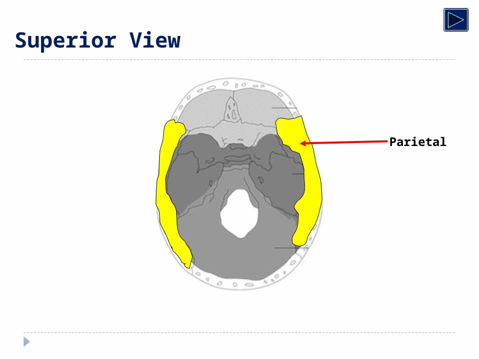
Two bones form the sides of the vault just behind the frontal bone on each side of the median plane. They articulate with each other at the sagital suture, with the frontal bone at the coronal suture, with the occipital bone at the lambdoid suture, and with the temporal bone at the squamous suture.
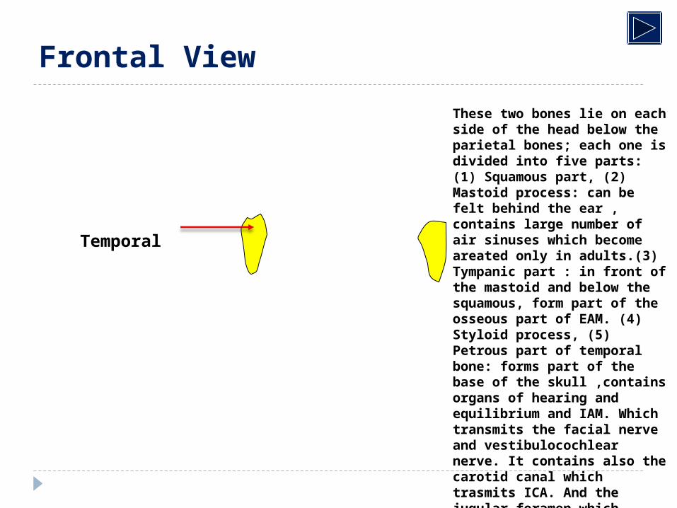
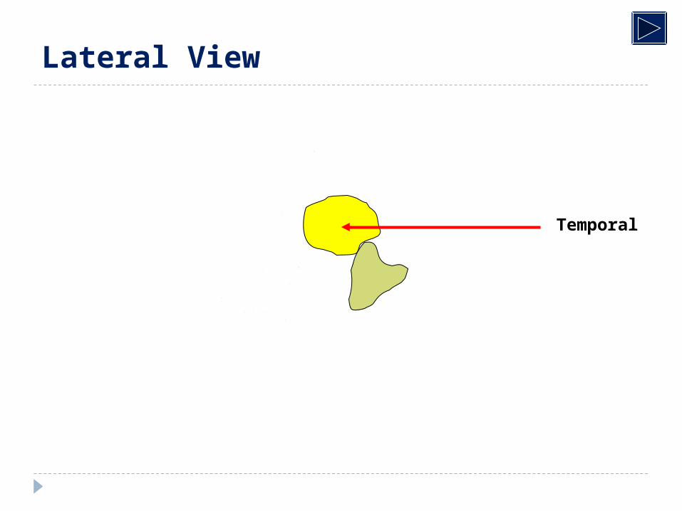
Temporal
Frontal View
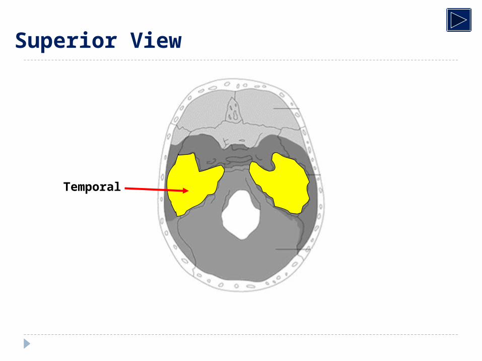
These two bones lie on each side of the head below the parietal bones; each one is divided into five parts: (1) Squamous part, (2) Mastoid process: can be felt behind the ear , contains large number of air sinuses which become areated only in adults.(3) Tympanic part : in front of the mastoid and below the squamous, form part of the osseous part of EAM. (4) Styloid process, (5) Petrous part of temporal bone: forms part of the base of the skull ,contains organs of hearing and equilibrium and IAM. Which transmits the facial nerve and vestibulocochlear nerve. It contains also the carotid canal which trasmits ICA. And the jugular foramen which transmits the IJV.
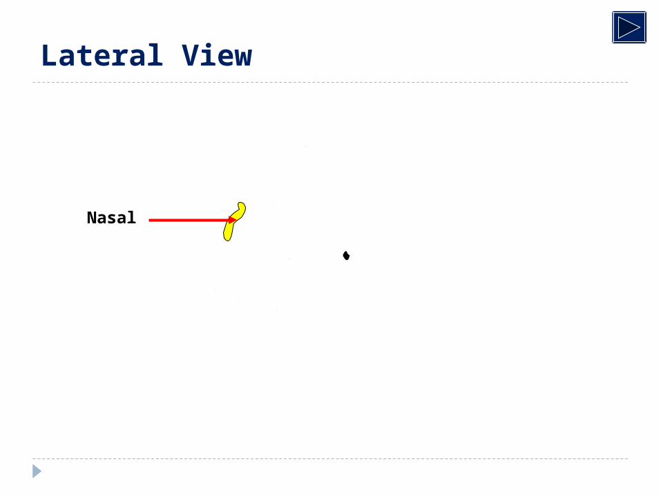
Nasal
Frontal View
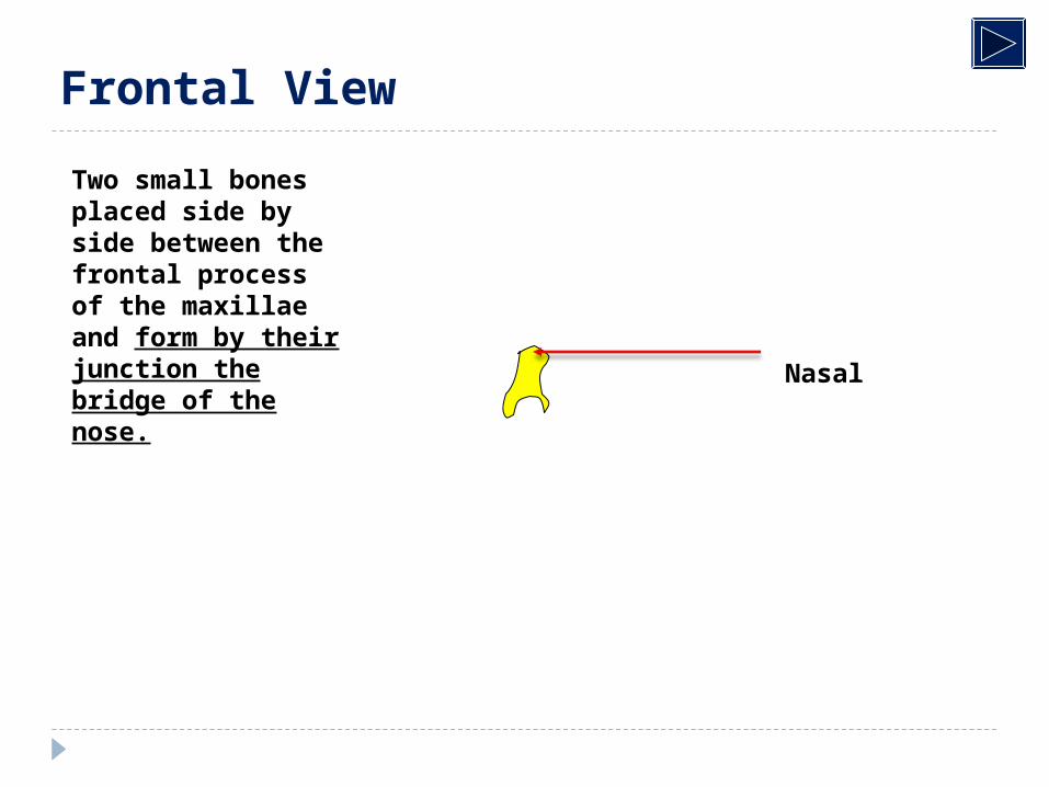
Two small bones placed side by side between the frontal process of the maxillae and form by their junction the bridge of the nose.
Vomer
Frontal View
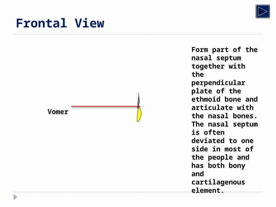
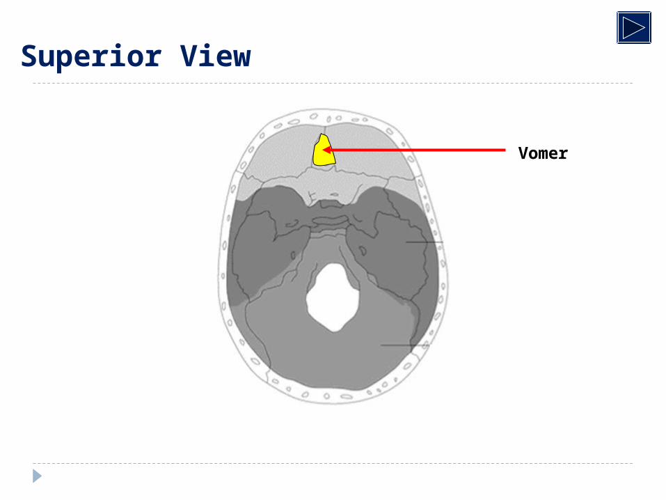
Form part of the nasal septum together with the perpendicular plate of the ethmoid bone and articulate with the nasal bones. The nasal septum is often deviated to one side in most of the people and has both bony and cartilagenous element.
Zygoma
Frontal View
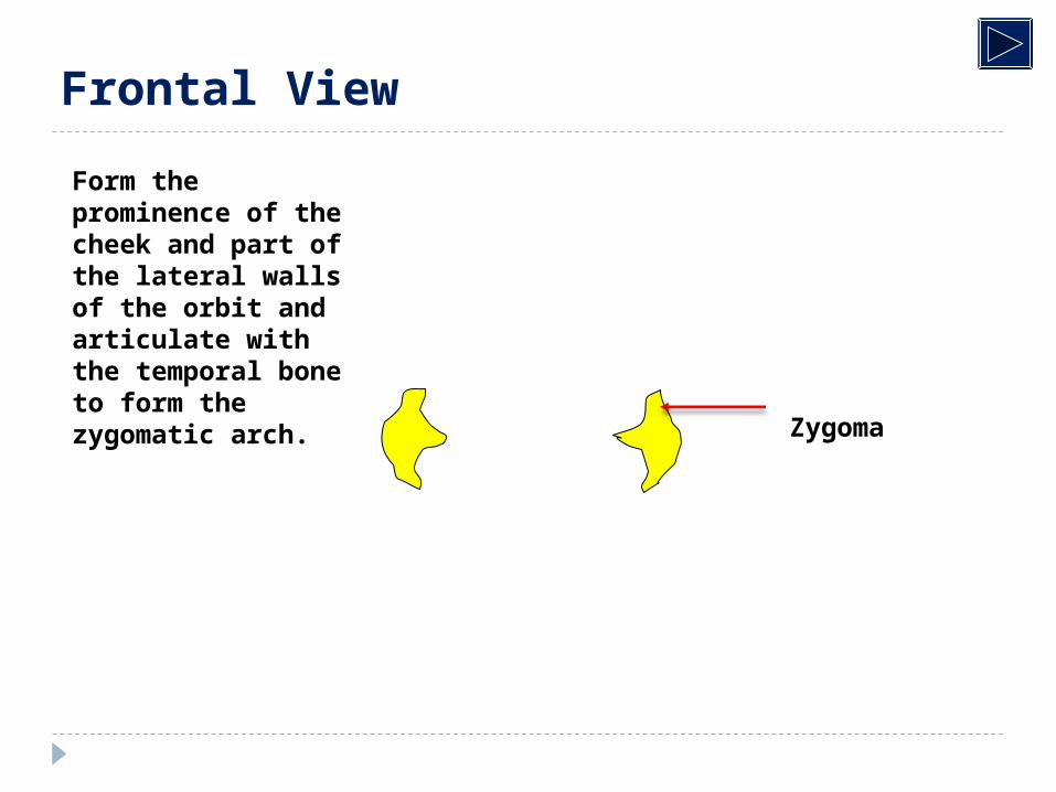
Form the prominence of the cheek and part of the lateral walls of the orbit and articulate with the temporal bone to form the zygomatic arch.
Maxilla
Frontal View
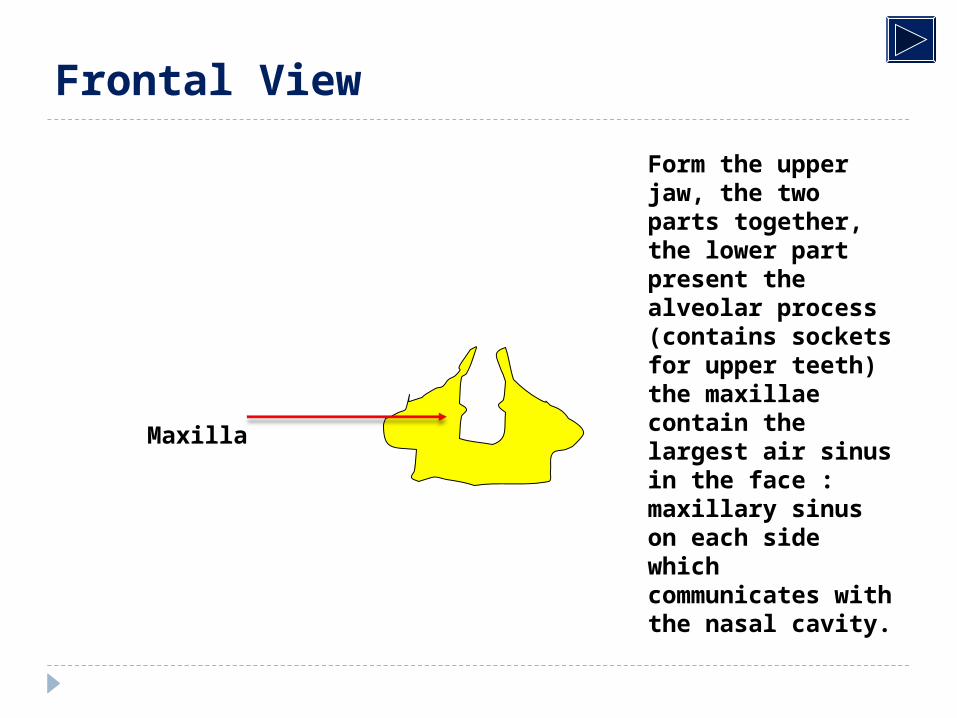
Form the upper jaw, the two parts together, the lower part present the alveolar process (contains sockets for upper teeth) the maxillae contain the largest air sinus in the face : maxillary sinus on each side which communicates with the nasal cavity.
Mandible
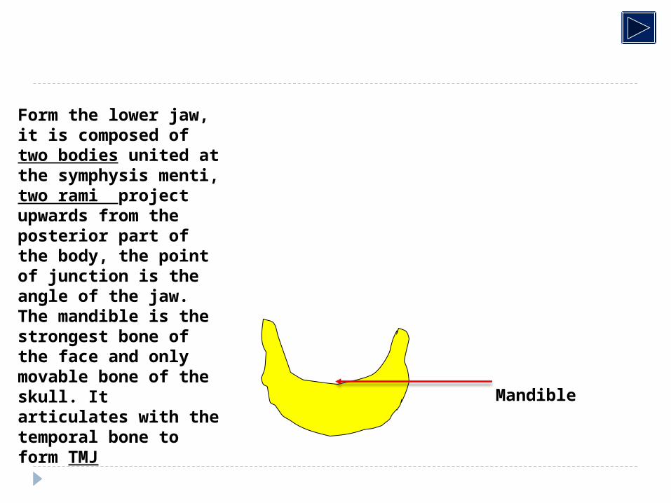
Form the lower jaw, it is composed of two bodies united at the symphysis menti, two rami project upwards from the posterior part of the body, the point of junction is the angle of the jaw. The mandible is the strongest bone of the face and only movable bone of the skull. It articulates with the temporal bone to form TMJ
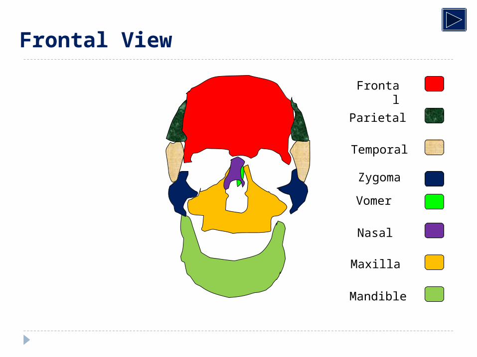
Frontal
Parietal
Temporal
Zygoma
Nasal
Vomer
Maxilla
Mandible
Frontal View
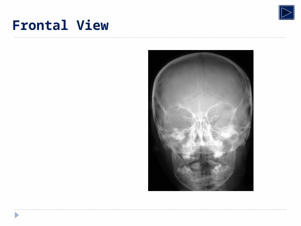
Frontal View
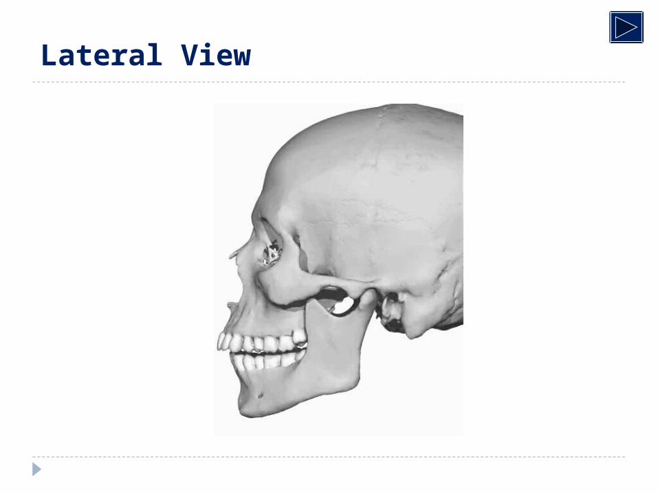
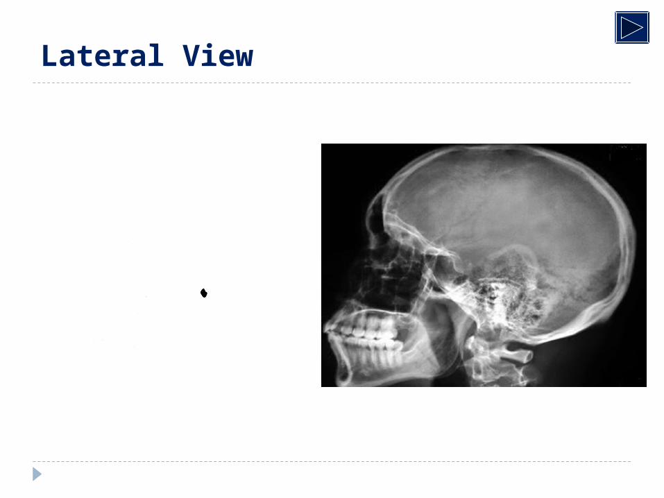
Lateral View
Frontal
Lateral View
Parietal
Lateral View
Temporal
Lateral View
Nasal
Lateral View
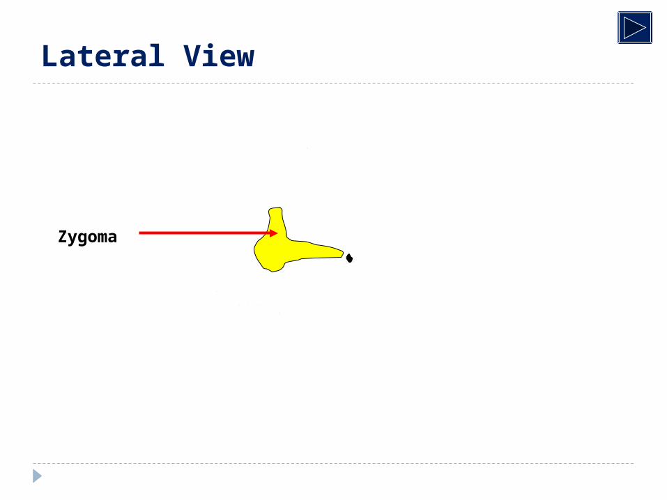
Zygoma
Lateral View
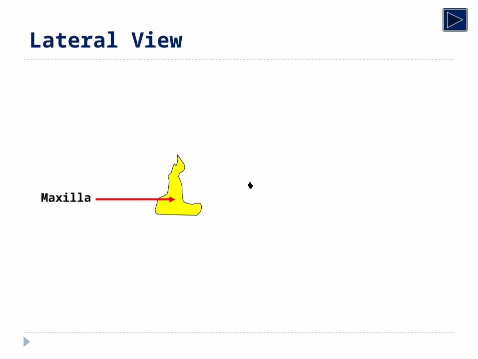
Maxilla
Lateral View
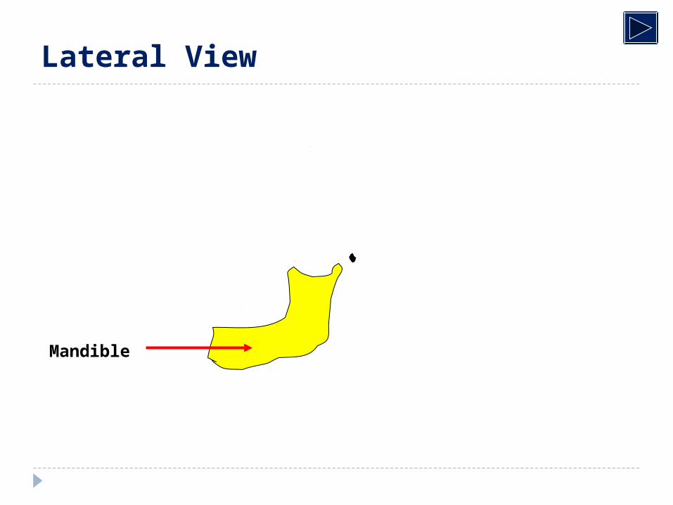
Mandible
Lateral View
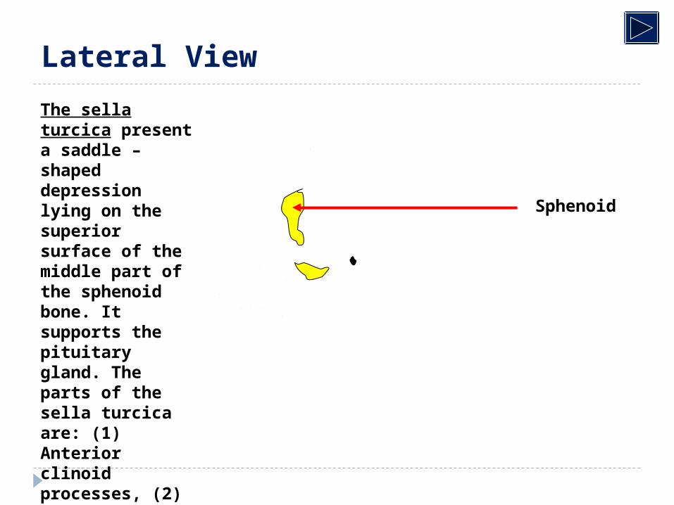
Sphenoid
Lateral View
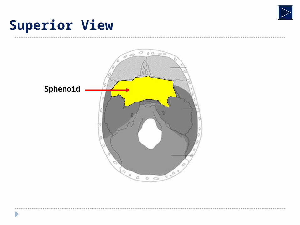
The sella turcica present a saddle –shaped depression lying on the superior surface of the middle part of the sphenoid bone. It supports the pituitary gland. The parts of the sella turcica are: (1) Anterior clinoid processes, (2) Dorsum sellae = posterior clinoid processes, (3) Lamina dura which cover the floor of sella, (4) Sphenoid air sinuses.
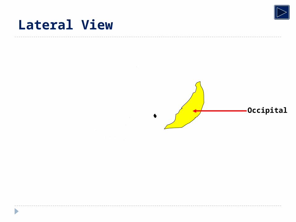
Occipital
Lateral View
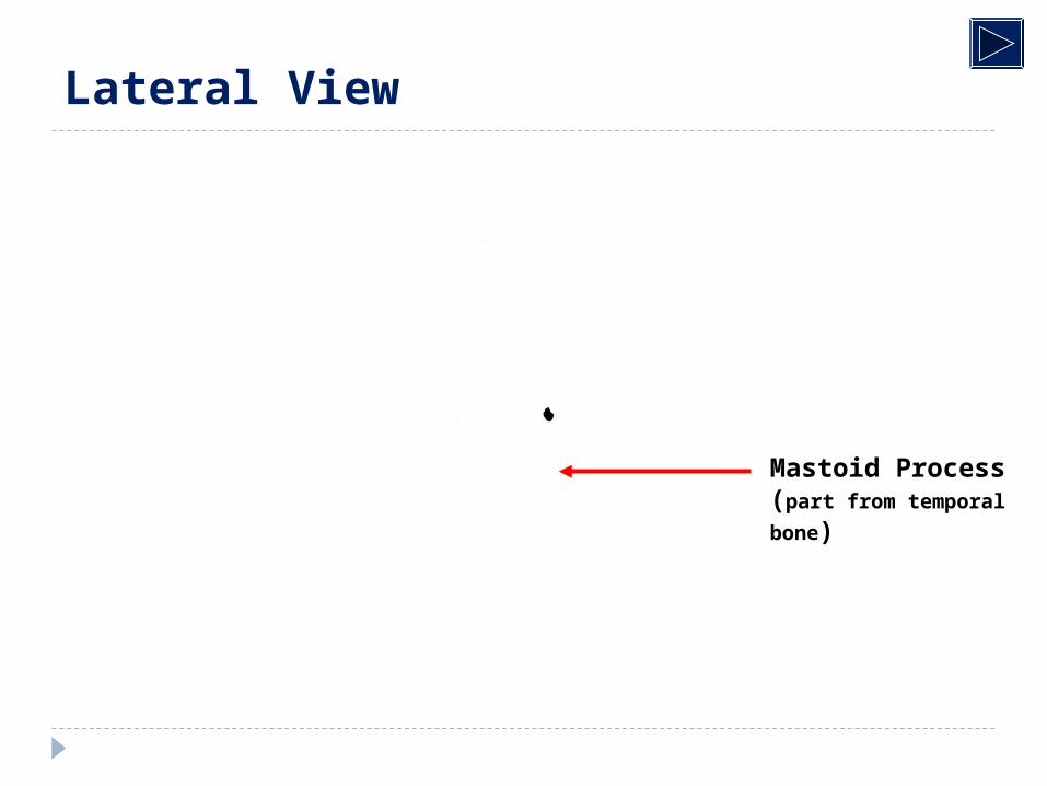
Mastoid Process(part from temporal bone)
Lateral View
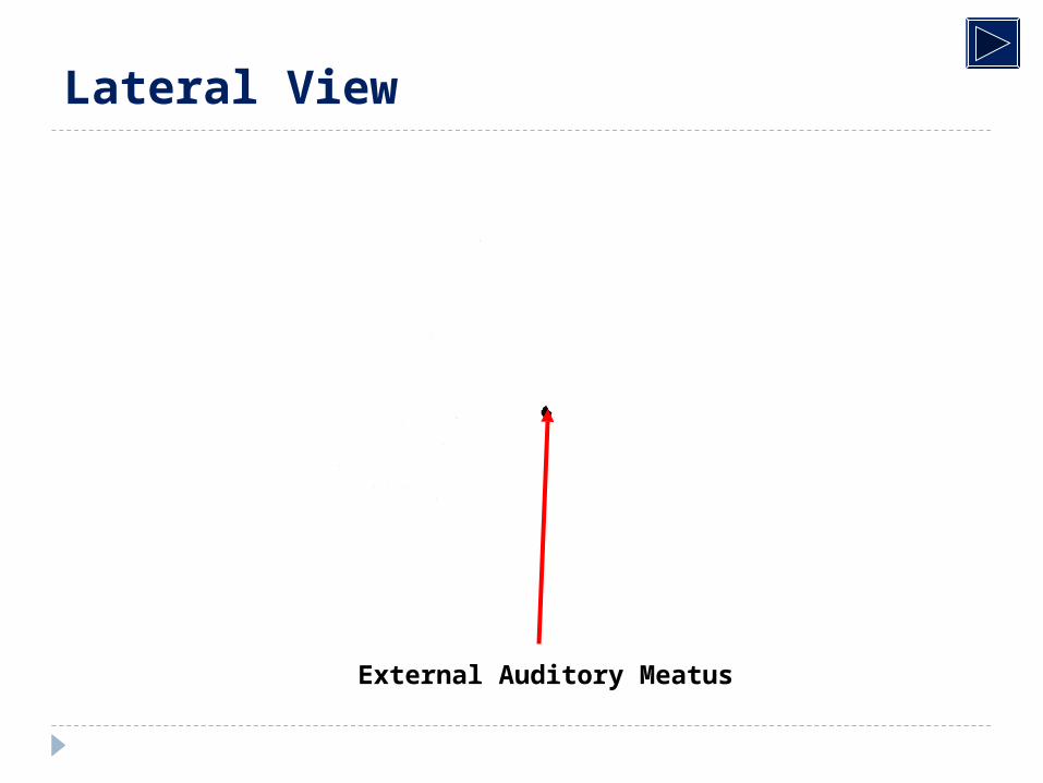
External Auditory Meatus
Lateral View
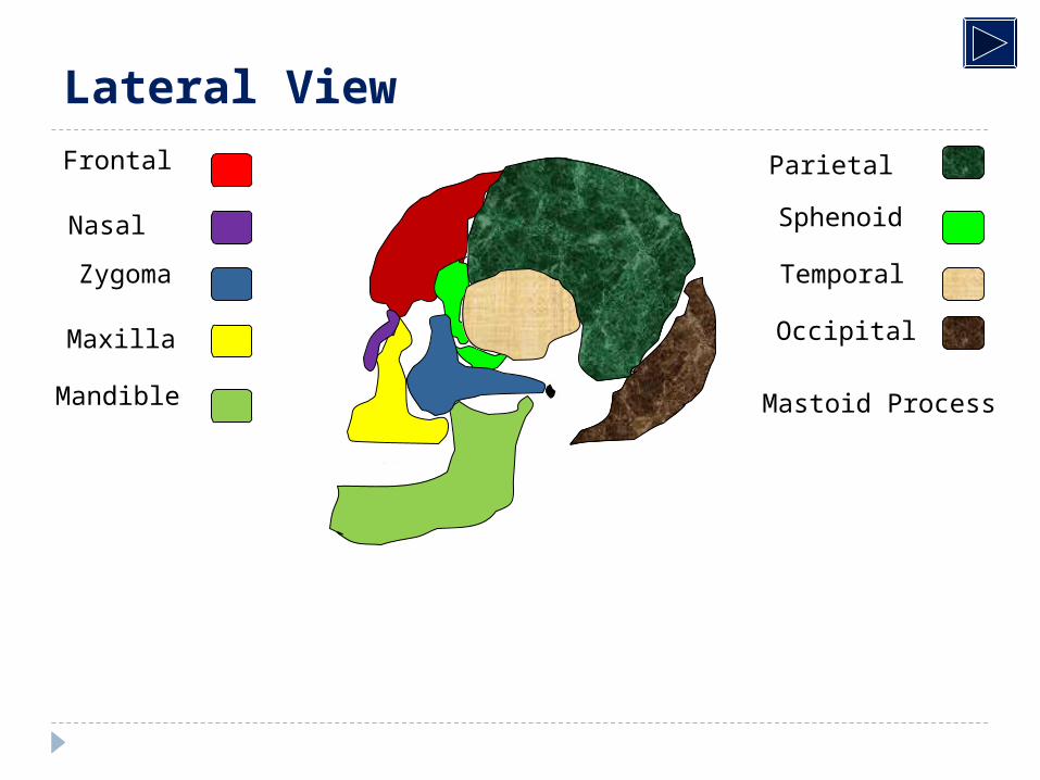
Frontal
Nasal
Zygoma
Maxilla
Mandible
Parietal
Sphenoid
Temporal
Occipital
Mastoid Process
Lateral View
Lateral View
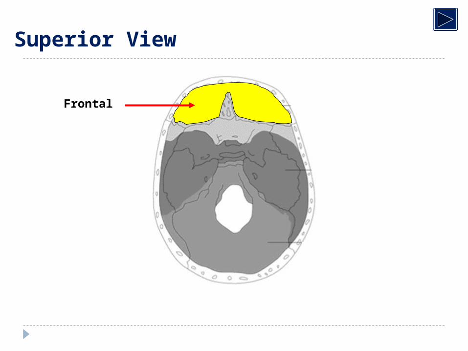
Frontal
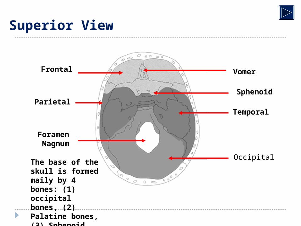
Superior View
Superior View
Parietal
Superior View
Temporal
Superior View
Vomer
Superior View
Sphenoid
Superior View
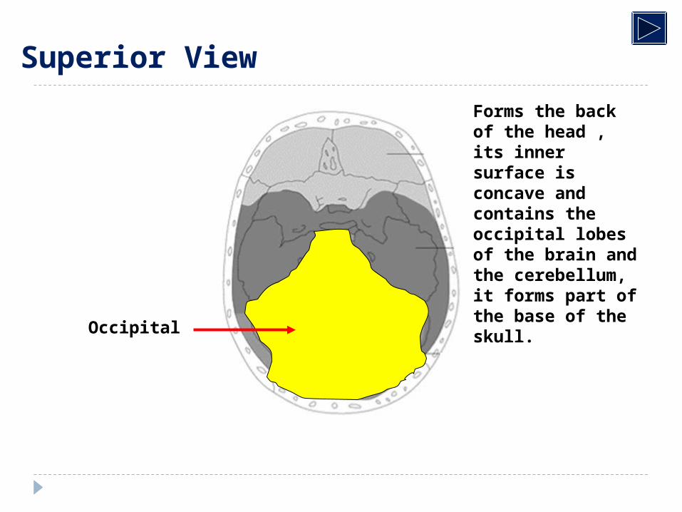
Occipital
Forms the back of the head , its inner surface is concave and contains the occipital lobes of the brain and the cerebellum, it forms part of the base of the skull.
Superior View
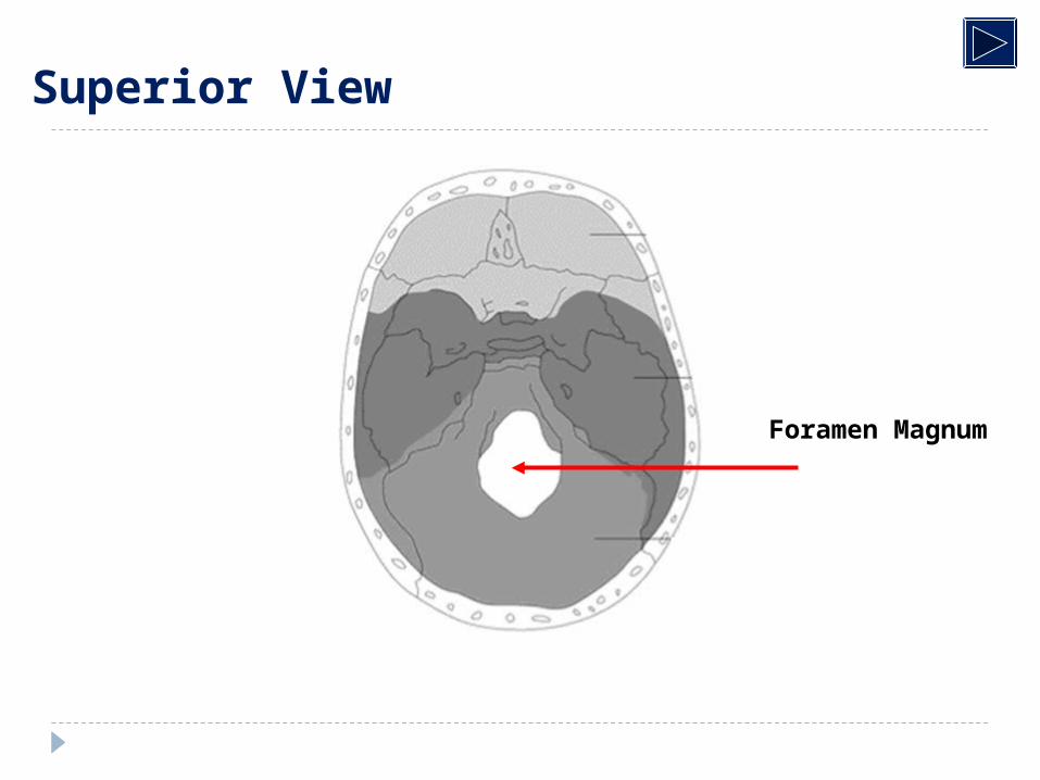
Foramen Magnum
VomerFrontal
Parietal
Occipital
Temporal
ForamenMagnum
Sphenoid
Superior View
The base of the skull is formed maily by 4 bones: (1) occipital bones, (2) Palatine bones, (3) Sphenoid bone, (4) Ethmoid bone.
Sutures
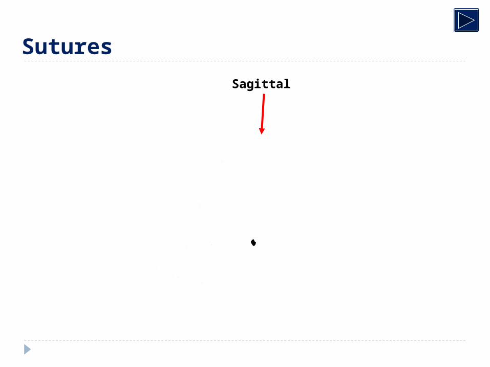
Sagittal
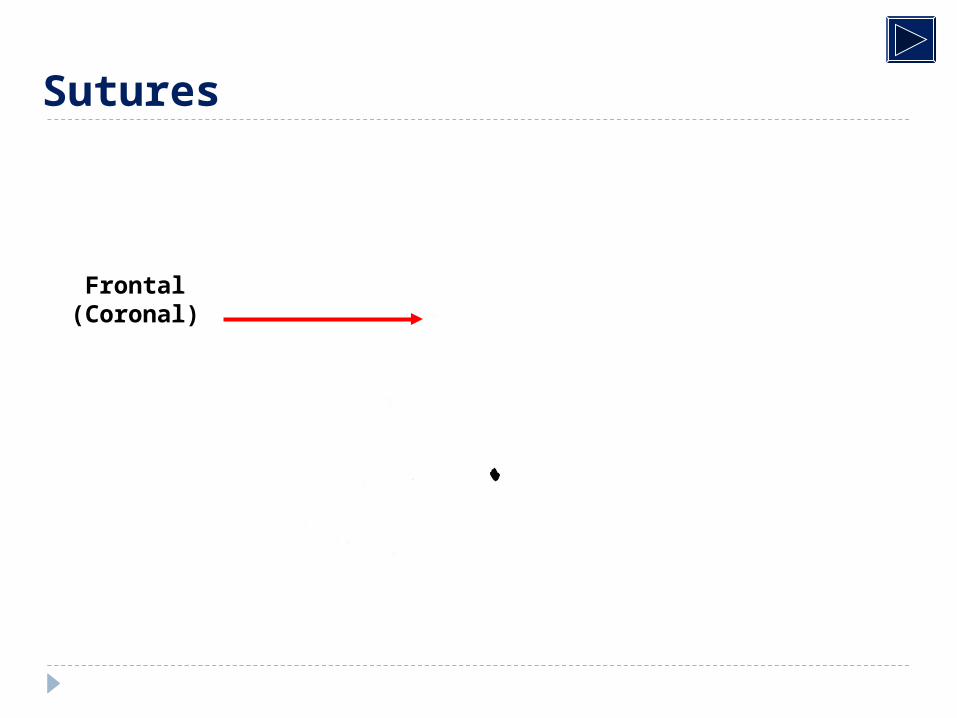
Sutures
Sutures
Frontal)Coronal(
Sutures
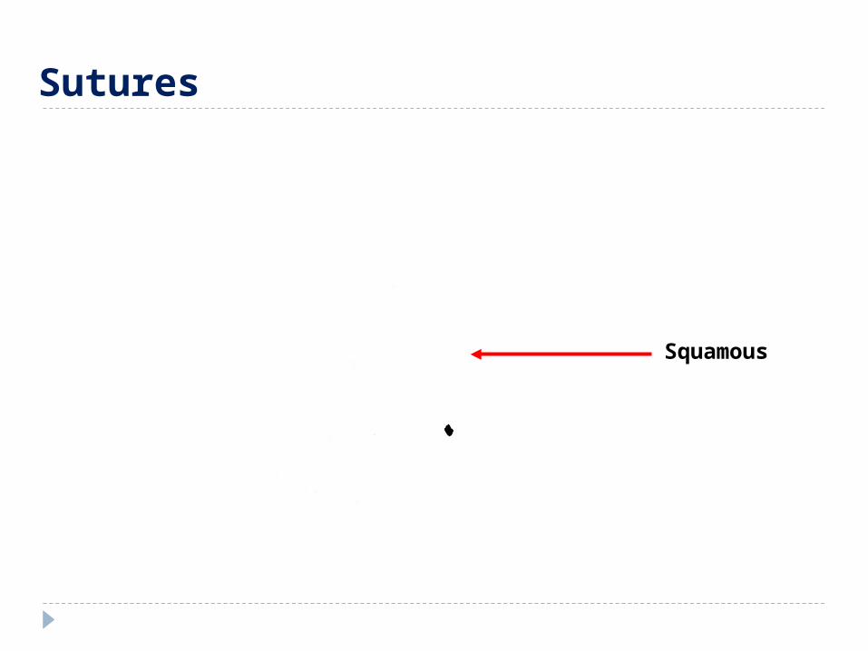
Squamous
Sutures
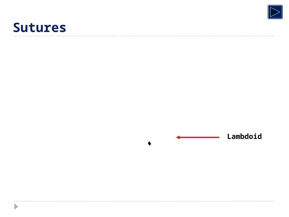
Lambdoid
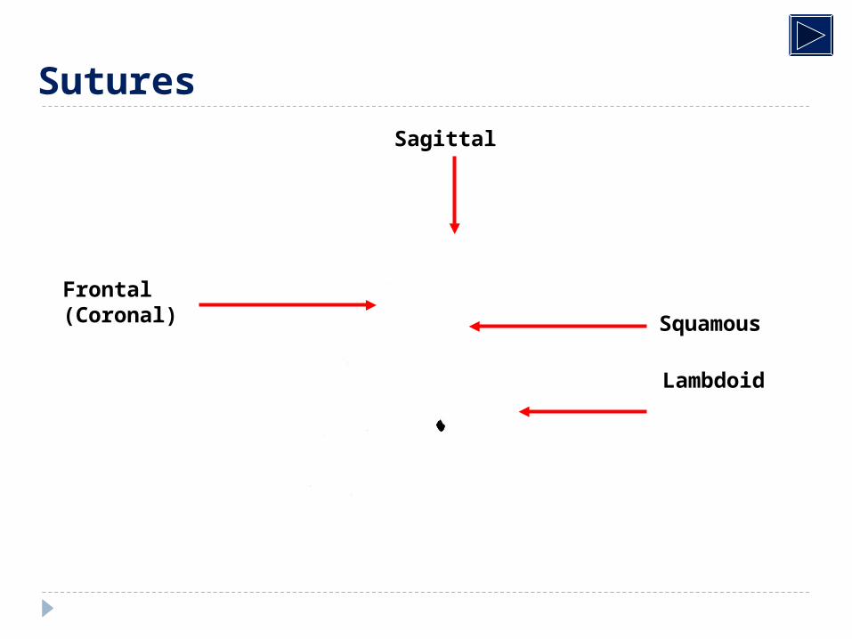
Frontal)Coronal(
Sagittal
Squamous
Lambdoid
Sutures
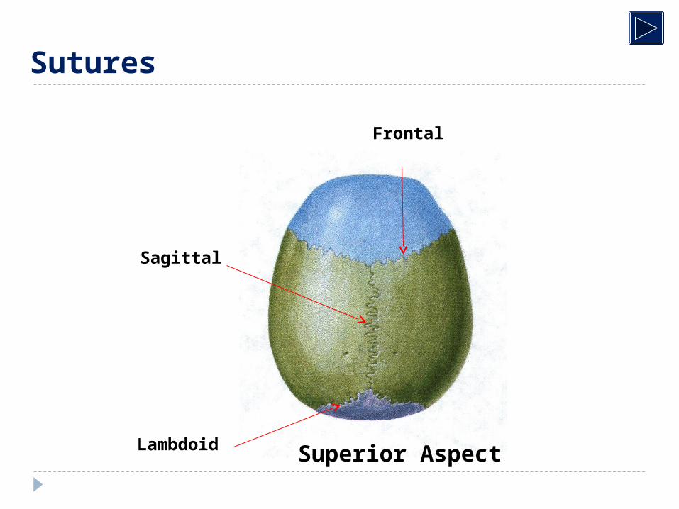
Sagittal
Lambdoid
Frontal
Superior Aspect
Sutures
49
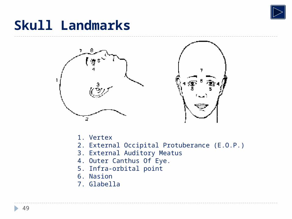
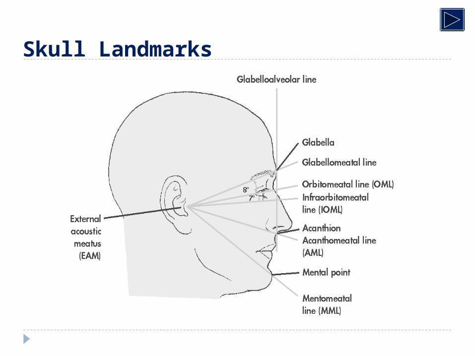
Skull Landmarks
1. Vertex 2. External Occipital Protuberance (E.O.P.) 3. External Auditory Meatus 4. Outer Canthus Of Eye. 5. Infra-orbital point 6. Nasion 7. Glabella
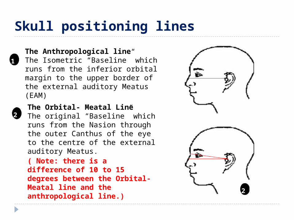
The Anthropological lineThe Isometric “Baseline” which runs from the inferior orbital margin to the upper border of the external auditory Meatus (EAM)
1
1
2
Skull positioning lines
The Orbital- Meatal LineThe original “Baseline” which runs from the Nasion through the outer Canthus of the eye to the centre of the external auditory Meatus.
( Note: there is a difference of 10 to 15 degrees between the Orbital- Meatal line and the anthropological line.) 2
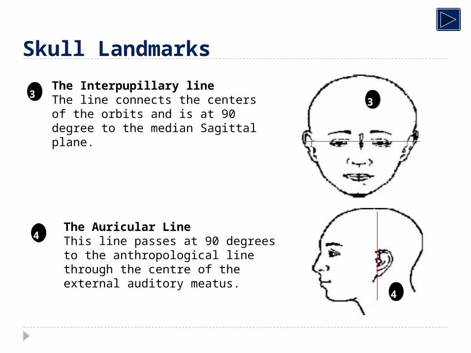
The Interpupillary lineThe line connects the centers of the orbits and is at 90 degree to the median Sagittal plane.
3
4
4
3
The Auricular LineThis line passes at 90 degrees to the anthropological line through the centre of the external auditory meatus.
Skull Landmarks
Skull Landmarks
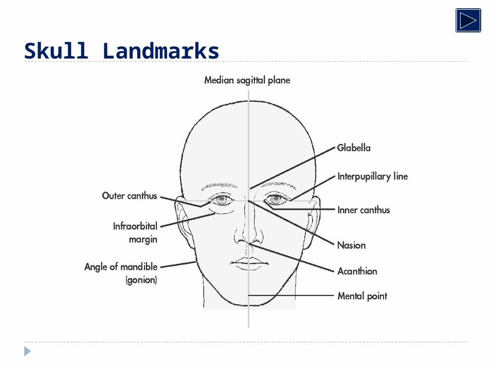
Skull Landmarks
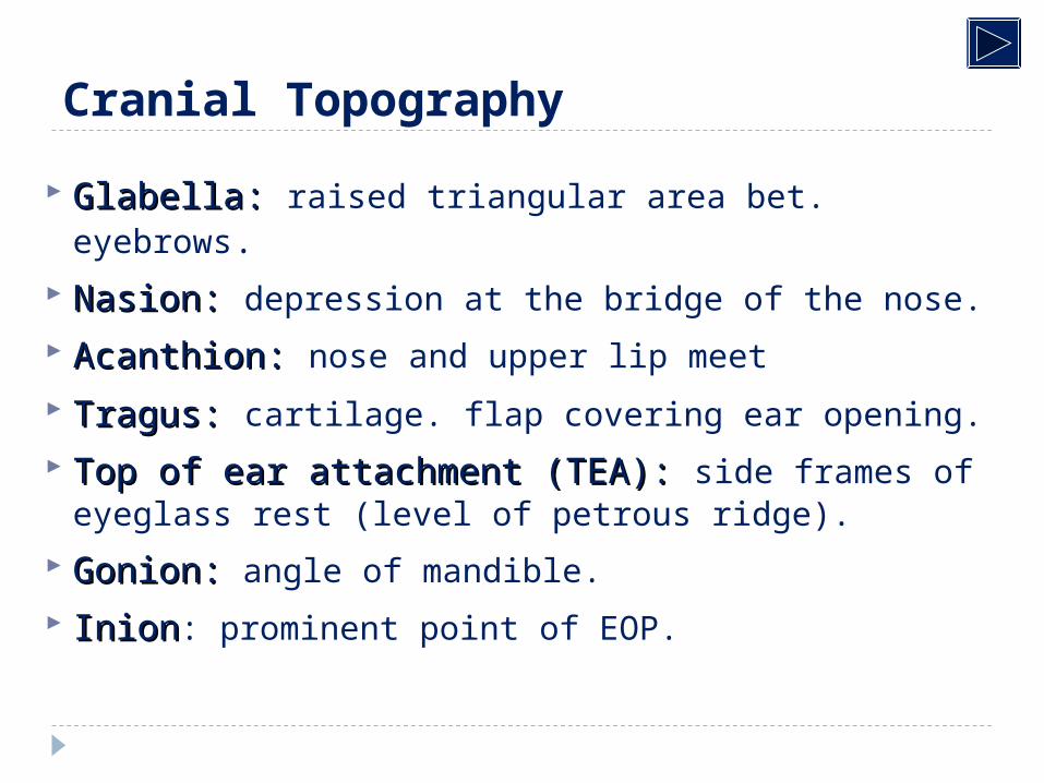
Cranial Topography
Glabella:Glabella: raised triangular area bet. eyebrows. Nasion:Nasion: depression at the bridge of the nose.
Acanthion:Acanthion: nose and upper lip meet
Tragus:Tragus: cartilage. flap covering ear opening. Top of ear attachment (TEA):Top of ear attachment (TEA): side frames of
eyeglass rest (level of petrous ridge).
Gonion:Gonion: angle of mandible.
InionInion: prominent point of EOP.
Skull Series: Basic Projections
1. PA 0º
2. PA axial 15º
3. AP axial
4. Lateral
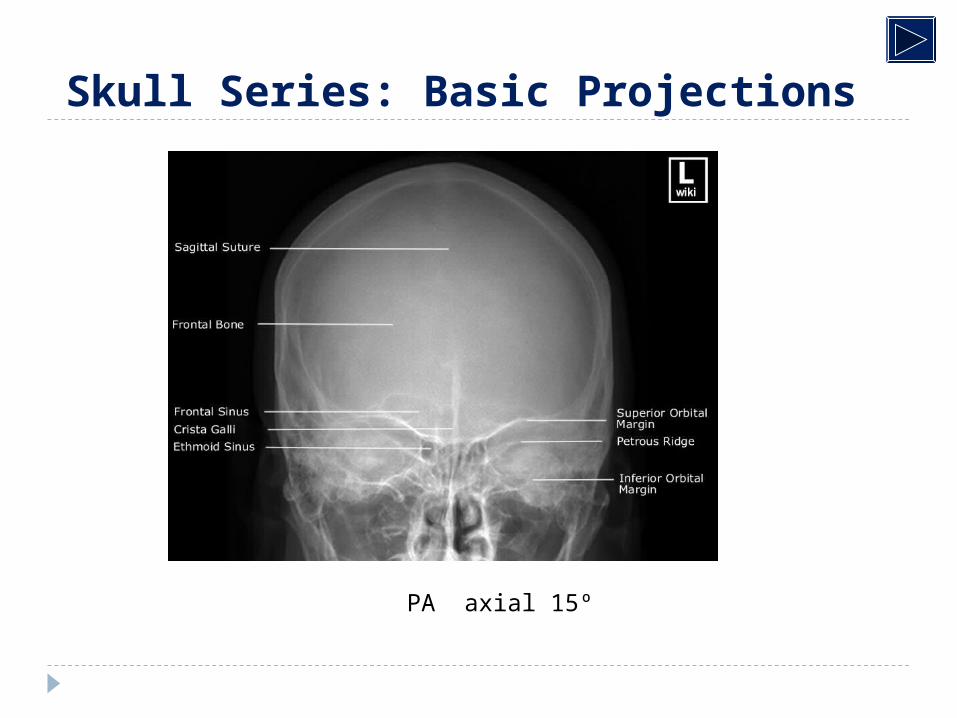
Skull Series: Basic Projections
PA axial 15º
Skull Series: Basic Projections
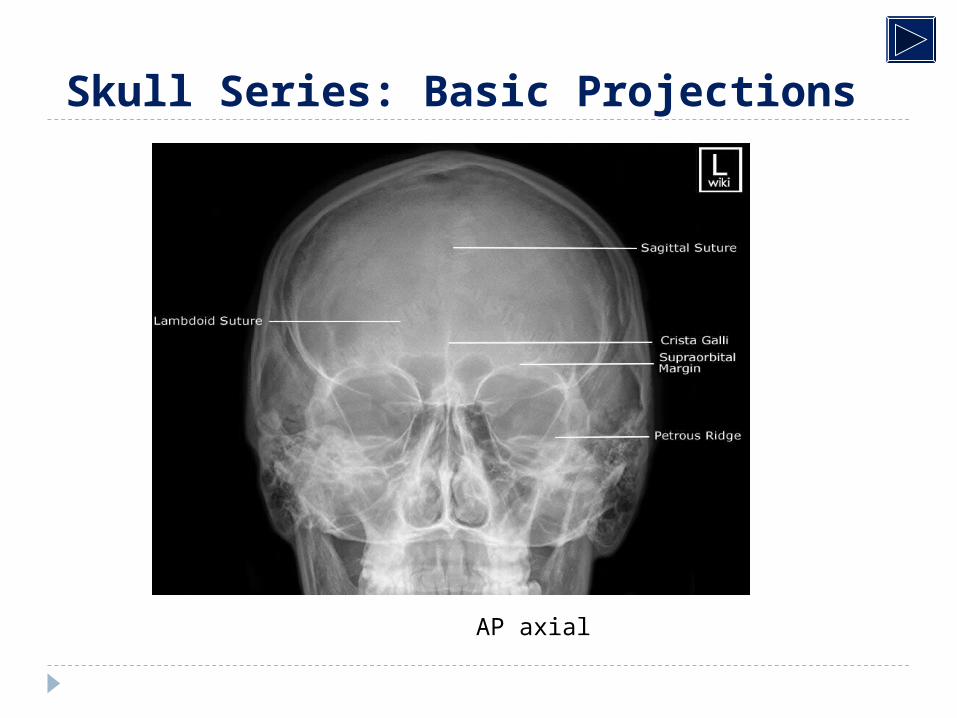
AP axial
Skull Series: Basic Projections
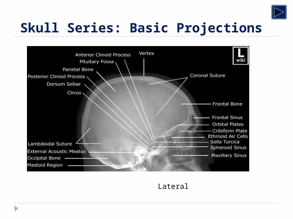
Lateral

THANK YOU