Embed Size (px)
DESCRIPTION
Kuliah radiografi
Citation preview
RADIIOGRAFI FRAKTUR KG
Oleh : Supriyadi,drg,M.Kes
Laboratorium Radiologi KG FKG Universitas Jember
≥
FRAKTUR : terjadi diskontinuitas tulang Sebagian besar kasus radiografi sangat penting: Site of the fracture The number of the fracture Direction of the fracture line Relationship of a tooth or root to the fracture line Displacement of the fracture fragment Any complicating Union or Non-Union Gambaran radiografi: Umum : Lenear radiolucency with rough border Radiographic investigation approach 3 katagori: 1. Injures of teeth and their supporting structures 2. Fractures of the mandible 3. Fractures of the middle third (1/3) of the facial skeleton
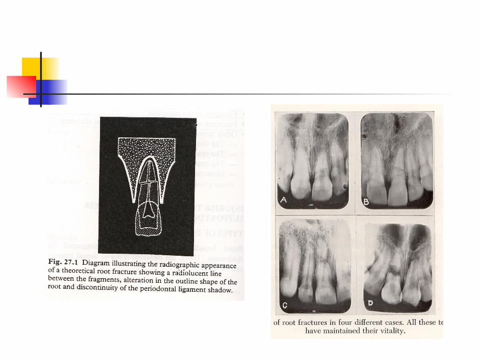
INJURES OF TEETH AND THEIR SUPPORTING STRUCTURESTYPES OF INJURE :1. Fractures of teeth : Coronal fractures Root fractures Coronal-root fractures2. Luxation; these including : concusion, subluxation, avulsion3. Fractures of the alveolar bone Fractures of the socket Fractures of the alveolar process Fractures of the associated jaw
PROYEKSI RADIOGRAFIidealnya 2 radiograf dari sudut/proyeksi yang berbeda; contoh: proyeksi periapikal dan oklusal
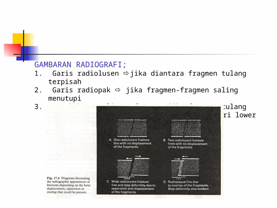
GAMBARAN RADIOGRAFI;1. Garis radiolusen jika diantara fragmen tulang terpisah2. Garis radiopak jika fragmen-fragmen saling menutupi3. Perubahan outline tulang jika fragmen tulang displaced,
menghasilkan step deformity dari lower border atau occlusal plane
RADIOGRAPHIC INFORMATION:• The type of injure to the teeth• Site of fractures• Displacement of the tooth fragments• The stage of root development• The condition of the apical tissues• The presence, site and displacement of alveolar bone fracture• The condition of adjacent or underlying teeth• Post-trauma complications : resorption, infection• Healing
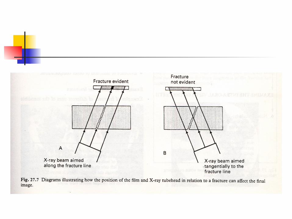
LIMITATION OF RADIOGRAPHIC INTERPRETATIONGambaran radiografi trauma gigi tidak selalu jelas; karena;• The position and severity of the fracture• The degree of fragment displacement• Posisi film dan arah sinar X hubungannya terhadap garis fraktur
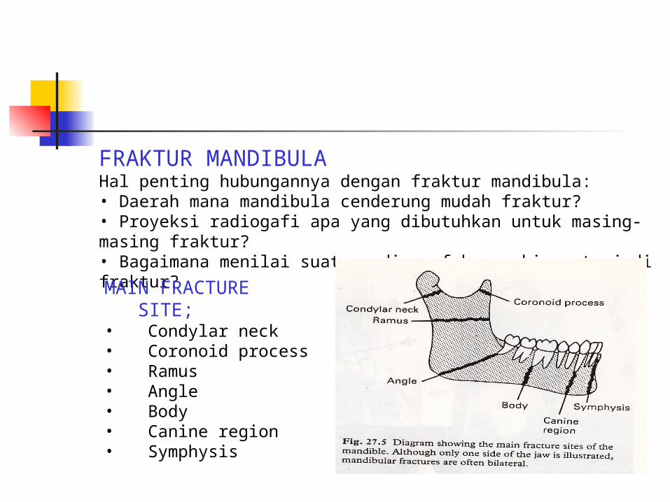
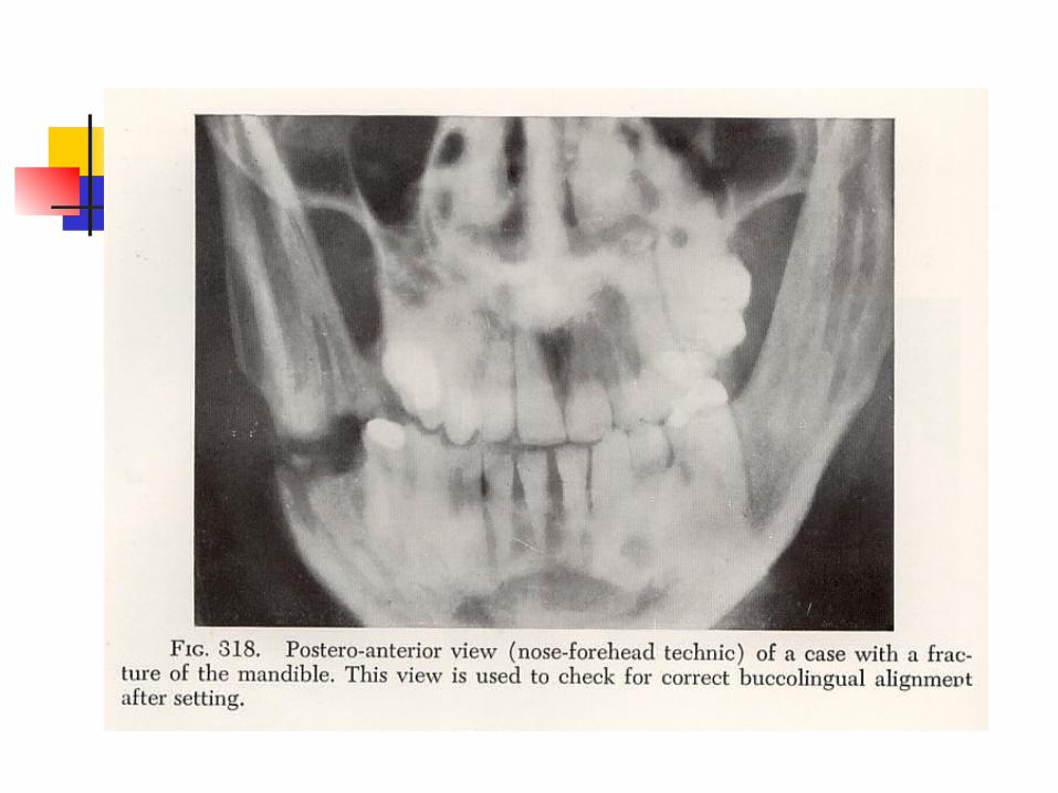
FRAKTUR MANDIBULAHal penting hubungannya dengan fraktur mandibula:• Daerah mana mandibula cenderung mudah fraktur?• Proyeksi radiogafi apa yang dibutuhkan untuk masing-masing fraktur?• Bagaimana menilai suatu radiograf kemungkinan terjadi fraktur?MAIN FRACTURE SITE;• Condylar neck• Coronoid process• Ramus• Angle• Body• Canine region• Symphysis
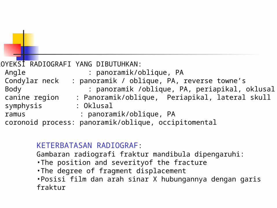
PROYEKSI RADIOGRAFI YANG DIBUTUHKAN:1. Angle : panoramik/oblique, PA2. Condylar neck : panoramik / oblique, PA, reverse towne’s3. Body : panoramik /oblique, PA, periapikal, oklusal4. canine region : Panoramik/oblique, Periapikal, lateral skull5. symphysis : Oklusal6. ramus : panoramik/oblique, PA7. coronoid process: panoramik/oblique, occipitomental
KETERBATASAN RADIOGRAF:Gambaran radiografi fraktur mandibula dipengaruhi:•The position and severityof the fracture•The degree of fragment displacement•Posisi film dan arah sinar X hubungannya dengan garis fraktur
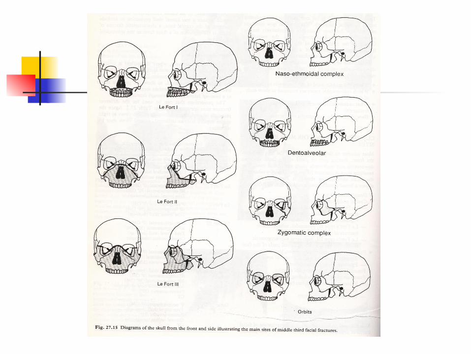
FRAKTUR 1/3 TULANG MUKA• Facial skeleton cukup kompleks knowledge of anatomi sangat dibutuhkan• Hal penting lain yang diperlukan:• Bagiam mana dari 1/3 tulang muka yang cendrung mudah terjadi fraktur?• Proyeksi radiografi apa yang dibutuhkan untuk masing-masing fraktur tulang muka?• Apakah gambaran radiografi menginikasikan adanya fraktur?• Bagaimana menentukan adanya fraktur pada radiograf?
KLASIFIKASI
1. Dento-alveolar fractures /proc.alveolaris maksila2. Central middle third fractures; terdiri dari: a. Le Fort I Garis fraktur : sebagian dinding sinus maksilaris(SM), palatum,
bagian bawah proc.pterygoideus, bawah proc. zygomatikus Klinis : maloklusi, RA seperti lepas b. Le Fort II Garis fraktur : bagian bawah os. Nasi, dinding orbita medial
dan inferior, dinding lateral SM dan septum nasi. Klinis : wajah bengkak (month face), perdarahan hidung c. Le Fort III Garis fraktur : os.nasi,dinding medial dan leteral cavum orbita,
arkus zigomatikus, septum nasi Klinis : edeme 1/3 tengah wajah (month face), edem
konjuntiva, kerusakan orbita(bleeding, diplopia, penglihatan
menurun), bleeding hidung3. Fractures of zygomatic complex4. Fractures of the naso-ethmoidal complex5. Fractures af the orbitale