Embed Size (px)
Citation preview
This article is protected by copyright. All rights reserved. This is an open access article under the terms of the Creative Commons Attribution-NonCommercial-NoDerivs License, which permits use and distribution in any medium, provided the original work is properly cited, the use is non-commercial and no modifications or adaptations are made.
Radiofrequency Ablation (RFA) of Renal Cell Carcinoma (RCC):
Experience in 200 Tumours1
Wah TM FRCR EBIRa
Irving HC FRCRa
Gregory W PhD b
Cartledge J FRCS c
Joyce AD FRCS c
Selby PJ MD DScd
Address for correspondence:
Tze M Wah
Diagnostic and Interventional Radiology
Lead for Percutaneous Tumor Ablation Program at LTHT
Institute of Oncology
Leeds Teaching Hospitals Trust
St. James’s University Hospital
Beckett Street
Leeds LS9 7TF
Tel: +441132065525 Fax +441132064640
Email: [email protected]
This article has been accepted for publication and undergone full peer review but has not been through the copyediting, typesetting, pagination and proofreading process, which may lead to differences between this version and the Version of Record. Please cite this article as doi: 10.1111/bju.12349 Acc
epte
d A
rticl
e
2
a. Current address: Department of Diagnostic and Interventional Radiology, Institute
of Oncology, Leeds Teaching Hospitals Trust, St. James’s University Hospital,
Beckett Street, Leeds, LS9 7TF, United Kingdom.
b. Professor of Statistical Methodology in Clinical Trials, Clinical Trials
Research Unit (CTRU), University of Leeds, Clinical Trials Research House
71-75 Clarendon Road, Leeds, LS2 9PH
c. Department of Urology, St. James’s University Hospital, Beckett Street, Leeds, LS9
7TF, United Kingdom.
d. Professor of Cancer Medicine, Leeds Institute of Molecular Medicine, St. James's
University Hospital, Beckett Street, Leeds LS9 7TF, UK
Author contributions:
Guarantor of integrity of entire study; TMW, HCI, ADJ, JC, PS
Study concepts and design; TMW, HCI, PS
Literature search; TMW, HCI
Clinical studies; TMW, HCI, JC, ADJ
Data acquisition; TMW, HCI
Data analysis; TMW, WG
Statistical analysis; TMW, WG
Manuscript preparation; all authors
Manuscript definition of intellectual content; all authors
Manuscript editing; all authors
Manuscript review; all authors Acc
epte
d A
rticl
e
3
Manuscript final version approval; all authors
ABSTRACT
Objectives
• We wish to report the evaluation of our clinical experience with percutaneous
image-guided RFA of 200 renal tumours in a large tertiary referral university
institution.
Patients and Methods
• Image-guided RFA (US or CT) of 200 renal tumours in 165 patients from June
2004 to 2012 was prospectively evaluated. Institutional review board approval
has been granted.
• The treatment response and technical success were defined by absence of
contrast enhancement within the tumour on contrast enhanced CT or MRI.
• Both major and minor complications, pre and post RFA glomerular filtration
rate (GFR), the management and outcomes of the complications as well as
oncologic outcome were prospectively documented. Multivariate analysis was
performed to determine variables associated with major complications and
also percentage GFR change post RFA.
• The overall, 5-year cancer specific, local recurrence-free and metastasis-free
survival rates are presented using the Kaplan Meier Curves.
Results Acc
epte
d A
rticl
e
4
• Two hundred tumours were RF ablated with a mean tumour size of 2.9cm
(size range 1-5.6cm) and the mean patient age was 67.7 years (age range 21-
88.6 years) with a mean follow-up period of 46.1 months.
• The primary technical and overall technical success rate was 95.5% and 98.5%
respectively. Two independent predictors of successful ablation in a single
sittings were tumour size (<3cm) and exophytic location in multivariate
logistic regression analysis.
• Major complications included ureteric injury (n=6), calyceal-cutaneous fistula
(n=1), acute tubular necrosis (n=1) and abscess (n=2). Two independent
predictors of ureteric injury were central location and lower pole position.
• Within this cohort of patients, only 4 patients developed significant renal
function deterioration i.e. >25% decreased in GFR. A total of 161 (98%)
patients out .of the 165 patients have preservation of renal function. Any
change in renal function post RFA was not influenced by tumours factors or
solitary kidney status.
• In our clinical series, this yields a 5- year overall, cancer specific, local
recurrence free and metastasis free survival rates of 75.8%, 97.9%, 93.5% and
87.7% respectively.
Conclusions
• Image-guided RFA is a safe, nephron sparing and effective treatment for small
RCC tumours with a low rate of recurrence and has good 5-year cancer
specific and metastasis free survival rates.
Keywords: Radiofrequency ablation, Renal Cell Carcinoma, complication, survival rates Acc
epte
d A
rticl
e
5
Introduction
Renal cell carcinoma (RCC) is the commonest cancer of the kidney. It accounts for
3% of all cancers in adults [1]. The detection of RCC has increased over the past
decade and each year there are around 270,000 cases worldwide [2, 3]. This has been
partly due to the increase of incidentally detected RCC from wider use of radiological
imaging [4] and it is also related to increased incidence of renal cancer in the general
population secondary to cigarette smoking and obesity [5-8].
Incidentally detected renal masses are smaller and at an earlier stage than those
tumours which present clinically with symptoms such as pain, hematuria and palpable
flank mass [9, 10]. Around 80% of these incidental detected solid masses are RCC on
histological diagnosis [11]. Historical data has suggested that 60% of these small
masses will grow gradually over a period of time [12]. Therefore, there remains a
clinical risk with adopting a ‘watchful waiting’ approach for younger patients as these
tumours may become symptomatic or metastasize [12].
Currently, there is general consensus that smaller renal tumours (< 4 cm), should be
treated with minimally invasive techniques in order to preserve renal function and
avoid unnecessary surgical removal of the entire kidney [13]. Nephron sparing
surgery (NSS) with either laparoscopic or open partial nephrectomy has replaced the
gold standard of radical nephrectomy whenever it is deemed technically possible to
remove the small renal tumour and to preserve the rest of the kidney. It has been
shown that radical nephrectomy leads to a higher incidence of chronic kidney disease, Acc
epte
d A
rticl
e
6
especially those with co-morbidity e.g. diabetes and this leads to increased mortality
and morbidity [14]. The NSS approach has demonstrated similar recurrence free and
long term survival outcomes as those with radical nephrectomy [15, 16] and has good
preservation of renal function [14, 17]. However, partial nephrectomy is technically
challenging and associated with significant morbidity [15, 16, 18, 19].
Given the surgical constraints, the initial exploratory work with the image guided
ablative treatment of the small renal tumours with RFA, cryotherapy and microwave
has evolved at a rapid pace over the last decade. Percutaneous tumour ablation has
proven to be a safe and effective treatment option for small renal tumours, and good
oncological outcome data is emerging for radiofrequency ablation (RFA) [20, 21] and
cryoablation [22].
Percutaneous RFA is now a well-established technique for treating small renal
tumours [23-25]. It uses a high-frequency, alternating current within the targeted
tissue to cause ionic agitation generating frictional heat which results in cancer cell
destruction when the temperatures exceed 600C. This paper evaluates our clinical
experience in a single university institution for the treatment of 200 renal tumours
with image-guided RFA.
Materials and Methods
Patients
Our Percutaneous Renal RFA program was established in 2004. All patients were
referred through our local urology multi-disciplinary team (MDT) meeting. The MDT Acc
epte
d A
rticl
e
7
panel consisted of at least one consultant urologist, a consultant oncologist, a
consultant radiologist and other supporting staff. The patient imaging was reviewed
and the management options were discussed and a consensus achieved within the
panel. In our institution, the diagnosis of renal tumour before nephrectomy has
historically been based on imaging criteria alone, as established on CT when the
tumour has a mean density of >20HU and shows >20 HU enhancement post contrast
[4, 26] or on MRI when there is appropriate enhancement (15% from threshold) post
Gadolinium [27]. The inclusion criteria for consideration of RFA treatment were:
non-surgical candidates (in our early experience this was defined by ASA>3, but
subsequently all patients with ASA>3 unsuitable for general anaesthesia were also a
relative contraindication for image guided renal ablation) with incidental stage T1
renal tumours, renal tumours in a solitary kidney, synchronous primary renal tumour,
patients with Von-Hippel-Lindau (VHL) disease or patients with impaired renal
function. In addition, depending on the clinical situation, the patient’s personal choice
for nephron sparing treatment, when partial nephrectomy was deemed technically
impossible by the urologist and occasionally, patients with metastatic RCC
undergoing immunotherapy were also considered.
Following the MDT meeting, the patients were seen in the out-patient consultation
clinic by both the consultant urologist and radiologist. A comprehensive consultation
was provided by the urologist covering the various treatment options available for the
patient. Patients who wished to consider percutaneous renal ablation would then be
seen by the interventional radiologist (IR) in the clinic. The IR would provide in-
depth discussion of the pros and cons of the treatment taking account of the size and
location of the renal tumours and explanation of the implications of the percutaneous Acc
epte
d A
rticl
e

8
renal ablation with the requirement for long term imaging follow up. In the beginning
of the development of this program (2004 to 2009), image guided renal RFA was the
only treatment option and since 2009, both image guided renal RFA and cryoablation
were offered as treatment options. Depending on the tumour size and location, heat or
cold based energy might be offered as a preferential technique. Informed written
consent for the treatment with RFA of their renal tumours was obtained in all patients.
In our program, we also have a clinical specialist nurse who was available to support
the patient’s journey. All patients were referred to the hospital pre-assessment clinic
to assess for their fitness for general anaesthesia and screened for day case admission.
Routine baseline laboratory investigations were performed, including a clotting screen
(INR < 1.5 was required at the time of treatment), renal function tests (creatinine
measurement) and full blood counts.
From June 2004 to 2012, we have performed image guided RFA of 200 renal tumours
in 165 patients. The patients’ prospectively collected clinical database was evaluated
for technical success, renal function, clinical complications and oncologic treatment
outcome. The review study has been granted approval by our institution’s ethics
committee chairman and informed consent was waived for the database review by the
institutional ethical board.
A total of 165 patients (109 men, 56 women; age range 21-88.6 years; mean age 67.7
years) underwent percutaneous RFA of the 200 renal tumours in 210 treatment
sessions. One hundred and two renal tumours were in the right kidney and 98 in the
left kidney. The tumours size ranged from 1 to 5.6cm (mean= 2.9cm). Amongst the
renal tumours RF ablated, tumour sizes greater than 3 cm and less than 3cm were 134 Acc
epte
d A
rticl
e
9
and 66 respectively. The polar position of the renal tumours was: upper (n=63),
middle (n=86) and lower (n=51). The tumour treatment classification according to the
criteria of Gervais et al [28] was: exophytic (n=43), mixed (n=100), parenchymal
(n=41) and central (n=16). All of the patients had a baseline renal function
immediately before and a repeat renal function test at 24 hour post RFA treatment.
Biopsy Procedure:
In our institution, all patients undergo biopsies at the time of ablation of the renal
tumour as part of their standard clinical care. The co-access sheath used is part of the
RFA LeVeen needle electrode system (Boston Scientific, MA, USA) and allows
biopsy without the need to reposition the sheath for RFA treatment. The co-access
sheath was inserted into the renal tumour under CT guidance and all biopsies were
performed with an 18 gauge core biopsy needle gun through a 16 gauge outer sheath
(Boston Scientific, MA, USA). At least two core biopsies of each renal tumour were
performed.
RF ablation
All the RFA procedures were performed under general anaesthesia as the preferred
option of the anaesthetist, apart from 3 patients who were treated with intravenous
conscious sedation at the patient’s choice and the discretion of the anaesthetist at the
time of treatment. All patients received broad spectrum intravenous antibiotics
amoxicillin trihydrate/ potassium clavulanate (Co-amoxiclav 1.2 g) during the
procedure and at 12 hour post treatment followed by a 10-day course of oral Acc
epte
d A
rticl
e
10
ciprofloxacin (500mg twice daily). This routine prophylaxis had been adopted as
policy following consultation with Breen et al. [29] but we acknowledged this remains
a controversial practice amongst operators performing image guided renal ablation.
RFA was performed as an elective procedure in all patients with a routine admission
the day before and observation overnight post procedure in our institution.
All ablations were performed by one or two of the two consultant radiologists
(T.M.W., H.C.I). A total of 200 renal tumours were ablated in 210 treatment sessions
with an impedance-controlled pulsed current from a 200-W RF 3000 generator
(Boston Scientific, MA, USA). Ablation was performed with a varying size (3, 3.5 or
4 cm) umbrella shaped multi-tines needle electrode (LeVeen CoAccess RFA needle
electrode, Boston Scientific, MA, USA), selected to match the size of the tumour.
During treatment, the number of overlapping ablations was dependent on the size and
geometry of the lesion. For this system, the timing of individual ablation was
impedance-controlled, depending upon the tissue vascularity and resistance. The RCC
target tissue cell death is achieved via tissue desiccation and lost its ability to conduct
current hence the rise in the impedance. ‘Roll off’ equates to clinical endpoint where
complete tissue coagulation is reached when the impedance reaches a clinically
relevant level and there is concurrent power shut down of the generator. A more
vascular tumour will cause more heat-sink effect and this will lead to longer treatment
time.
From June 2004 to 2006, due to the initial set up of the program, most of the renal
RFA were performed in an operating theatre under US guidance (n=31). Since June
2006, the treatment sessions were moved into our CT interventional suite, and all Acc
epte
d A
rticl
e
11
renal tumours were subsequently ablated under CT guidance (n= 179) with US
available to compliment guidance if necessary. The availability of CT imaging during
treatment allows better assessment of the safety margin of the treatment, particularly
to assess the proximity of the surrounding organ such as bowel and ureter in relation
to the multi-tine needle electrode. In our institution, the CT guided RFA involves a
contrast enhanced study with 100mls of iodinated contrast medium given at a rate of
3ml/second at the beginning to assist targeting of the renal tumour and a subsequent
intra-procedural CT to guide RF electrode positioning which was performed at 3-mm-
collimation spiral acquisition. We do not perform immediate post procedural contrast
enhanced CT to assess treatment effect as this is performed at one month post
treatment to allow the early post RFA changes to resolve.
During treatment, the number of overlapping ablations was dependent on the size and
geometry of the lesion and multi-planar reformatting of the multi-tines electrode
positioning was important to determine the overall tumour coverage (Figures 1a, b &
c). The ablated renal tumour size ranged from 1 to 5.6 cm (mean size= 2.9 cm). In
each patient, the total overlapping ablations ranged from 1 to 5 (mean ablation cycle=
2.5) and the total ablation time was between 6 and 63.6 mins (mean ablation time=
26.3 mins).
Post RFA, all patients were monitored in the theatre recovery area and then
transferred to the ward for overnight observation and discharged home if clinically
stable the following day as the standard care. All the patients were monitored
clinically and followed up with radiology imaging centrally by our institution during
this period. Complications of the procedure were prospectively collated. Acc
epte
d A
rticl
e
12
Complications were classified as major or minor based on the classification of the
Society of Interventional Radiology with major complications requiring treatment or
hospitalization and minor complications needing only conservative monitoring. Their
average hospital stay was 2.8 days.
Cold Pyelo-perfusion Technique
This technique was used when the renal tumour was centrally located or the treated
tumour margin was close to the ureter and there was concern for renal pelvis or
proximal ureteric injury [30, 31]. This was initially described by us for patients had
treatment in theatre [31] and since 2006, we had modified the technique to
accommodate for the fact that all treatments are now performed in CT interventional
suite [30]. This in essence involved patient lying supine on our CT table and
retrograde cannulation of the ureter in the CT suite by the urologist. A guide wire was
passed under direct visualisation using flexible cystoscope into the renal pelvis. A 6
French open end flushing catheter (70cm, COOK, Bloomington, IN) was then passed
over the guide wire and positioned into the renal pelvis. The position was then
confirmed by performing a CT scout view with small amount contrast instilled
retrogradely to confirm the location. A 14 French Foley catheter was placed into the
bladder, to which the ureteric catheter was taped in order to prevent its displacement.
Cold 5% Dextrose at 60C was perfused via the ureteric catheter by gravity (80 cm
H2O) and drained via the ureter into the bladder. At the end of the procedure,
depending on the clinical requirement, we sometimes exchanged the ureteric catheter
for a 7.5F Optipur (Ettlingen, Germany) ureteric stent in the fluoroscopic suite under
fluoroscopic guidance. The Foley catheter was removed 24 hours after the procedure. Acc
epte
d A
rticl
e
13
Thirteen RFA treatment sessions in 10 patients with centrally located renal tumours
were RF ablated using the cold pyeloperfusion technique. Two patients were at very
high risk for ureteral injury as their tumours were obliterating the PUJ and ureter and
despite the risks were keen to go ahead with the treatment with the cold
pyeloperfusion technique.
Hydro-dissection Technique
Some patients had renal tumours that were in close proximity to bowel loops during
treatment, either because of the location of the tumour (anterior and mid-polar
location) or due to lack of intra-abdominal fat. The hydrodissection technique was
used in these instances when there was <1cm between the treatment margin and the
bowel loop in order to avoid thermal injury to the small or large bowel or any other
surrounding vital organ [29, 32]. We routinely performed hydro-dissection with a 16G
sheathed needle (Boston Scientific, MA, USA) and 150 to 500 mls of 5% Dextrose
solution at room temperature was instilled to displace the bowel. In this series, this
technique was performed in 25 patients who had 26 RFA treatment sessions (mean
volume= 287.5 mls of 5% Dextrose).
Post RFA Clinical and Radiology Follow-Up
All patients had dynamic contrast enhanced (DCE) cross sectional (CT or MRI)
imaging before and after RF ablation. We routinely performed DCE-MRI at baseline,
1, 3 and 6 months post RF ablation and annual CT for full staging (chest and kidneys) Acc
epte
d A
rticl
e
14
for a period of 10 years as part of our Yorkshire Cancer Network (YCN) protocol. In
patients with serum Cr >200 µmol/L, DCE-MRI was performed to assess the kidneys
and un-enhanced CT for chest staging annually. Triple phase DCE-CT of kidneys
(which includes unenhanced, arterial and portal-venous phases) was performed to
assess the zone of ablation if MRI was contra-indicated (e.g. cardiac pacemaker) or
patient was claustrophobic. Occasionally, additional imaging was performed as a
result of patients’ clinical symptoms.
All MRI examinations were acquired on a 1.5 T system (Symphony; Siemens Medical
Systems, Erlangen, Germany). A dedicated four element body array coil and
integrated spine coil were used for signal reception. The MRI examination included
T1, T2, true-FISP sequences in the axial, sagittal and coronal planes and pre and post
gadolinium enhancement TI VIBE sequences in both coronal and axial planes.
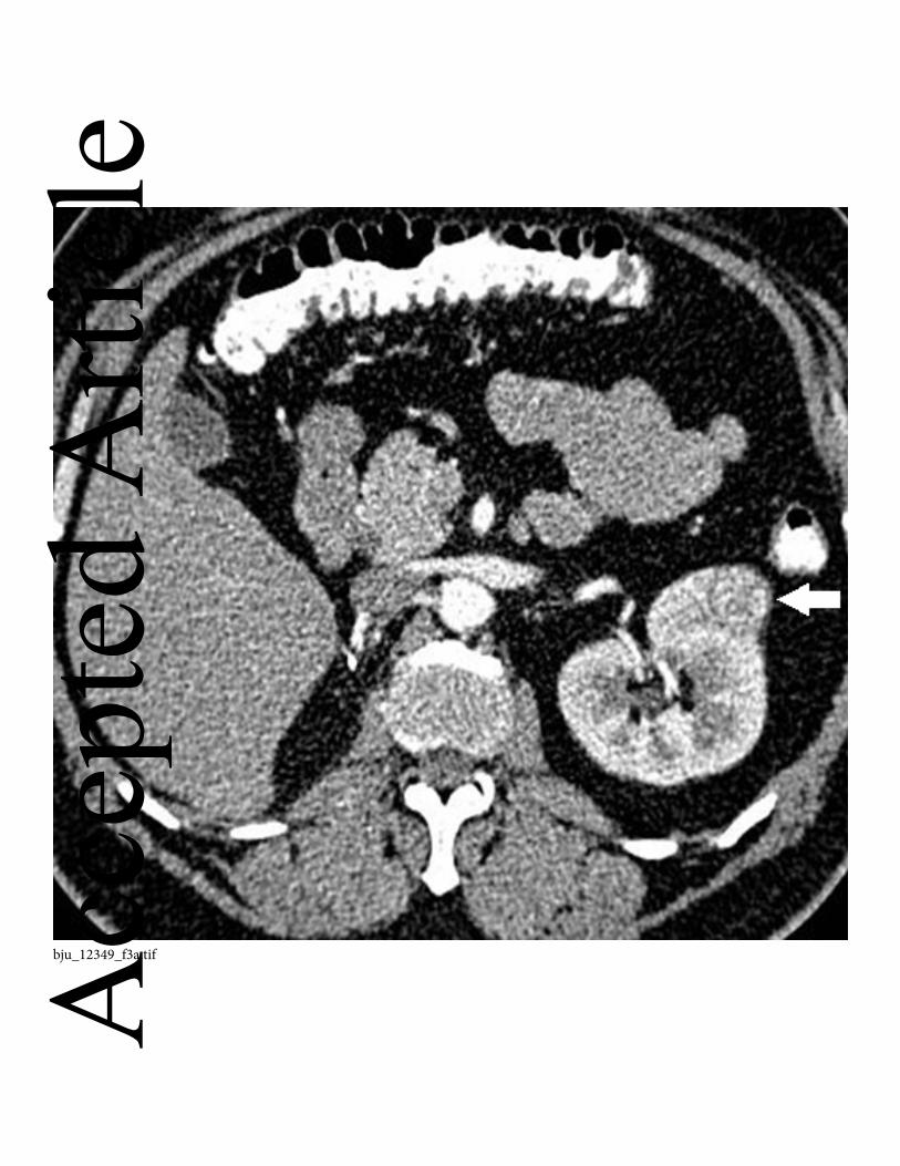
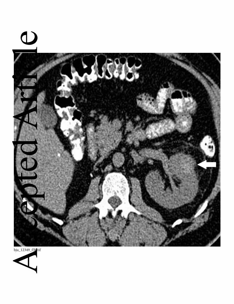
Technical success was defined by absence of contrast enhancement within the tumour
on CT or MRI (Figures 3a and b). Residual disease was defined as persistent
enhancement in an area of tumour after RF ablation seen on imaging at least one
month post treatment. This usually presents as nodular and crescent enhancement
around the periphery of the RF ablated zone [28, 33]. Recurrent disease was defined
as new area of enhancement in the zone of ablation after at least one imaging (> 3
months) had shown complete lack of enhancement in the treated area (i.e. complete
ablation). The imaging was reviewed by one of the two consultant radiologists
(T.M.W., H.C.I.) and consensus was achieved if there was any uncertainty regarding
the imaging findings.
Acc
epte
d A
rticl
e
15
Both major and minor complications, pre and post RFA glomerular filtration rate
(GFR), the management and outcomes of the complications as well as oncologic
outcome were prospectively documented. The overall, cancer specific, local-
recurrence free and metastasis-free rates were also documented.
Data Analysis and Statistics
Statistical analysis was performed using Stata (StataCorp. 2009. Stata: release 11.
Statistical Software. College Station, TX: StataCorp LP). Descriptive statistics (e.g.
mean, SD and variance) were reported and differences with a p value of less than 0.05
were considered to be statistically significant.
Univariate analysis was performed by using the Fisher exact test to assess the tumour
size and location as predictors of technical success. The t test was used to evaluate
differences between the group means.
Multivariate logistic regression analysis was performed to determine any association
between the change of pre and post treatment GFR (% GFR change) with the tumour
size, polar position (upper, middle and lower pole of the kidney), tumour treatment
location (exophytic, mixed, parenchymal and central), the total size of the tumour
treated per ablation session, number of tumours treated and the solitary kidney status.
Multivariate logistic regression analysis was performed to determine any association
between the importance of tumour position in the kidney (upper, mid or lower),
tumour treatment location (central, mixed, parenchymal and expohytic) and tumour Acc
epte
d A
rticl
e
16
size in influencing the technical success of the treatment or complication. Kaplan
Meier curves have been used to determine the overall, cancer specific, local
recurrence free and metastasis-free survival and these were also documented.
Results
Renal Tumor Morphology and Histology
One hundred ninety six (196) renal tumours were solid and four (4) had cystic
components within the renal tumour. The ablated renal tumour size ranged from 1 to
5.6 cm (mean size= 2.9 cm) and they were all clinical stage T1 renal tumours.
One hundred and eighty eight (188) core biopsies yielded an adequate sample for
histological diagnosis with inadequate samples in 12 (6%) renal tumours. Amongst
them, the histological subtype was: clear cell carcinoma (n=160), papillary (n=8),
distal nephron tumour (chromophobe or oesinophilic variant) (n=14), oncocytoma
(n=1), fibrosis (n=3), metastasis from GI tumor (n=1) and angiomyolipoma (n=1).
Therefore, in our cohort of patients, 183 (91.5%) renal tumors had histological
confirmation of malignancy and all were proven RCCs with various histological
subtypes apart from the one metastasis from oesophageal cancer. The five (2.5%)
patients with benign histological diagnosis did not need long term follow up. The 12
(6%) renal tumours with inconclusive biopsies, mainly due to the small sample size,
were placed in an indeterminate group.
Acc
epte
d A
rticl
e
17
Technical Success (Primary and Overall)
Of the 200 treated tumours, 197 (98.5%) were completely ablated (191 in a single
ablation session, 3 after a second ablation session and 3 after a third session). Three
patients declined re-treatment. Therefore the primary and overall technical success
rates were 95.5 % and 98.5% respectively. The local repeat RF ablation rate for the
individual renal tumour was 3%.
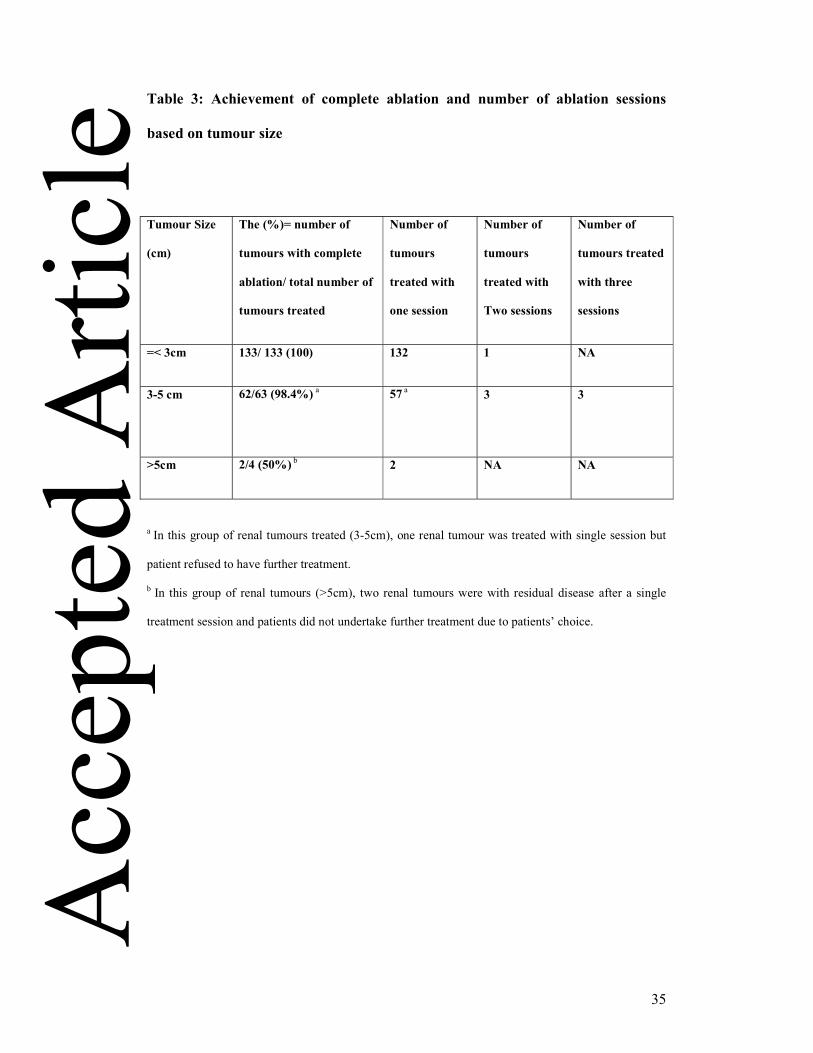
Technical Success vs. Tumor Location, Position and Size
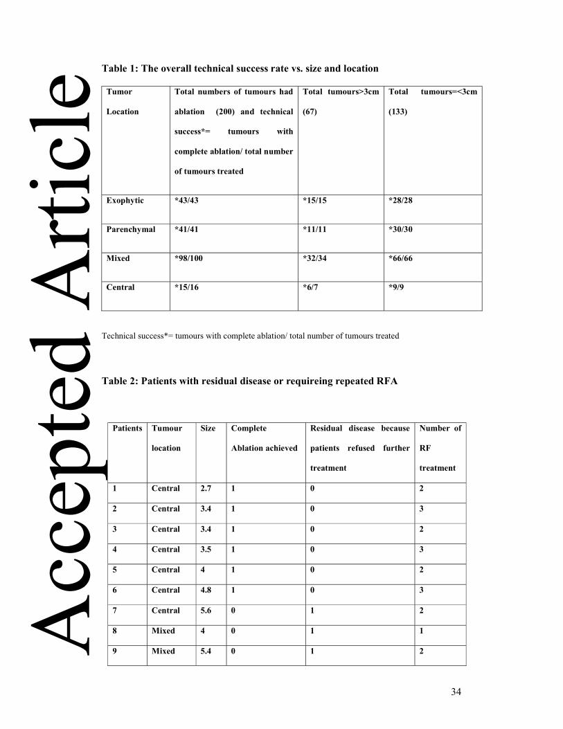
The overall technical success of the RF ablation of all renal tumours is summarized in
the Table 1. The results were categorized into tumour location as well as tumour size.
All exophytic and parenchymal renal tumours, regardless of location and size, were
completely ablated in single treatment session. All 6 patients who required more than
one treatment to achieve complete ablation had centrally located renal tumours (Table
2). In our series, a strong statistical association was seen between central vs. non
central locations (which includes exophytic and parenchymal location) and the
primary technical success rate i.e. successful ablation in a single sitting (p<0.0001,
Fisher’s exact test). Similar results were also seen when we compared the renal
tumour location for central (n=16) vs. exophytic (n=43) (p=0.0002, Fisher’s exact
test). Three patients with residual disease refused further treatment, one had surgery
and two were deemed unfit for surgery from the outset, the tumours location were
central (n=1) and mixed (n=2) location. In addition, the size of the tumour is also a
strong independent predictor in achieving complete RF ablation in a single treatment Acc
epte
d A
rticl
e
18
session (Table 3), between renal tumours <3cm vs. >3cm (p=0.0008, Fisher’s exact
test). Size and location were shown to be independent predictors in multivariate
logistic regression analysis. However, the primary technical success rate was shown to
be not influenced by the tumour polar position (upper, middle or lower) (p>0.7,
Fisher’s exact test).
Technical success vs. Interventional Consultant Radiologists (IR) Experience
During the eight year period, a total of 128 renal tumours were RF ablated from June
2004 to 2008 (< 4 year experience) and a further 72 renal tumours were RF ablated
from June 2008 to 2012 (>4 year experience). All the patients with residual disease
(n=9) were treated in the first 4 years. In the subsequent 4 years, the primary technical
success rate was 100%. Therefore, in our series, the IR experience does influence the
primary technical success rate when we compared the IR’s experience < 4 year vs. > 4
year period (p=0.03, Fisher’s exact test). When experience of the operator is included
in the multivariate logistic regression analysis, along with size and location, IR
experience is no longer significant, though still with a similar trend (p=0.18, Fisher’s
exact test). It is likely that this merely reflects that the experience of the operator
results in a better selection of tumour for treatment, with the larger and central
tumours all occurring in the first 4 years.
Patients with residual (incompletely treated) disease
At the time of reporting, a total of 3 patients within this cohort were incompletely
treated, 1 refused further treatment and two had repeated treatment but had residual Acc
epte
d A
rticl
e
19
disease and it was deemed inappropriate to treat further. The patient who refused
further treatment has a metastasis free survival of 80.2 months and is still alive. One
patient had ankylosing spondylitis (AS) and was treated initially under US guidance.
He then had a repeat treatment under CT guidance but could not be positioned
optimally in the CT scanner due to his AS so that only part of the residual disease was
treated. He subsequently opted for open radical nephrectomy. Another elderly patient
was deemed inappropriate for a third ablation as the MDT decided that surveillance
should be advocated instead and the patient had metastasis free survival of 94.6
months after the second ablation before dying of ischaemic heart disease (IHD). All 3
patients were treated during our early experience (<3years) and in our first 50 cohort
of patients.
RF Procedural Complications
Complications were classified as major or minor based on the classification of the
Society of Interventional Radiology with major complications requiring treatment or
hospitalization and minor complications needing only conservative monitoring. There
have been a total of 11 major complications directly relating to our treatment
technique, including ureteral stricture (n=7), acute tubular necrosis resulting in
permanent renal failure in a patient with solitary kidney (n=1), calyceal cutaneous
fistula (n=1) and renal abscesses (n=2). There were 12 minor complications that
include lateral cutaneous nerve paraesthesia (n= 5), skin burn (n=2), self-limiting
subcapsular haematoma (n=4) and self-limiting pneumothorax (n=1). In this cohort,
one patient developed myocardial infarction post general anaesthesia but recovered
after appropriate medical treatment. Acc
epte
d A
rticl
e
20
Haemorrhage
There was no major haemorrhage in this cohort of patients requiring intervention such
as blood transfusion or vascular embolization. There were 4 patients who developed
self-limting subcapsular haematoma post renal RF ablation which were present at the
time of treatment completion.
Ureteral stricture
There were 7 (3.5%) ureteral strictures in 210 RF treatment sessions. All the ureteral
injuries occurred in the upper 1/3 of the ureter and pelvi-ureteric junction (PUJ). The
management for the ureteric stricture was: retrograde ureteric stenting (n=4),
conservative management (n=2) and radical nephrectomy (n=1). One patient had
opted for radical nephrectomy as recurrent urinary tract infections from the
subsequent ureteric stent insertion resulted in persistent psoas abscess formation.
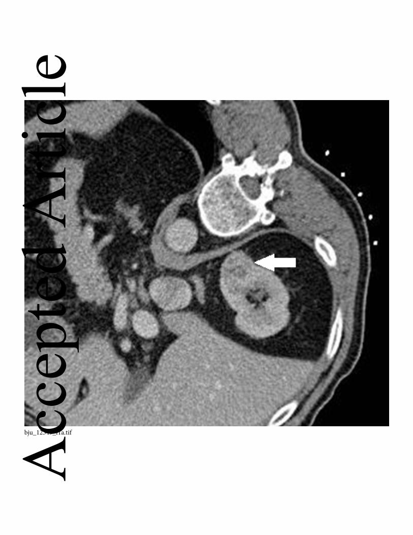
For the 16 central tumours, cold pyeloperfusion technique was used during 13
treatment sessions in 10 centrally located renal tumours and 8 (80%) patients were
treated successfully without the development of ureteric stricture. As we had
predicted, the 2 high risk patients who were deemed likely to develop ureteric
strictures (tumour abutting the ureter with zero distance to the treatment margin)
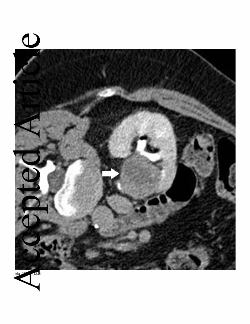
(Figure 2) had developed strictures in-spite of using the protective mechanism. The
remaining five patients also developed ureteric stricture without cold pyeloperfusion
technique were in retrospect, all <1cm margin from the ureter and the locations of the Acc
epte
d A
rticl
e
21
tumours were: central (n=1) and lower pole (n=4). Therefore, in this cohort (excluding
the two high risk patients), the use of cold pyeloperfsuion technique when treating
renal tumours in close proximity of the ureter (<1cm) would protect the ureter from
injury (p=0.0008, Fisher’s exact test) but the protection is impossible if the margin
between tumour and ureter is completely obliterated. In addition, two independent
predictors of likelihood of developing ureteric stricture are central vs. non central
(exophytic and parenchymal) location (p=0.004, Fisher’s exact test) and lower pole
vs. non lower pole location (p=0.01, Fisher’s exact test).
Renal Abscess
There were two patients who developed renal abscesses- one patient had a pre-
existing ileal-conduit [34] and the other patient appeared to be immune-compromised.
Both were treated with parenteral antibiotics and local drainage.
Calyceal-cutaneous fistula and Acute Tubular Necrosis
One patient who developed a calyceal cutaneous fistula had a pre-existing ileal
conduit formed for previous cystectomy and had suffered from recurrent urinary tract
infections. Another patient developed irreversible ATN post renal RFA. We have
reported these cases previously [34, 35].
Renal Function Measurement: Pre and Post RFA GFR Acc
epte
d A
rticl
e
22
The mean GFR before and after renal RFA were: 54.7 ml/min/1.73m2 (+/- SD 18.2
ml/min/1.73m2) vs. 52.7 ml/min/1.73m2 (+/- SD 18.5 ml/min/1.73m2). There was a
significant difference between the pre and post GFR measurement with a mean
difference of 2.03 where the pre GFR was higher before treatment (p <0.0001,
Wilcoxon signed rank test). Within this cohort of patients, only 4 patients developed
significant renal function deterioration (>25% decreased in GFR). A total of 161
(98%) patients out .of the 165 patients have preservation of renal function.
The mean percentage change of GFR before and after RFA was – 3.1% (+/- SD
15.2%), i.e. worsened by 3.1% in GFR measurement post RFA treatment. However,
using multivariate logistic regression analysis there was no association between the
percentage of GFR change with tumour size, polar position (upper, middle and lower
pole of the kidney), tumour treatment location (exophytic, mixed, parenchymal and
central), the size of the tumour treated per ablation session, number of tumours treated
and the solitary kidney status.
RFA Oncologic Outcome
All the patients were followed up both radiologically and clinically (range 2.6 to 96
months) (mean=47.6 months). The majority of our elderly patients (n=20) in the early
cohort had succumbed to ischaemic heart disease (IHD) or respiratory infection. In the
overall cohort, 9 patients presented with existing renal metastasis or pre-existing GI
cancer but were in remission at the time of initial RF ablation and they were excluded
from the Kaplan Meier Curve analysis. The reason for treating the primary renal
tumor in the setting of renal metastasis was with the intention to debulk the primary Acc
epte
d A
rticl
e
23
tumor bulk before anti-angiogenic therapy or immunotherapy. In our clinical series,
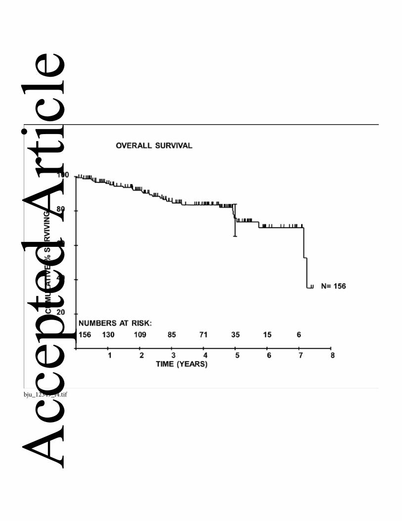
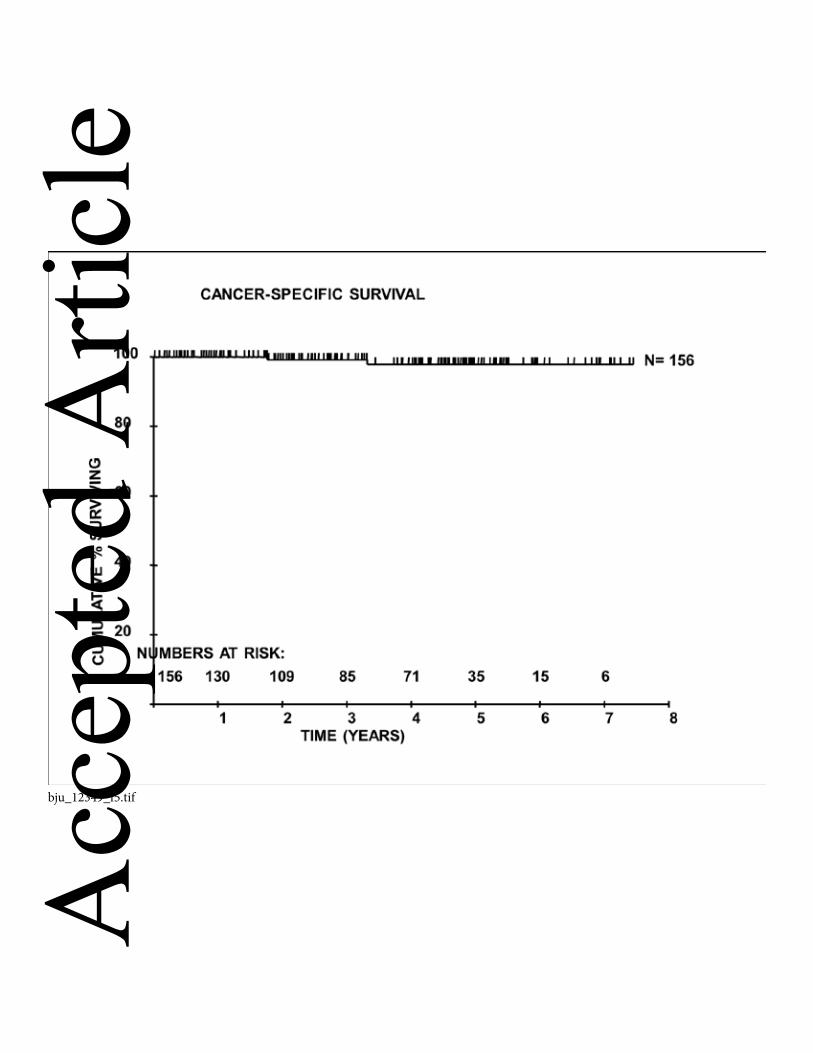
this yields a 5- year overall and cancer specific survival rates of 75.8% and 97.9%
respectively (Figures 4 & 5).
Local Recurrence and Distant Metastasis
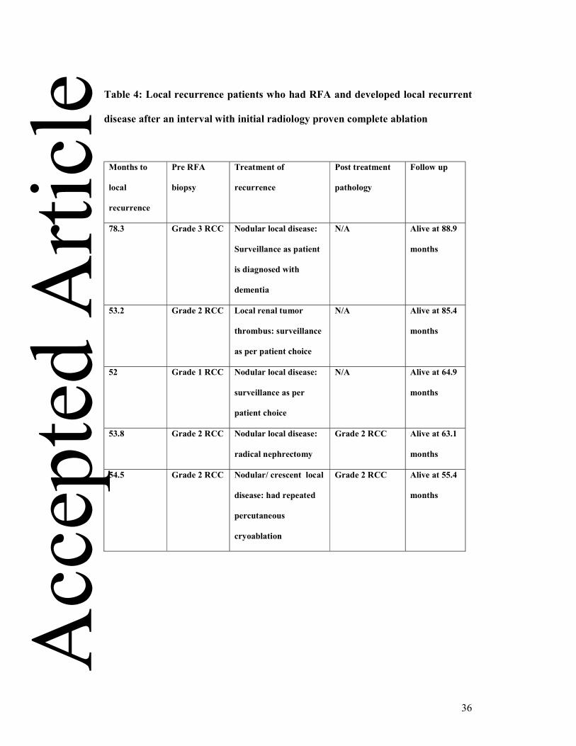
In our cohort, there were 5 (2.5%) local recurrences and all were late recurrences (> 4
year follow up) with mean detection at 58.3 months (Table 4). All the local
recurrences recurred at the inner margin of the zone of the ablation.
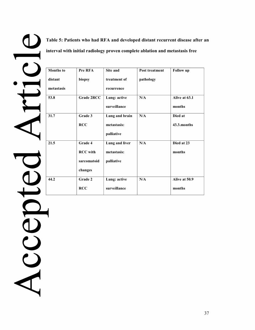
A total of 4 (2%) patients developed distant metastasis with mean detection at 37.8
months (table 5). Two patients have died from metastatic RCC and the other two
patients are still alive.
One of these patients died 23 months after treatment having developed both
pulmonary and liver metastases which were detected at 21.5 months during an acute
CT examination after she had presented with abdominal pain. She had RFA of her 3.5
cm renal tumour which had a histological diagnosis of Grade 4 sarcomatoid changes
conventional RCC. Another patient known to have chronic renal impairment
presented with pulmonary metastasis at 31.7 months following RFA of two renal
tumours in the left kidney (4cm and 2.5cm), with histological diagnosis of grade 3
conventional RCC. He died 43 months later when he developed brain metastases.
A further two patients with distant metastasis are still alive. One patient developed
local recurrence in the zone of ablation of the treated right grade 2 conventional RCC
(3.3cm) and a pulmonary metastasis (<10mm) at 53.8 months. He underwent radical Acc
epte
d A
rticl
e
24
right nephrectomy at 55.3 months and his pulmonary metastasis was kept under
surveillance. He developed bony metastases (ribs, iliac wing and sacrum) at 63.5
months and he is currently undergoing anti-angiogenic therapy with Sutent. One
patient developed a presumed pulmonary metastasis at 44.2 months post RFA of a
right grade 2 conventional RCC (4.2cm) but no conclusive CT guided pulmonary
biopsies had been achieved at 55.2 months. He had three negative histological
biopsies to date (last one in Oct 2012).
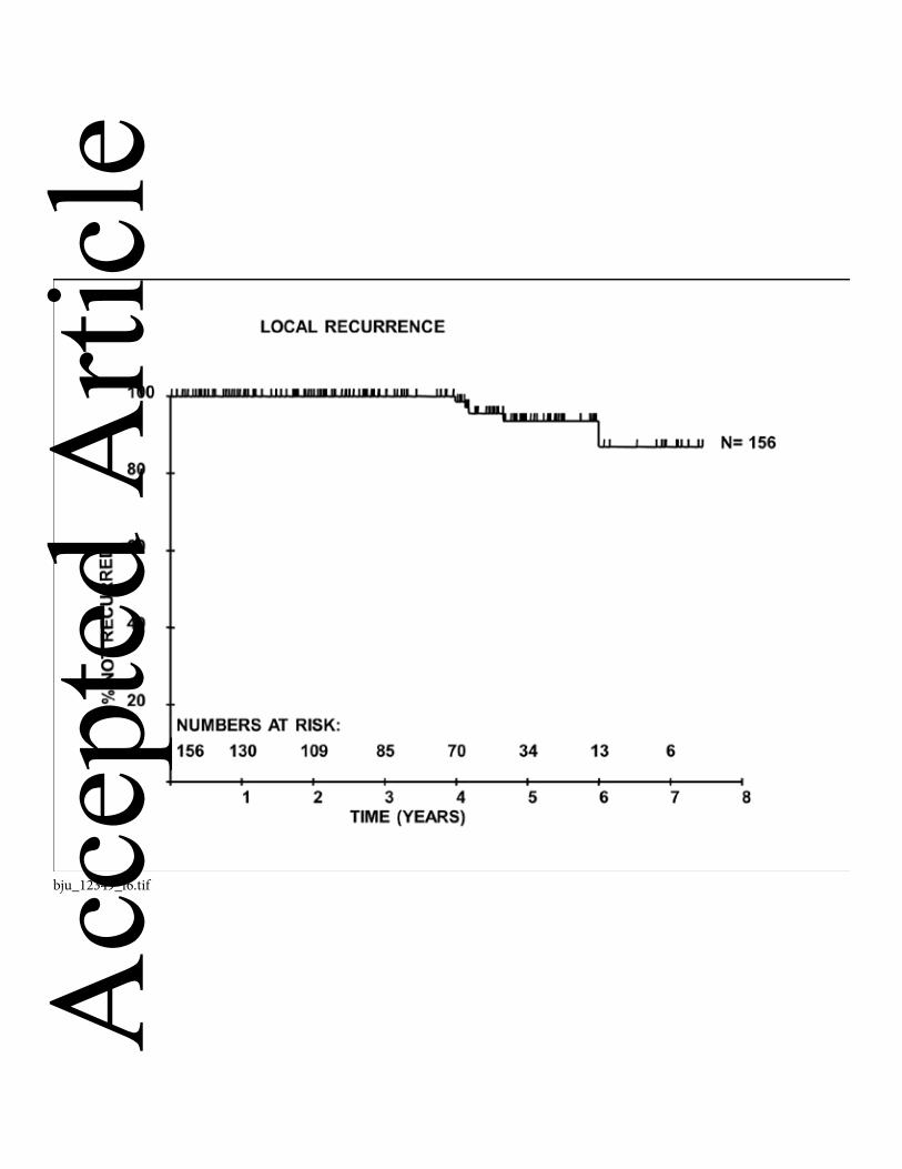
Our 5-year local recurrence free and metastasis free survival rates of 93.5% and
87.7% respectively (Figures 6 & 7).
Discussion
The new accepted standard treatment for small renal tumours (<4cm) is a nephron
sparing technique with either a surgical approach or image guided ablative therapy
[13].
We have reviewed our experience in a large tertiary university institution over an
eight years period from June 2004 to 2012 with the treatment of 200 renal tumours
using percutaneous RFA and compared to a range of 41 to 185 patients reported in
previous series with long term follow up [20, 21, 36-38]. Our image guided renal
ablation program is a supra-regional centre for the whole of the Yorkshire Cancer
Network (YCN) as well as outside YCN regions e.g. South Yorkshire, Lincolnshire
and Tyneside. Whilst our urologists received referral from a smaller referral base e.g. Acc
epte
d A
rticl
e
25
Leeds and Harrogate, they had treated 129 patients with T1 renal tumour and these
patients underwent either radical or partial nephrectomy during the same period.
We have evaluated our technical success, treatment complications, treatment effect on
the renal function and the 5-year overall, cancer specific, local recurrence as well as
metastasis free survival rates in this cohort of patients. In addition, operator
experience as well as tumor factors such as size and location were assessed to
determine its influence on the primary treatment technical success rate (i.e. achieve
complete RF ablation after one setting). For renal tumour in close proximity to the
ureter (<1cm), we also reviewed our experience in the use of protective techniques
with cold pyeloperfusion technique and assessed whether this step provided protection
from ureteral injury.
In our series, the primary and overall technical success rate was 95.5 % (191/200) vs.
98.5% (197/200) respectively for the 200 renal tumours which were RF ablated. The
local repeat RF ablation rate was 3%. This overall technical success rate is
comparable to other published series with reported rates that ranged from 90 to 100%
[21, 29, 39, 40]. In addition, it is interesting to note that our primary technical success
rate (100%) has improved considerably after the initial years of learning curve (> 4
years) and as stated earlier this is likely to be related to the better case selection by the
team. Similar observations were also reported by Poon et al - there is a definite
learning curve in acquiring this specialist skill by a dedicated team in order to achieve
good technical outcomes and minimal complication rate [41].
Acc
epte
d A
rticl
e
26
We have also demonstrated that tumour factors such as size (<3cm) and exophytic
location are two important independent predictors in achieving complete ablation with
a single treatment [29, 39, 42]. This is because a smaller vascular pedicle in the
exophytic renal tumour as well as surrounding peri-renal fat allows more effective
treatment than in central tumours.
In this cohort, our major and minor complication rates are also comparable to other
published series [29, 39, 42]. Interestingly, we did not experience significant
haemorrhage post RF ablation in this cohort, as this is one of the commonest major
complications reported previously [43].
The ureteral stricture complication in this cohort was 3.5% and is similar to the
reported range of around 2-3% [42, 43]. This was in part related to our early success
in treating central tumour with cold pyeloperfusion technique [30, 31] following
which we have performed more RF ablations of renal tumours in closer proximity to
ureters. Our overall experience has indicated that cold pyeloperfusion is effective in
protecting most of the ureters (80%) but does not always confer protection to the
ureter and PUJ during RF ablation especially when there is zero distance between the
tumour and the ureter. It is crucial during case selection and especially when
providing consultation to patients, to highlight the potential higher risks of ureteral
injury when the renal tumour has a central or lower pole location, so the patients are
consented accordingly and aware of the potential risks that the procedure entails.
We have been meticulous in protecting vital organs such as colon from the RF
ablation treatment margin with the hydrodissection technique with 5% Dextrose, and Acc
epte
d A
rticl
e
27
to date, we have not encountered any bowel injury during RF ablation in our cohort of
200 renal tumours. This is similarly reported by other institutions and the use of
protective techniques such as hydro-dissection is becoming increasingly routine when
the distance between treatment margin to bowel is <1cm [32, 43].
Development of chronic kidney disease when GFR <60 ml/min/ 1.73m2 is associated
with higher incidence of death, cardiovascular events and hospital admissions[44].
Therefore it is important that nephron sparing procedure can be performed in patients
with RCC that have compromised renal function such as solitary kidney status or with
pre-existing chronic kidney disease, with the aim to preserve renal function and
maintain well-being. The published literatures have confirmed that renal preservation
can be achieved with image guided renal ablation [17, 45-47]. However, this is the
first clinical series ever examine the relationship between the change of pre and post
treatment GFR (% GFR change) with the tumour characteristics (tumour size, polar
position and location), the total bulk of the tumour treated per ablation session,
number of tumours treated and the solitary kidney status and we have confirmed that
there is no association of renal function change with any of these factors [48]. Our
result has also confirmed that there is a 3.1% worsening of GFR after RFA treatment
when compared to pre-treatment GFR which is statistically significant between the
pre and post GFR (p<0.0001). However, there are only 4 patients developed
significant deterioration of renal function (>25% decreased in GFR post treatment).
Majority (98%) of our cohort have preservation of their renal function. It is important
to note that the measurement of renal function at day 1 post procedure is a routine
clinical test in our institution and we are aware that in majority of the patients the
renal function does recover and stabilize over a period of 3-6 months post RFA. The Acc
epte
d A
rticl
e
28
immediate renal function in our practice was a clinical guide for us to decide which
patients, especially the high risks patients e.g. with single kidney or impaired renal
function whether it was safe to discharge immediately or if further renal function
monitoring was required.
There are only limited series reporting the longer term oncological efficacy of RFA of
renal tumour with a total of 417 patients reported [20, 21, 36-38]. Our clinical series
has shown overall, 5-year cancer specific, local recurrence free and metastasis free
survival rates of: 75.8%, 97.9%, 93.5% and 87.7% respectively. This is comparable to
both Tracy et al and Zagoria et al cohorts. Tracy et al have reported the overall, 5-year
cancer specific and recurrence free survival rates that were: 85%, 99% and 93%. In
addition, Zagoria et al reported similar findings where 5-year overall survival, local
recurrence free and disease free rates were: 66%, 88% and 83%. The overall survival
rate in our cohort is a reflection of elderly and unfit patients in our early experience.
This is also comparable to the long term outcome of laparoscopic renal cryoablation
for small renal tumor by Aron et al, where 5-year overall, cancer specific and
recurrence free survival rates were: 83%, 95% and 78% [49].
Our experience also demonstrated a local repeat RFA rate (3%), local tumour
progression (2.5%) and metastatic progression potential (2%). These results also
compare favorably with a meta-analysis of renal cryoablation vs. RFA where the
cryoablation has local re-ablation, local tumor progression and metastatic progression
rates of: 1%, 5% and 2% respectively [50]. Similar results also seen in other RFA
published series: 3%, 7% and 5% respectively by Tracy et al [20]and Zagoria et al Acc
epte
d A
rticl
e
29
have also shown 12% local progression and 7% metastatic progression in their longer
term cohort [21].
Overall it is reassuring to confirm that our longer term survival outcome is within the
reported range of the gold standard ‘partial nephrectomy’ where the 5-year metastasis
free survival post partial nephrectomy for T1 renal tumours was 86-97% [16] and the
local recurrence rate post partial nephrectomy is around 1-3% [51, 52]. However, it is
prudent that we continue to monitor our technical success, and complication rate, as
well as local disease and metastatic disease progression potential following local
ablative therapy, and also adopts the Nephrometry score to assess the complexity of
case selection increasingly used in NSS (either the RENAL / PADUA score) [53, 54],
so that we may collectively provide long term 10 year oncological data in the near
future.
Conclusions
Whilst there have been multiple previous publications on RFA in renal cancer, this is
one of the largest series by far and increases the total number of cases reported from
approximately 400 to 600. This, together with our ability to analyse risk factors in the
numbers available to us, means that it is now possible to begin to draw firm
conclusions about the safety and efficacy of RFA and to reassure patients that, in the
hands of experienced operators, they can be confident that the results are comparable
to any alternative approach to nephron sparing surgical or non-surgical treatment for
renal cancer.
Acc
epte
d A
rticl
e
30
Conflict of Interest
None declared
Legends
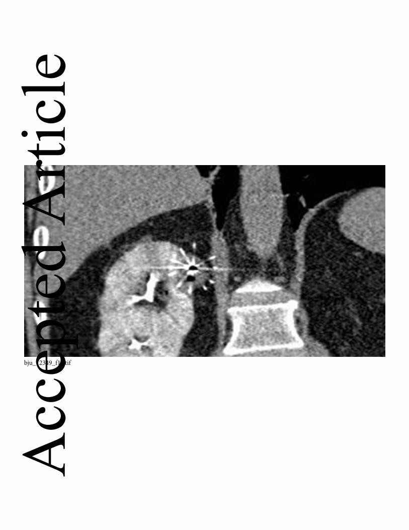
Figure1 (a) - Pre RFA axial contrast enhanced CT showed a 2.5cm enhancing renal
tumour at the upper pole of the right kidney (white arrow) (b)- Sagittal reformatting
showed the forward RF burn and (c)- coronal reformatting showed the overall
coverage of the tumour by the multi-tines RF electrode
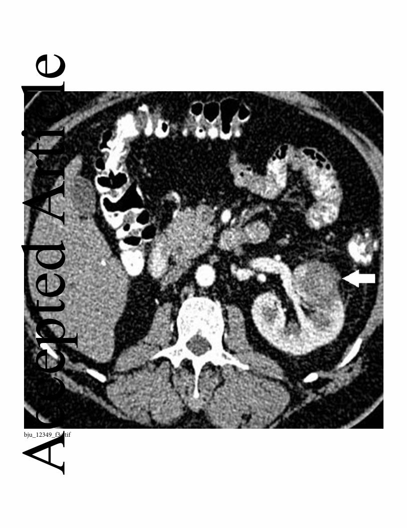
Figure 2- Pre RFA axial CT showed a centrally located renal tumour (white arrow)
where it was abutting the ureter and PUJ.
Figure 3- Axial contrast enhanced CT showed a small enhancing left renal tumour
(white arrow) at the anterior cortex of the kidney pre-RFA (a) and the zone of ablation
had high attenuation HU post RFA consistent with coagulation necrosis (white arrow)
on the unenhanced CT (b) and displayed no enhancement (white arrow) post contrast
administration (c).
Figure 4- The overall survival curve
Figure 5- The 5-year cancer specific survival curve
Figure 6- The 5-year local recurrence free survival curve
Figure 7- The 5-year distant metastasis free survival curve
Acc
epte
d A
rticl
e
31
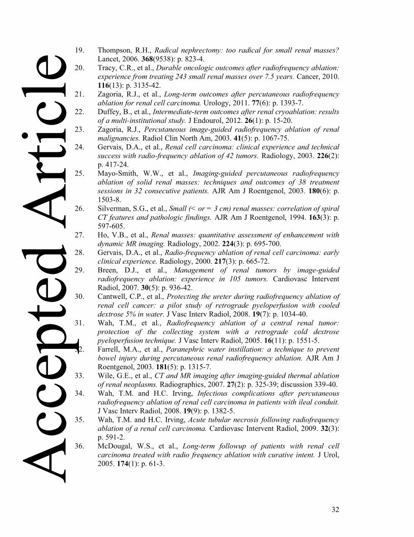
References
1. Jemal, A., et al., Cancer statistics, 2004. CA Cancer J Clin, 2004. 54(1): p. 8-
29. 2. Statistics, O.f.N., Cancer Statistics registrations: Registrations of cancer
diagnosed in 2008, England. 2010, Office for National Statistics, National Statistics London.
3. Globocan, 2008. 4. Zagoria, R.J., Imaging of small renal masses: a medical success story. AJR
Am J Roentgenol, 2000. 175(4): p. 945-55. 5. Jayson, M. and H. Sanders, Increased incidence of serendipitously discovered
renal cell carcinoma. Urology, 1998. 51(2): p. 203-5. 6. Hollingsworth, J.M., et al., Rising incidence of small renal masses: a need to
reassess treatment effect. J Natl Cancer Inst, 2006. 98(18): p. 1331-4. 7. Nguyen, M.M., I.S. Gill, and L.M. Ellison, The evolving presentation of renal
carcinoma in the United States: trends from the Surveillance, Epidemiology, and End Results program. J Urol, 2006. 176(6 Pt 1): p. 2397-400; discussion 2400.
8. Pantuck, A.J., A. Zisman, and A.S. Belldegrun, The changing natural history of renal cell carcinoma. J Urol, 2001. 166(5): p. 1611-23.
9. Pantuck, A.J., et al., Incidental renal tumors. Urology, 2000. 56(2): p. 190-6. 10. Luciani, L.G., R. Cestari, and C. Tallarigo, Incidental renal cell carcinoma-
age and stage characterization and clinical implications: study of 1092 patients (1982-1997). Urology, 2000. 56(1): p. 58-62.
11. Remzi, M. and M. Marberger, Renal tumor biopsies for evaluation of small renal tumors: why, in whom, and how? Eur Urol, 2009. 55(2): p. 359-67.
12. Abouassaly, R., B.R. Lane, and A.C. Novick, Active surveillance of renal masses in elderly patients. J Urol, 2008. 180(2): p. 505-8; discussion 508-9.
13. Ljungberg, B., et al., EAU guidelines on renal cell carcinoma: the 2010 update. Eur Urol, 2010. 58(3): p. 398-406.
14. Huang, W.C., et al., Chronic kidney disease after nephrectomy in patients with renal cortical tumours: a retrospective cohort study. Lancet Oncol, 2006. 7(9): p. 735-40.
15. Van Poppel, H., et al., Treatment of localised renal cell carcinoma. Eur Urol, 2011. 60(4): p. 662-72.
16. Uzzo, R.G. and A.C. Novick, Nephron sparing surgery for renal tumors: indications, techniques and outcomes. J Urol, 2001. 166(1): p. 6-18.
17. Lucas, S.M., et al., Renal function outcomes in patients treated for renal masses smaller than 4 cm by ablative and extirpative techniques. J Urol, 2008. 179(1): p. 75-9; discussion 79-80.
18. Hafez, K.S., A.C. Novick, and B.P. Butler, Management of small solitary unilateral renal cell carcinomas: impact of central versus peripheral tumor location. J Urol, 1998. 159(4): p. 1156-60. Acc
epte
d A
rticl
e
32
19. Thompson, R.H., Radical nephrectomy: too radical for small renal masses? Lancet, 2006. 368(9538): p. 823-4.
20. Tracy, C.R., et al., Durable oncologic outcomes after radiofrequency ablation: experience from treating 243 small renal masses over 7.5 years. Cancer, 2010. 116(13): p. 3135-42.
21. Zagoria, R.J., et al., Long-term outcomes after percutaneous radiofrequency ablation for renal cell carcinoma. Urology, 2011. 77(6): p. 1393-7.
22. Duffey, B., et al., Intermediate-term outcomes after renal cryoablation: results of a multi-institutional study. J Endourol, 2012. 26(1): p. 15-20.
23. Zagoria, R.J., Percutaneous image-guided radiofrequency ablation of renal malignancies. Radiol Clin North Am, 2003. 41(5): p. 1067-75.
24. Gervais, D.A., et al., Renal cell carcinoma: clinical experience and technical success with radio-frequency ablation of 42 tumors. Radiology, 2003. 226(2): p. 417-24.
25. Mayo-Smith, W.W., et al., Imaging-guided percutaneous radiofrequency ablation of solid renal masses: techniques and outcomes of 38 treatment sessions in 32 consecutive patients. AJR Am J Roentgenol, 2003. 180(6): p. 1503-8.
26. Silverman, S.G., et al., Small (< or = 3 cm) renal masses: correlation of spiral CT features and pathologic findings. AJR Am J Roentgenol, 1994. 163(3): p. 597-605.
27. Ho, V.B., et al., Renal masses: quantitative assessment of enhancement with dynamic MR imaging. Radiology, 2002. 224(3): p. 695-700.
28. Gervais, D.A., et al., Radio-frequency ablation of renal cell carcinoma: early clinical experience. Radiology, 2000. 217(3): p. 665-72.
29. Breen, D.J., et al., Management of renal tumors by image-guided radiofrequency ablation: experience in 105 tumors. Cardiovasc Intervent Radiol, 2007. 30(5): p. 936-42.
30. Cantwell, C.P., et al., Protecting the ureter during radiofrequency ablation of renal cell cancer: a pilot study of retrograde pyeloperfusion with cooled dextrose 5% in water. J Vasc Interv Radiol, 2008. 19(7): p. 1034-40.
31. Wah, T.M., et al., Radiofrequency ablation of a central renal tumor: protection of the collecting system with a retrograde cold dextrose pyeloperfusion technique. J Vasc Interv Radiol, 2005. 16(11): p. 1551-5.
32. Farrell, M.A., et al., Paranephric water instillation: a technique to prevent bowel injury during percutaneous renal radiofrequency ablation. AJR Am J Roentgenol, 2003. 181(5): p. 1315-7.
33. Wile, G.E., et al., CT and MR imaging after imaging-guided thermal ablation of renal neoplasms. Radiographics, 2007. 27(2): p. 325-39; discussion 339-40.
34. Wah, T.M. and H.C. Irving, Infectious complications after percutaneous radiofrequency ablation of renal cell carcinoma in patients with ileal conduit. J Vasc Interv Radiol, 2008. 19(9): p. 1382-5.
35. Wah, T.M. and H.C. Irving, Acute tubular necrosis following radiofrequency ablation of a renal cell carcinoma. Cardiovasc Intervent Radiol, 2009. 32(3): p. 591-2.
36. McDougal, W.S., et al., Long-term followup of patients with renal cell carcinoma treated with radio frequency ablation with curative intent. J Urol, 2005. 174(1): p. 61-3. Acc
epte
d A
rticl
e
33
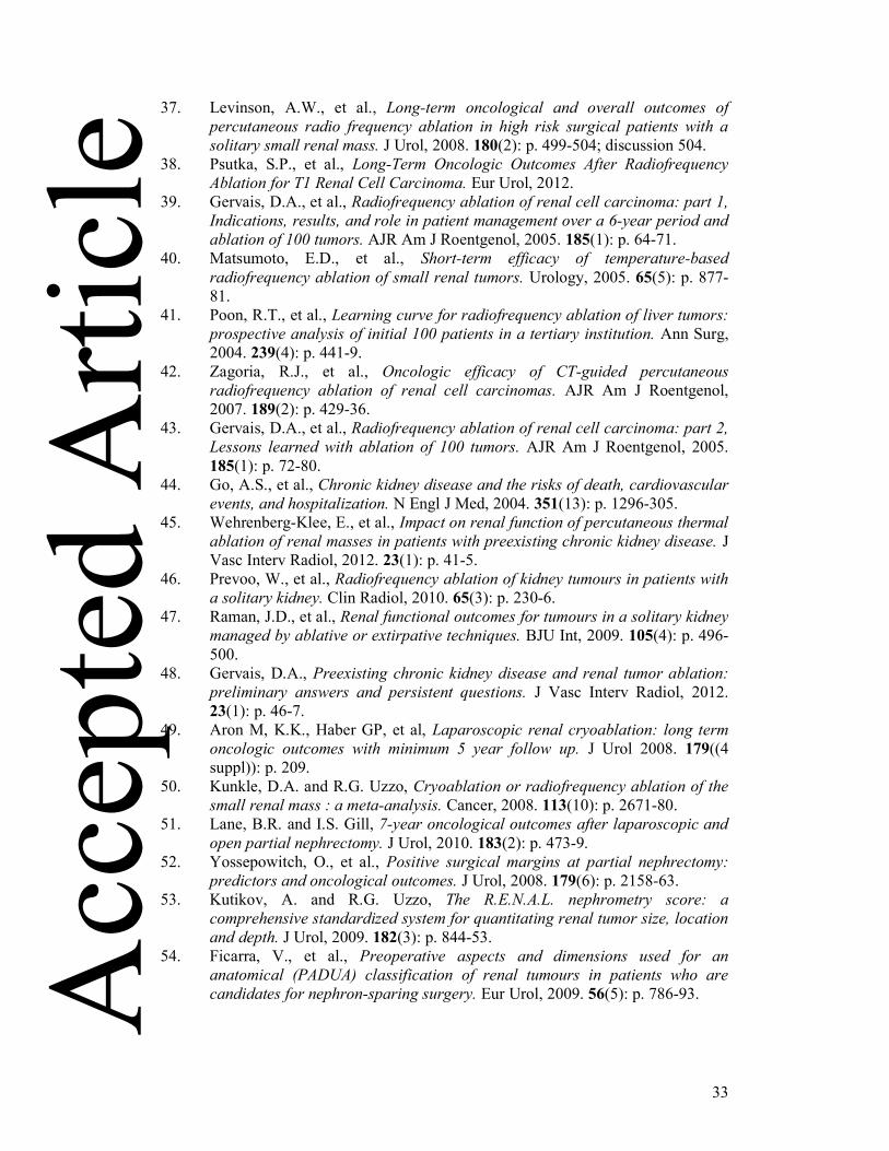
37. Levinson, A.W., et al., Long-term oncological and overall outcomes of percutaneous radio frequency ablation in high risk surgical patients with a solitary small renal mass. J Urol, 2008. 180(2): p. 499-504; discussion 504.
38. Psutka, S.P., et al., Long-Term Oncologic Outcomes After Radiofrequency Ablation for T1 Renal Cell Carcinoma. Eur Urol, 2012.
39. Gervais, D.A., et al., Radiofrequency ablation of renal cell carcinoma: part 1, Indications, results, and role in patient management over a 6-year period and ablation of 100 tumors. AJR Am J Roentgenol, 2005. 185(1): p. 64-71.
40. Matsumoto, E.D., et al., Short-term efficacy of temperature-based radiofrequency ablation of small renal tumors. Urology, 2005. 65(5): p. 877-81.
41. Poon, R.T., et al., Learning curve for radiofrequency ablation of liver tumors: prospective analysis of initial 100 patients in a tertiary institution. Ann Surg, 2004. 239(4): p. 441-9.
42. Zagoria, R.J., et al., Oncologic efficacy of CT-guided percutaneous radiofrequency ablation of renal cell carcinomas. AJR Am J Roentgenol, 2007. 189(2): p. 429-36.
43. Gervais, D.A., et al., Radiofrequency ablation of renal cell carcinoma: part 2, Lessons learned with ablation of 100 tumors. AJR Am J Roentgenol, 2005. 185(1): p. 72-80.
44. Go, A.S., et al., Chronic kidney disease and the risks of death, cardiovascular events, and hospitalization. N Engl J Med, 2004. 351(13): p. 1296-305.
45. Wehrenberg-Klee, E., et al., Impact on renal function of percutaneous thermal ablation of renal masses in patients with preexisting chronic kidney disease. J Vasc Interv Radiol, 2012. 23(1): p. 41-5.
46. Prevoo, W., et al., Radiofrequency ablation of kidney tumours in patients with a solitary kidney. Clin Radiol, 2010. 65(3): p. 230-6.
47. Raman, J.D., et al., Renal functional outcomes for tumours in a solitary kidney managed by ablative or extirpative techniques. BJU Int, 2009. 105(4): p. 496-500.
48. Gervais, D.A., Preexisting chronic kidney disease and renal tumor ablation: preliminary answers and persistent questions. J Vasc Interv Radiol, 2012. 23(1): p. 46-7.
49. Aron M, K.K., Haber GP, et al, Laparoscopic renal cryoablation: long term oncologic outcomes with minimum 5 year follow up. J Urol 2008. 179((4 suppl)): p. 209.
50. Kunkle, D.A. and R.G. Uzzo, Cryoablation or radiofrequency ablation of the small renal mass : a meta-analysis. Cancer, 2008. 113(10): p. 2671-80.
51. Lane, B.R. and I.S. Gill, 7-year oncological outcomes after laparoscopic and open partial nephrectomy. J Urol, 2010. 183(2): p. 473-9.
52. Yossepowitch, O., et al., Positive surgical margins at partial nephrectomy: predictors and oncological outcomes. J Urol, 2008. 179(6): p. 2158-63.
53. Kutikov, A. and R.G. Uzzo, The R.E.N.A.L. nephrometry score: a comprehensive standardized system for quantitating renal tumor size, location and depth. J Urol, 2009. 182(3): p. 844-53.
54. Ficarra, V., et al., Preoperative aspects and dimensions used for an anatomical (PADUA) classification of renal tumours in patients who are candidates for nephron-sparing surgery. Eur Urol, 2009. 56(5): p. 786-93.
Acc
epte
d A
rticl
e
34
Table 1: The overall technical success rate vs. size and location
Tumor
Location
Total numbers of tumours had
ablation (200) and technical
success*= tumours with
complete ablation/ total number
of tumours treated
Total tumours>3cm
(67)
Total tumours=<3cm
(133)
Exophytic *43/43 *15/15 *28/28
Parenchymal *41/41 *11/11 *30/30
Mixed *98/100 *32/34 *66/66
Central *15/16 *6/7 *9/9
Technical success*= tumours with complete ablation/ total number of tumours treated
Table 2: Patients with residual disease or requireing repeated RFA
Patients Tumour
location
Size Complete
Ablation achieved
Residual disease because
patients refused further
treatment
Number of
RF
treatment
1 Central 2.7 1 0 2
2 Central 3.4 1 0 3
3 Central 3.4 1 0 2
4 Central 3.5 1 0 3
5 Central 4 1 0 2
6 Central 4.8 1 0 3
7 Central 5.6 0 1 2
8 Mixed 4 0 1 1
9 Mixed 5.4 0 1 2 Acc
epte
d A
rticl
e
35
Table 3: Achievement of complete ablation and number of ablation sessions
based on tumour size
Tumour Size
(cm)
The (%)= number of
tumours with complete
ablation/ total number of
tumours treated
Number of
tumours
treated with
one session
Number of
tumours
treated with
Two sessions
Number of
tumours treated
with three
sessions
=< 3cm 133/ 133 (100) 132 1 NA
3-5 cm 62/63 (98.4%) a
57 a 3
3
>5cm 2/4 (50%) b 2 NA NA
a In this group of renal tumours treated (3-5cm), one renal tumour was treated with single session but
patient refused to have further treatment.
b In this group of renal tumours (>5cm), two renal tumours were with residual disease after a single
treatment session and patients did not undertake further treatment due to patients’ choice.
Acc
epte
d A
rticl
e
36
Table 4: Local recurrence patients who had RFA and developed local recurrent
disease after an interval with initial radiology proven complete ablation
Months to
local
recurrence
Pre RFA
biopsy
Treatment of
recurrence
Post treatment
pathology
Follow up
78.3 Grade 3 RCC Nodular local disease:
Surveillance as patient
is diagnosed with
dementia
N/A Alive at 88.9
months
53.2 Grade 2 RCC Local renal tumor
thrombus: surveillance
as per patient choice
N/A Alive at 85.4
months
52 Grade 1 RCC Nodular local disease:
surveillance as per
patient choice
N/A Alive at 64.9
months
53.8 Grade 2 RCC Nodular local disease:
radical nephrectomy
Grade 2 RCC Alive at 63.1
months
54.5 Grade 2 RCC Nodular/ crescent local
disease: had repeated
percutaneous
cryoablation
Grade 2 RCC Alive at 55.4
months
Acc
epte
d A
rticl
e
37
Table 5: Patients who had RFA and developed distant recurrent disease after an
interval with initial radiology proven complete ablation and metastasis free
Months to
distant
metastasis
Pre RFA
biopsy
Site and
treatment of
recurrence
Post treatment
pathology
Follow up
53.8 Grade 2RCC Lung: active
surveillance
N/A Alive at 63.1
months
31.7 Grade 3
RCC
Lung and brain
metastasis:
palliative
N/A Died at
43.3.months
21.5 Grade 4
RCC with
sarcomatoid
changes
Lung and liver
metastasis:
palliative
N/A Died at 23
months
44.2 Grade 2
RCC
Lung: active
surveillance
N/A Alive at 50.9
months
Acc
epte
d A
rticl
e
bju_12349_f1a.tif
Acc
epte
d A
rticl
e
bju_12349_f1b.tif
Acc
epte
d A
rticl
e
bju_12349_f1c.tif
Acc
epte
d A
rticl
e
bju_12349_f2.tifAcc
epte
d A
rticl
e
bju_12349_f3a.tif
Acc
epte
d A
rticl
e
bju_12349_f3b.tifAcc
epte
d A
rticl
e
bju_12349_f3c.tifAcc
epte
d A
rticl
e
bju_12349_f4.tif
Acc
epte
d A
rticl
e
bju_12349_f5.tif
Acc
epte
d A
rticl
e
bju_12349_f6.tif
Acc
epte
d A
rticl
e

bju_12349_f7.tif
Acc
epte
d A
rticl
e

![Radiofrequency Ablation of Lung Cancer at Okayama University … · results of RFA for the treatment of lung cancer in 2004 [1]. At the time of that report, we had per-formed RFA](https://img.pdfslide.us/doc/110x75/60509535341f544fe72779ba/radiofrequency-ablation-of-lung-cancer-at-okayama-university-results-of-rfa-for.jpg)