Embed Size (px)
Citation preview
Bioorganic & Medicinal Chemistry 21 (2013) 6699–6707
Contents lists available at ScienceDirect
Bioorganic & Medicinal Chemistry
journal homepage: www.elsevier .com/locate /bmc
Radiochemical and radiobiological assessment of a pyridyl-S-cysteinefunctionalized bombesin derivative labeled with the 99mTcðCOÞþ3 core
0968-0896/$ - see front matter � 2013 Elsevier Ltd. All rights reserved.http://dx.doi.org/10.1016/j.bmc.2013.08.010
Abbreviations: Boc, t-butyloxycarbonyl; BSA, bovine serum albumin; Cys,cysteine; DIEA, N,N-diisopropylethylamine; DIC, diisopropylcarbamide; DMEM,Dulbecco’s modified Eagle medium; DMF, dimethylformamide; EDTA, ethylenedi-aminetetraacetic acid; ESI-MS, electrospray ionization mass spectrometry; EtOH,ethanol; FBS, fetal bovine serum; Fmoc, 9-fluorenylmethyloxycarbonyl; HOBt, 1-hydroxybenzotriazole; ID, injected dose; I.D., internal diameter; iv, intravenous;PBS, phosphate buffered saline; p.i., postinjection; PET, positron emission tomog-raphy; PMSF, phenylmethanesulfonyl fluoride; PSPMT, position sensitive photo-multiplier tube; ROI, regions of interest; RP/SE-HPLC, reversed-phase/size-exclusionhigh-performance liquid chromatography; RT, room temperature; SCID, severecombined immunodeficiency; SPECT, single photon emission computerized tomog-raphy; SPPS, solid phase peptide synthesis; TFA, trifluoroacetic acid; TIPS,triisopropylsilane.⇑ Corresponding authors. Tel.: +30 210 6503687; fax: +30 210 6543526.
E-mail addresses: [email protected] (P. Bouziotis), [email protected] (D. Psimadas).
� Present address: Department of Nuclear Medicine, University Hospital Freiburg,Hugstetterstrasse 55, 79106 Freiburg, Germany.
� Present address: Department of Nuclear Medicine, University Hospital of Larissa,41110 Mezourlo, Larissa, Greece.
§ Present address: Department of Radiology and Nuclear Medicine, UniversityHospital Basel, 4031 Basel, Switzerland.
Penelope Bouziotis a,⇑, Eleni Gourni a,�, George Patsis a, Dimitrios Psimadas a,⇑,�, Christos Zikos a,Melpomeni Fani a,§, Stavros Xanthopoulos a, George Loudos b, Maria Paravatou-Petsotas a,Evangelia Livaniou a, Alexandra D. Varvarigou a, Ioannis Pirmettis a, Minas Papadopoulos a
a Institute of Nuclear and Radiological Sciences and Technology, Energy and Safety, N.C.S.R. ‘Demokritos’, 15310 Aghia Paraskevi, Greeceb Department of Medical Instruments Technology, Technological Educational Institute of Athens, Aghiou Spyridonos 28, 12210 Egaleo, Greece
a r t i c l e i n f o a b s t r a c t
Article history:Received 23 May 2013Revised 12 July 2013Accepted 5 August 2013Available online 12 August 2013
Keywords:BombesinRadiolabelingRadiopharmaceuticalsTechnetiumTricarbonyl precursor
Bombesin is a neuropeptide widely studied due to its ability to target various types of cancers. Techne-tium-99m on the other hand is ideal for diagnostic tumor targeting. The aim of the present study is theinvestigation of the coupling of the ligand (S)-(2-(20-pyridyl)ethyl)-D,L-cysteine with the BN-peptide Gln-Arg-Leu-Gly-Asn-Gln-Trp-Ala-Val-Gly-His-Leu-Met(CONH2) through the spacer aminohexanoic acidandthe labeling of the resulting derivative MBN with the synthon [M(CO)3(H2O)3]+ (M = 99mTc, Re). The pep-tide was synthesized according to the SPPS method, purified and characterized by ESI-MS. The new99mTc-labeled biomolecule was stable in vitro, showed high affinity for the human GRP receptorexpressed in PC3 cells and the rate of internalization was found to be time-dependent tissue distributionof the radiopeptide was evaluated in normal mice and in prostate cancer experimental models and sig-nificant radioactivity uptake was observed in the pancreas of normal mice as well as in PC3 tumors.Dynamic studies of the radiopeptide showed satisfactory tumor images.
� 2013 Elsevier Ltd. All rights reserved.
1. Introduction
There has been remarkable progress in reducing cancer deathrates worldwide.1 Most patients diagnosed with early-stage dis-
ease will survive their illness. Despite these advances, cancer stillremains the number two cause of death in the United States.
The detection and treatment of cancer are desirable, especiallyin its early stages. A very promising group of tumor-targeting li-gands are regulatory peptides, because they act on multiple targetsin the human body at extremely low concentrations and, mostimportantly, because of the over-expression of their receptors onthe surface of different kinds of cancer cells.2 One peptide withgreat interest in the field of nuclear oncology is the amphibian tet-radecapeptide bombesin (BN) which was first isolated in 1971 byAnastasi et al. from the skin of the frog Bombina bombina.3 BNhas high affinity for gastrin-releasing-peptide (GRP) receptors.GRP, a 27-amino acid peptide, is the mammalian analogue of theamphibian BN. Having almost the same C-terminal amino-acid se-quences, BN and GRP present nearly identical biological and recep-tor-binding properties.4 The physiological actions of BN are: (a)stimulation of gastric secretion, (b) pancreatic enzyme secretion,(c) hypothermia, (d) hyperglycaemia, (e) disruption of peristalticactivity, (f) release of enteric peptides and (g) satiety.5
The BN receptor family belongs to the G-protein receptor super-family and includes four different subtypes: the GRP-R subtype(BB2), the neuromedin (NMB) receptor subtype (BB1), the orphanreceptor subtype (BB3), and the bombesin receptor subtype 4(BB4).6,7 The most extensively studied receptor subtype is BB2.
6700 P. Bouziotis et al. / Bioorg. Med. Chem. 21 (2013) 6699–6707
The BB2 receptor has been isolated on the hypothalamus, pituitarygland, vagal ganglia, as well as on glioblastoma, small-cell lung car-cinoma, ovarian epithelial cancer, melanoma, colon, prostatecancer, and several other tumors.5,8 Of special interest is that tu-mor autoradiography-based studies have reported that GRP-Rsare expressed at very high density in invasive prostatic carcinomaswhereas normal and hyperplastic prostate tissue were predomi-nantly GRP-R negative.8
The co-ordination chemistry of technetium and its surrogaterhenium is the focus of intense research activity, mainly due tothe extensive use of the radioactive isotopes of these elements innuclear medicine. Technetium-99m (t1/2 = 6 h, Ec = 140 keV) isthe radioisotope of choice for nuclear medical imaging, due to itsideal nuclear properties, its low cost, and widespread availabilitywhile 188Re (t1/2 = 0.7 d, Emax = 2.12 MeV) finds growing applicationin radiation therapy. Both isotopes are readily available as permet-allates MðVIIÞO�4 from a 99Mo/99mTc or 188W/188Re generator sys-tem, respectively. New metal cores and ligand systems arecontinuously emerging in the literature, aiming at complexes withhigh in vivo stability, favorable pharmacokinetic properties andtarget tissue specificity. The introduction of the air-stable fac-[M(CO)3(H2O)3]+ (M = 99mTc or 188Re) synthon produced by thegentle reduction of M(VII) to M(I) under 1 atm of CO, establishedthe MðIÞðCOÞþ3 core as an easily accessible platform in the searchfor new radiopharmaceuticals.9,10 The small size and kinetic inert-ness of the fac-[M(CO)3(H2O)3]+ synthon make it suitable for thelabeling of biologically active molecules with minimum disruptionof their activity or specificity.11,12 Furthermore, the three aqua li-gands of the MðIÞðCOÞþ3 core are labile and readily substituted bya variety of functional groups including amines, thioethers, imines,thiols, and phosphines to give stable hexaco-ordinated com-plexes.12–14 Previous studies on the coordination chemistry of thefac �MðIÞðCOÞþ3 core showed that the tridentate ligands NSN areideal for the tricarbonyl precursor complexation, since they formcomplexes even at very low concentrations (approximately10�5 M).14–16
Following previous work on pyridyl derivatives as bifunctionalchelators for labeling peptides with the fac-[M(CO)3]+ moiety,13,14
in the present study we focus on the investigation of the couplingof the ligand (S)-(2-(2-pyridyl)ethyl)-D,L-cysteine with the BN-pep-tide Gln-Arg-Leu-Gly-Asn-Gln-Trp-Ala-Val-Gly-His-Leu-Met(CONH2) via the Aca (6-aminohexanoic acid) moiety and thelabeling of the coupled derivative (MBN) with the synthon[M(CO)3(H2O)3]+ (M = 99mTc or 185/187Re). The coupled derivative(MBN) was synthesized with the SPPS method, by following theFmoc strategy, and its reaction with [185/187Re(CO)3Br3]2� affordeda single product, which was characterized by mass spectroscopyand RP-HPLC. At the 99mTc level, the corresponding complex wasobtained by incubating MBN with the fac-[99mTc(CO)3(H2O)3]+ pre-cursor in water. In vitro studies followed, to determine the bindingproperties as well as the internalization efficiency of the 99mTc-MBN complex into the cells as a function of time. These studieswere performed in PC-3 human prostate cancer cells, a cell linethat expresses the GRP receptor in very high numbers(2.7 ± 0.6 � 105 receptors per cell).17 Studies of the behavior ofthe 99mTc-labeled derivative were assessed in normal mice andSCID mice bearing prostate cancer xenografts and dynamic evalu-ation was also performed
2. Materials and methods
2.1. Materials and instruments
The flow rate was 3.5 mL/min. The pure peptide was identifiedby a Waters analytical RP-HPLC System (pump 616, detector 996
PDA) using a Lichrospher RP C18 column (250 � 4.6 mm I.D.;5 lm particle size; Merck). The solvent system contained 0.05%TFA in 0.1 M NaCl (solvent A) and 90% ACN, 10%, 0.05% TFA in0.1 M NaCl (solvent B). A linear gradient from 10% to 60% solventB was applied within 20 min, at a flow rate of 1 mL/min. Peptidepeaks were detected photometrically (220 nm). RP-HPLC analysesof all radiolabeled species were performed on a Waters l-Bonda-pack C18 (3.9 � 50 mm I.D.) cartridge column (Waters, Germany).RP-HPLC solvents consisted of 0.1% TFA in MeOH (solvent A) and0.1% TFA in H2O (solvent B). Elution was achieved by applying a lin-ear gradient from 100% B to 20% B in 30 min. The flow rate was1.0 mL/min. [Tyr4]-BN and KReO4 was purchased from Sigma–Al-drich (GmbH, Austria). [125I-Tyr4]-BN was purchased from Amer-sham (100 lL, 10 lCi, 2000 Ci/mmol). 99mTechnetium, in the formof Na99mTcO4 in 0.9% NaCl, was eluted from a commercial99Mo-99mTc generator (Mallinckrodt Medical B.V.). Solvents forHPLC were of analytical grade; they were further filtered through0.22 lm membrane filters (Millipore, Milford, USA) and degassed.Radioactivity measurements were conducted in a automatedwell-type gamma-counter (NaI(Tl) crystal, Canberra-Packard,auto-gamma 5000 series model) calibrated for 99mTc.
The human prostate adenocarcinoma cell line PC3 was obtainedfrom American Type Culture Collection (ATCC). DMEM, FBS, L-glu-tamine, penicilline/streptomycin and trypsin/EDTA solution werepurchased from PAA Laboratories (GmbH, Austria). BSA, glutamax,HEPES, bacitracin, aprotinin and PMSF used were obtained fromSigma–Aldrich (GmbH, Austria).
Animal experiments were carried out in compliance to Euro-pean and Greek regulations. Female Swiss albino mice and maleSCID mice of 6 weeks of age on the day of inoculation, were ob-tained from the breeding facilities of the NCSR ‘Demokritos’. Theanimals were kept under aseptic conditions until the day ofbiodistribution.
The small mouse-sized camera used is based on a pair of twosquare H8500 flat-panel PSPMT with external dimensions of52 � 52 and 34 mm thick. The two H8500 PSPMTs are gently cou-pled to NaI(Tl) crystal array (Bicron-St. Gobain) with an active areaof approximately 98 � 48 mm. The pixel dimensions are1 � 1 � 5 mm with a pitch of 1.2 mm. The array is viewed througha 3 mm glass window and encapsulation is completed by an alumi-num cover 50 lm thick. An optical grease (RX-688 Optical Cou-pling Grease, Rexon Inc., Beachwood, OH, 44122) has been usedfor optical coupling of the PSPMTs to the scintillation array win-dow. The camera is equipped with a parallel-hole lead collimatorwith hexagonal holes 1.2 mm in diameter and 0.2 mm thick sep-tum walls (Tecomet Inc.). The collimator is 25 mm thick, and hasan active area 52 � 105 mm. The system is enclosed in an 8 mmtungsten (W) housing box 140 � 82 � 107 mm deep. The front faceentrance window is of 0.5 mm thick, graphite-based compositematerial, manufactured principally for use in medical imaging ta-bles. Each PSPMT provides four analog signals X+, X�, Y+ and Y�.These signals are collected using two National InstrumentsPCI6110 ADCs which have four channels each. Data acquisition,as well as preprocessing and system calibration, are carried outusing custom software written in Kmax (Sparrow Corp., Port Or-ange, FL, 32128) environment. The spatial resolution of the systemis approx. 1.5 mm in 0 mm distance from collimator surface. En-ergy resolution is approx. 15% and sensitivity has been found tobe approx. 5000 counts/min/mCi/cm2. These characteristics aresuitable for short dynamic studies of 2–3 min per frame.
2.2. Solid-phase peptide synthesis (SPPS)
Coupling was performed by dissolving excessive quantity(4 mol equiv) of Fmoc-protected amino acid and HOBt in DMF.
P. Bouziotis et al. / Bioorg. Med. Chem. 21 (2013) 6699–6707 6701
The solution was cooled on ice and then DIC (4 mol equiv) wasadded. The reaction mixture was left on ice (10 min) and then at25 �C (10 min). Afterwards, it was added to the resin and allowedto react. Coupling efficiency was checked by using the Kaiser nin-hydrin test. Removal of the N-terminal Fmoc group (deprotection)was performed within 30 min using a 20% (v/v) solution of piperi-dine in DMF. After the removal of the Fmoc group of the N-terminalamino acid the resin was washed 4� DMF, 6� DCM and 2� petro-leum ether and then dried under vacuum (P2O5) for 18 h. The pep-tide synthesized was cleaved from the resin using an appropriatequantity (�20 mL/g resin) of a cleavage mixture consisting ofTFA/H2O/EDT/TIPS 95:2.5:2.5:1 (vol). The cleavage reaction lasted2 h. The reaction mixture was filtered in a centrifuge flask, the re-sin was washed with TFA (2�, 2 min), and the peptide precipitatedas a solid material by adding Et2O. After centrifugation the crudepeptide was isolated and dried under vacuum (P2O5) for 18 h.The purification of the crude peptide was achieved by semiprepar-ative RP-HPLC while the identification of the pure peptide by ana-lytical RP-HPLC. In 2.1., a detailed description of both HPLCsystems, the solvent systems and the linear gradient, is given.
2.3. [185/187Re(CO)3]-MBN
The 185/187Re complex of the peptide derivative was prepared byaddition of a solution of the derivative in H2O to an equimolaraqueous solution of [NEt4]2[ReBr3(CO)3], as previously described.14
The mixture was stirred at 70 �C, and the solution mixture wasperiodically checked in time. The reaction afforded a single prod-uct, which was identified by mass spectroscopy and HPLC. ESI-MS spectroscopy of MBN and 185/187Re-labeled-MBN was per-formed at the ‘Mass Spectrometry and Dioxin Analysis Lab’, NCSR‘Demokritos’. The test solution in 50% aqueous ACN was infusedinto an electrospray interface mass spectrometer (AQA Navigator,Finnigan) at a flow rate of 0.1 mL/min, using a Harvant Syringepump. Negative or positive ion ESI-MS spectra were acquired byadjusting the needle and cone voltages accordingly. Hot nitrogengas (Dominic–Hunter UHPLCMS-10) was used for desolvation at170 �C.
2.4. [99mTc(H2O)3(CO)3]
Preparation of [99mTc(H2O)3(CO)3] was performed according toa published method with minor modifications.9 Briefly, a penicillinvial (10 mL) containing 5.5 mg NaBH4, 4 mg Na2CO3 and 20 mg Na/K-tartrate was sealed with an aluminium-capped rubber stopperand flushed for 20 min with CO gas. Two-milliliters of 99mTc-gener-ator eluate were added by a syringe and the mixture was gentlystirred. The solution was heated for 20 min at 75–80 �C. After cool-ing to RT, 100 lL of a 1 N HCl solution were added to adjust the pHat a range of 7.0–7.5. Quality control of the precursor was per-formed by RP-HPLC.
2.5. Radiolabeling and stability
The vial was sealed and the mixture was stirred and incubatedat 75–80 �C for 20 min. After cooling to RT, radiochemical analysiswas performed by gradient RP-HPLC using the conditions de-scribed for the quality control of the [99mTc(H2O)3(CO)3] precursor.The stability of the radiolabeled peptide was assessed up to 6 h atRT. Furthermore, His and Cys challenge experiments were per-formed, in order to assess the stability of the new peptide conju-gate against high concentrations of L-His and L-Cys. Briefly,aliquots of 100 lL of the complex were added to 900 lL of a10�2 M (10�3 M) L-His and 10�2 M (10�3 M) L-Cys solutions in
0.9% NaCl. The samples were incubated at RT for 24 h. At the 1, 6and 24 h time-points, 100 lL of the sample were removed and ana-lyzed by gradient RP-HPLC.
2.6. Cell binding studies
The human PC3 adenocarcinoma cell line was maintained inDMEM-HIGH GLUCOSE supplemented with 10% FBS, 1% L-gluta-mine, 1% penicillin/streptomycin, 1% glutamax. Cells were incu-bated at 37 �C in a humidified atmosphere containing 5% CO2 andsubcultured twice a week after detaching with trypsin–EDTA. Eval-uation of competitive binding was performed using the PC3 cellline. [Tyr4]-BN served as the control peptide, while [125I-Tyr4]-BNwas the radioligand. Briefly, cells were placed at confluence in24-well plates with 35,000 cpm [125I-Tyr4]-BN in the presence ofincreasing concentrations of MBN and 185/187Re-MBN (0–1000 nM). The total volume of the binding buffer in which this as-say was performed was 500 lL (DMEM-HIGH GLUCOSE supple-mented with 1% FBS, 1% L-glutamine, 1% glutamax, 1% penicillin/streptomycin, 50 mM HEPES, 1 lg/mL aprotinin, 0.25 mM PMSFand 0.125% BSA, pH 7.4). After 1 h at 37 �C, the supernatant was re-moved and cells were washed twice with 1 mL of cold binding buf-fer in order to remove the unbound radioligand. The solubilizationof the cells was achieved by adding 1 mL of NaOH, 1 N. Radioactiv-ity of surface bound radioligand was determined in a gamma scin-tillation counter. IC50 values were calculated by applying thePRISM 4 program (Graph Pad Software, San Diego, CA). All experi-ments were carried out 2–3 times in triplicate.
2.7. Internalization studies
For the in vitro internalization analyses, subtype II receptor po-sitive PC3 cells were distributed in 6-well plates. In brief, 1 millioncells per well were washed with internalization medium (bindingbuffer–DMEM–HIGH GLUCOSE supplemented with 1% FBS, 1% L-glutamine, 1% penicillin/streptomycin, 1% glutamax, 50 mMHEPES, 1 lg/mL aprotinin, 0.25 mM PMSF and 0.125% BSA, pH7.4). Afterward, in each well 1.2 mL binding buffer and 150 lL ofa PBS/0.5% BSA solution containing 100,000–300,000 cpm of theradiolabeled peptide were added. This amount of radioactivity cor-responds to 200 fmol of the total peptide (the concentration of thetotal peptide was maintained the same during all the internaliza-tion experiments). To determine nonspecific internalization, anexcess of [Tyr4]-BN (150 lL of a solution PBS/0.5% BSA, final con-centration of [Tyr4]-BN 1 lM) was added. The cells were incubatedat 37 �C in 5% CO2. Internalization was stopped at appropriate timepoints (5, 15, 30, 60 and 120 min) by removal of the medium. Thecells were washed twice with 1 mL of ice-cold PBS (pH 7.4) andthen washed twice with 1 mL of glycine buffer (0.05 mM glycinesolution, pH adjusted to 2.8 with 1 N HCl) for 5 min at 0 �C to dis-tinguish between cell surface-bound (acid-releasable) and inter-nalized (acid-resistant) radioligand. Finally, cells were solubilizedwith 1 mL of NaOH, 1 N, at 37 �C for 10 min to detach them fromthe plates. Total-added, surface-bound and internalized radioctivi-ty were measured in a gamma-counter. Considering that the totalcell-associated radioactivity comprises membrane-bound plusinternalized radioactivity, the percent internalized radioactivityfor the different time points was calculated by applying MS Excel.All experiments were carried out 2–3 times in triplicate.
2.8. Biodistribution studies in normal mice
The in vivo behavior of the radiolabeled compound was as-sessed in normal mice (25 ± 5 g) in groups of three animals per
6702 P. Bouziotis et al. / Bioorg. Med. Chem. 21 (2013) 6699–6707
time-point. For the biodistribution studies the radiolabeled peptidewas purified by RP-HPLC. After removal of methanol by passing ofa stream of nitrogen, the purified product was diluted with PBS0.1 M. In groups of three animals, 100 lL (�1–2 lCi) of the labeledcompound was administrated via the tail vein. The animals weresacrificed 15, 30, 60 and 120 min p.i. The organs of interest, as wellas blood and muscle samples, were removed, weighed and countedin a gamma scintillation counter, together with collected urinesamples. The percentage of the injected dose per organ (% ID/or-gan) and the injected dose per gram of tissue (% ID/g) were calcu-lated, in reference to a standard of the injected solution.
2.9. Biodistribution and dynamic imaging studies in PC3bearing SCID mice
Each athymic SCID mouse (average weight: 20 g) was subcuta-neously inoculated into the right shoulder with a bolus injection of107 cells human PC3 cells (100 lL). The tumors were allowed togrow for 3 weeks. Tissue distribution studies of 99mTc-MBN werecarried out after iv administration of 100 lL (approximately 1–2 lCi) of radiolabeled peptide per animal, via the tail vein. The ani-mals were sacrificed in groups of three at 30, 60 and 90 min p.i. byether anesthesia. The main organs were removed and, togetherwith samples of muscles and urine, were weighed and counted.The animals of an additional group were each co-injected with100 lL (100 lg) of native BN (1 mg/mL) and 100 lL of the labeledpeptide. These animals were sacrificed at 60 min p.i. Biodistribu-tion data were calculated as the percent injected dose per gram(% ID/g), using an appropriate standard.
For the dynamic imaging studies mice were iv injected into thetail vein with 100 lL (approximately 100 lCi) of the radiolabeledpeptide per animal. An initial stock solution of the anesthetic isprepared by dissolving 1.0 g of 2,2,2-tribromomethanol in 1.0 mLof 2-methyl-2-butanol. This solution is kept in the dark at 4 �C.On the day of the experiment, 50 lL of the above solution were dis-solved in 1 mL of NaCl 0.9%. The animals were subsequently anes-thetized by intraperitoneal injection of this solution, at a dose of10 lL/g of body weight. Mice were placed in a prone position di-rectly under the camera head approx. 5 min after tracer injection.At this position the distance between camera and mouse is mini-mized and maximum resolution and sensitivity can be achieved,which are better than 2 mm with the present system. Tracer kinet-ics was not fast, so data acquisition was performed for 3 min perframe, in order to improve image statistics. The images were storedin raw format and processed with ImageJ software.46 Regions ofinterest (ROIs) were drawn in the tumor region and on the contra-lateral region (upper left hand). The total counts were corrected for99mTc decay and plotted as a function of time (acquisition framenumber) and were proportional to % per organ. Normally, ROI anal-ysis is carried out in SPECT mode, however, in this case, the lack ofother organs in the shoulder region and the symmetry of the twoshoulders gave a satisfactory approximation of radiopharmaceuti-cal concentration as a function of time.
3. Results
3.1. Synthesis and radiolabeling
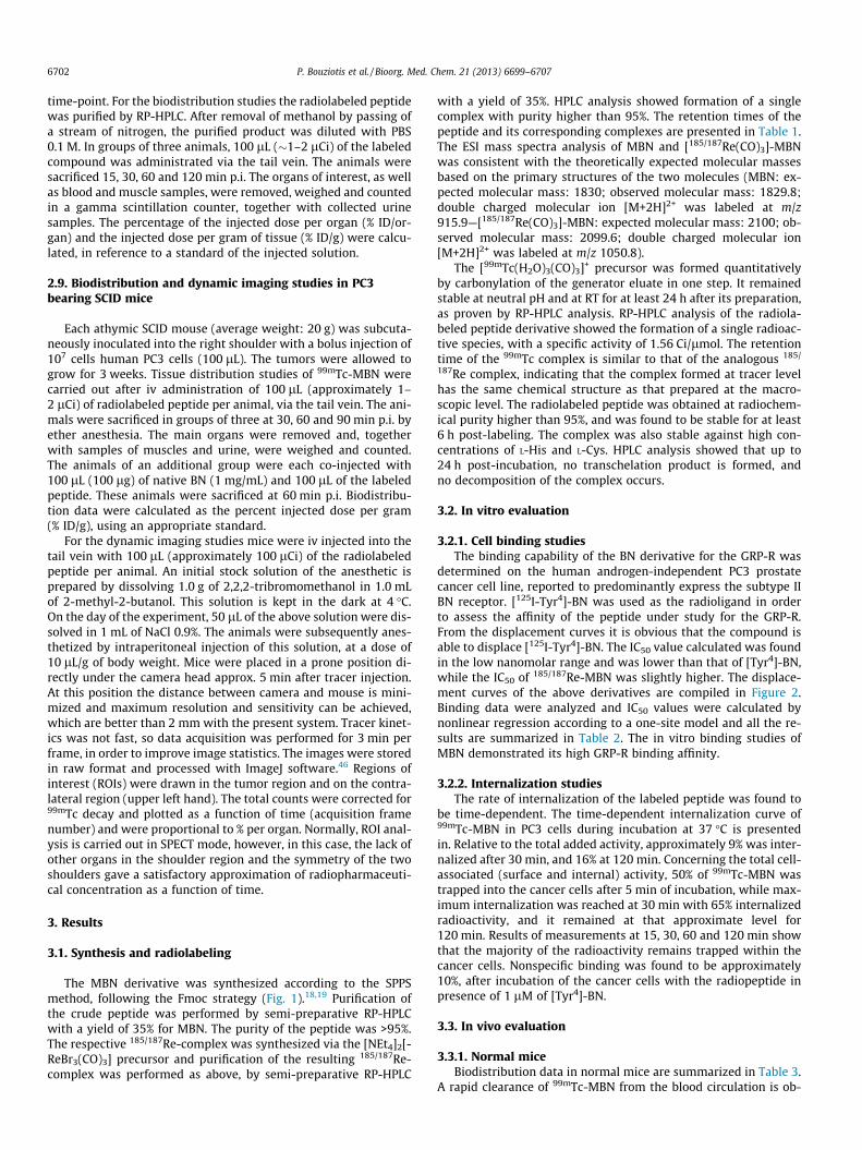
The MBN derivative was synthesized according to the SPPSmethod, following the Fmoc strategy (Fig. 1).18,19 Purification ofthe crude peptide was performed by semi-preparative RP-HPLCwith a yield of 35% for MBN. The purity of the peptide was >95%.The respective 185/187Re-complex was synthesized via the [NEt4]2[-ReBr3(CO)3] precursor and purification of the resulting 185/187Re-complex was performed as above, by semi-preparative RP-HPLC
with a yield of 35%. HPLC analysis showed formation of a singlecomplex with purity higher than 95%. The retention times of thepeptide and its corresponding complexes are presented in Table 1.The ESI mass spectra analysis of MBN and [185/187Re(CO)3]-MBNwas consistent with the theoretically expected molecular massesbased on the primary structures of the two molecules (MBN: ex-pected molecular mass: 1830; observed molecular mass: 1829.8;double charged molecular ion [M+2H]2+ was labeled at m/z915.9—[185/187Re(CO)3]-MBN: expected molecular mass: 2100; ob-served molecular mass: 2099.6; double charged molecular ion[M+2H]2+ was labeled at m/z 1050.8).
The [99mTc(H2O)3(CO)3]+ precursor was formed quantitativelyby carbonylation of the generator eluate in one step. It remainedstable at neutral pH and at RT for at least 24 h after its preparation,as proven by RP-HPLC analysis. RP-HPLC analysis of the radiola-beled peptide derivative showed the formation of a single radioac-tive species, with a specific activity of 1.56 Ci/lmol. The retentiontime of the 99mTc complex is similar to that of the analogous 185/
187Re complex, indicating that the complex formed at tracer levelhas the same chemical structure as that prepared at the macro-scopic level. The radiolabeled peptide was obtained at radiochem-ical purity higher than 95%, and was found to be stable for at least6 h post-labeling. The complex was also stable against high con-centrations of L-His and L-Cys. HPLC analysis showed that up to24 h post-incubation, no transchelation product is formed, andno decomposition of the complex occurs.
3.2. In vitro evaluation
3.2.1. Cell binding studiesThe binding capability of the BN derivative for the GRP-R was
determined on the human androgen-independent PC3 prostatecancer cell line, reported to predominantly express the subtype IIBN receptor. [125I-Tyr4]-BN was used as the radioligand in orderto assess the affinity of the peptide under study for the GRP-R.From the displacement curves it is obvious that the compound isable to displace [125I-Tyr4]-BN. The IC50 value calculated was foundin the low nanomolar range and was lower than that of [Tyr4]-BN,while the IC50 of 185/187Re-MBN was slightly higher. The displace-ment curves of the above derivatives are compiled in Figure 2.Binding data were analyzed and IC50 values were calculated bynonlinear regression according to a one-site model and all the re-sults are summarized in Table 2. The in vitro binding studies ofMBN demonstrated its high GRP-R binding affinity.
3.2.2. Internalization studiesThe rate of internalization of the labeled peptide was found to
be time-dependent. The time-dependent internalization curve of99mTc-MBN in PC3 cells during incubation at 37 �C is presentedin. Relative to the total added activity, approximately 9% was inter-nalized after 30 min, and 16% at 120 min. Concerning the total cell-associated (surface and internal) activity, 50% of 99mTc-MBN wastrapped into the cancer cells after 5 min of incubation, while max-imum internalization was reached at 30 min with 65% internalizedradioactivity, and it remained at that approximate level for120 min. Results of measurements at 15, 30, 60 and 120 min showthat the majority of the radioactivity remains trapped within thecancer cells. Nonspecific binding was found to be approximately10%, after incubation of the cancer cells with the radiopeptide inpresence of 1 lM of [Tyr4]-BN.
3.3. In vivo evaluation
3.3.1. Normal miceBiodistribution data in normal mice are summarized in Table 3.
A rapid clearance of 99mTc-MBN from the blood circulation is ob-
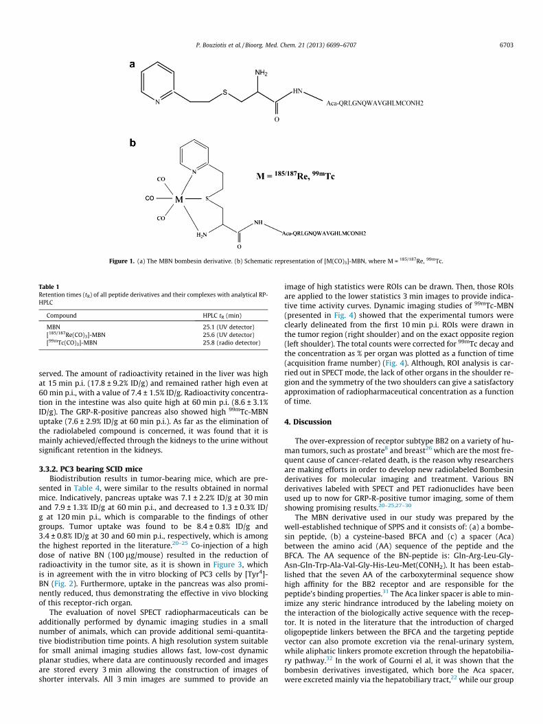
Figure 1. (a) The MBN bombesin derivative. (b) Schematic representation of [M(CO)3]-MBN, where M = 185/187Re, 99mTc.
Table 1Retention times (tR) of all peptide derivatives and their complexes with analytical RP-HPLC
Compound HPLC tR (min)
MBN 25.1 (UV detector)[185/187Re(CO)3]-MBN 25.6 (UV detector)[99mTc(CO)3]-MBN 25.8 (radio detector)
P. Bouziotis et al. / Bioorg. Med. Chem. 21 (2013) 6699–6707 6703
served. The amount of radioactivity retained in the liver was highat 15 min p.i. (17.8 ± 9.2% ID/g) and remained rather high even at60 min p.i., with a value of 7.4 ± 1.5% ID/g. Radioactivity concentra-tion in the intestine was also quite high at 60 min p.i. (8.6 ± 3.1%ID/g). The GRP-R-positive pancreas also showed high 99mTc-MBNuptake (7.6 ± 2.9% ID/g at 60 min p.i.). As far as the elimination ofthe radiolabeled compound is concerned, it was found that it ismainly achieved/effected through the kidneys to the urine withoutsignificant retention in the kidneys.
3.3.2. PC3 bearing SCID miceBiodistribution results in tumor-bearing mice, which are pre-
sented in Table 4, were similar to the results obtained in normalmice. Indicatively, pancreas uptake was 7.1 ± 2.2% ID/g at 30 minand 7.9 ± 1.3% ID/g at 60 min p.i., and decreased to 1.3 ± 0.3% ID/g at 120 min p.i., which is comparable to the findings of othergroups. Tumor uptake was found to be 8.4 ± 0.8% ID/g and3.4 ± 0.8% ID/g at 30 and 60 min p.i., respectively, which is amongthe highest reported in the literature.20–25 Co-injection of a highdose of native BN (100 lg/mouse) resulted in the reduction ofradioactivity in the tumor site, as it is shown in Figure 3, whichis in agreement with the in vitro blocking of PC3 cells by [Tyr4]-BN (Fig. 2). Furthermore, uptake in the pancreas was also promi-nently reduced, thus demonstrating the effective in vivo blockingof this receptor-rich organ.
The evaluation of novel SPECT radiopharmaceuticals can beadditionally performed by dynamic imaging studies in a smallnumber of animals, which can provide additional semi-quantita-tive biodistribution time points. A high resolution system suitablefor small animal imaging studies allows fast, low-cost dynamicplanar studies, where data are continuously recorded and imagesare stored every 3 min allowing the construction of images ofshorter intervals. All 3 min images are summed to provide an
image of high statistics were ROIs can be drawn. Then, those ROIsare applied to the lower statistics 3 min images to provide indica-tive time activity curves. Dynamic imaging studies of 99mTc-MBN(presented in Fig. 4) showed that the experimental tumors wereclearly delineated from the first 10 min p.i. ROIs were drawn inthe tumor region (right shoulder) and on the exact opposite region(left shoulder). The total counts were corrected for 99mTc decay andthe concentration as % per organ was plotted as a function of time(acquisition frame number) (Fig. 4). Although, ROI analysis is car-ried out in SPECT mode, the lack of other organs in the shoulder re-gion and the symmetry of the two shoulders can give a satisfactoryapproximation of radiopharmaceutical concentration as a functionof time.
4. Discussion
The over-expression of receptor subtype BB2 on a variety of hu-man tumors, such as prostate8 and breast26 which are the most fre-quent cause of cancer-related death, is the reason why researchersare making efforts in order to develop new radiolabeled Bombesinderivatives for molecular imaging and treatment. Various BNderivatives labeled with SPECT and PET radionuclides have beenused up to now for GRP-R-positive tumor imaging, some of themshowing promising results.20–25,27–30
The MBN derivative used in our study was prepared by thewell-established technique of SPPS and it consists of: (a) a bombe-sin peptide, (b) a cysteine-based BFCA and (c) a spacer (Aca)between the amino acid (AA) sequence of the peptide and theBFCA. The AA sequence of the BN-peptide is: Gln-Arg-Leu-Gly-Asn-Gln-Trp-Ala-Val-Gly-His-Leu-Met(CONH2). It has been estab-lished that the seven AA of the carboxyterminal sequence showhigh affinity for the BB2 receptor and are responsible for thepeptide’s binding properties.31 The Aca linker spacer is able to min-imize any steric hindrance introduced by the labeling moiety onthe interaction of the biologically active sequence with the recep-tor. It is noted in the literature that the introduction of chargedoligopeptide linkers between the BFCA and the targeting peptidevector can also promote excretion via the renal-urinary system,while aliphatic linkers promote excretion through the hepatobilia-ry pathway.32 In the work of Gourni el al, it was shown that thebombesin derivatives investigated, which bore the Aca spacer,were excreted mainly via the hepatobiliary tract,22 while our group
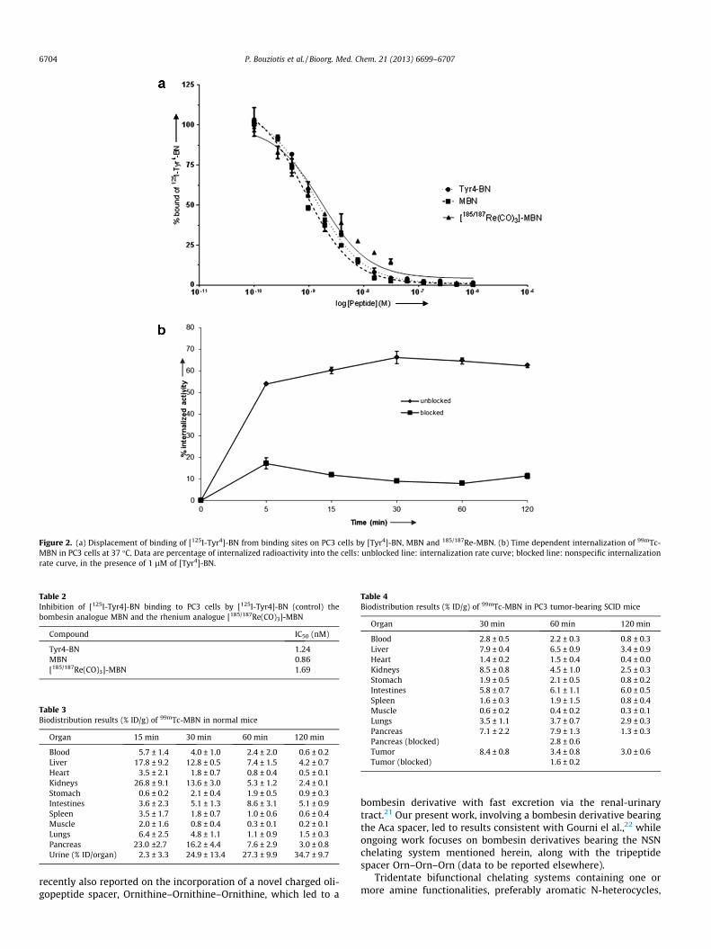
Figure 2. (a) Displacement of binding of [125I-Tyr4]-BN from binding sites on PC3 cells by [Tyr4]-BN, MBN and 185/187Re-MBN. (b) Time dependent internalization of 99mTc-MBN in PC3 cells at 37 �C. Data are percentage of internalized radioactivity into the cells: unblocked line: internalization rate curve; blocked line: nonspecific internalizationrate curve, in the presence of 1 lM of [Tyr4]-BN.
Table 4Biodistribution results (% ID/g) of 99mTc-MBN in PC3 tumor-bearing SCID mice
Organ 30 min 60 min 120 min
Blood 2.8 ± 0.5 2.2 ± 0.3 0.8 ± 0.3Liver 7.9 ± 0.4 6.5 ± 0.9 3.4 ± 0.9Heart 1.4 ± 0.2 1.5 ± 0.4 0.4 ± 0.0Kidneys 8.5 ± 0.8 4.5 ± 1.0 2.5 ± 0.3Stomach 1.9 ± 0.5 2.1 ± 0.5 0.8 ± 0.2Intestines 5.8 ± 0.7 6.1 ± 1.1 6.0 ± 0.5Spleen 1.6 ± 0.3 1.9 ± 1.5 0.8 ± 0.4Muscle 0.6 ± 0.2 0.4 ± 0.2 0.3 ± 0.1Lungs 3.5 ± 1.1 3.7 ± 0.7 2.9 ± 0.3Pancreas 7.1 ± 2.2 7.9 ± 1.3 1.3 ± 0.3Pancreas (blocked) 2.8 ± 0.6Tumor 8.4 ± 0.8 3.4 ± 0.8 3.0 ± 0.6Tumor (blocked) 1.6 ± 0.2
Table 3Biodistribution results (% ID/g) of 99mTc-MBN in normal mice
Organ 15 min 30 min 60 min 120 min
Blood 5.7 ± 1.4 4.0 ± 1.0 2.4 ± 2.0 0.6 ± 0.2Liver 17.8 ± 9.2 12.8 ± 0.5 7.4 ± 1.5 4.2 ± 0.7Heart 3.5 ± 2.1 1.8 ± 0.7 0.8 ± 0.4 0.5 ± 0.1Kidneys 26.8 ± 9.1 13.6 ± 3.0 5.3 ± 1.2 2.4 ± 0.1Stomach 0.6 ± 0.2 2.1 ± 0.4 1.9 ± 0.5 0.9 ± 0.3Intestines 3.6 ± 2.3 5.1 ± 1.3 8.6 ± 3.1 5.1 ± 0.9Spleen 3.5 ± 1.7 1.8 ± 0.7 1.0 ± 0.6 0.6 ± 0.4Muscle 2.0 ± 1.6 0.8 ± 0.4 0.3 ± 0.1 0.2 ± 0.1Lungs 6.4 ± 2.5 4.8 ± 1.1 1.1 ± 0.9 1.5 ± 0.3Pancreas 23.0 ±2.7 16.2 ± 4.4 7.6 ± 2.9 3.0 ± 0.8Urine (% ID/organ) 2.3 ± 3.3 24.9 ± 13.4 27.3 ± 9.9 34.7 ± 9.7
Table 2Inhibition of [125I-Tyr4]-BN binding to PC3 cells by [125I-Tyr4]-BN (control) thebombesin analogue MBN and the rhenium analogue [185/187Re(CO)3]-MBN
Compound IC50 (nM)
Tyr4-BN 1.24MBN 0.86[185/187Re(CO)3]-MBN 1.69
6704 P. Bouziotis et al. / Bioorg. Med. Chem. 21 (2013) 6699–6707
recently also reported on the incorporation of a novel charged oli-gopeptide spacer, Ornithine–Ornithine–Ornithine, which led to a
bombesin derivative with fast excretion via the renal-urinarytract.21 Our present work, involving a bombesin derivative bearingthe Aca spacer, led to results consistent with Gourni el al.,22 whileongoing work focuses on bombesin derivatives bearing the NSNchelating system mentioned herein, along with the tripeptidespacer Orn–Orn–Orn (data to be reported elsewhere).
Tridentate bifunctional chelating systems containing one ormore amine functionalities, preferably aromatic N-heterocycles,
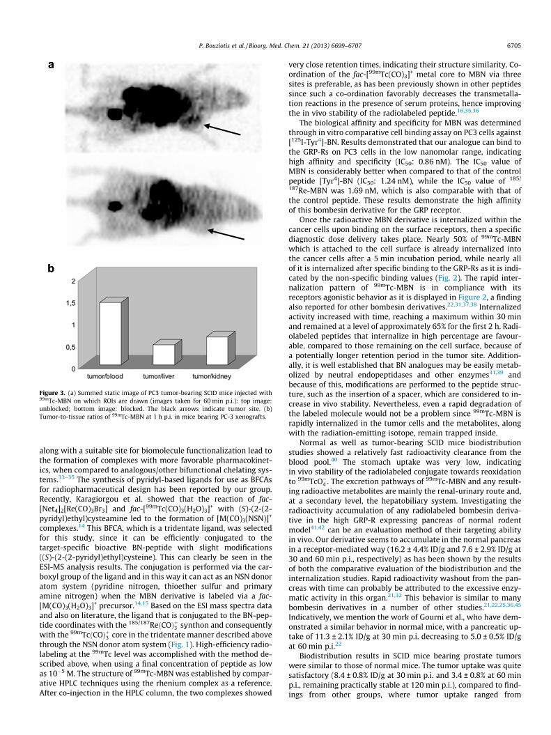
Figure 3. (a) Summed static image of PC3 tumor-bearing SCID mice injected with99mTc-MBN on which ROIs are drawn (images taken for 60 min p.i.): top image:unblocked; bottom image: blocked. The black arrows indicate tumor site. (b)Tumor-to-tissue ratios of 99mTc-MBN at 1 h p.i. in mice bearing PC-3 xenografts.
P. Bouziotis et al. / Bioorg. Med. Chem. 21 (2013) 6699–6707 6705
along with a suitable site for biomolecule functionalization lead tothe formation of complexes with more favorable pharmacokinet-ics, when compared to analogous/other bifunctional chelating sys-tems.33–35 The synthesis of pyridyl-based ligands for use as BFCAsfor radiopharmaceutical design has been reported by our group.Recently, Karagiorgou et al. showed that the reaction of fac-[Net4]2[Re(CO)3Br3] and fac-[99mTc(CO)3(H2O)3]+ with (S)-(2-(2-pyridyl)ethyl)cysteamine led to the formation of [M(CO)3(NSN)]+
complexes.14 This BFCA, which is a tridentate ligand, was selectedfor this study, since it can be efficiently conjugated to thetarget-specific bioactive BN-peptide with slight modifications((S)-(2-(2-pyridyl)ethyl)cysteine). This can clearly be seen in theESI-MS analysis results. The conjugation is performed via the car-boxyl group of the ligand and in this way it can act as an NSN donoratom system (pyridine nitrogen, thioether sulfur and primaryamine nitrogen) when the MBN derivative is labeled via a fac-[M(CO)3(H2O)3]+ precursor.14,15 Based on the ESI mass spectra dataand also on literature, the ligand that is conjugated to the BN-pep-tide coordinates with the 185/187ReðCOÞþ3 synthon and consequentlywith the 99mTcðCOÞþ3 core in the tridentate manner described abovethrough the NSN donor atom system (Fig. 1). High-efficiency radio-labeling at the 99mTc level was accomplished with the method de-scribed above, when using a final concentration of peptide as lowas 10�5 M. The structure of 99mTc-MBN was established by compar-ative HPLC techniques using the rhenium complex as a reference.After co-injection in the HPLC column, the two complexes showed
very close retention times, indicating their structure similarity. Co-ordination of the fac-[99mTc(CO)3]+ metal core to MBN via threesites is preferable, as has been previously shown in other peptidessince such a co-ordination favorably decreases the transmetalla-tion reactions in the presence of serum proteins, hence improvingthe in vivo stability of the radiolabeled peptide.16,35,36
The biological affinity and specificity for MBN was determinedthrough in vitro comparative cell binding assay on PC3 cells against[125I-Tyr4]-BN. Results demonstrated that our analogue can bind tothe GRP-Rs on PC3 cells in the low nanomolar range, indicatinghigh affinity and specificity (IC50: 0.86 nM). The IC50 value ofMBN is considerably better when compared to that of the controlpeptide [Tyr4]-BN (IC50: 1.24 nM), while the IC50 value of 185/
187Re-MBN was 1.69 nM, which is also comparable with that ofthe control peptide. These results demonstrate the high affinityof this bombesin derivative for the GRP receptor.
Once the radioactive MBN derivative is internalized within thecancer cells upon binding on the surface receptors, then a specificdiagnostic dose delivery takes place. Nearly 50% of 99mTc-MBNwhich is attached to the cell surface is already internalized intothe cancer cells after a 5 min incubation period, while nearly allof it is internalized after specific binding to the GRP-Rs as it is indi-cated by the non-specific binding values (Fig. 2). The rapid inter-nalization pattern of 99mTc-MBN is in compliance with itsreceptors agonistic behavior as it is displayed in Figure 2, a findingalso reported for other bombesin derivatives.22,31,37,38 Internalizedactivity increased with time, reaching a maximum within 30 minand remained at a level of approximately 65% for the first 2 h. Radi-olabeled peptides that internalize in high percentage are favour-able, compared to those remaining on the cell surface, because ofa potentially longer retention period in the tumor site. Addition-ally, it is well established that BN analogues may be easily metab-olized by neutral endopeptidases and other enzymes31,39 andbecause of this, modifications are performed to the peptide struc-ture, such as the insertion of a spacer, which are considered to in-crease in vivo stability. Nevertheless, even a rapid degradation ofthe labeled molecule would not be a problem since 99mTc-MBN israpidly internalized in the tumor cells and the metabolites, alongwith the radiation-emitting isotope, remain trapped inside.
Normal as well as tumor-bearing SCID mice biodistributionstudies showed a relatively fast radioactivity clearance from theblood pool.40 The stomach uptake was very low, indicatingin vivo stability of the radiolabeled conjugate towards reoxidationto 99mTcO�4 . The excretion pathways of 99mTc-MBN and any result-ing radioactive metabolites are mainly the renal-urinary route and,at a secondary level, the hepatobiliary system. Investigating theradioactivity accumulation of any radiolabeled bombesin deriva-tive in the high GRP-R expressing pancreas of normal rodentmodel41,42 can be an evaluation method of their targeting abilityin vivo. Our derivative seems to accumulate in the normal pancreasin a receptor-mediated way (16.2 ± 4.4% ID/g and 7.6 ± 2.9% ID/g at30 and 60 min p.i., respectively) as has been shown by the resultsof both the comparative evaluation of the biodistribution and theinternalization studies. Rapid radioactivity washout from the pan-creas with time can probably be attributed to the excessive enzy-matic activity in this organ.21,32 This behavior is similar to manybombesin derivatives in a number of other studies.21,22,25,36,45
Indicatively, we mention the work of Gourni et al., who have dem-onstrated a similar behavior in normal mice, with a pancreatic up-take of 11.3 ± 2.1% ID/g at 30 min p.i. decreasing to 5.0 ± 0.5% ID/gat 60 min p.i.22
Biodistribution results in SCID mice bearing prostate tumorswere similar to those of normal mice. The tumor uptake was quitesatisfactory (8.4 ± 0.8% ID/g at 30 min p.i. and 3.4 ± 0.8% at 60 minp.i., remaining practically stable at 120 min p.i.), compared to find-ings from other groups, where tumor uptake ranged from
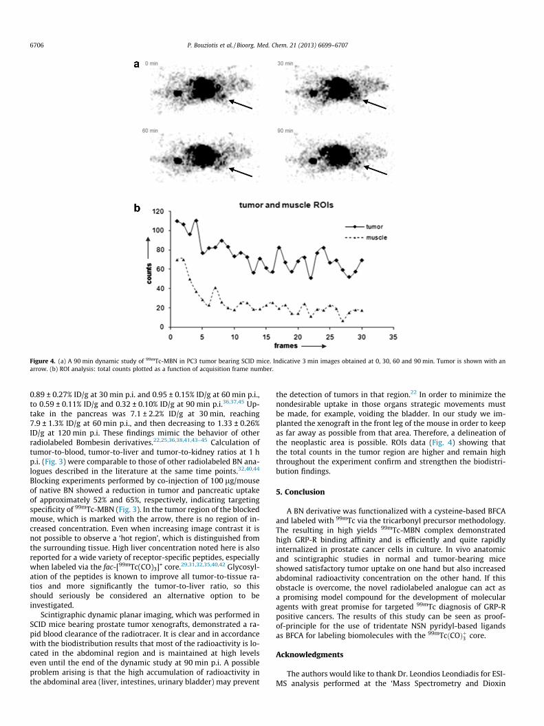
Figure 4. (a) A 90 min dynamic study of 99mTc-MBN in PC3 tumor bearing SCID mice. Indicative 3 min images obtained at 0, 30, 60 and 90 min. Tumor is shown with anarrow. (b) ROI analysis: total counts plotted as a function of acquisition frame number.
6706 P. Bouziotis et al. / Bioorg. Med. Chem. 21 (2013) 6699–6707
0.89 ± 0.27% ID/g at 30 min p.i. and 0.95 ± 0.15% ID/g at 60 min p.i.,to 0.59 ± 0.11% ID/g and 0.32 ± 0.10% ID/g at 90 min p.i.36,37,45 Up-take in the pancreas was 7.1 ± 2.2% ID/g at 30 min, reaching7.9 ± 1.3% ID/g at 60 min p.i., and then decreasing to 1.33 ± 0.26%ID/g at 120 min p.i. These findings mimic the behavior of otherradiolabeled Bombesin derivatives.22,25,36,38,41,43–45 Calculation oftumor-to-blood, tumor-to-liver and tumor-to-kidney ratios at 1 hp.i. (Fig. 3) were comparable to those of other radiolabeled BN ana-logues described in the literature at the same time points.32,40,44
Blocking experiments performed by co-injection of 100 lg/mouseof native BN showed a reduction in tumor and pancreatic uptakeof approximately 52% and 65%, respectively, indicating targetingspecificity of 99mTc-MBN (Fig. 3). In the tumor region of the blockedmouse, which is marked with the arrow, there is no region of in-creased concentration. Even when increasing image contrast it isnot possible to observe a ‘hot region’, which is distinguished fromthe surrounding tissue. High liver concentration noted here is alsoreported for a wide variety of receptor-specific peptides, especiallywhen labeled via the fac-[99mTc(CO)3]+ core.29,31,32,35,40,42 Glycosyl-ation of the peptides is known to improve all tumor-to-tissue ra-tios and more significantly the tumor-to-liver ratio, so thisshould seriously be considered an alternative option to beinvestigated.
Scintigraphic dynamic planar imaging, which was performed inSCID mice bearing prostate tumor xenografts, demonstrated a ra-pid blood clearance of the radiotracer. It is clear and in accordancewith the biodistribution results that most of the radioactivity is lo-cated in the abdominal region and is maintained at high levelseven until the end of the dynamic study at 90 min p.i. A possibleproblem arising is that the high accumulation of radioactivity inthe abdominal area (liver, intestines, urinary bladder) may prevent
the detection of tumors in that region.22 In order to minimize thenondesirable uptake in those organs strategic movements mustbe made, for example, voiding the bladder. In our study we im-planted the xenograft in the front leg of the mouse in order to keepas far away as possible from that area. Therefore, a delineation ofthe neoplastic area is possible. ROIs data (Fig. 4) showing thatthe total counts in the tumor region are higher and remain highthroughout the experiment confirm and strengthen the biodistri-bution findings.
5. Conclusion
A BN derivative was functionalized with a cysteine-based BFCAand labeled with 99mTc via the tricarbonyl precursor methodology.The resulting in high yields 99mTc-MBN complex demonstratedhigh GRP-R binding affinity and is efficiently and quite rapidlyinternalized in prostate cancer cells in culture. In vivo anatomicand scintigraphic studies in normal and tumor-bearing miceshowed satisfactory tumor uptake on one hand but also increasedabdominal radioactivity concentration on the other hand. If thisobstacle is overcome, the novel radiolabeled analogue can act asa promising model compound for the development of molecularagents with great promise for targeted 99mTc diagnosis of GRP-Rpositive cancers. The results of this study can be seen as proof-of-principle for the use of tridentate NSN pyridyl-based ligandsas BFCA for labeling biomolecules with the 99mTcðCOÞþ3 core.
Acknowledgments
The authors would like to thank Dr. Leondios Leondiadis for ESI-MS analysis performed at the ‘Mass Spectrometry and Dioxin
P. Bouziotis et al. / Bioorg. Med. Chem. 21 (2013) 6699–6707 6707
Analysis Lab’ of NCSR ‘Demokritos’. Financial support provided bythe General Secretariat of Research and Technology Grant ‘Re-search in Excellence’ is gratefully acknowledged.
References and notes
1. Cancer facts and figures 2013, American Cancer Society.2. Reubi, J. C. Endocr. Rev. 2003, 24, 389.3. Anastasi, A.; Erspamer, V.; Nucci, M. Arch. Biochem. Biophys. 1972, 148, 443.4. Cornelio, D. B.; Roesler, R.; Schwartsmann, G. Ann. Oncol. 2007, 18, 1457.5. Van De Wiele, C.; Dumont, F.; Van Belle, S.; Slegers, G.; Peers, S. H.; Derricks, R.
A. Nucl. Med. Commun. 2001, 22, 5.6. Nagalla, S. R.; Barry, B. J.; Falicks, A. M.; Gibson, B. W.; Taylor, J. E.; Dong, J. Z.;
Spindel, E. R. J. Biol. Chem. 1996, 271, 7731.7. Ohki-Hamazaki, H.; Iwabuchi, M.; Maekawa, F. Int. J. Dev. Biol. 2005, 49, 293.8. Markwalder, R.; Reubi, J. C. Cancer Res. 1999, 59, 1152.9. Alberto, R.; Schibli, R.; Egli, A.; Schubiger, A. P.; Abram, U.; Kaden, T. A. J. Am.
Chem. Soc. 1998, 120, 7987.10. Schibli, R.; Schwarzbach, R.; Alberto, R.; Ortner, K.; Schmalle, H.; Dumas, C.;
Egli, A.; Schubiger, P. A. Bioconjugate Chem. 2002, 13, 750.11. Chiotellis, A.; Tsoukalas, C.; Pelecanou, M.; Raptopoulou, C.; Terzis, A.;
Papadopoulos, M.; Papadopoulou-Daifoti, Z.; Pirmettis, I. Inorg. Chem. 2008,47, 2601.
12. Schibli, R.; La Bella, R.; Alberto, R.; Garcia-Garayoa, E.; Ortner, K.; Abram, U.;Schubiger, P. A. Bioconjugate Chem. 2000, 11, 345.
13. Papagiannopoulou, D.; Tsoukalas, C.; Makris, G.; Raptopoulou, C. P.; Psycharis,V.; Leondiadis, L.; Gniazdowska, E.; Kozminski, P.; Fuks, L.; Pelecanou, M.;Pirmettis, I.; Papadopoulos, M. S. Inorg. Chim. Acta 2011, 378, 333.
14. Karagiorgou, O.; Patsis, G.; Pelecanou, M.; Raptopoulou, C. P.; Terzis, A.; Siatra-Papastaikoudi, A.; Alberto, R.; Pirmettis, I.; Papadopoulos, M. Inorg. Chem. 2005,44, 4118.
15. He, H.; Morley, J. E.; Twamley, B.; Groeneman, R. H.; Bucar, D. K.; MacGillivray,L. R.; Benny, P. D. Inorg. Chem. 2009, 48, 10625.
16. Psimadas, D.; Fani, M.; Gourni, E.; Loudos, G.; Xanthopoulos, S.; Zikos, C.;Bouziotis, P.; Varvarigou, A. D. Bioorg. Med. Chem. 2012, 20, 2549.
17. Rogers, B. E.; Bigott, H. M.; McCarthy, D. W.; Manna, D. D.; Kim, J.; Sharp, T. L.;Welch, M. L. Bioconjugate Chem. 2003, 14, 756.
18. Amblard, M.; Fehrentz, J.-A.; Martinez, J.; Subra, G. Mol. Biotechnol. 2006, 33,239.
19. Evangelou, A.; Zikos, C.; Livaniou, E.; Evangelatos, G. P. J. Peptide Sci. 2004, 10,631.
20. Fani, M.; Maecke, H. R. Eur. J. Nucl. Med. Mol. Imaging 2012, 39, S11.21. Fragogeorgi, E. A.; Zikos, C.; Gourni, E.; Bouziotis, P.; Paravatou-Petsotas, M.;
Loudos, G.; Mitsokapas, N.; Xanthopoulos, S.; Mavri-Vavayanni, M.; Livaniou,E.; Varvarigou, A. D.; Archimandritis, S. C. Bioconjugate Chem. 2009, 20, 856.
22. Gourni, E.; Bouziotis, P.; Benaki, D.; Loudos, G.; Xanthopoulos, S.; Paravatou-Petsotas, M.; Mavri-Vavagianni, M.; Pelecanou, M.; Archimandritis, S. C.;Varvarigou, A. D. J. Med. Chem. 2009, 52, 4234.
23. Van De Wiele, C.; Dumont, F.; Dierckx, R. A.; Peers, S. H.; Thornback, J. R.;Slegers, G.; Thierens, H. J. Nucl. Med. 2001, 42, 1722.
24. Varvarigou, A.; Bouziotis, P.; Zikos, C.; Scopinaro, F.; De Vincentis, G. CancerBiother. Radiopharm. 2004, 19, 219.
25. Nock, B.; Nikolopoulou, A.; Chiotellis, E.; Loudos, G.; Maintas, D.; Reubi, J. C.;Maina, T. Eur. J. Nucl. Med. 2003, 30, 247.
26. Halmos, G.; Wittliff, J. L.; Schally, A. V. Cancer Res. 1995, 55, 280.27. Zhang, X.; Cai, W.; Cao, F.; Schreibmann, E.; Wu, Y.; Wu, J. C.; Xing, L.; Chen, X. J.
Nucl. Med. 2006, 47, 492.28. Veerendra, B.; Sieckman, G. L.; Hoffman, T. J.; Rold, T.; Retzloff, L.; McCrate, J.;
Prasanphanich, A.; Smith, C. J. Synth. React. Inorg. Met. -Org. Chem. 2006, 36,481.
29. Parry, J. J.; Kelly, T. S.; Andrews, R.; Rogers, B. E. Bioconjugate Chem. 2007, 18,1110.
30. Nanda, P. K.; Pandey, U.; Bottenus, B. N.; Rold, T. L.; Sieckman, G. L.;Szczodroski, A. F.; Hoffman, T. J.; Smith, C. J. Nucl. Med. Biol. 2012, 39, 461.
31. García Garayoa, E.; Rüegg, D.; Bläuenstein, P.; Zwimpfer, M.; Khan, I. U.; Maes,V.; Blanc, A.; Beck-Sickinger, A. G.; Tourwé, D. A.; Schubiger, P. A. Nucl. Med.Biol. 2007, 34, 17.
32. Alves, S.; Correia, J. D. G.; Santos, I.; Veerendra, B.; Sieckman, G. L.; Hoffman, T.J.; Rold, T.; Figueroa, S. D.; Retzloff, L.; McCrate, J.; Prasanphanich, A.; Smith, C.J. Nucl. Med. Biol. 2006, 33, 625.
33. Schibli, R.; Netter, M.; Scapozza, L.; Birringer, M.; Schelling, P.; Dumas, C.;Schoch, J.; Schubiger, A. P. J. Organomet. Chem. 2003, 668, 67.
34. Vitor, R. F.; Alves, S.; Correia, J.; Paulo, A.; Santos, I. J. Organomet. Chem. 2004,689, 4764.
35. Fani, M.; Psimadas, D.; Zikos, C.; Xanthopoulos, S.; Loudos, G. K.; Bouziotis, P.;Varvarigou, A. D. Anticancer Res. 2006, 26, 431.
36. Lane, S. R.; Veerendra, B.; Rold, T. L.; Sieckman, G. L.; Hoffman, T. G.; Jurisson, S.S.; Smith, C. J. Nucl. Med. Biol. 2008, 35, 263.
37. La Bella, R.; Garcia-Garayoa, E.; Langer, M.; Bläuenstein, P.; Beck-Sickinger, A.G.; Schubiger, P. A. Nucl. Med. Biol. 2002, 29, 553.
38. Nock, B.; Nikolopoulou, A.; Galanis, A.; Cordopatis, P.; Waser, B.; Reubi, J. C.;Maina, T. J. Med. Chem. 2005, 48, 100.
39. Bläuenstein, P.; Garayoa, E. G.; Rüegg, D.; Blanc, A.; Tourwé, D.; Beck-Sickinger,A.; Schubiger, P. A. Cancer Biother. Radiopharm. 2004, 19, 181.
40. Schweinsberg, C.; Maes, V.; Brans, L.; Bläuenstein, P.; Tourwé, D. A.; Schubiger,P. A.; Schibli, R.; García Garayoa, E. Bioconjugate Chem. 2008, 19, 2432.
41. Fanger, B. O.; Wade, A. C.; Cardin, A. D. Regul. Pept. 1991, 32, 241.42. Huang, S. C.; Yu, D. H.; Wank, S. A.; Gardner, J. D.; Jensen, R. T. Peptides 1990, 11,
1143.43. Faintuch, B. L.; Teodoro, R.; Duatti, A.; Muramoto, E.; Faintuch, S.; Smith, C. J.
Nucl. Med. Biol. 2008, 35, 401.44. Smith, C. J.; Sieckman, G. L.; Owen, N. K.; Hayes, D. L.; Mazuru, D. G.; Kannan,
R.; Volkert, W. A.; Hoffman, T. J. Cancer Res. 2003, 63, 4082.45. La Bella, R.; Garcia-Garayoa, E.; Bahler, M.; Blauenstein, P.; Schibli, R.; Conrath,
P.; Tourwe, D.; Schubiger, P. A. Bioconjugate Chem. 2002, 13, 599.46. Loudos, G.; Majewski, S.; Wojcik, R.; Weisenberger, A.; Sakellios, N.; Nikita, K.;
Uzunoglu, N.; Bouziotis, P.; Xanthopoulos, S.; Varvarigou, A. IEEE Trans. Nucl.Soc. 2007, 54, 454.


![Humanlung small-cell carcinomacontains bombesin · methodofGrimelius (29) onBouin's fixed paraffin sections. Radioimmunoassays(RIAs). [Tyr8]Bombesin (donatedbyJ. E. Rivier, the Salk](https://img.pdfslide.us/doc/110x75/5f4a8f32d06af4400036e022/humanlung-small-cell-carcinomacontains-bombesin-methodofgrimelius-29-onbouins.jpg)
![methoxy-pyridyl)]-benzimidazole derivatives Supporting ... · Novel bright blue emissions IIB group complexes constructed with various polyhedron-induced 2-[2′-(6-methoxy-pyridyl)]-benzimidazole](https://img.pdfslide.us/doc/110x75/611dc45d3b745e14fc5b42aa/methoxy-pyridyl-benzimidazole-derivatives-supporting-novel-bright-blue-emissions.jpg)


![[Bis(2-pyridyl-[kappa]N)amine]chlorido([eta]6 … · 2017. 3. 23. · [Bis(2-pyridyl-jN)amine]chlorido-(g6-hexamethylbenzene)ruthenium(II) hexafluoridophosphate dichloromethane solvate](https://img.pdfslide.us/doc/110x75/60ee35de6a41681269719553/bis2-pyridyl-kappanaminechloridoeta6-2017-3-23-bis2-pyridyl-jnaminechlorido-g6-hexamethylbenzenerutheniumii.jpg)