Embed Size (px)
Citation preview
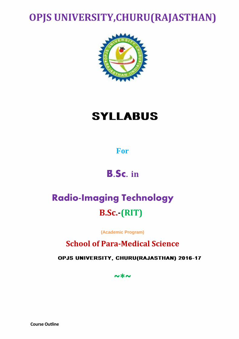
OPJS UNIVERSITY,CHURU(RAJASTHAN)
SYLLABUS
For
B.Sc. in
Radio-Imaging Technology
B.Sc.-(RIT)
(Academic Program)
School of Para-Medical Science
OPJS UNIVERSITY, CHURU(RAJASTHAN) 2016-17
~*~
Course Outline

First Year
Course Code Course Title Distribution of Marks Total Marks
(800) Theory/
Practical
Continuous
Assessment
BSRIT-101 English 70 30 100
BSRIT-102 Basic of Computer 70 30 100
BSRIT-103 Human Anatomy, Physiology and Pathology Relevant to Radiology
70 30 100
BSRIT-104 General Physics, Radiation Physics and Physics of Diagnostic Radiology
70 30 100
BSRIT-105 Radio Diagnosis Equipments, Maintenance and Quality Control
70 30 100
BSRIT-106 Hospital Training* (Internal Practical Evaluation) 200 100 300
*INTERNAL PRACTICAL EVALUATION
BSRIT-102 Basic of Computer 50 25 75
BSRIT-104 General Physics, Radiation Physics and Physics of Diagnostic Radiology
50 25 75
BSRIT-103 Human Anatomy, Physiology and Pathology Relevant to Radiology
50 25 75
BSRIT-105 Radio diagnosis Equipments, Maintenance and Quality Control
50 25 75
TOTAL MARKS 200 100 300
Second Year
Course Code Course Title Distribution of Marks Total Marks
(800) Theory/
Practical
Continuous
Assessment
BSRIT-201 Patient Care & Medical Ethics 70 30 100
BSRIT-202 Principles of Medical Emergencies 70 30 100
BSRIT-203 Clinical Radiography 70 30 100
BSRIT-204 X-ray Film/Image Processing Technique Including Dark Room
Techniques
70 30 100
BSRIT-205 Contrast & Special Radiography Procedures 70 30 100
BSRIT-206 Hospital Training * (Internal Practical Evaluation) 200 100 300
*INTERNAL PRACTICAL EVALUATION
BSRIT-202 Principles of Medical Emergencies 50 25 75
BSRIT-203 Clinical Radiography 50 25 75
BSRIT-204 X-ray Film/Image Processing Technique Including Dark Room
Techniques
50 25 75
BSRIT-205 Contrast & Special Radiography Procedures 50 25 75
TOTAL MARKS 200 100 300
Third Year
Course Code Course Title Distribution of Marks Total Marks
(800 Marks) Theory/
Practical
Continuous
Assessment
BSRIT-301 Equipments of Modern Imaging Modalities 70 30 100
BSRIT-302 Modern Imaging Techniques and Recent Trends in Imaging 70 30 100
BSRIT-303 Quality Control in Radiology, Radiation Safety in Radio
Diagnosis/Imaging
70 30 100
BSRIT-304 Project/Seminar 100 100 200
BSRIT-305 Hospital Training-I 200 100 300
DETAILED SYLLABUS
Bachelor of Radiation Technology
First Year
Course Code: BSRIT-101
Course Title: ENGLISH
Unit1. Parts of speech:
Introduction to all parts of speech along with examples and their use in language, definite
Indefinite articles, use of prepositions like a, an, and, "the" along with examples,
Unit2. Noun:
Definition, Types Uses in routine conversation
Unit3. Pronouns: Definition, Types and Uses, Personal, reflexive, emphatic, demonstrative, relative,
indefinite, interrogative and distributive pronouns and their suitable examples
Unit4. Adjective:
Definition, types, uses with suitable examples
Unit5. Verb:
Definition, form, phrases, adjective, adverb and noun phrases
Unit6. Anonym and Synonyms
Introduction, using Synonym and Anonyms, types of tense and their uses, transformation of sentences
Unit7. Speech and parts of Speech
Active and Passive voice, Direct and Indirect Speeches, transformation of speech
Unit8. Tense Definition, types of tense, present, past and future tense and their sub types along with suitable
examples
Unit9. Business Correspondence-I:
Paragraph writing, Drafting a Professional Letter, Writing Orders, Circulars and Tenders, Purchase Orders Etc. . Unit10. Business Correspondence II: Drafting notices, Issue Agendas and proceedings of a meeting, drafting minutes, application letter, drafting the application etc Unit11. Reading an unseen passage and answering the questions from the given text Unit12. Reading and understanding an interesting story. Writing short composition expressing your views on a particular topic Learning Resources: Reference Text Books, Lecture Notes and SLM List of reference books: 1. Functional English Grammar: S K Sinha
2. English Grammar and Composition-Wren & Martin @ All Copy Rights 2017 Reserved
Course Code: BSRIT-102
Course Title: BASIC OF COMPUTER
Unit 1 Computer Fundamentals:
1.1. Introduction to computers
1.2. History, generations & classification of computers
1.3. Computer memories, input & output devices
1.4. Virus & antivirus
1.5. Block diagram of computer
1.6. Programming languages in computer(HLL & LLL)
1.7. Translation(assembler, interpreter & compiler)
Unit 2 Operating systems:
2.1 Introduction to operating system
2.1 Attachment Unit Interface (AUI)
2.3 Graphical User Interface (GUI)
2.4 Character User Interface (CUI).
Unit 3 Disk Operating System (DOS):
3.1 Introduction to DOS
3.2 Commands used in DOS & functioning
Unit 4 MS-Window:
4.1 Introduction & features of window
4.1 Window accessories like Notepad
4.3 MS-paint, Word pad, Calculator etc.
Unit 5 Word Processor (MS-Word):
5.1 Introduction, creating, saving & editing a document
5.2 Tabular techniques, uses of tools & menu operations in MS-word
Unit 6 Spread Sheet (MS- Excel):
6.1 Introduction to MS-Excel
6.2 Parts of MS-Excel screen
6.3 Formatting techniques, use of functions & formulae
6.4 Linking & sharing of work sheets
6.5 Uses of Charts & Graphs.
Unit 7 Presentations (MS-Power Point):
7.1 Introduction
7.2 Working with tool bar
7.3 Editing a slide, slide layouts, working with slides, slide Transition & Animation
7.4 Inserting sounds in a power point presentation
7.5 Creating tables & organization charts.
@ All Copy Rights 2017 Reserved
Unit 8 Internet:
8.1 Introduction
8.2 Creating an e-mail ID on internet
8.3 Uses of internet in e-mail
8.4 World Wide Web (www), chats etc.
Learning Resources: Reference Text Books, Lecture Notes and SLM
List of Reference Books 1. Fundamentals of Computer-C.B. Gupta 2. Computer Fundamentals-P.K.Sinha
@ All Copy Rights 2017 Reserved
Course Code: BSRIT-103
Course Title: HUMAN ANATOMY, PHYSIOLOGY AND PATHOLOGY RELEVANT TO RADIOLOGY
Unit1: Introduction Definition of Anatomy and Physiology, land marks of the body, common anatomical terminologies, anatomical positions & anatomical Planes, definition of Cell & Tissues, Various types of cells and tissues- a brief description of cell structure with functions of cell organelles, Unit2: Skeletal System Introduction and definition of bone & joints, different types of bones, their structure and functions of bones, joints and their types, cartilaginous joint, fibrous joints and synovial joints, sub types of synovial joints, various types of movements at the bony joints, clinical notes
Unit3: Blood & Lymphatic System Blood as a connective tissue, composition & functions of blood, Lymph & Lymphatic System- brief overview, Clinical terminologies Unit4: Cardiovascular System Anatomical structure of heart, Systemic or body circulation, Pulmonary or lesser circulation, cardiac cycle, cardiac output, stroke volume, heart sounds, brief description of blood supply in upper & lower limb, circle of Willis or blood supply in brain, clinical notes. Unit5: Digestive System Introduction to digestion, brief anatomical description of various parts of the digestive system & their functions, nine regions of the abdominal cavity, an overview of the mechanism of digestion, digestive Juices, outlines of the Hepato-biliary system, clinical notes Unit6: Excretory System Definition of excretion & excretory system, gross anatomy of Kidney, Ureters & Urinary Bladder & their functions, formation of Urine, composition .of urine, structure of nephron, clinical notes
Unit7: Respiratory System
Introduction & definition of respiration, inspiration & expiration, various parts of the respiratory system &
their functions with emphasis on the lungs, mechanism of gaseous exchange, clinical terminologies.
Units8: Reproductive System Introduction & definition of reproduction, brief anatomical description of the male and female reproductive system and their functions, male & female reproductive hormones, related clinical notes
Unit9: Endocrine System Definition & classification of glands, various endocrine glands & their hormones with special emphasis on pituitary gland
Unit10: Nervous System Introduction to nervous system, various parts of brain & their functions, Meninges as protective coverings of brain, different regions of brain with their specific function, C,S.F, ventricles of brain, hypothalamus as specialized contra of brain, cranial & spinal nerves
@ All Copy Rights 2017 Reserved

Unit11: The Sensory System
Brief overview of structure & functi.ons of eye, ear, nose, tongue & skin
Learning Resources: Reference Text Books, Lecture Notes and SLM
List of reference books:
1. Text Book of Human Anatomy- Dr. B.D. Chaurasia, Vol-I&II
2. A Text Book of Physiology- Dr. Chatterjee 3. Graaff, Kent Van de and et al, Schaum's Outline of Human Anatomy and Physiology: Fourth
Edition, (2013), McGraw-Hill
@ All Copy Rights 2017 Reserved
Course Code: BSRIT-104
Course Title: GENERAL PHYSICS, RADIATION PHYSICS AND PHYSICS OF DIAGNOSTIC RADIOLOGY
Unit1. Radiation: Definition, General and ionizing Radiation, Sources of Radiation, Radiation Hazards, Principles of Radiation Protection, Units of Radiation, Roentgen, Curie, RAD, REM, Gray & Sieverts, Unit2. Basic and Radiation Physics: Matter, Atom, Molecule, structure of atom in the light of Bohr's model & Rutherford's Atomic model, Electronic configuration, Neutron, Mass number, Atomic number, Isotopes, Isobars & Isotones with suitable examples, binding energy, mass energy, potential energy, kinetic energy, capacitance and capacitor, quantum nature of radiation. Unit3. Units and Measurements: Introduction & definition, types of units & their examples, CGS, FPS & MKS system of units, Units of Work, Power, Energy, Mass, Velocity, Acceleration, Resistance, Current, Temperature and Heat. Unit4. Magnet & Magnetism: Introduction & definition, types of magnet, Magnetic classification of matter, paramagnetic, ferromagnetic, diamagnetic & Non-magnetic materials & their examples, properties of magnet, magnetic lines of force, units of magnetism, Faraday's law of magnetism, Electromagnetic induction & laws of Electromagnetic Induction Unit5. Transformer:
Introduction, definition & classification, types & structure of various transformers, efficiency ratio of a transformer, advantages of transformer, disadvantages or power losses of transformer, Single phase generator, Three phase generator, Constant potential generator, Fuses, switches and interlocks, Exposure switching and timers, AEC (Automatic Exposure Control) Unit6. Rectification: Introduction, definition & types of rectification, rectifier, a brief overview of diode as a rectifier, full wave & half wave rectification, mode of functioning, advantages & disadvantages of each Unit7. Electricity: Introduction to the flow of electricity, electric classification of matter i.e. conductor, semiconductor, Insulator & non-conductor & their examples, potential difference, resistance, Impedance, volts, coulomb, Ohm's law, unit of current etc. Unit8. X-rays: Production, Properties and Interaction with Matter
Electromagnetic waves, sources .of electromagnetic radiation, definition & discovery of x-rays, properties of x-rays, interaction of matter with x-rays (Photoelectric Absorption, Bremstrauhlung's Radiation, Compton Effect & Pair Production), Linear Energy Transfer (LET) Unit9. X-ray Tube: Detailed structure of rotary anode X-ray tube, historical overview of previously used X-ray tubes i,e, Coolidge & Cold cathode Gas tube, structure of rotary & stationary anode & features of the anode material, cathode, housing, glass envelope, power supply, target, focal spot, line focus principle, anode angle, methods of heat dissipation in modem X-ray tubes, a comparative study of the diagnostic & therapeutic X- ray tubes, Tube Rating and Tube Charts, Tube Voltage (kVp) and Tube Current (mAs), Quality and Quantity of X-ray beam' and factors affecting them Unit10. Biological Effects of Radiation: Ionization & ionizing radiation, ionization effect of x-rays, sources of ionizing radiation, definition of biological effects, their types, causes, symptoms & protective measurements against ionizing radiation, units of radiation, RAD, REM, MPD, occupational & occasional dose etc, @ All Copy Rights 2017 Reserved
Unit11. Methods of Radiation Measurement: A brief overview of structure & function of Geiger Muller counter, ionization chamber & Thermo Luminescence Dosimeter (TLD) Unit12. Radioactivity
Definition, discovery, types, Units of Radioactivity, Radium, Thorium and Uranium series
Radioactive disintegration, Half life period, Decay Period, Production of radioisotopes, Cyclotron,
Neutron fission and fusion, Chain' reaction, Half value layer, Linear Attenuation Coefficient, Interactions of x
and gamma rays in the body fat, Soft tissue and Bone, Total attenuation coefficient, Relative clinical
importance
Learning Resources: Reference Text Books, Lecture Notes and SLM
List of Reference Books
1.Equipments for Student Radiographer-Chesney & Chesney
1.Fundamental of X-ray and Radium Physics-Joseph Selman
3. A Text Book of Radiology for Technicians- Dr. S.K Bhargava, CBS Publication
@ All Copy Rights 2017 Reserved
Course Code: BSRIT-105
Course Title: RADIODIAGNOSIS EQUIPMENTS, MAINTENANCE AND QUALITY CONTROL
Unit1. Instrumentation in X-ray
1.1 Structure of rotary and stationary anode X-ray tube
1.2 LBD
1.3 Cones and cylinders
1.4 X-ray table, grid, lead protection devices, immobilization devices, compression bands
1.5 Fluoroscopy
1.6 Image intensifiers
1.7 Processing units
1.8 Cassettes & hangers
Unit2. Instrumentation in Computed Tomography
2.1 CT Gantry
2.2 X-ray tube used in Ct
2.3 Scintillation detectors
2.4 CT couch
2.5 Hardware and software components of CT
Unit3. Instrumentation in MRI
3.1 Magnet used in MRI
3.2 Fourier transformer
3.3 MRI coils
3.4 Open and closed core MRI
3.5 Software and hardware components of MRI
Unit4. Instrumentation in Ultrasound
4.1 The Ultrasound machine
4.2 Probe
4.3 Various key functions of Ultrasound machine
4.4 Artifacts in USG and their elimination

SECOND YEAR
Course Code: BSRIT-201
Course Title: PATIENT CARE AND MEDICAL ETHICS
Unit1. The Hospital and the Radiographer
1.1 Role of a radiographer in patient care delivery mechanism
1.2 Duties and responsibilities of radiographer
1.2.1 Clinical responsibilities
1.2.2 Ethical responsibility
1.2.3 Legal responsibility
Unit2. General Patient Care
2.1 General preliminaries
2.2 Moving chair and stretcher patients
2.2.1 The patient in a wheel chair
2.2.2 The patient on a stretcher
2.2.3 The patient on a bed
2.3 Dealing with anaesthetized patient
2.4 Patient preparation before anesthesia
2.5 Hygiene in X-ray department
Unit3. Patient Preparation for Diagnostic Procedures
3.1 General abdominal preparation
3.2 Purgatives
3.2.1 Irritant purgatives
3.2.2 Lubricant purgatives
3.2.3 Side effect of purgative
3.3 Sand suppositories
Unit4. Nosocomial Infection and Isolation Precautions
4.1 Definition and causes
4.2 Routes of Transmission
4.3 Factors affecting nosocomial infection
4.4 Susceptible host
4.5 Means of transmission of nosocimal infection
4.6 Hand washing and Gloving
4.7 Patient Isolation
4.8 Transport of infected patient
4.9 Patient care equipment and articles
Unit5. Hospital Waste & Their Management
5.1 .1 Infectious waste
5.1.2 Pathological waste
5.1.3 Pharmaceutical waste
5.1.4 Genotoxic waste
5.1.5 Chemical waste
5.1.6 Radioactive waste
5.2 Waste collection and disposal of waste
@ All Copy Rights 2017 Reserved

Unit6 Sterilization Technique
6.1 Methods of sterilization
6.1.1 Sterilization by boiling
6.1.2 Sterilization by steam under pressure
6.1.3 Sterilization by dry heat in an oven
6.2 Central Sterile Supply Department
6.3 Disinfection of X-ray equipments in ward radiography
Unit7. Management in Emergencies
7.1 Emergencies in Radiology Department
7.2 Primary approach
7.2.1 Cardio-pulmonary resuscitation
7.23 Secondary approach
7.3.1 Use of drugs and defibrillator
7.4 ABCD Management
7.5 Overview of emergency drugs
Unit8. Medico-Legal Aspects
8.1 Breach of professional Conduct
8.2 Negligence and responsibility for negligence
8.3 Procedure to record the event of an accident
8.4 Importance of records
Unit9. Radiation Hazards & Safety
9.1 Sources of radiation hazard in radiology department
9.2 Radiation protection measure
9.2.1 The Inverse Square law
9.2.2 Principles of radiation protection
9.2.3 Use of lead aprons, gloves and gonadal shields
9.2.4 Collimation
9.2.5 Use of LBD
Learning Resources: Reference Text Books, Lecture Notes and SLM
List of Reference Books
1. Priorities in Critical Care Nursing: Moosby London
2. Love of the patient in Pragmatic Radiography: N.D Chesney & MM Chesney
@ All Copy Rights 2017 Reserved
Course Code: BSRIT-202
Course Title: PRINCIPLES OF MEDICAL EMERGENCIES
Unit1. Medical Emergencies 1.1 Respiratory Emergencies 1.1.1 Dyspnoea 1.1.2 Suffocation 1.1.3 Smoke Inhalation 1.2 Cardiovascular Emergencies 1.2.1 Heart Attacks 1.2.2 Angina 1.2.3 Heart Failure 1.3 Epilepsy 1.4 Hysteria
Unit2. Anaphylactic Shock 2.1 Insect Bites 2.2 Scorpion Bites 2.3 Snake Bite 2.4 Dog Bite
Unit3. Emergencies due to Road Accidents
Unit4. Surgical Emergencies
4.1 First Aid in Muscle Injury 4.2 First Aid in Joint Injury
Unit5. Emergencies during Trauma 5.1 Emergencies during Head Trauma 5.2 Emergencies during Spine Trauma 5.3 Emergencies in Burns and Scalds 5.4 Care of a Trauma Patient
@ All Copy Rights 2017 Reserved

Course Code: BSRIT-203
Course Title: CLINICAL RADIOGRAPHY
Unit 1 Definitions & Anatomical Terminologies
1.1 Exposure factors
1.2 FFD, OFD, SSD, AEC, grid, scattered radiation, attenuation, Macro & Micro-radiography
1.3 Collimation, LBD, Anode Heel effect, magnification, distortion
1.4 Radiation protection devices
1.5 Tomography
1.6 Sharpness, Unsharpness and its types, contrast & density
1.7 Anatomical terminologies
1.7.1 AP, PA, Lateral, Oblique, Lateral Decubitus
1.7.2 Different anatomical movements at joints-flexion, extension, abduction, adduction, rotation,
inversion, eversion, circumduction etc
1.7.3 Various types of anatomical planes etc.
1.7.4 Positioning Terminologies viz. Supine, Prone, Semi Prone, Recumbent, Semi recumbent, erect,
lateral decubitus
Unit2. Factors affecting Image Quality
2.1 kVp 2.2 mAs 2.3 FFD, Collimation 2.4 Grid, Compression Device 2.5 Developing chemicals, temperature etc.
Unit 3 Radiography of Lower Limb:
3.1 Radiography of the followings along with the Clinical Indications:
3.1.1 Radiography of Foot, Ankle & Leg- AP, Lateral & Oblique views
3.1.2 “Calcaneal Spur” view
3.1.3 AP, Lateral & Skyline view of Patella
3.1.4 Basic views of femur, AP, Lateral, Translateral & Frog’s Leg view of Femur.
Unit 4 Radiography of Upper Limb
4.1 AP, lateral & oblique views of hand & fore ar
4.2 Four view protocol of wrist joint-AP, Lateral, Oblique, Ulnar deviation and radial deviation view
4.3 Carpal-Tunnel view of wrist joint
4.4. Elbow Joint- AP, lateral, semi flexion & full flexion views
4.5 Humerus or Upper Arm- AP & Lateral views
4.6 Shoulder Joint- AP, lateral, axial, Stryker’s view, “y” view of scapula & Infero-superior view
Unit 5 Radiography of Skull & CV Junction
5.1 Basic landmarks, imaginary lines & planes of skull
5.2 Clinical indications of skull radiography
5.3 AP. PA, Lateral, Translateral & Towne’s view of skull
5.3.1 Full Towne’s, Half Towne’s, Reverse Towne’s view
5.3.2 Submento Vertical (SMV) view
@ All Copy Rights 2017 Reserved
Unit 6 Radiography of Mastoid Air Cells, Petrous Part & Styloid Process:
6.1 Anatomical introduction to Mastoid Air Cells
6.2 Clinical indications
6.3 Recommended projections of Mastoid radiography:
6.3.1 Full Towne’s AP view
6.3.2 Lateral oblique view or “Schuller’s projection”
6.4 Radiography of Petrous Portion of Temporal Bone: Stenver’s View
6.5 Radiography of Styloid process of Temporal Bone: AP & Lateral views (Open Mouth)
Unit 7 Radiography of Para Nasal Sinuses & Nasal Bone
7.1 A brief introduction to Para Nasal Sinuses (PNS) and its types
7.2 Clinical indications of PNS radiography
7.3 Occipito-frontal 10◦ caudad and Occipito-frontal 15◦ cranial view
7.3.1 Water’s view & Caldwell’s view of PNS radiography, anterior oblique view with head tilted laterally
7.4 Radiography of Nasal Bone: Occlusal view or supero-inferior view, Lateral view
Unit 8 Radiography of Facial bones & Orbit:
8.1 Clinical indications & basic projections
8.2 Full Towne’s view for Zygomatic Arch
8.3 Lateral projection for facial bones
8.4 Radiography of Orbital Cavity: Clinical Indications
8.5 PA view and Occipito-frontal modified view
Unit9. Radiography of TM Joint
9.1 Introduction to TM joint and articulation process
9.2 Clinical Indication for radiography of TM Joint
9.3 Full Towne’s and “Open Moth Projections of TM Joint
Unit10. Radiography of Sella Turcica
10.1 Introduction to basic anatomy of Sella Turcica
10.2 Clinical Indications
10.3 Full Towne’s AP View & Lateral cone view of Sella Turcica
Unit 11 Radiography of Chest:
11.1 Introduction to chest radiography
11.2 Clinical indications, advantages of PA view of chest over AP view
11.3 Common errors in Chest Radiography and their elimination
11.4 Recommended Projections for Chest Radiography:
11.4.1 Chest PA, AP, Lateral & Lateral Decubitus View
11.4.2 Apicogram, chest radiography in case of Pneumonitis
11.4.3 Chest radiography in case of Pleural Effusion & rib injury
Unit12. Radiography of Abdomen:
12.1 A brief introduction to nine regions of abdominal cavity
12.1 Classification of patients according to body built: sthenic, asthenic, and slender patients
12.2 Clinical indications of radiography of abdomen
12.3 Abdomen supine, erect, semi recumbent & K.U.B. view
12.4 Techniques for better image quality in abdomen radiography
@ All Copy Rights 2017 Reserved

Unit 13 Radiography of Vertebral Column:
13.1 Introduction to basic landmarks & surface anatomy of vertebral column
13.1 Clinical indications of cervical radiography
13.1.1 AP & Lateral views of Cranio-vertebral junction or Atlanto-Occipital Junction
13.2 Radiography of Cervical vertebrae
13.2.1 AP, Lateral & Trans-lateral views
13.2.2 Lateral flexion and extension view
13.2.3 AP Axial view, anterior oblique and posterior oblique views
13.2.4 Pillar’s projection
13.3 Radiography of Cervico-thoracic junction
13.3.1 Swimmer’s view and AP view
13.4 Radiography of Thoracic Vertebrae
13.4.1 AP, Lateral and oblique views
13.5 Lumbar radiography: introduction, clinical indications and landmarks
13.5.1 Bowel preparation of patient
13.5.2 AP, PA & Lateral views, Translateral view, Cone AP view of L.S. Junction
13.5.3 Posterior oblique and anterior oblique views
13.5.4 Lateral flexion and extension view
13.5.5 Lumbar radiography in case of scoliosis
Unit 14 Radiography of SI Joint & Sacro-coccygeal Region:
14.1 Clinical indications
14.2 Basic projections & radiation protection in radiography of SI Joint
14.3 AP and oblique views of SI Joint
14.4 AP & Lateral views of Sacrum and Coccyx
Learning Resources: Reference Text Books, Lecture Notes and SLM
List of Reference Books
1. Clarke’s Positioning in Radiography
2. Manual of Radiographic Positioning – Merill’s
@ All Copy Rights 2017 Reserved

Course Code: BSRIT-204
Course Title: X-RAY FILM/IMAGE PROCESSINGTECHNIQUES INCLUDING DARK ROOM TECHNIQUES
Unit1. Basic Terminologies: Contrast, types of contrast, sharpness, unsharpness and its types, density, densitometer, fog, artifact, scattered radiation, attenuation, grid, types of grid etc. Unit2. Dark Room Layout: Introduction & definition of dark room, formation of latent image & conversion into visible image, the sensitivity speck, light reduction, chemical reduction and necessity of chemical reduction, size, location, Door, walls, Illumination, electric supply, ventilation & entrances of dark room, pass box, wet bench & dry bench, Dim hangers, Dim hoppers etc. Unit3. X-ray film: Definition & types of medical X-ray film, radiosensitive material ,spectral sensitivity, structure of a single emulsion and double emulsion mm, H&D curve, advantages of double emulsion Dim, anti-halo layer, the halo effect, anti curling back, a brief overview of speed of x-ray films, factors affecting the film speed, storage of exposed and unexposed Dims, identification of films, the trimming legends, Unit4. Cassette: Introduction, definition & structure of cassette, orientation to various types of cassettes used in diagnostic radiology with emphasis on single screen cassettes and curve cassettes and their uses, care of cassette, test for light leakage, storage of cassettes Unit5. Intensifying screen: Luminescence, fluorescence & phosphorescence, introduction, definition & structure of intensifying screen, conventional phosphor, rare earth materials, intensification factor, factors affecting the speed of the intensifying Screen, advantages & disadvantages of IF screen, screen mottle, care of Intensifying screen, artifacts related to intensifying screen, test for Dim screen contact, test to show the effect of intensifying screen in cutting-off the scattered radiation, Unit6. Developer & Development:
Introduction to developer & development, latent image, light and chemical reduction, components of manual developer with their functions, PQ & MQ type developer, Radium and high contrast developer, developer used in automatic processing and theater processing, time-temperature development, rate of development and factors affecting rate of development, replenisher & replenishment Unit7. Fixer & Fixing: Introduction and definition, components of fixing & their function, rapid fixer, aim of fixing, factors affecting fixing rate, importance of fixing, silver recovery-necessity and methods. Unit8. Rinsing, Washing & Drying: Various steps of film processing in manual and automatic processing, OT processing, dish units, importance & methods of developing, rinsing, fixing, washing & drying static rinsing and jet bath, open air drying and drying using drier, common technical faults during Dim processing Unit9. Automatic Processor: Introduction to automatic processing, characteristic features of automatic processor, advantages of automatic processing over manual processing Unit10. Artifacts: Definition, types & remedies of various types of artifacts in diagnostic radiography viz, water bubble artifact, streak artifact, electrostatic artifact, air bell artifact, motion artifact etc. @ All Copy Rights 2017 Reserved
Unit11. Accessories Used in Dark Room Viewing boxes, Spot light illuminator, Projectors and viewing screens for miniature radiography, magnifiers, Lead letters and numbers, Actinic marker embossing machine, Film Corner cutters Dental mounts and cutter, Filling units
Learning Resources: Reference Text Books, Lecture Notes and SLM
List of Reference Books 1. Radiographic Photography- M.O. Chesney & W.O. Chesney
2. Medical Radiographic Technique and Dark Room Practices- Krishnamurthy
@ All Copy Rights 2017 Reserved

Course Code: BSRIT-205
Course Title: CONTRAST AND SPECIAL RADIOGRAPHY PROCEDURES
Unit1 Contrast Media: Introduction, definition, classifications & reactions of contrast media. Overview
of the contrast media used in X-ray & CT, Ultrasonography and MRI, Routes of administration of
Contrast Media, properties of an ideal contrast media, Neurotoxic, immunologic and chemotoxic effects
of Contrast Media. Contrast Media used in Gut Studies, preparation of Barium Sulphate Solution using
w/v method, properties of an ideal Gut Contrast Media
Unit 2 Short Terminologies: Catheter, Scalp Vein Set, Chiba Needle, Seldinger’s Needle, Guide Wire,
Cannula, Ambu bag, Pressure injector, Suction Machine, Defibrillator, Crash cart, Cleaning agents,
Emergency drugs, Preparatory drugs, Mammography, tomography, CT Scan, MRI, Ultrasound, ABCD
Management etc.
Unit 3 Special Radiological Procedures of Urinary System
●Intra-Venous Urography (IVP)- Clinical indications, contraindications, patient preparation, accessories
and equipments, plain film, contrast media, special techniques and filming involved in IVU, delayed
films, IVU in case of young child, complications and after care
●Micturating Cyst-Urethrography (MCU)- Clinical indications, contraindications, patient preparation,
accessories and equipments, plain film, contrast media, special techniques and filming involved in MCU,
Choke MCU, complications and after care
● Retrograde Urethrography (RGU)- Clinical indications, contraindications, patient preparation,
accessories and equipments, plain film, contrast media, special techniques and filming involved in RGU,
complications and aftercare
Unit 4 Special procedures of Female Reproductive System: Detail study of Hystero-salpingography (HSG)
including indications, contraindications, patient preparation, accessories required, contrast media,
special technique, complications & after care
Unit 5 Special Radiological Procedures of Hepato-Biliary System
●Oral Cholecystography (OCG)- Cholecystographic contrast agents, indications and contraindications,
patient preparation, need of plain film, techniques and filming, the four day test, complications and
aftercare
●Endoscopic Retrograde Choledocho-Pancreatography (ERCP)- Clinical indications, contraindications,
patient preparation, accessories and equipments, plain film, contrast media, special techniques and
filming involved in ERCP, complications and aftercare
●Percutaneous Trans-hepatic Cholangiography (PTC) - Clinical indications, contraindications, patient
preparation, accessories and equipments, plain film, contrast media, special techniques and filming
involved in PTC, complications and aftercare
● T-Tube Cholangiography (TTC)- Clinical indications, contraindications, patient preparation, accessories
and equipments, plain film, contrast media, special techniques and filming involved in TTC,
complications and aftercare
Unit 6 Barium Studies: Gross anatomy of Gastro-Intestinal Tract (GIT), various types of Barium Studies,
detail studies of Barium Swallow, Barium Meal, Barium Meal Follow Through (BMFT) & Double Contrast
Barium Enema (DCBE)
@ All Copy Rights 2017 Reserved
Unit 7 Angiography: Introduction to various puncture sites, Seldinger technique of femoral puncture,
complication & after care in Angiography
Unit 8 Radiological Procedures of salivary glands: detail study of Sialography
Unit 9 Radiological Procedures of Respiratory system: a brief study of Bronchography
Unit 10 Radiological Procedures of Synovial joints: a brief study of Arthrography
Learning Resources: Reference Text Books, Lecture Notes and SLM List of Reference Books
1. A Guide to Special Radiological Procedures –Nakielny & Chapman
2. Radiological Procedures (Vol-1)- Dr. S.K. Bhargava; Jaypee Pub
3. Radiological Procedures-Dr. Bhushan N. Lakhkar; Arya Publications
@ All Copy Rights 2017 Reserved
THIRD YEAR
Course Code: BSRIT-301
Course Title: EQUIPMENTS OF MODERN IMAGING MODALITIES
Unit1. Ultrasound Imaging
1.1 Basic acoustics
1.2 Orientation to wavelength, frequency, acoustic impedance, acoustic enhancement, resolution
etc.
1.3 Characteristics of sound waves
1.4 Piezo-electric effect and Piezo electric crystals
1.5 Principles of ultrasound
1.6 Various modes of ultrasound
1.7 Parts of an ideal ultrasound machine and overview of their functions
1.8 Types of Transducer
1.9 Choosing Ultrasound transducer for various examinations
1.10 Artifacts in Ultrasound and their remedies
Unit2. Computerized Tomography Scanning 2.1 Overview of Tomography
2.2 Basic Principles of Computed Tomography
2.3 Components of a CT Machine
2.3.1 X-ray tube
2.3.2 CT Couch
2.3.3 Gantry
2.3.4 Scan acquisition system
2.3.5 Computer monitors
2.4 Overview of collimators, detectors, window width window level, pitch ratio, CT no.,
Pixel, Voxel, SNR, slip ring technology etc.
2.5 Artifacts in CT system
Unit3. MRI 3.1 Introduction to magnet, magnetism, electromagnetism & properties of magnet
3.2 Types of magnet
3.3 Commonly used magnets in MRI
3.4 Basic principles of MRI
3.5 Overview of Larmor frequency, torque, resonance & excitation
3.6 Brief description of main magnetic field, RF pulse, gradient coils & spin density
3.7 T1 relaxation, T2 relaxation and their effect
3.8 Advantages and disadvantages of MRI
3.9 Safety concerns in MRI
@ All Copy Rights 2017 Reserved
Unit4. Digital Imaging
4.1 Introduction to digital imaging
4.2 Fluoroscopy and fluorography
4.3 Conventional fluoroscopy
4.4 Image intensified fluoroscopy Image intensifier-structure and mode of functioning
4.5 advantages of image intensifier
4.6 Magnetic tapes and other storage devices in image intensifier
4.7 PACS
4.7 Digital Subtraction Angiography
4.8 Flat Panel Detectors
4.9 Basic principles of Xeroradiography
4.10 Overview of Digital Image Processing
Learning Resources: Reference Text Books, Lecture Notes and SLM
List of Reference Books:
2. Digital Imaging System for Plain Radiography: Luic Lanca and Augusto Silva
@ All Copy Rights 2017 Reserved
Course Code: BSRIT-302
Course Title: MODERN IMAGING TECHNIQUES AND RECENT TRENDS IN IMAGING
PART-A: COMPUTED TOMOGRAPHY Unit1. Introduction and Instrumentation 1.1 Introduction to CT Instrumentation 1.2 CT Generations-a brief overview of salient features of each generation 1.3 System Components-Gantry, Computer, Operating Console, CT Couch, X-ray tube used in CT Detectors used in CT 1.4 Image Characteristics-Matrix, Voxel, Pixels, CT No, Image Reconstruction, MIP, SSD, VR, Quantum
Mottle, Contrast and Spatial Resolution 1.5 Principles of Spiral CT
Unit2. Multislice or Multidetector CT (MSCT or MDCT) 2.1 Multislice detector array 2.2 Data Acquisition System 2.3 Multipitch 2.4 Slice Acquisition Rate 2.5 Tissue Volume Covered
Unit3. Image Artifacts
3.1 Motion Artifacts 3.2 Streak Artifact 3.3 Beam hardening, partial and full volume artifact 3.4 Ring Artifact
Unit4. Plain and Contrast CT Techniques of Different Body Parts
4.1 Routine and Contrast CT Techniques of Head & Neck, Para-Nasal Sinuses, Temporal Bone & TM Joint 4.2 CECT-Pituitary Fossa and Orbit 4.3 General CT of Liver, Pancreas, Kidney, Adrenal Glands, GI Tract, Pelvis and Whole Abdomen CT 4.4 CT of Cervical Spine, Lumbar Spine, Thoracic Spine-Plain and CECT 4.5 Oral Contrast & Guidelines for CT Patients 4.6 Rectal Contrast & Guidelines for CT Patients 4.7 IV Contrast & Guidelines for CT Patients 4.8 Intra-thecal Contrast & Contrast Guidelines for CT Patients
PART-B: MAGNETIC RESONANCE IMAGING (MRI)
Unit1. Introduction and Instrumentation
1.1 Introduction to magnet and magnetic properties
1.2 Types of Magnets, magnetic classification of matter and Electro magnetism
1.3 Magnets used in MRI; Primary and Secondary MRI magnets
1.4 Basic Principles of MRI
1.5 Excitation, Resonance, Larmour frequency, Torque, T1 relaxation and T2 relaxation
1.6 Coils used in MRI- Shim Coil, Gradient Coils, Body Coils, RF Coils and their uses
Unit2. MR Signals
2.1 Alignment and Precession 2.2 Magnetic Signals 2.3 Net Magnetization 2.4 Precession and Precessional Frequency 2.5 Resonance 2.6 Signal Generation

Unit3. MRI Contrast Agents
3.1 Introduction, Uses and methodology 3.2 Review of weighting 3.3 Mechanism of action, Dipole-dipole interactions 3.4 Magnetic susceptibility, Relaxivity; 3.5 Gadolinium safety, Feridex safety, 3.6 Current applications of contrast agents. Unit4. Image Contrast
4.1 Contrast Mechanism 4.2 T1 Recovery 4.3 T2 Decay 4.4 T1 Weighted Image 4.5 T2 Weighted Image 4.6 Proton Density Weighting
Uni5. Pulse Sequence
5.1 Saturation recovery 5.2 Spin Echo 5.3 Inversion Recovery 5.4 Gradient Echo 5.5 Echo Planner Imaging 5.6 Various Image artifacts in MRI
PART-C-ULTRASONOGRAPHY
Unit1. Introduction 1.1 Basic Principles of Ultrasound
1.2 Piezo-electric effect
1.3 Transducer and their types
1.4 Phased arrays
1.5 Steering and Focusing the beam
1.6 Basics components of USG machine and their functions
1.6.1 Amplifiers
1.6.2 Time Gain Compensation, Presets, Brightness
1.6.3 Edge enhance, Line Density, Echo Delay, Rejection
Unit2. Echoes-Reflections 1.1 Reflections & Transmission 1.2 Specular Reflections 1.3 Scattering 1.4 Ray Leigh Scattering 1.5 Penetration & Resolution Unit3. Resolution 1.2 Longitudinal Resolution 1.3 Lateral Resolution 1.4 Elevation Resolution 1.5 Temporal Resolution 1.6 Resolution phantoms
Unit4. Artifacts 4.1 Slice Thickness 4.2 Refraction 4.3 Multi path 4.4 Reverberation 4.5 Acoustic Shadow 4.6 Acoustic Enhancement
Unit5. Doppler Ultrasound 5.1 Doppler Effect 5.2 Doppler Shift 5.3 Spectral Analysis 5.4 Aliasing 5.5 CW Doppler 5.6 PW Doppler 5.7 PW & CW Spectrums 5.8 Color Flow Imaging
PART-D-NUCLEAR MEDICINE
Unit1. Introduction 1.1 Composition of a nucleus 1.2 Radioactivity, radioactive materials and their sources, radioactive decay, exponential growth 1.3 Nuclear fusion and nuclear fission 1.4 Units of radioactivity, half life period Unit2. Radiation Detectors: 2.6 Introduction 2.7 Gas chambers 2.8 Ionization chambers 2.9 Proportional counters 2.10 Geiger Muller counters 2.11 Semiconductor detectors 2.12 Scintillation detectors Unit3. Radio Nuclides 3.1 Nuclear Reactors 3.2 Reactor produced radionuclide 3.3 Principles of Nuclear Reactor 3.4 Accelerator produced radionuclide 3.5 Radionuclide generators. Unit4. Instrumentation 4.1 The Anger Camera-basic principle 4.2 System components- Detector system and electronics 4.3 Collimators, Image display and recording systems, Scanning camera. Unit5. Radio -pharmaceutics: 5.1 Introduction, 5.2 Basic principle of tracer technique; 5.3 Preparation of different labelled compounds with technetium-99m isotope; Cold kits. Unit6. Nuclear Imaging Technique: 6.1 Static and dynamic studies; 6.2 Thyroid imaging 6.3 Imaging of bones 6.4 Nuclear Imaging of Respiratory system, Urinary system, G.I. system & Cardiovascular system 6.5 Iodine 131 uptake studies; Iodine 131 therapy for thyrotoxicosis and thyroid ablation List of Reference Books
1. Step by Step CT- Dr. Karthikeyan and Deepa Cheghu
2. CT & MRI Protocol)- Dr. S.K. Bhargava
3. MRI Made Easy- Govind P. Chavhan; Jaypee Publications MRI Guide for Technologists-A Step by
Step Approach- Mootoo S. Chunasamy
4. MRI and Spectroscopy-NR Jagannathan; Jaypee Publications 5. Principles & Practices of Ultrasound-Dr. S. K. Bhargava; Jaypee Publications
@ All Copy Rights 2017 Reserved
Course Code: BRT0303 Course Title: Quality Control in Radiology, Radiation Safety in Radio Diagnosis/Imaging Unit1. Biological Effects of Radiation
1.1Sources of radiation 1.7 Ionization radiation and biological effects of radiation 1.8 Stochastic and Non stochastic radiation 1.9 Somatic effects
1.9.1 Immediate somatic effects 1.9.2 Intermediate somatic effects 1.9.3 Delayed somatic effects
1.10 Genetic Effects 1.11 AERB, BARC, ICRP, WHO & their role in radiation protection 1.12 Units of radiation measurement 1.13 RAD, REM, MPD, half value layer, occupational dose & occasional dose 1.14 Principle of ALARA
Unit2. Methods of Radiation Protection 5.7 Principles of time, distance and shielding 5.8 Effective communication, use of filters & use of light beam diaphragms 5.9 Use of radiation protection devices 5.10 Radiation protection in case of paediatric patient 5.11 Radiation protection in ward radiography 5.12 Radiation protection in dental radiography 5.13 Radiation protection in OT radiography and ICU 5.14 Protection methodology in CT scan & Fluoroscopy 5.15 Ten days rule 5.16 Use of wedge filters, wedge angle and hinge angle 5.17 Radiation protection in radiotherapy 5.18 Area monitoring and survey 5.19 Survey meters Unit3. Personnel Monitoring Devices & radiation Measurement 3.1 Introduction 3.2 Working principles, advantages and disadvantages of 3.2.1 Film Badges 3.2.2 TLD Badges 3.2.3 Pocket Dosimeters 3.3 Overview of various methods of radiation measurement 3.3.1 Geiger –Muller Counter 3.3.2 Proportional Counter 3.3.3 Gas Chamber
Unit4.Radiation emergencies 4.1 Situation preparedness 4.2 Safety and prevention-legal requirements 4.3 Recent developments in radiation safety related topics
Learning Resources: Reference Text Books, Lecture Notes and SLM List of Reference Books
1. Priorities in Critical Care Nursing: Moosby London
2. Love of the patient in Pragmatic Radiography: N.D Chesney & MM Chesney
@ All Copy Rights 2017 Reserved