Embed Size (px)
Citation preview
5DGLDWLRQ�3URWHFWLRQ����
*8,'(/,1(6�21�('8&$7,21
$1'�75$,1,1*�,1�5$',$7,21
3527(&7,21�)25�0(',&$/
(;32685(6
(XURSHDQ�&RPPLVVLRQ
European Commission
5DGLDWLRQ�3URWHFWLRQ����
*8,'(/,1(6�21�('8&$7,21�$1'�75$,1,1*�,1�5$',$7,21
3527(&7,21�)25�0(',&$/�(;32685(6
2000
Directorate-GeneralEnvironment
3
&217(17
Page
FOREWORD .............................................................................................................4
1. Introduction ...........................................................................................5
2. General recommendations for Training Programmes in RadiationProtection ..............................................................................................8
3. Recommendations for the credentialing process in radiationprotection.............................................................................................12
4. Recommendations for radiation protection of the patient forindividuals undergoing training programmes in health centres .........12
5. Recommendations for continuing education and training afterqualification and on implementation of new techniques....................13
6. Recommendations regarding the course on radiation protection in thebasic curriculum of medical and dental schools .................................14
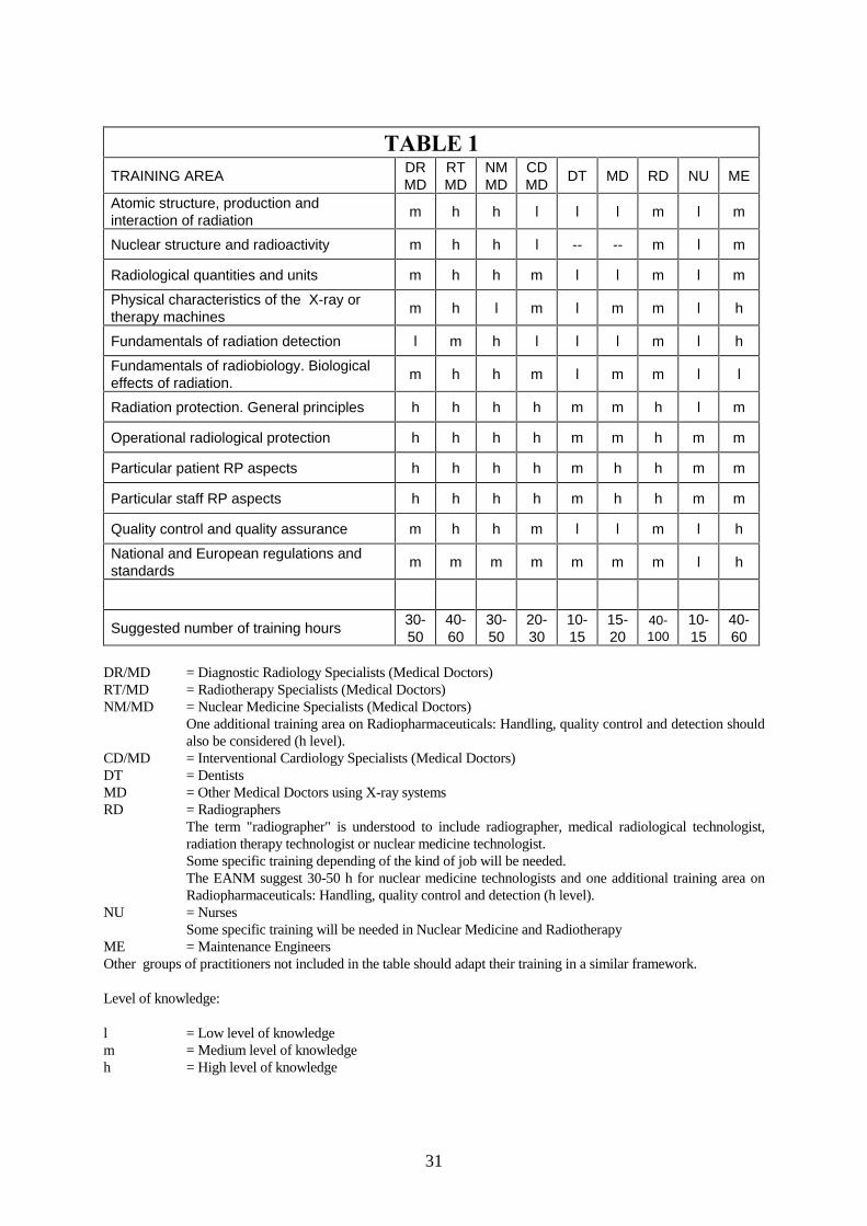
ANNEXES...........................................................................................................16
1. Outline for specific training in Radiation Protection for InterventionalRadiology ............................................................................................16
2. Outline of specific educational objectives for mammography...........18
3. Outline of specific educational objectives for paediatric radiology...20
4. Addendum for paediatric nuclear medicine........................................24
5. Outline of specific educational objectives for radiotherapy...............24
6. Training modules on radiation safety .................................................27
7. Council Directive 97/43/EURATOM of 30 June 1997. Extracts ofArticles concerning training aspects...................................................28
ACKNOWLEDGEMENTS .........................................................................................30
TABLE 1 .............................................................................................................31
REFERENCES .........................................................................................................32
4
)25(:25'
The EURATOM Treaty and its implementing Council Directives govern the work of theEuropean Commission in the field of radiation protection.
The framework directive is the Basic Safety Standards Directive (BSS), on the protection ofworkers and the general population against the dangers arising from ionizing radiation(80/836/Euratom), as last revised by Council Directive 96/29/EURATOM of 13 May 1996that comes into force on 13 May 2000.
In 1984, the Council of Ministers issued a Directive, supplementing the BSS, on theprotection of persons undergoing medical exposures (84/466/Euratom), the so-called "Patientdirective". This was revised in 1997 by Council Directive 97/43/Euratom of 30 June 1997 onhealth protection of individuals against the dangers of ionizing radiation in relation to medicalexposure, known as the Medical Exposure Directive (MED). The MED has to be transposedinto national law no later than 13 May 2000.
According to Article 7 of the Medical Exposure Directive, Member States shall ensure that thepractitioner and those individuals that are mentioned in Article 5(3) and 6(3) have adequatetheoretical and practical training for the purposes of radiological practices, as well as relevantcompetence in radiation protection. Individuals undergoing relevant training programmes mayparticipate in practical aspects for the procedures mentioned in Article 5(3). Member Statesshall ensure that continuing education and training after qualification is provided and shallencourage the introduction of a course on radiation protection in the basic curriculum ofmedical and dental schools. Article 9 requires Member States to ensure that practitionersconducting special practices receive appropriate training.
These guidelines contain some specific recommendations for the application of the Directiveand were developed with the assistance of the group of health experts established underArticle 31 of the Euratom Treaty. The guidelines are not binding on Member States, and formpart of a number of technical guides drawn up to facilitate implementation of the MED.
The document is structured as follows:
A general introduction providing background information and indication of the required levelof training in radiation protection. This is followed by a chapter on general recommendationsfor training programmes in radiation protection. The third chapter gives recommendations forthe establishing of credentials in radiation protection. Chapter 4 lays down recommendationsfor radiation protection of the patient during training programmes in health centres. Chapter 5provides recommendations for continuing education and training after qualification and whennew techniques are implemented. Chapter 6 includes recommendations for introducing thecourse on radiation protection in the basic curriculum of medical and dental schools and isfollowed by seven annexes presenting examples of specific educational objectives to beincluded in some of the training activities.
6X]DQQH�)5,*5(1Director Nuclear Safety and Civil
Protection
5
��� ,QWURGXFWLRQ
(1) Training in general and specific training in radiation protection are widely recognised asone of the basic components of optimisation programmes for medical exposures. All theinternational bodies, e.g. the International Commission on Radiological Protection(ICRP), the World Health Organisation (WHO), the International Atomic EnergyAgency (IAEA) etc., along with several guidelines published by the EuropeanCommission (EC), recognise the importance of education and training in reducingpatient doses while maintaining the desired level of quality in medical exposures (goodtherapeutic treatments and images of sufficient quality for diagnosis).
(2) The ICRP, in its publication 73 entitled “Radiological Protection and Safety inMedicine” (ICRP, 1996), states (paragraph 128) that “one important need is to provideadequate resources for the education and training in radiological protection for futureprofessional and technical staff in medical practice. The training programme shouldinclude initial training for all incoming staff and regular updating and retraining”.
(3) Article 7 of the Council Directive 97/43/EURATOM of 30 June 1997 on healthprotection of individuals against the dangers of ionising radiation in relation to medicalexposure (EC, 1997) lays down requirements for education and training. Certain aspectsof this Article may require some clarification and orientation for Member States and thisguideline contains some specific recommendations for the application of the Directive.
Individuals undergoing medical exposures due to diagnosis or treatment are notnecessarily patients but could also by healthy individuals (e.g. in screeningprogrammes). However, for simplicity, in these guidelines they will be called patients.
(4) This document presumes that some “adequate theoretical and practical training for thepurpose of radiological practices” already exists under national requirements, togetherwith national mechanisms to control this level of training.
(5) Training for radiographers, medical radiological technologists, radiotherapytechnologists or nuclear medicine technologists and other staff who work in areas wherethey may be exposed to radiation is required by Article 5(3) of the Directive. Medicalphysics experts are covered in Article 6(3). Maintenance engineers and other auxiliariesinvolved in medical exposures are not specifically mentioned, but obviously they alsoneed training in radiation protection.
(6) Article 9(2) of the Directive (special practices) requires Member States to ensure thatpractitioners conducting medical exposures of children, screening programmes and highdose procedures (interventional radiology amongst others) receive appropriate training.The recommendations of the World Health Organisation, produced at a workshop heldin Munich in October 1996 (BAUML, 1997), provide a good example of the trainingand credentialing requirements for people involved in interventional radiology practice.
6
(7) Some indication of the required level of training in radiation protection (RP) would beuseful to ensure appropriate curricula in the different Member States. All staff withresponsibility for medical exposures will need training in radiation protection. Thefollowing groups of professionals have been identified:
7.1 Diagnostic radiology specialists7.2 Nuclear medicine specialists7.3 Radiotherapy specialists7.4 Cardiologists7.5 Other medical doctors using X-ray systems (specially fluoroscopy systems)
such as urologists, vascular surgeons, traumatologists, etc.7.6 Dentists7.7 Podiatrists7.8 Radiographers and radiological technologists7.9 Nurses (especially oncological nurses)7.10 Technicians performing quality control in radiology installations7.11 Medical physicists7.12 Maintenance engineers and maintenance technicians7.13 Chiropractors
(8) Nevertheless, some specific cases could require revision, for example radiographers. Inthis case, the differences in contents and in hours of training between Member States aresignificant and some complementary efforts to harmonise this specific training will beneeded. The International Society of Radiographers and Radiological Technologists(ISRRT) has published a relevant document on Professional Standards for the Educationof Radiographers (ISRRT, 1996).
(9) In certain Member States, the training of Medical Physicists could also require somesupport to promote actions in the specific fields of diagnostic radiology, nuclearmedicine and radiotherapy, and continuing education activities. These actions should begeared to improving the training of Medical Physicists and improving the participationof these specialists in the training of medical doctors. The European Federation ofOrganisations for Medical Physics (EFOMP) has published relevant guidelines(EFOMP, 1996, 1998, 1999) on this issue.
It is the experience of the European Society for Therapeutic Radiology and Oncology(ESTRO), which has organised regular multidisciplinary training in radiotherapy sincethe mid-1980s, that support for such training is not uniform in all Member States despiteevidence that standards in participating hospitals have been progressively raised.
(10) The experience of the European Association of Radiology (EAR, 1997) and theAmerican College of Radiology (ACR, 1997) could be valuable in this field. Guidelinesfor the basic aspects of continuing education programmes are needed in Europe andACR standards can be relevant sources of information.
(11) Practitioners and prescribers need to be considered differently for training in RP.Prescribers need a basic knowledge of some aspects of patient RP. A number of relevantguidelines have been published by the Radiology Societies (RCR, 1998), and thenadapted by the European Commission and experts representing European Radiology andNuclear Medicine (EC, 2000).
7
(12) In the last few years, the European Commission Radiation Protection Actions haveproduced a series of guidelines concerning image quality criteria (EUR, 1996 a, b, c, d;EUR 1999), which are intended to be widely used as part of training programmes toimprove the radiation protection of the patient.
(13) The ERPET (European Radiation Protection Education and Training) courses are also asource of good training material. The European Commission sometimes publishes theproceedings of these courses (ERPET, 1997).
(14) During the workshop held in Grado (Italy) in September 1993, sponsored by theEuropean Commission, some relevant conclusions about training in radiation protectionfor medical exposures were reached (EUR, 1995). One of the conclusions was “the needand demand to improve training in radiation protection and in quality assurance.Common training programmes at European level must be continued. A practical way tomake progress in the harmonisation of training could be the elaboration of specificeducational objectives, as has been made within the EC VALUE Programme forradiologists and radiographers”.
(15) In July 1998, the European Commission sent a questionnaire to Member States,professional organisations and scientific societies, requiring information on theirtraining recommendations regarding radiation protection during medical exposures. Aneed for harmonisation is evident and some of the Member States asked theCommission to produce guidelines. The relevant aspects of the answers to thisquestionnaire have been taken into account in this document. In July 1999, a draft of thisGuideline was submitted for comments to the European professional organisations andthey have been also taken into account.
8
��� *HQHUDO� UHFRPPHQGDWLRQV� IRU� 7UDLQLQJ� 3URJUDPPHV� LQ� 5DGLDWLRQ3URWHFWLRQ
(17) A list of topics to be included in the training programmes in RP for the different groupsof professionals should be established. The following training areas could provideexamples of radiation protection programmes in diagnostic radiology (VAÑO 1993):
17.1 The atomic structure and interaction of radiation17.2 Radiological quantities and units17.3 Physical characteristics of X-ray machines17.4 Fundamentals of radiation detection17.5 Detectors used in diagnostic installations17.6 Fundamentals of radiobiology: cell, systemic and whole body responses17.7 Radiation protection. General criteria17.8 Operational radiological protection17.9 General RP aspects in diagnostic radiology17.10 Particular aspects of patient and staff RP17.11 Quality control and quality assurance17.12 National and European regulations and standards17.13 Practical training
But these other topics should also be considered:
½ Radiation effects½ Definitions of the variety of terms used for dose½ Relationship of equipment characteristics to dose and image quality½ Relationship of exposure factors to dose and image quality½ Concept of risk, comparative risk through age range and period of pregnancy½ Protocols for over exposure and accidents½ Clear communication at the appropriate level with patient, staff, comforters and
carers and the public½ Diagnostic reference levels
(18) The WHO recommendations for interventional radiology (IR) require a specific secondlevel of training in RP for these specialists (BAUML, 1997) in the following areas:
18.1 X-ray systems for IR.18.2 Dosimetric quantities specific for IR.18.3 Radiobiology: risks in IR.18.4 Radiological protection of patient and staff in IR.18.5 Quality assurance in IR.18.6 Local and international rules concerned with IR.18.7 Procedures optimisation in IR.
(19) The European Society for Therapeutic Radiology and Oncology recommends that thefollowing topics should be included in the training syllabus for radiotherapy:
19.1 Radiotherapy equipment - safety and accuracy19.2 Dosimetric and geometric quantities for accuracy in radiotherapy
9
19.3 Radiobiology and radiation risks19.4 Radiation treatment planning for optimising delivery of radiation dose19.5 Optimal and safe use of radionuclides in radiotherapy19.6 Radiation hazards in radiotherapy facilities
(20) There are also British recommendations for areas of training in RP for nuclear medicine(HARDING, 89):
20.1 Nature of ionising radiation and its interaction with tissue20.2 Genetic and somatic effects and how to assess their risks20.3 Patient doses20.4 Quality assurance and quality control20.5 Dose limitation20.6 Pregnancy and breast feeding20.7 Unsealed sources20.8 Organisation for radiation protection20.9 Statutory responsibilities
(21) The European Association of Nuclear Medicine (EANM) states that nuclear physicianshave to be familiar with and have a knowledge of Radiological Protection (RP),amongst other topics. Nuclear physicians must have gained practical experience inpatient dosimetry (diagnosis and therapy) and radiation protection (decontamination,waste disposal, staff dosimetry, etc.), amongst other topics. EANM recommends 120hours for assessing the basic science training but “courses on radiation protection andregulation issues are not included due to different national rules”. The EANM also statesthat practical training has to be added to the courses and has to be formally controlled(EANM, 1997).
(22) In addition, it is obvious that the topics to be included in training activities and the level ofknowledge of the topics should be tailored to the various specialities (diagnostic radiology,radiotherapy, cardiology, dentistry, etc.) and the different kinds of work and responsibility(medical doctors, medical physicists, maintenance engineers, radiographers, etc.). At theWHO Munich meeting (BAUML, 1997), different levels of training were proposed forinterventional radiology. Lists of topics and levels of knowledge were drawn up formedical doctors, radiographers, nurses and maintenance engineers. Medical Physicistsshould know all topics in greater detail.
(23) The guidelines produced during recent years under the Radiation Protection Actions ofthe European Commission concerning image quality criteria (EUR, 1996 a, b, c, d, e)are good examples of training material for diagnostic radiology specialists, medicalphysicists and radiographers. The American Association of Physicists in Medicine hasalso published relevant guidelines concerning the teaching of clinical radiologicalphysics to residents in diagnostic and therapeutic radiology (AAPM, 1999).
(24) For those involved in medical exposures, table 1 presents a proposal for training areasand levels of knowledge. The areas and levels suggested in table 1 should be consideredas core knowledge. More detailed additional training for some of the groups could berequired. The practical application of radiological protection specific to modality shouldbe included in "operational radiological protection”. Medical physics experts should
10
know all the training areas at the highest level in addition to physics and all relevantaspects of quality assurance programmes.
(25) The number of hours indicated in table 1 should be considered as being in addition tothe basic training for prescribers and could be included in different training periods suchas basic residency programmes and special training courses.
(26) Training programmes should include in any case details of the procedures to be followedoccurring accidental or unintended doses to patients from radiological practices.
(27) A formal recommendation on the number of hours for the training programmes (theoryand practical work) should be established. This training period will depend on the previousknowledge of radiation physics, radiobiology, etc., among the different groups ofprofessionals in the different countries. A good tool for defining the number of hoursneeded for training could be the use of guidelines containing the specific educationalobjectives for adapting the training time to achieving these objectives.
(28) A common core of knowledge in RP throughout Europe for the different groups of healthworkers in relation to their roles and responsibilities within their health care system wouldbe desirable. It is easier to agree a list of topics for training and a catalogue of specificeducational objectives than a certain number of hours of training. As part of training and inaddition to attending a course self-training activities should be promoted. Appropriatetraining material will be required for self-training.
(29) Practical exercises and practical sessions should be included in the programmes fortraining in radiation protection. A minimum of 1-2 hours practical session in a clinicalinstallation should be included in the most simple training programmes, while 20-40% ofthe total time scheduled in more extensive courses should be devoted to practicalexercises.
(30) An important aspect of these training activities is the availability of training material.Books, slide collections, videos, interactive CD-ROM, are scarce and not always adaptedto the level needed in the different courses. The EC will encourage publication of thecontent of the different training courses and promote the preparation of specific trainingmaterial for a set of basic courses (e.g. basic training in radiation protection forcardiologists). Profiting from the experience of the different European countries in thisfield could also be very positive. The EFOMP has co-sponsored the EC actionEMERALD dedicated to education and training in Medical Physics (information availablefrom EFOMP, http://www.efomp.org/index.html).
(31) The lecturers must have previous experience in radiation protection in medicalinstallations and in practical work in a clinical environment. Installations wherepractical training is provided should be medical installations and not only laboratory orsimulation exercises.
(32) Paediatric radiology, screening mammography and computed tomography also requiresome specific training in RP for radiologists and radiographers involved in theseexaminations. Some relevant guidelines for quality assurance for paediatrics have beenpublished (EUR, 1996b; COOK, 1998, NCRP, 1981). Some proposals for specificeducational objectives are included as examples in these guidelines.
11
(33) Some special practices like interventional radiology require, as recommended by theWHO (BAUML, 1997), a second level of specific training in radiation protection inaddition to the general level received by diagnostic radiology specialists.
(34) In addition, some special consideration is required in the case of medical doctors (non-radiologists) using fluoroscopy X-ray systems regularly (urologists, vascular surgeons,traumatologists, etc.). A basic training in RP should be defined as indicated in table 1(MD column).
(35) In some states in the USA, these doctors require specific credentialing in radiationprotection (WAGNER, 1998) and in Europe a similar system should be established.
(36) In September 1999, The “Institut National des Sciences et Techniques Nucléaires”organised in Saclay (France) an International Conference on “Radiation protection:What are the future training needs?” A list of training modules on radiation safety hasbeen agreed for the medical field (see Annex).
12
3. 5HFRPPHQGDWLRQV�IRU�WKH�FUHGHQWLDOLQJ�SURFHVV�LQ�UDGLDWLRQ�SURWHFWLRQ
(37) A system for credentialing RP training programmes should be established at national orregional level. This process should be undertaken by the Regulatory Authority, with thehelp of Academic Institutions (Universities) and scientific or professional societies. Aregister of accredited bodies should be established. For Medical Physicists, EFOMP haslaid down a well-structured system of recommendations for education and training,continuing professional development and requirements for National Registration Schemes(EFOMP, 1998).
(38) The minimum requirements for credentialing a training programme should consider all theaspects involved: enough administrative support, guarantees for the archiving of files,diplomas, etc., for a minimum number of years (20 years), enough didactic support(classroom, audio-visual support, etc.), teachers qualified in the topics to be imparted andwith experience in hospital medical physics work, instrumentation for practical exercises,availability of clinical installations for practical sessions, etc.
(39) Various alternatives should be proposed for credentialing different professionals withdifferent duties and responsibilities (examination at the end of the course, residency,continuous evaluation, etc.).
(40) Basic details should be given in the diplomas or certificates received by people attending atraining programme in RP: centre conducting the training, number of accredited traininghours, process of credentialing: examination or other form of assessment, date of thetraining, name of the academic staff with responsibility for the training programme, etc.
(41) The second level of training in RP for Interventional Radiology, recommended by theWHO (BAUML, 1997) would also require a specific accreditation, and the same appliesto the special case of medical doctors using fluoroscopy on a regular basis.
��� 5HFRPPHQGDWLRQV� IRU� UDGLDWLRQ� SURWHFWLRQ� RI� WKH� SDWLHQW� IRULQGLYLGXDOV�XQGHUJRLQJ�WUDLQLQJ�SURJUDPPHV�LQ�KHDOWK�FHQWUHV
(42) Patient RP requires special consideration during the training of residents andradiographers. The criteria of justification and optimisation should be applied carefullyand all the procedures should be performed under the responsibility of a senior specialist.
(43) Some specific recommendations could also be made for planning practical RP training inmedical installations (e.g. X-ray systems must remain in operative condition after thetraining sessions) if patients are involved, simple procedures - low doses - must beselected; It is not permitted to give some additional irradiation to the patient just fortraining purposes.
(44) In the case of high patient dose procedures (interventional and some vascular diagnosticprocedures), strict patient dose control should be performed to guarantee that nosignificant additional doses due to the training are imparted. In paediatrics, screeningmammography and computed tomography also no significant additional doses due totraining should be imparted.
13
��� 5HFRPPHQGDWLRQV� IRU� FRQWLQXLQJ� HGXFDWLRQ� DQG� WUDLQLQJ� DIWHUTXDOLILFDWLRQ�DQG�RQ�LPSOHPHQWDWLRQ�RI�QHZ�WHFKQLTXHV
(45) The ACR Continuous Medical Education (CME) Standard (for physicians and medicalphysicists) requires a minimum of 150 hours of approved education in category 1 and 2every three years (to be renewed in a three year cycle) (ACR, 1997). The experience ofthe European Association of Radiology (EAR, 1997) also is relevant for this topic.
45.1 Category 1 (designated by ACR or other recognised organisations). Theminimum number of hours is 60. Accredited residencies and fellowships up to50 hours per year can be included.
45.2 Category 2. The maximum number of hours allowed is 90. Activities accepted:medical meetings, lectures, course syllabuses, study of authoritative medicalliterature, teaching radiology-related services to medical students, preparationand publication of scientific papers, presentation of papers, courses, orscientific exhibits, clinical consultations, use of computer-assisted learningmaterials designed to enhance patient care, review of manuscripts for peer-reviewed journals and review of abstracts for scientific meetings.
(46) Some training directed towards continuous education in RP should be included (togetherwith practical education, particularly on the installation of new equipment), the extentdepending on the kind of work. A radiographer, radiotherapist or medical physicistwould need more time dedicated to continuous education in RP than a dentist, forexample.
(47) This continuous training education in RP should be promoted by the Regulatory andHealth Authorities, and some basic courses should, where necessary, be organised by thehealth centres, academic institutions or professional and scientific societies. In this field,the actions of the European Society of Radiology (ESR), the European Society forTherapeutic Radiology and Oncology (ESTRO), the European Federation ofOrganisations for Medical Physics (EFOMP), the European Association of NuclearMedicine (EANM) and the International Society of Radiographers and RadiologicalTechnologists (ISRRT) are also relevant.
(48) Whenever a new radiation equipment is introduced in a hospital or clinic, specifictraining should be provided before clinical use of the system and the participation of theengineers of the firm supplying the system should be required. This training should bepart of the commissioning process of the new radiation system. It is important toconsider the responsibility of the supplier for the availability of full and understandableinstructions in the local language.
(49) Specific training for new people arriving at installations practising medical exposuresshould be also provided before the clinical work begins.
(50) Additionally, whenever a new technique is implemented in a centre, prior training forstaff should be required. In this case, the training should be provided at another centrewith previous experience in the technique, taking into account the considerationsmentioned in the previous section on “Recommendations for radiation protection of thepatient for individuals undergoing training programmes in health centres”. A certain
14
number of examinations and/or procedures which should be performed under the controlof an experienced physician could be considered in some cases.
��� 5HFRPPHQGDWLRQV�UHJDUGLQJ�WKH�FRXUVH�RQ�UDGLDWLRQ�SURWHFWLRQ�LQ�WKHEDVLF�FXUULFXOXP�RI�PHGLFDO�DQG�GHQWDO�VFKRROV
(51) According to the EC Medical Exposure Directive (MED), “Member States shallencourage the introduction of a course on radiation protection in the basic curriculum ofmedical and dental schools”.
(52) This training should include all the basic RP knowledge needed by the prescriber (amedical doctor, dentist or other health professional who is entitled to refer individualsfor medical exposure to a practitioner, in accordance with national requirements).Prescribers should be educated in the basic aspects of radiation protection, speciallyjustification and optimisation. Article 5 of the MED states that both the prescriber andthe practitioner shall be involved as specified by Member States in the justificationprocess at the appropriate level. Article 4(1) is dealing with optimisation andArticle 4(3) with the right choice of equipment. This basic training should beindependent of the complementary training received where some of the doctors becomepractitioners.
(53) These courses should have a different orientation and content for medical and dentalstudents. In medical schools, the main topics should be the general aspects of patientprotection such as biological effects, justification of medical exposures, risk benefitanalysis, typical doses per examination, etc., together with some basic knowledge of theadvantages and disadvantages of the use of ionising radiation in medicine (includingobjective information about radioactive waste and its safe management). Medicalstudents do not need specific training in the design and operation of the medicalinstallations required for radiodiagnosis, nuclear medicine and radiotherapy. Thisspecific RP training will form part of their training programme as residents to becomespecialists.
(54) The case of dental schools is different. In addition to the basic aspects mentioned formedical schools, the course on radiation protection should also include all the specifictraining for the safe operation of X-ray systems for diagnostic purposes, such as theprinciples of X-ray tube operation, radiographic imaging, film processing, qualityassurance programmes, occupational and patient dose control, etc.
(55) Assuming that a basic knowledge of radiation physics forms part of preclinical training(basic Medical Physics or equivalent), the general part of the recommended RP coursecould concentrate on topics addressing patient protection. A possible outline could bethe content of the ICRP 73, Radiological Protection and Safety in Medicine (ICRP,1996).
55.1 Introduction55.2 The Quantification of Radiation Dose and Risks (including radiation effects)55.3 The Framework of Radiological Protection55.4 The Justification of a Practice55.5 The Optimisation of Protection
15
55.6 Individual Dose Limits55.7 Practical Methods of Protection55.8 Operational Guides and Reference Levels55.9 Accidents and Emergencies55.10 Institutional Arrangements
(56) Some practical sessions and seminars could be focussed on the following topics:
56.1 Justification of medical exposures for some specific diagnosis (advisability ofsimple radiography or CT; consideration of alternatives such as ultrasound ormagnetic resonance, etc.).
56.2 Responsibility of the referring physician regarding medical exposure.56.3 Different levels of risk as a function of the age of the patients.56.4 Different levels of doses for different kinds of procedures (chest, abdomen and
spine examinations, CT, nuclear medicine examinations, etc.).56.5 Recommendations addressed to pregnant and breast feeding patients who need
radiological examinations (with X-ray and with radionuclides).56.6 Importance of diagnostic reference levels in optimisation programmes and in
standard risk estimation.56.7 Why and how a hospital produces radioactive waste and the safe management
of this kind of waste.56.8 Practical examples of how to inform patients (and helpers) about the risk of
medical exposure. Comparison with other kinds of risks.
(57) Part of this training could be merged with the basic training in radiology during theclinical period. This should have as part of its aim “to explain basic radiation protectionin the light of EU Directive 97/43/Euratom of 30 June 1997” (ERIKSON, 1998). Theproblem of educating radiologists in radiation protection is a separate postgraduateissue.
(58) A basic radiation protection course should also be introduced in nursing and podiatryschools.
(59) The duration of this RP training should be between 20 and 40 hours, assuming a priorknowledge of radiation physics. A percentage of 20-30% should consist of practical orseminar sessions analysing typical cases presented in clinical practice.
(60) This training in RP should be encouraged and provided at the end of the preclinicalperiod or during the clinical period.
16
$11(;(6
These annexes present only examples of topics to be included in some of the trainingactivities. Their coverage and level will obviously depend on the kind of job andresponsibility. These specific educational objectives will be of interest to different groups ofprofessionals (radiologists and radiographers involved in interventional procedures,mammography, or in paediatric radiology; some examples are also presented for paediatricnuclear medicine and for radiotherapy). Similar catalogues of specific educational objectivesfor other techniques (e.g. Computed Tomography, Digital Radiology, etc.) should be drawnup.
��� 2XWOLQH�IRU�VSHFLILF�WUDLQLQJ�LQ�5DGLDWLRQ�3URWHFWLRQ�IRU�,QWHUYHQWLRQDO5DGLRORJ\
(1) As an example of the usefulness of specific educational objectives in preparing trainingactivities, some items defined by a group of DIMOND experts (VAÑO, 1997), for areasproposed during the WHO meeting in Munich, are presented:
(2) X-ray systems for interventional radiology.
2.1 To explain the effect of a high additional filtration (e.g. copper filters) onconventional X-ray beams.
2.2 To explain the operation of continuous and pulsed X-ray emission modes.2.3 To explain the benefits of the grid controlled X-ray tube when using pulsed
beams.2.4 To explain road mapping.2.5 To explain temporal integration and its benefits in terms of image quality.2.6 To analyse the changes on the dose rate when varying the distance from image
intensifier to patient.
(3) Dosimetric quantities specific for interventional radiology.
3.1 To define the dose-area product (DAP) and its units.3.2 To define entrance dose and entrance dose rate in fluoroscopy.3.3 To discuss the correlation between surface dose and DAP.3.4 To discuss the relationship between DAP and effective dose.3.5 To correlate the dose upon entry into the patient with the dose at the exit
surface and the dose at the intensifier input surface.
(4) Radiological risks in interventional radiology.
4.1 To describe deterministic effects which may be observed in IR.4.2 To analyse the risks of deterministic effect induction as a function of the
surface doses received by the patients.4.3 To analyse the relationship between received doses and deterministic effects in
the lens of the eye.4.4 To be aware of the likely time intervals between irradiation and occurrence of
the different deterministic effects, the required follow-up and control ofpatients.
17
4.5 To analyse the stochastic risks in interventional procedures and their agedependence.
(5) Radiological protection of the staff in interventional radiology.
5.1 To comment on the most important factors which influence staff doses in IRlaboratories.
5.2 To analyse the influence of the X-ray C-arm positioning on occupational doses.5.3 To analyse the effects of using different fluoroscopy modes on occupational
doses.5.4 To analyse the effects of using personal protection (e.g. leaded aprons, gloves,
eyeglasses, thyroid protectors, etc.).5.5 To analyse the benefits and drawbacks of using articulated screens suspended
from the ceiling.5.6 To understand the importance of the suitable location of personal dosimeters.
(6) Radiological protection of patients in interventional radiology.
6.1 To analyse the correlation between fluoroscopy time and number of imagestaken in a procedure and dose received by patients.
6.2 To discuss the effects of the focus to skin distance and patient image intensifierinput distance.
6.3 To analyse the dose reductions attainable by modifying the image rate in cineor in digital acquisition.
6.4 To give typical examples of patient entrance dose value per image in differentprocedures.
6.5 To analyse the effect of using different magnifications in the patient dose.6.6 To discuss the parameters which should be recorded in the patient history
regarding (or with reference to data on) the doses received.
(7) Quality assurance (QA) in interventional radiology.
7.1 To discuss the difference between parameters that usually do not downgradewith time and those which could require periodical control.
7.2 To discuss the importance of establishing simple criteria to compare doses atthe patient or intensifier entrance in different situations.
7.3 To note the importance in QA programs of the periodical control of patientdose and its comparison with reference dose levels.
(8) Local and international rules for interventional radiology.
8.1 To discuss the different national regulations which apply in IR installations.8.2 To describe the international recommendations for IR (WHO, IAEA, ICRP,
EC, etc.).8.3 To provide information on the international recommendations concerning the
limitation of high-dose modes.
(9) Procedure optimisation in interventional radiology.
9.1 To note the importance of optimisation in IR radiation procedures.
18
9.2 To discuss the importance of reference levels related to the patient dose atlocal, national and international levels.
9.3 To analyse the importance of periodical patient dose control in each room.9.4 To discuss the possibility of using different C-arm orientations during long
procedures in which the threshold for deterministic effects may be attained.9.5 To analyse the importance of recording the dose imparted to every patient.
��� 2XWOLQH�RI�VSHFLILF�HGXFDWLRQDO�REMHFWLYHV�IRU�PDPPRJUDSK\
(1) X-ray and image systems for mammography.
1.1 To discuss the effect of the generator in the quality and intensity of the X-raybeam (power, wave form, etc.).
1.2 To analyse the importance of the generator power in exposure times.1.3 To describe the several focus sizes for the same X-ray tube and the differences
between the power associated with each of them.1.4 To discuss the focus size to be employed with the conventional and
magnification techniques.1.5 To discuss the X-ray beam characteristics employed in mammography.1.6 To discuss the effect of the anode type on the quality and intensity of the X-ray
beam.1.7 To discuss the effect of the filter type on the quality and intensity of the X-ray
beam.1.8 To describe the different anode/filter combinations that are available in modern
X-ray mammography units.1.9 To explain the employment of different anode/filter combinations depending
on breast features.1.10 To describe the most important grid parameters (grid ratio, number of grid
lines/cm, interspace material and focus distance).1.11 To discuss the use of the grid and its dependence on breast size and breast
composition.1.12 To describe the basic elements and performance of automatic exposure control
(AEC).1.13 To discuss the AEC sensor position with regard to breast size.1.14 To analyse the most important problems related to the routine use of AEC.1.15 To discuss the effect of breast compression on X-ray beam attenuation.1.16 To describe the film parameters (base+fog, speed, contrast, average gradient
and latitude).
(2) Dosimetric quantities specific for mammography.
2.1 To define the entrance surface air-kerma (ESAK).2.2 To define backscatter and the backscatter factor.2.3 To define the entrance surface dose (ESD).2.4 To define the average glandular dose (AGD).2.5 To establish the relation between ESAK and AGD.2.6 To describe the breast features that affect the ESAK-AGD relationship.2.7 To describe the X-ray beam parameters that affect the ESAK-AGD
relationship.
19
2.8 To explain the methods for estimating the ESAK and AGD values.
(3) Radiobiology: risks in mammography.
3.1 To describe the increment in the stochastic effects as a function of the AGD.3.2 To discuss the factors proposed by the ICRP-60 for stochastic effects in the
breast (fatal and curable cancers).3.3 To discuss the increment in organ dose with breast size and breast composition.
(4) Radiological protection of staff in mammography.
4.1 To comment on the most important factors that influence staff doses inmammography installations.
4.2 To analyse some typical values of occupational dose for mammographyinstallations with and without protection screen.
4.3 To correlate the occupational dose values with workload.
(5) Radiological protection of patients in mammography.
5.1 To analyse the effect of mammography equipment (generator, FFD, anode/filtercombination, dose rate, etc.) on the dose to the patient.
5.2 To analyse the effect of the radiological technique (kV, grid, type of view,optical density of the film, exposure time, etc.) on the dose received by thepatient.
5.3 To discuss the effect of breast compression on dose values.5.4 To discuss the influence of the film-screen combination on dose values.5.5 To discuss the influence of developer temperature and extended processing
time on dose values.5.6 To analyse the typical ESAK and AGD values for an average breast.5.7 To analyse the reference values for the ESAK and AGD.5.8 To discuss the potential dose reduction to be obtained with digital
mammography.
(6) Image Quality in mammography.
6.1 To discuss the more relevant features of a mammography image by comparingwith the radiological images from other types of examination.
6.2 To discuss the effect of the X-ray equipment characteristics (generator, FFD,anode/filter combination, focus size, etc.) on the quality of the mammographyimage.
6.3 To point out the importance of “low exposure times”.6.4 To analyse the effect of the radiological technique (kV, grid, type of view,
optical density of the film, compression, patient positioning etc.) on imagequality.
6.5 To discuss the influence of the film parameters (contrast, average gradient,latitude) on image quality.
6.6 To discuss the influence of developer temperature and extended processingtime on image quality.
6.7 To describe the several methods for evaluating image quality.
20
6.8 To discuss the limiting values proposed for the parameters associated withimage quality.
6.9 To point out the image quality criteria for clinical mammography images.6.10 To discuss the potential improvement in image quality to be obtained with
digital mammography.
(7) Quality assurance in mammography.
7.1 To discuss the difference between parameters that usually do not downgradewith the passing of time and those which could do and require periodicalcontrol.
7.2 To discuss the additional QA requirements for mammography screening.7.3 To analyse the importance of periodic control of all components of the X-ray
equipment.7.4 To analyse the importance of periodic control of the processor.7.5 To analyse the importance of periodic control of the film/screen system.7.6 To analyse the importance of periodic control of the viewing box.7.7 To note the importance in QA programmes of the periodical control of patient
dose and its comparison with reference dose values.7.8 To analyse the importance of periodical image quality control.7.9 To analyse the importance of the periodical evaluation of image quality based
on clinical criteria.
(8) Local and international rules and recommendations concerning mammography.
8.1 To discuss the national and European recommendations which apply inmammography installations.
8.2 To discuss some examples of accreditation programmes for mammography.8.3 To analyse the content of the guidelines published by the EC which apply to
mammography.
(9) Procedure optimisation in mammography.
9.1 To note the importance of optimisation in mammography.9.2 To discuss the importance of using the reference dose values at the local,
national and international levels.9.3 To analyse the importance of recording periodically the dose values and
radiographic techniques.9.4 To point out the need for frequently reviewing the tolerances or limiting values
proposed in the quality control protocols.
��� 2XWOLQH�RI�VSHFLILF�HGXFDWLRQDO�REMHFWLYHV�IRU�SDHGLDWULF�UDGLRORJ\
(1) General, equipment and installation considerations.
1.1 To justify the requirements concerning the power of the generator and itsrelationship with the need for short exposure times (3 milliseconds).
1.2 To explain the convenience of high frequency generators in relation to theaccuracy and reproducibility of exposures in paediatrics.
21
1.3 To discuss the advantages and limitations of automatic exposure controldevices in paediatrics.
1.4 To justify the specific technical requirements of the automatic exposure controldevices for paediatrics.
1.5 To explain that careful manual selection of exposure factors usually results inlower doses.
1.6 To explain the design aspects to be considered in paediatric X-ray rooms forimproving the child’s cooperation (control panel with easy patient visibility andcontact, etc.).
1.7 To discuss the advantages and limitations of fast film-screen combinations.1.8 To discuss the advantages of using low-absorbing materials in cassettes, tables,
etc.1.9 To analyse the limited improvement in image quality when using the antiscatter
grid in paediatrics and the increase in patient dose.1.10 To analyse the specific technical requirements of antiscatter grids for
paediatrics.1.11 To explain how the antiscatter grid should be removable in paediatric
equipment, particularly fluoroscopic systems.1.12 To explain the convenience of using image intensifiers with high conversion
factors for reducing patient dose in fluoroscopic systems.1.13 To justify the convenience of specific kV-mA dose rate curves for automatic
brightness control in fluoroscopic systems used for paediatrics.1.14 To explain that it is preferable not to use the ABC unless there is an automatic
cut-off device.1.15 To discuss the importance of using specific technical radiographic parameters
for CT examinations in paediatrics (lower mAs than for adults, and lower kV insome cases).
1.16 To analyse the special problems with the use of mobile X-ray units inpaediatrics.
1.17 To explain the advantages and disadvantages of under-couch and over-couchfluoroscopy units for paediatrics.
1.18 To discuss the advantages and role of pulsed fluoroscopy.1.19 To compare conventional and digital equipment and the role/use of frame-grab
technique in digital imaging.1.20 To discuss value of cine playback (digital) and video playback
(digital/conventional fluoroscopy) in screening examinations.1.21 To discuss the role of additional tube filtration.
(2) Reduction of exposure
2.1 To analyse the most frequent causes of repeating films in paediatrics - rejectanalysis, audit and feedback.
2.2 To discuss how immobilisation can reduce the repeating film rate.2.3 To analyse the different immobilisation devices available for paediatric
radiology to make application atraumatic. The role of simple aids such as stickytape, sponge wedges and sand bags.
2.4 To explain how short exposure times can improve image quality and reduce thenumber of films repeated.
22
2.5 To explain the inconvenience of using mobile X-ray units for paediatrics andthe difficulty in getting short exposure times.
2.6 To explain the importance of having radiographers with specific training inpaediatric radiology.
2.7 To discuss the importance of gonadal protection in paediatric radiology andvalue of having various sizes and types.
2.8 To analyse the importance of the collimation (in addition to the basiccollimation corresponding to the film size) in paediatric patients, particularlywindow protection for hips and lateral collimation devices for follow-upscoliosis.
2.9 To discuss the importance of the correct patient positioning and collimation,particularly for excluding the gonads from the direct beam.
2.10 To discuss the importance of establishing whether adolescent girls might bepregnant when abdominal examinations are contemplated.
2.11 To discuss use of the 28 and 10 day rule in children over 12 years or younger ifrelevant.
2.12 To discuss the fact that motion is a greater problem in children and couldrequire specific adjustment of radiographic techniques.
2.13 To discuss the importance of a proper consultative relationship between thereferring physician and the radiologist. Role of agreed protocols and diagnosticpathways.
2.14 To discuss some examples of radiological examination of questionable value inchildren (like some follow-up chest radiographs in simple pneumonia,abdominal radiographs in suspected constipation, etc.).
2.15 To explain that the repetition of a radiological examination in paediatricsshould always be decided by the radiologist.
2.16 To discuss the convenience of using appropriate projections for minimisingdose in high risk tissues (PA projections should replace AP where possible forspinal examinations).
2.17 To discuss the convenience of having additional filters available to enable themto be easily changed (1 mm Al; 0.1 and 0.2 mm Cu should be available).
2.18 To discuss the value of having a dedicated paediatric room or completesessions dedicated to paediatric radiology. Experienced staff who can obtainthe child’s confidence and cooperation in a secure and child-friendlyenvironment are of paramount importance in reducing radiation doses inpaediatrics.
2.19 To discuss the importance of having specific referral criteria, e.g. for headinjury where the incidence of injury is low.
2.20 To discuss referral criteria for all X-ray examination of children, especiallythose which may be age-related, e.g. scaphoid not ossified, below age of 6years, nasal bones cartilaginous below age of 3 years.
2.21 To discuss high kV techniques.2.22 To explain the value of using long focus patient distances.2.23 To explain the importance of using the light beam diaphragm to move the
patient into position rather than screening during overcouch fluoroscopyprocedures.
2.24 To discuss the role of audit and quality assurance in maintaining or improvingimage quality and dose.
23
(3) Risk factors
3.1 To discuss the fact that longer life expectancy in children means a greaterpotential for manifestation of possible harmful effects of radiation.
3.2 To consider that the radiation doses used to examine young children shouldgenerally be smaller than those employed in adults.
3.3 To explain that the risk factor for cancer induction in children is between 2 and3 times higher than for adults, with emphasis on the developing breast andgonads and the more widespread distribution of red bone marrow in thedeveloping skeleton.
3.4 To discuss the risk factor for genetic effects in children.3.5 To compare the risk factors for radiological examination in children with other
common risks like travelling by air or by car.3.6 To relate with the natural occurrence of congenital abnormalities.3.7 To relate with the natural incidence of cancer.
(4) Patient dosimetry. Reference dose values.
4.1 To explain the specific difficulties of measuring patient doses in paediatrics.4.2 To discuss the dosimetric techniques available for patient dosimetry in
paediatrics.4.3 To discuss how patient dose values are related to patient size.4.4 To analyse some typical patient reference dose values in paediatrics and their
relation with patient size.4.5 To analyse the reference dose values available for paediatrics.4.6 To discuss how to use reference dose values in paediatric radiology.
(5) Protection of personnel and parents
5.1 To analyse the possibility of parents cooperating in the radiologicalexamination of their children and the precautions to be taken.
5.2 To clarify that the parents’ exposure in this situation can be considered as amedical exposure but that optimisation criteria must be applied.
5.3 To highlight that the parents or helpers should know exactly what is required ofthem.
5.4 To explain that pregnant women should not be allowed to help duringpaediatric examinations.
5.5 To explain the importance of using lead aprons and lead gloves (if the handsare in the direct radiation field) in these situations.
(6) European guidelines and international recommendations
6.1 To explain the content of the European Guidelines on Quality Criteria forDiagnostic Radiographic Images in Paediatrics.
6.2 To take into account the existence of relevant documents published by theICRP, NCRP and WHO concerning radiation protection in paediatricradiology.
24
��� $GGHQGXP�IRU�SDHGLDWULF�QXFOHDU�PHGLFLQH
Some of the previous objectives could also be of interest for nuclear medicine specialistsperforming paediatric procedures. Some additional items proposed by the EANM are asfollows:
(1) General considerations
1.1 To explain the importance of having nuclear medicine technologists withspecific training in paediatric radiology.
1.2 To discuss the fact that motion is a greater problem in children and couldrequire specific adjustment of nuclear medicine techniques.
1.3 To discuss the importance of a proper consultative relationship between thereferring physician and the nuclear medicine specialist.
1.4 To explain that the repetition of a nuclear medicine examination in paediatricsshould always be decided by the nuclear medicine specialist.
(2) Risk factors
2.1 To compare the risk factors for nuclear medicine examinations in children withother common risks like travelling by air or by car.
(3) Patient dosimetry. Reference dose values.
3.1 To discuss how to use the reference dose values in paediatric nuclear medicine.
(4) Protection of personnel and parents
4.1 To explain how to deal with radioactivity in body fluids, especially urine.
(5) Reduction of exposure
5.1 To discuss how to determine the amount of activity to be administered topaediatric patients.
5.2 To discuss how to enhance elimination of radiopharmaceuticals in order toreduce exposure.
��� 2XWOLQH�RI�VSHFLILF�HGXFDWLRQDO�REMHFWLYHV�IRU�UDGLRWKHUDS\
The practice of radiation oncology (radiotherapy) encompasses the clinical care of patients aswell as the technical aspects of radiotherapy. Benefits to patients accruing from radiotherapydepend upon the accurate delivery of high doses to the tumour with doses to normal tissuesbeing kept to a minimum. In addition to these patient-centred aspects of radiation protection inradiotherapy, appropriate measures must also be taken to reduce the amount of radiation tostaff and the general public to as low a level as is reasonably attainable.
In order to achieve these aims, a broad basic training is required in all of the disciplinesinvolved in the delivery of ionising radiation. ESTRO has recommendations for core curriculafor the disciplines involved, and this outline lists the elements from these curricula whichrelate specifically to radiation protection.
25
It is important to reiterate that the extent of training required will depend upon the existinglevels of knowledge and training of different groups of professionals in physics, radiobiologyetc., and this may vary across the Member States.
(1) Radiotherapy equipment - safety and accuracy.
1.1 To show that the principles of operation and details of construction of therapyX-ray generators, including treatment head, are designed for safe and accuratedelivery of radiation to the target volume with minimal collateral radiationdose.
1.2 To discuss how filtration and factors affecting output of kV X-ray unitsdetermine the radiation dose to skin and target volume.
1.3 To discuss how the construction of cobalt-60 units and methods of safetycontrol minimise the risk of radiation accidents.
1.4 To describe the production of MV X-rays in a linear accelerator, and thearrangements for limiting X-ray head leakage.
1.5 To describe kV X-ray applicators, electron applicators, conventional linearaccelerator collimators, multi-leaf collimators, the effect of collimators onpenumbra size, shielding materials and dose under shielding materials, and therelevance in restricting radiation dose to the target volume.
1.6 To describe equipment controls and interlocks, and select/confirm systems, andtheir role in hazard control.
1.7 To explain the role of commissioning measurements and quality control checksin determining the accuracy of radiation dose delivered to the patient.
1.8 To discuss the merits of equipment and limitations of use with respect to theoptimal and safe delivery of radiation to the patient.
1.9 To discuss the merits of verification in respect of the information needed toensure accurate and safe delivery of radiation to the treatment volume.
(2) Dosimetric and geometric quantities for accuracy in radiotherapy
2.1 To discuss the use of percentage depth dose curves, backscatter and peak-scatter factors, tissue phantom ratios, tissue standard factors and equivalentsquares in determining the radiation dose delivered to a patient.
2.2 To discuss the role of beam geometry, magnification and beam penumbra indetermining the extent of the radiation field which treats a patient.
2.3 To explain the definition of field size and its use in ensuring correct coverageof the target volume.
2.4 To explain the variation of depth-dose characteristics with energy and to relatethese to the optimum choice of energy in delivering radiation to a patient.
2.5 To explain the general features of isodose charts and their dependence uponFSD and energy with regard to ensuring the adequate and homogeneousirradiation of the target volume.
2.6 To describe the acquisition and use of beam data for radiotherapy treatmentplanning and to analyse the limitations of the algorithms used.
2.7 To explain calibration protocols and the uncertainties in the calibration processand to relate these to the overall uncertainty of patient radiation dosage.
26
(3) Radiobiology and radiation risks
3.1 To discuss the justification and use of radiotherapy in malignant and benigndisease.
3.2 To contrast the use of external beam therapy and brachytherapy in the treatmentof disease and to discuss the relative benefits of both modalities to the patient.
3.3 To relate the response to radiation at the molecular and cellular level, includingcellular injury and cell survival curves, to the macroscopic response of tissue toradiation.
3.4 To discuss the response of tumours and normal tissue to therapeutic levels ofradiation, including dependence on fractionation, dose rate, radiosensitisation,reoxygenation.
3.5 To consider radiation reactions - early and late.3.6 To discuss the role of radiobiological modelling including linear-quadratic
model in explaining the effects of radiation injury to tissues.3.7 To discuss therapeutic ratio and its role in optimising dose delivered to
patients.3.8 To discuss the effects of radiation on the embryo and fetus, leukaemogenesis
and carcinogenesis, genetic and somatic hazards for exposed individuals andpopulations.
3.9 To explain the assessment of the efficacy of radiotherapy and its role in thejustification of radiation treatment.
(4) Radiation treatment planning for optimising delivery of radiation dose
4.1 To describe the delineation of target volumes including ICRU50 and ICRU62.and its role in optimising radiation treatment.
4.2 To contrast fixed-SSD and isocentric radiotherapy, and to discuss the relativebenefits of the two methods.
4.3 To describe beam modification including oblique incidence, inhomogeneities,wedges, compensators and interface effects in the context of achievingaccurate, homogeneous irradiation of the target volume.
4.4 To discuss the combination of fields to produce homogeneous irradiation of thetarget volume.
4.5 To discuss how 3-D treatment planning and optimisation can be used to limitthe radiation exposure of normal tissues.
4.6 To discuss how the use of conformal radiotherapy can optimise the irradiationof the target volume with respect to normal tissue.
4.7 To explain how treatment verification and in-vivo dosimetry can enhance theaccuracy of the dosage and targeting of the radiation field.
4.8 To explain how Intensity Modulated Radiotherapy (IMRT) can be used to limitthe radiation dose delivered to vulnerable organs.
4.9 To explain how stereotactic radiotherapy can limit collateral radiation damage.4.10 To explain the role of Monte Carlo treatment planning in enhancing the
accuracy of dose estimation.4.11 To discuss the role of different imaging modalities in radiotherapy including
CT and MRI in enhancing the accuracy of target volume delineation.4.12 To describe methods of patient alignment and immobilisation and their role in
enhancing the geometric accuracy of dose delivery to the patient.
27
4.13 To discuss the risks and benefits of special techniques: total-body Irradiation(TBI), intra-operative radiotherapy (IORT) and total-skin electron irradiation(TSEI).
(5) Optimal and safe use of radionuclides in radiotherapy
5.1 To discuss the types of sources used in radiotherapy and their construction,with regard to their efficacy in irradiating target volumes.
5.2 To relate the specification of source strength to the radiation dose delivered topatients.
5.3 To discuss the hazards of specific sources.5.4 To discuss the principles of clinical use and the associated radiation hazards.5.5 To discuss the control and testing of sealed sources in relation to the radiation
hazard.5.6 To discuss afterloading including benefits and hazards.5.7 To discuss the use of unsealed radionuclides for radiotherapy and radiation
protection requirements.
(6) Radiation hazards in radiotherapy facilities
6.1 To discuss current national legislation.6.2 To discuss the design of treatment rooms, including primary and secondary
barriers and the effects of leakage and scatter radiation.6.3 To discuss the design of sealed source storage and dispensing facilities.6.4 To discuss the measurement of radiation around treatment rooms.
��� 7UDLQLQJ�PRGXOHV�RQ�UDGLDWLRQ�VDIHW\
These modules, recommended for medical applications of ionising radiation, were agreed inSeptember 1999 at the International Conference on “Radiation protection: What are the futuretraining needs?” organised by the “Institut National des Sciences et Techniques Nucléaires” inSaclay (France). For Medical Physicists, all the modules are recommended. For medicaldoctors and paramedical personnel, all the modules are recommended apart from 15, 16 and20.
1.1 Basic physics, mathematics and biology for radiation protection.1.2 Radiation sources of exposure.1.3 Interaction of radiation with matter.1.4 Dosimetric quantities and units.1.5 Theory of radiation detection and measurement.1.6 Dosimetric calculations and measurements.1.7 Biological effects of ionising radiation.1.8 External dose assessment.1.9 Internal dose assessment.1.10 The role of International Organisations in radiation protection (not essential).1.11 Conceptual framework of radiation protection.1.12 Occupational radiation protection.1.13 Waste safety.1.14 Physical protection and security of sources.
28
1.15 Transport of radioactive material.1.16 Public exposure control.1.17 Intervention for protection of the public in chronic and acute exposure
situations.1.18 Medical exposures.1.19 Regulatory control.1.20 Communications on nuclear radiation transport and waste safety.1.21 Emergency preparedness and response. Accident analysis.1.22 Safe use of radiation sources in relation to specific practices.
��� &RXQFLO� 'LUHFWLYH� ������(85$720� RI� ��� -XQH� ������ ([WUDFWV� RI$UWLFOHV�FRQFHUQLQJ�WUDLQLQJ�DVSHFWV
Article 7 (covering training) of Council Directive 97/43/EURATOM of 30 June 1997 onhealth protection of individuals against the dangers of ionising radiation in relation to medicalexposure. Official Journal of the European Communities, L 180:22-27; 9.7.97, contains someaspects that might require some clarification and orientation for Member States.
The content of Article 7 is as follows:
1. Member States shall ensure that PRACTITIONERS and INDIVIDUALS MENTIONEDin Articles 5(3) and 6(3) have adequate theoretical and practical training for the purposeof radiological practices, as well as relevant competence in radiation protection.
For this purpose Member States shall ensure that appropriate curricula are established andshall recognise the corresponding diplomas, certificates or formal qualifications.
[Practitioner: a medical doctor, dentist or other health professional, who isentitled to take clinical responsibility for an individual medical exposure inaccordance with national requirements.
5(3) The practical aspects for the procedure or part of it may be delegated bythe holder of the radiological installation or the practitioner, as appropriate, toone or more INDIVIDUALS ENTITLED TO ACT in this respect in arecognised field of specialisation.
6(3) In radiotherapeutic practices, a MEDICAL PHYSICS EXPERT shall beclosely involved. In standardised therapeutic nuclear medicine practices and indiagnostic nuclear medicine practices, a medical physics expert shall beavailable. For other radiological practices, a medical physics expert shall beinvolved, as appropriate, for consultation on optimisation including patientdosimetry and quality assurance including quality control, and also to giveadvice on matters relating to radiation protection concerning medical exposure,as required.]
2. Individuals undergoing relevant training programmes may participate in practicalaspects for the procedures mentioned in Article 5(3).
29
3. Member States shall ensure that continuing education and training after qualification isprovided and, in the special case of the clinical use of new techniques, the organisationof training related to these techniques and the relevant radiation protection requirements.
4. Member States shall encourage the introduction of a course on radiation protection inthe basic curriculum of medical and dental schools.
Article 9 (Special Practices) also contains some references to training questions:
Article 9(2): Member States shall ensure that practitioners and those individuals referred to inArticle 5(3) performing the exposure referred to in the first paragraph [exposure of children,exposure as part of a health screening programme, and exposure involving high doses topatient, such as interventional radiology, computed tomography or radiotherapy], obtainappropriate training on these radiological practices as required by Article 7(1) and 7(2).
30
$&.12:/('*(0(176
The working group producing this document (C. Zuur, D. Teunen, E. Vaño, L.K.Harding, C. Back, W. Leitz, E. Marshall-Depompier, G. O’Reilly, P. Smeesters,O. Hjardemaal, A. Farulla, C. Reiners and H.G. Menzel) acknowledge the helpand comments of the European Society for Therapeutic Radiology and Oncology(ESTRO, A. Barret), the European Federation of Organisations for MedicalPhysics (EFOMP, F. Nüsslin), the European Association of Nuclear Medicine(EANM, A. Bischof-Delaloye), the European Association of Radiology (EAR, H.Ringertz), the International Society of Radiographers and RadiologicalTechnologists (ISRRT, A. Hembise), B. Bauer, J.V. Cook, M. Chevalier, K.Faulkner, C. Maccia, P. Moran, V. Neofotistou, R. Padovani, R. Van Loon, B.Wall and H. Zoetelief.
31
7$%/(��TRAINING AREA
DRMD
RTMD
NMMD
CDMD
DT MD RD NU ME
Atomic structure, production andinteraction of radiation
m h h l l l m l m
Nuclear structure and radioactivity m h h l -- -- m l m
Radiological quantities and units m h h m l l m l m
Physical characteristics of the X-ray ortherapy machines
m h l m l m m l h
Fundamentals of radiation detection l m h l l l m l h
Fundamentals of radiobiology. Biologicaleffects of radiation.
m h h m l m m l l
Radiation protection. General principles h h h h m m h l m
Operational radiological protection h h h h m m h m m
Particular patient RP aspects h h h h m h h m m
Particular staff RP aspects h h h h m h h m m
Quality control and quality assurance m h h m l l m l h
National and European regulations andstandards
m m m m m m m l h
Suggested number of training hours30-50
40-60
30-50
20-30
10-15
15-20
40-100
10-15
40-60
DR/MD = Diagnostic Radiology Specialists (Medical Doctors)RT/MD = Radiotherapy Specialists (Medical Doctors)NM/MD = Nuclear Medicine Specialists (Medical Doctors)
One additional training area on Radiopharmaceuticals: Handling, quality control and detection shouldalso be considered (h level).
CD/MD = Interventional Cardiology Specialists (Medical Doctors)DT = Dentists MD = Other Medical Doctors using X-ray systemsRD = Radiographers
The term "radiographer" is understood to include radiographer, medical radiological technologist,radiation therapy technologist or nuclear medicine technologist.Some specific training depending of the kind of job will be needed.The EANM suggest 30-50 h for nuclear medicine technologists and one additional training area onRadiopharmaceuticals: Handling, quality control and detection (h level).
NU = NursesSome specific training will be needed in Nuclear Medicine and Radiotherapy
ME = Maintenance EngineersOther groups of practitioners not included in the table should adapt their training in a similar framework.
Level of knowledge:
l = Low level of knowledgem = Medium level of knowledgeh = High level of knowledge
32
5()(5(1&(6
AAPM, 1999A guide to the teaching of clinical radiological physics to residents in diagnosticand therapeutic radiology. AAPM report No. 64. January 1999.
ACR, 1997ACR Standard for continuing medical education. Revised 1996. ACR Standards,1997.
BAUML, 1997Bäuml A, Bauer B, Bernhardt JH, Stieve FE, Veit R, Zeitlberger I, editors.Efficacy and Radiation Safety in Interventional Radiology: BfS-ISH-178/97.Proceedings of the 1995 Joint WHO/ISH Workshop on Efficacy and RadiationSafety in Interventional Radiology; October 9-13; Munich-Neuherberg. Salzgitter,Germany: Bundesamt für Strahlenschutz, 1997.
COOK, 1998Cook JV, Shah K, Pablot S, Kyriou J, Pettet A, Fitzgerald M. Guidelines of bestpractice in the X-ray imaging of children. Edited by the Queen Mary’s Hospital ofChildren. London 1998.
EANM, 1997EANM. Syllabus update 1997. Eur J Nucl Med 1998; 25:BP9-BP10.
EAR, 1997European Association of Radiology. CME Guidelines. Eur. Radiol. 1997; 7:454-458.
EC, 1997European Commission. Council Directive 97/43/EURATOM of 30 June 1997 onhealth protection of individuals against the dangers of ionising radiation inrelation to medical exposure. Official Journal of the European Communities, L180:22-27; 9.7.97. (http://europa.eu.int/comm/environment/radprot.
EC, 2000European Commission. Referral Criteria for Imaging, adapted by the EuropeanCommission and experts representing European Radiology and Nuclear Medicinein conjunction with the UK College of Radiologists. Office for OfficialPublications of the European Communities (in press), Luxembourg, 2000.
EFOMP, 1996EFOMP discussion paper. The need for continuing education for the MedicalPhysicist. Physica Medica 1996; XII (4): 279-282.
EFOMP, 1998EFOMP Guideline for training (WWW server:http://www.efomp.org/index.html).The European Federation of Organisations forMedical Physics (EFOMP). Policy Statement:. Continuing ProfessionalDevelopment for the Medical Physicist. N.F.Sheahan, S.Sherriff, I.L. Lamm.1998.
EFOMP, 1999Policy Statement No 9: Radiation Protection of the Patient in Europe: TheTraining of the Medical Physics Expert in Radiation Physics and RadiationTechnology. Physica Medica 1999; XV (3); 149-153.
ERIKSON, 1998Pre-graduate training in Radiology in Europe. Erikson U. Eur Radiol 1998;8:1067-1068.
33
ERPET, 1997Radiation protection in interventional radiology. ERPET Course. Madrid, 12-14May 1997. European Commission. Complutense University (RadiologyDepartment) and Ciemat. Proceedings published by the EC (Ref. XII-237-98).
EUR, 1995Data Analysis and Optimisation in Quality Control and Radiation Protection of thePatient in Diagnostic Radiology and Nuclear Medicine, Proceedings of theWorkshop jointly organised by the CEC, the Unitá Sanitaria Locale N' 7, Udine (I)and the World Health Organisation, held in Grado (Italy), 29 September- 1October, 1993. Edited by G. Contento, B. Wall, H. Schibilla and D. Teunen.Report EUR 15257, EN, Radiation Protection Dosimetry, Vol. 57 Ns. 1-4, 1995.
EUR, 1996aEuropean guidelines on quality criteria for diagnostic radiographic images. EUR16260-EN. Office for Official Publications of the European Communities,Luxembourg. June 1996.
EUR, 1996bQuality Criteria for Diagnostic Radiographic Images in Paediatrics. EUR 16261.Office for Official Publications of the European Communities. Luxembourg,1996.
EUR, 1996cEuropean Guidelines for Quality Assurance in Mammography Screening. 2ndedition. European Commission. Europe against Cancer Programme. RadiationProtection Actions. June 1996. Luxembourg.
EUR, 1996dEuropean Protocol on Dosimetry in Mammography. EUR-16263-EN. June 1996.
EUR, 1999Quality Criteria for Computed Tomography. EUR 16262. Office for OfficialPublications of the European Communities. Luxembourg 1999.
HARDING, 1989Harding LK. Training in radiation protection for those physically directingmedical exposures (nuclear medicine). Newsletter. Nucl Med Comm 1989; 10:531-532.
ICRP, 1996ICRP 73, Radiological Protection and Safety in Medicine. Annals of the ICRP,Vol. 26, Num. 2, 1996. Pergamon. U.K.
ISRRT, 1996Professional Standards for the Education of Radiographers. International Societyof Radiographers and Radiological Technologists. 170 The Donway West, Suite404, Don Mills, Ontario, M3C 2G3, Canada, 1996.
NCRP, 1981NCRP Report 68. Radiation Protection in Pediatric Radiology, 1981.
RCR, 1998Making the best use of a Department of Clinical Radiology. Guideline forDoctors. The Royal College of Radiologists. London. Fourth edition, 1998,
VAÑO, 1993Vaño E, Gonzalez L, Maccia C, Padovani R. Specific Educational Objectives inRadiological Protection and Quality Assurance for Diagnostic RadiologyInstallation Personnel. Radiologists and Radiographers. CE VALUE Programme.
34
Edited by Cátedra de Física Médica. Facultad de Medicina. UniversidadComplutense. 28040 Madrid. Spain. June 1993.
VAÑO, 1997Vaño E, Gonzalez L, Faulkner K, Padovani R, Malone JF. Specific trainingobjectives in Radiation Protection for medical staff in Interventional Radiologyinstallations Draft document. DIMOND European Concerted Action. January1997.
WAGNER, 1998Wagner LK, Archer BR. Minimizing Risks from Fluoroscopy X-rays. ACredentialing Programm for Anesthesiologists, Cardiologists, Gastroenterologists,Interventionalists, Orthopedists, Pulmonologists, Radiologists, Surgeons andUrologists. 1996, Partners in Radiation Management, Houston, USA. R.M.Partnership; 3E Wedgemere Circle. The Woodlands, TX 77381, USA. SecondEdition, 1998.
35
$%675$&7
The medical exposure directive (97/43/Euratom) stipulates that Member States shall ensurethat practitioners and the rest of the staff involved in a radiological procedure have adequatetheoretical and practical training for the purpose of radiological practices, as well as relevantcompetence in radiation protection. It requires Member States to establish the appropriatecurricula and the recognition of corresponding diplomas, certificates or formal qualifications,and to encourage the introduction of a radiation protection course in the basic curriculum ofmedical and dental schools.
These guidelines contain some specific recommendations on education and training for thedifferent groups of professionals involved in medical exposures, the aim being to providesome clarification and orientation for the application of the Directive. The annexes presenttopics to be included in some of the training activities.