Embed Size (px)
Citation preview

Chapter 17
Radiation Detection and
Measurement
Presented by Mingxiong Huang, Ph.D.

Basic Concepts
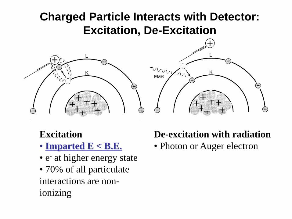
Charged Particle Interacts with Detector:
Excitation, De-Excitation
Excitation
• Imparted E < B.E.
• e- at higher energy state
• 70% of all particulate
interactions are non-
ionizing
De-excitation with radiation
• Photon or Auger electron

• Imparted E > B.E.
• Ion pair results
• Secondary ionization
Charged Particle
Interacts with Detector:
Ionization
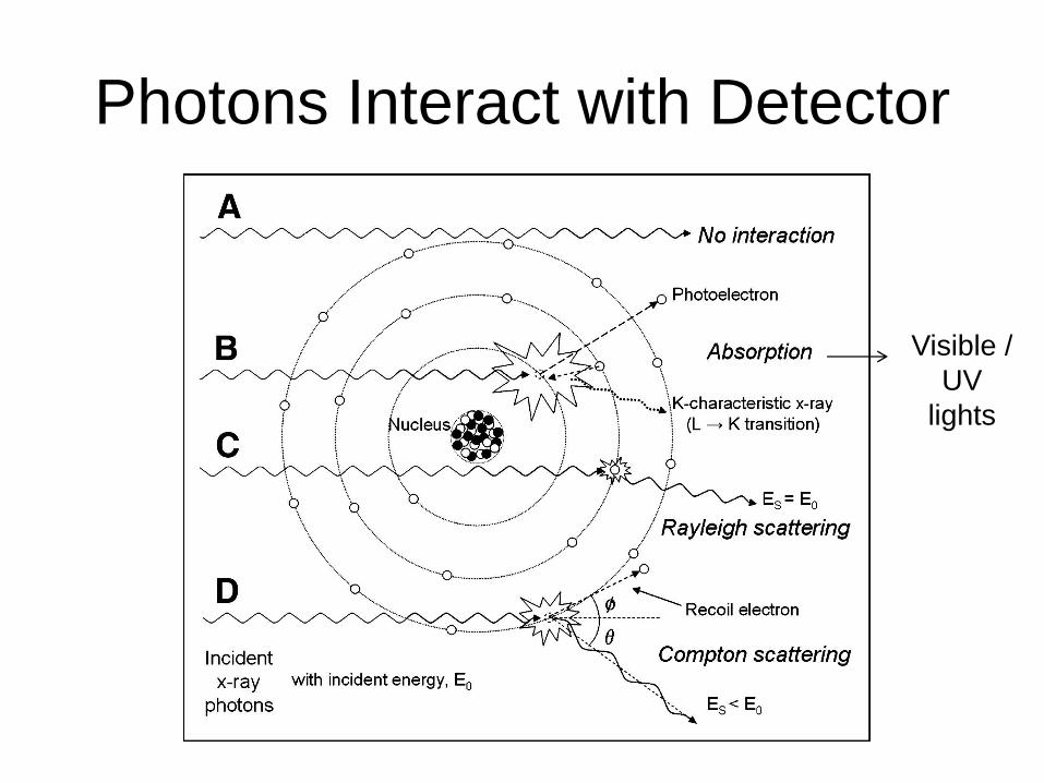
Photons Interact with Detector
Visible /
UV
lights
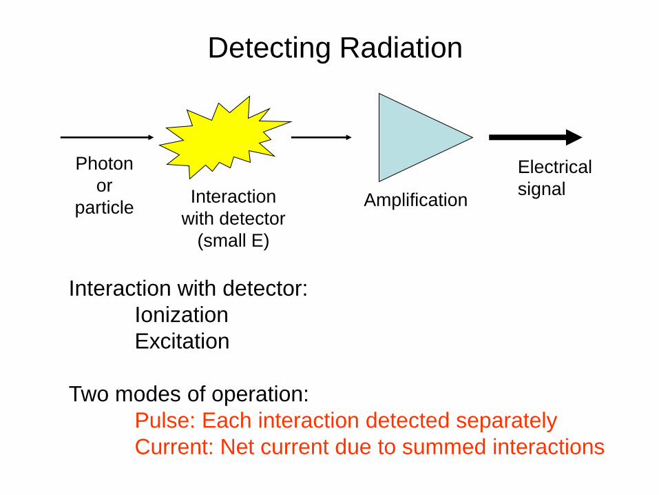
Detecting Radiation
Photon
or
particle Interaction
with detector
(small E)
Amplification
Electrical
signal
Interaction with detector:
Ionization
Excitation
Two modes of operation:
Pulse: Each interaction detected separately
Current: Net current due to summed interactions

Detector Properties
Dead time effect in Pulse Mode
•Current mode avoids dead time:
•e.g., CR, Image intensifier, CT, dose calibrator
•But info about individual interactions is lost

Spectroscopy
Pulse Height Spectrum
•Pulse mode: Amplitude is proportional to energy deposited
•Provides spectrum of energy deposited in detector
•E Not necessarily the same as the incident energy spectrum
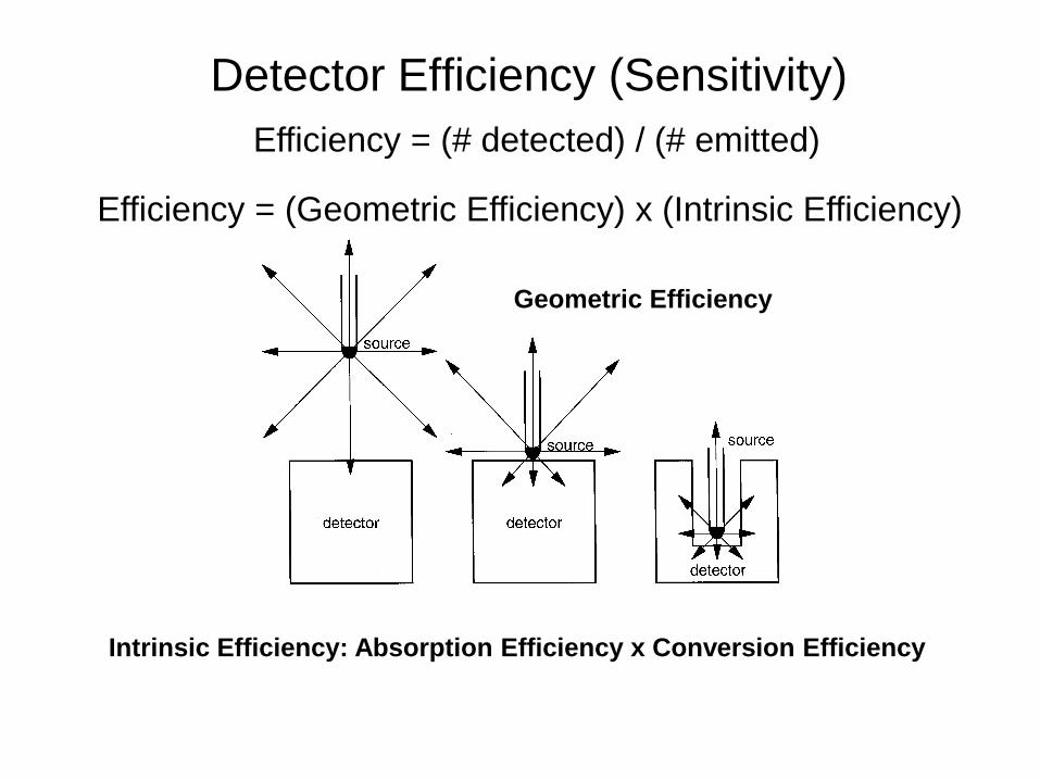
Detector Efficiency (Sensitivity)
Efficiency = (Geometric Efficiency) x (Intrinsic Efficiency)
Efficiency = (# detected) / (# emitted)
Geometric Efficiency
Intrinsic Efficiency: Absorption Efficiency x Conversion Efficiency
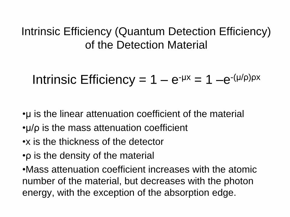
Intrinsic Efficiency (Quantum Detection Efficiency)
of the Detection Material
Intrinsic Efficiency = 1 – e-μx = 1 –e-(μ/ρ)ρx
•μ is the linear attenuation coefficient of the material
•μ/ρ is the mass attenuation coefficient
•x is the thickness of the detector
•ρ is the density of the material
•Mass attenuation coefficient increases with the atomic
number of the material, but decreases with the photon
energy, with the exception of the absorption edge.
Gas-Filled Detectors
(Ionization chamber, GM counter, proportional counter)

Gas-Filled Detector for Charged Particles
(Ionization chamber, GM counter, proportional counter)
(thin wall)
incident
photon is
blocked
(thick wall)

Gas-Filled Detector for Charges Particles:
Applied voltage determines type of operation
No current
flows
Gas
multiplication
“Avalanche”
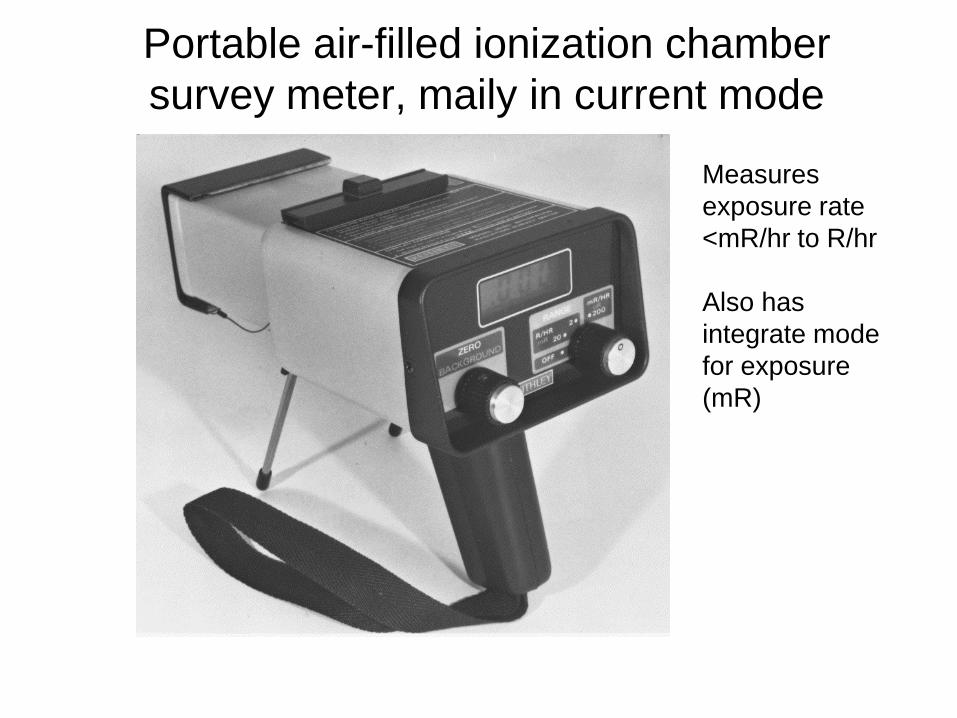
Portable air-filled ionization chamber
survey meter, maily in current mode
Measures
exposure rate
<mR/hr to R/hr
Also has
integrate mode
for exposure
(mR)

Port
able
air
-fill
ed io
niz
atio
n c
ha
mbe
r
su
rve
y m
ete
r, m
aily
in
cu
rre
nt
mo
de

Dose Calibrator, current mode, no dead-
time issue
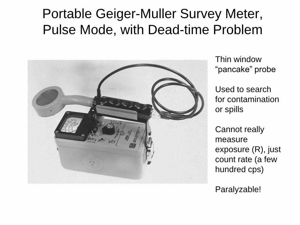
Portable Geiger-Muller Survey Meter,
Pulse Mode, with Dead-time Problem
Thin window
“pancake” probe
Used to search
for contamination
or spills
Cannot really
measure
exposure (R), just
count rate (a few
hundred cps)
Paralyzable!

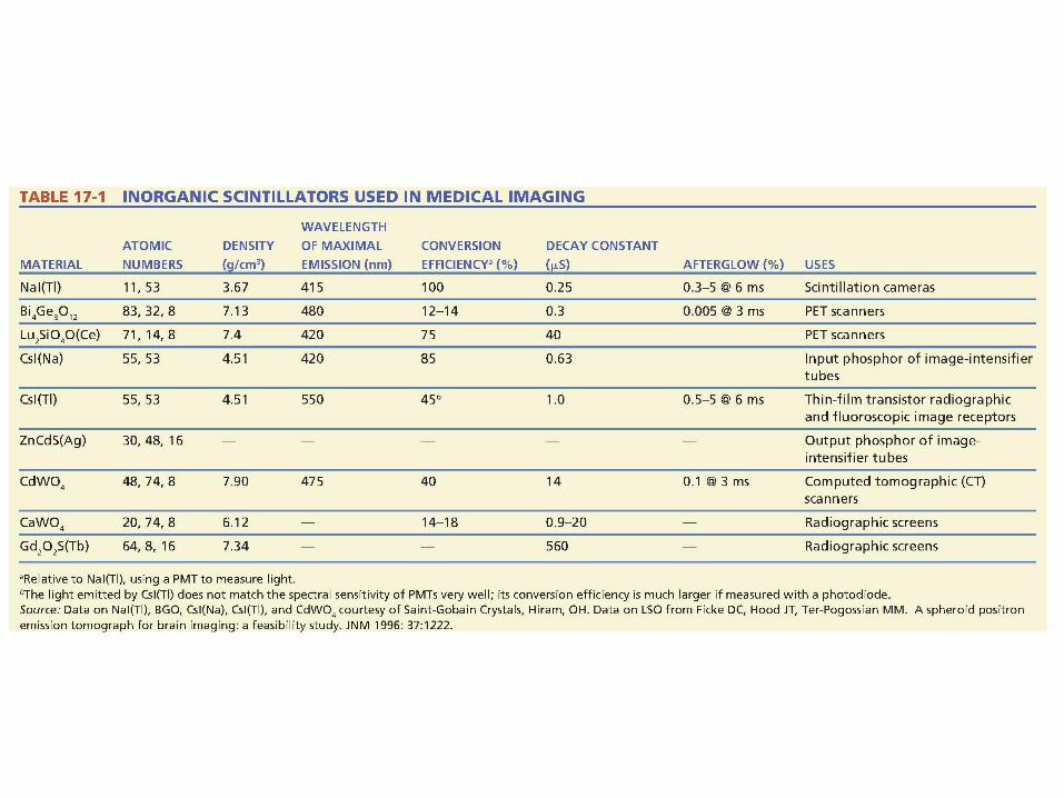
Scintillation Detectors, High Efficiency
Detectors for X-ray and Gamma-ray Photons
•Some common materials:
•NaI(Tl): Nuclear Medicine
•CsI: Image intensifiers, CT, DR
•Bi4Ge3O12: PET
•Gd2O2S(Tb): Radiographic screens
•ZnCdS(Ag): Image intensifier output phosphor
•CdWO4: CT
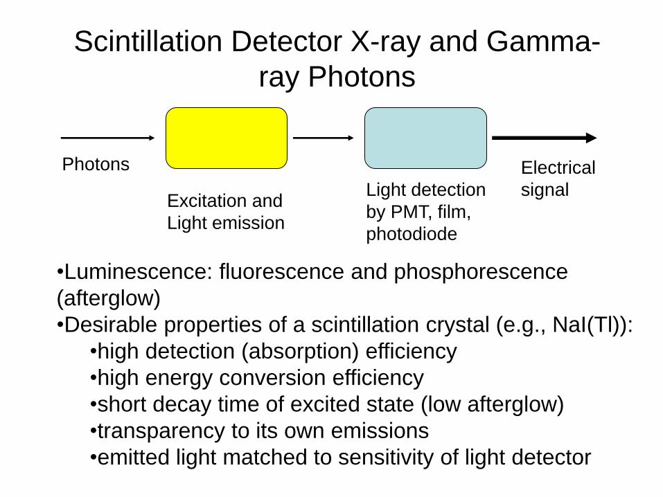
Scintillation Detector X-ray and Gamma-
ray Photons
Photons
Excitation and
Light emission
Light detection
by PMT, film,
photodiode
Electrical
signal
•Luminescence: fluorescence and phosphorescence
(afterglow)
•Desirable properties of a scintillation crystal (e.g., NaI(Tl)):
•high detection (absorption) efficiency
•high energy conversion efficiency
•short decay time of excited state (low afterglow)
•transparency to its own emissions
•emitted light matched to sensitivity of light detector
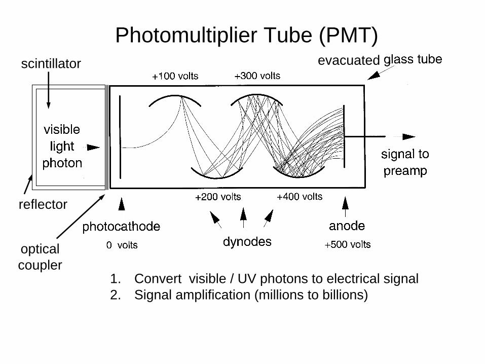
Photomultiplier Tube (PMT)
1. Convert visible / UV photons to electrical signal
2. Signal amplification (millions to billions)
scintillator
optical
coupler
reflector
evacuated
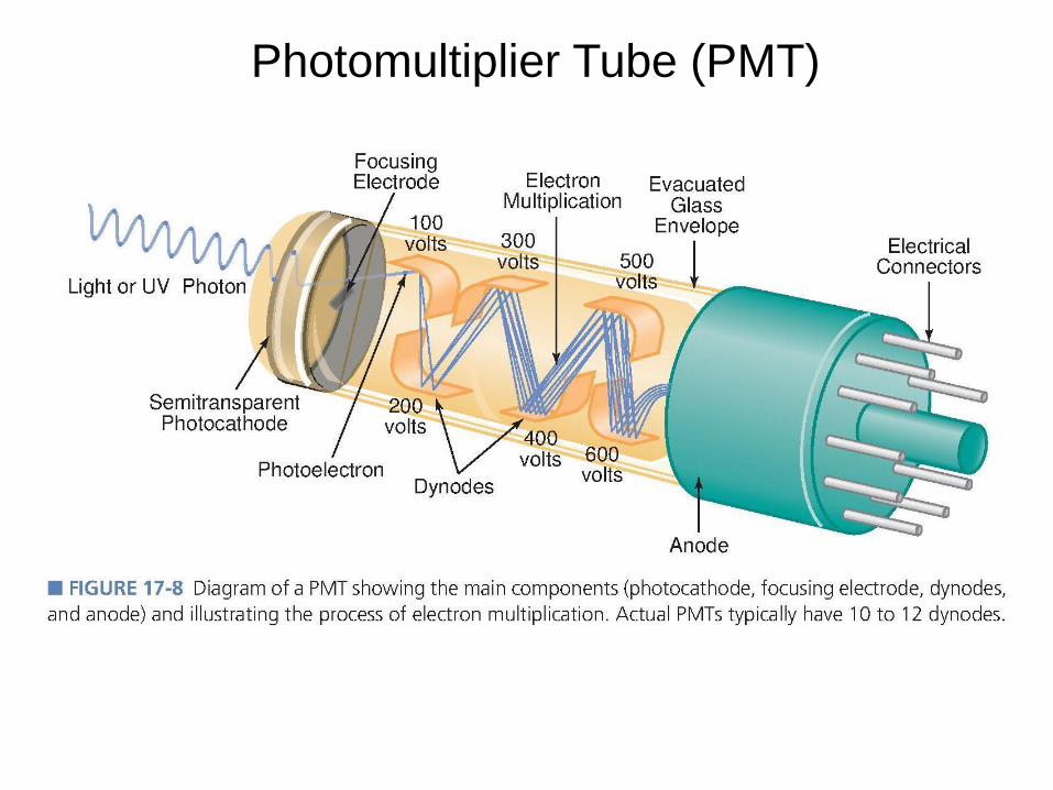
Photomultiplier Tube (PMT)
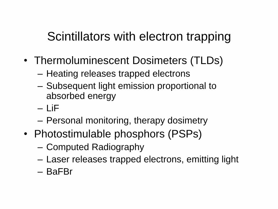
Scintillators with electron trapping
• Thermoluminescent Dosimeters (TLDs) – Heating releases trapped electrons
– Subsequent light emission proportional to absorbed energy
– LiF
– Personal monitoring, therapy dosimetry
• Photostimulable phosphors (PSPs) – Computed Radiography
– Laser releases trapped electrons, emitting light
– BaFBr

Semiconductor Crystal Detectors, mainly
for X-ray
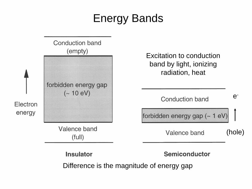
Energy Bands
Difference is the magnitude of energy gap
Excitation to conduction
band by light, ionizing
radiation, heat
e-
(hole)
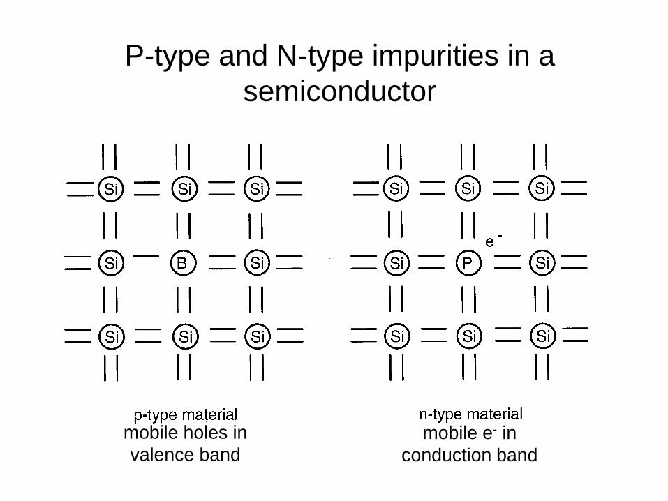
P-type and N-type impurities in a
semiconductor
mobile e- in
conduction band
mobile holes in
valence band
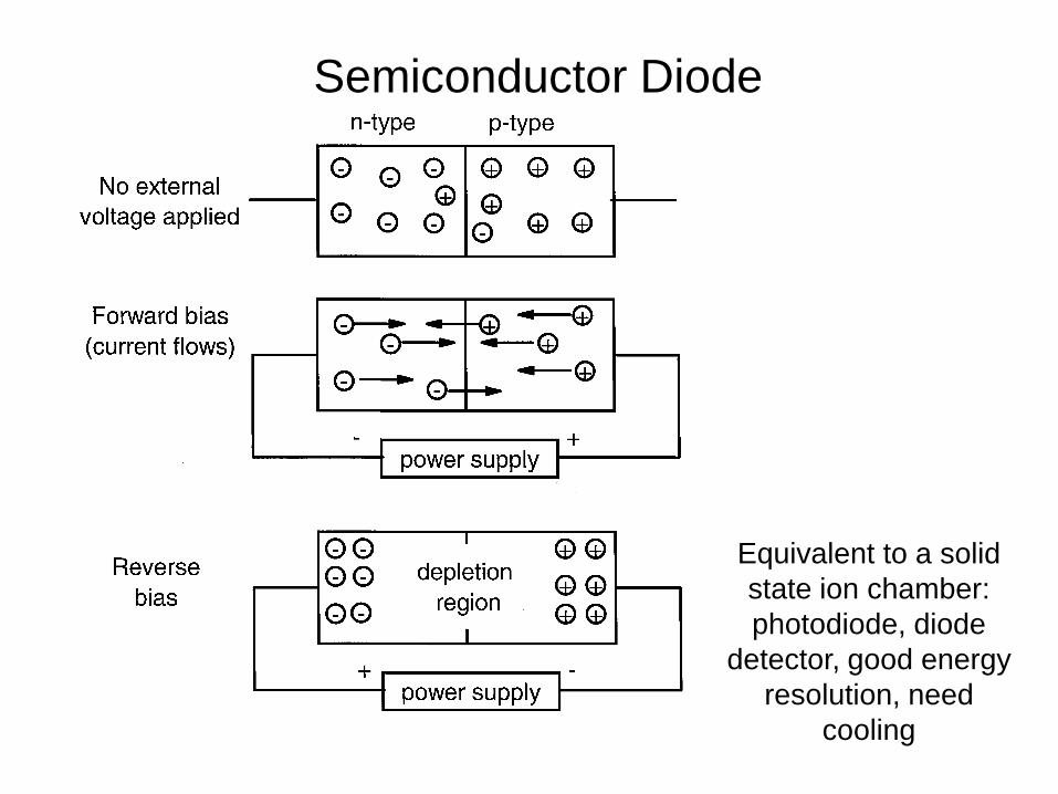
Semiconductor Diode
Equivalent to a solid
state ion chamber:
photodiode, diode
detector, good energy
resolution, need
cooling

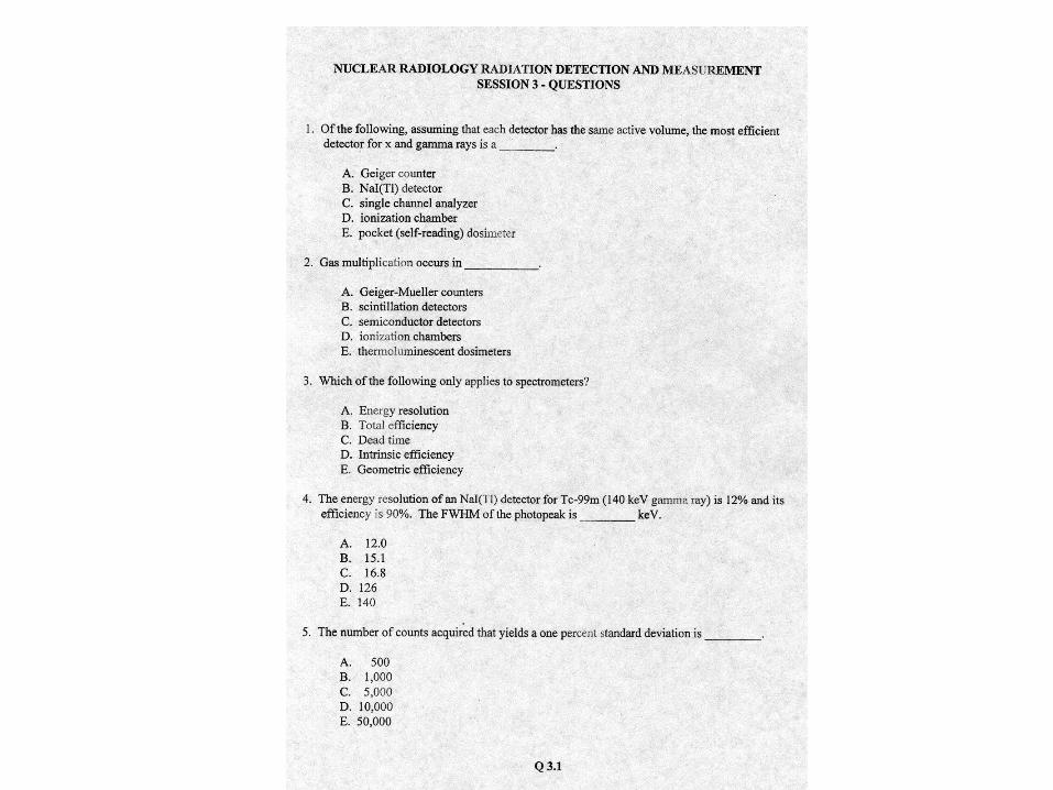
Gas multiplication occurs in _____.
a. Geiger-Mueller counters
b. Scintillation detectors
c. Semiconductor detectors
d. Ionization chambers
e. Thermoluminescent dosimeters
Gas multiplication occurs in _____.
a. Geiger-Mueller counters
b. Scintillation detectors
c. Semiconductor detectors
d. Ionization chambers
e. Thermoluminescent dosimeters
Of the following, assuming each detector has the
same active volume, the most efficient detector
for x- and gamma-rays is a _____.
a. Geiger Mueller counter
b. NaI(Tl) detector
c. Single channel analyzer
d. Ionization chamber
e. Dose calibrator

Of the following, assuming each detector has the
same active volume, the most efficient detector
for x- and gamma-rays is a _____.
a. Geiger Mueller counter
b. NaI(Tl) detector
c. Single channel analyzer
d. Ionization chamber
e. Dose calibrator

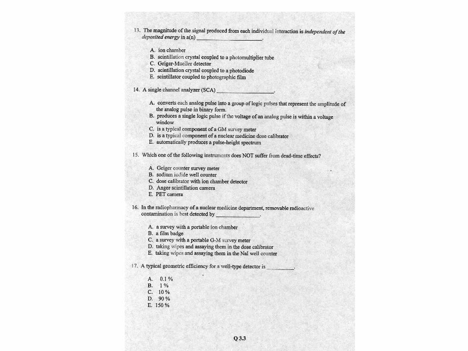
Which of the following instruments does NOT suffer
from dead-time effects?
a. Geiger-Mueller counter survey meter
b. NaI(Tl) well counter
c. PET camera
d. Scintillation camera
e. Dose calibrator with ion chamber detector
Which of the following instruments does NOT suffer
from dead-time effects?
a. Geiger-Mueller counter survey meter
b. NaI(Tl) well counter
c. PET camera
d. Scintillation camera
e. Dose calibrator with ion chamber detector
Pulse Height Spectroscopy
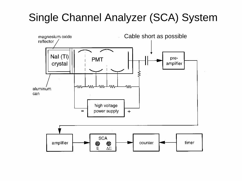
Single Channel Analyzer (SCA) System
Cable short as possible

Energy Discrimination
A logic pulse is
transmitted when
conditions of UL/LL
are met.
Calibration
(peaking) involves
matching V to E.

Multi-Channel Analyzer (MCA)
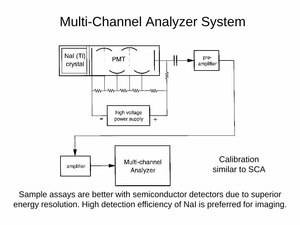
Multi-Channel Analyzer System
Calibration
similar to SCA
Sample assays are better with semiconductor detectors due to superior
energy resolution. High detection efficiency of NaI is preferred for imaging.
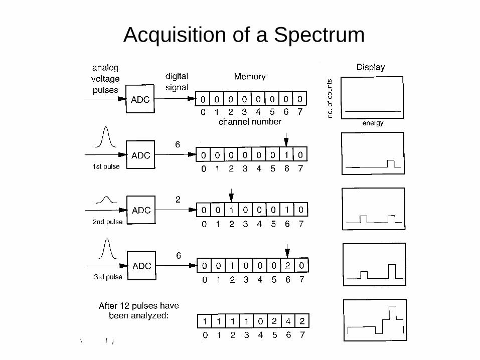
Acquisition of a Spectrum
X-ray and Gamma-Ray Spectroscopy with
Sodium Iodide Detectors
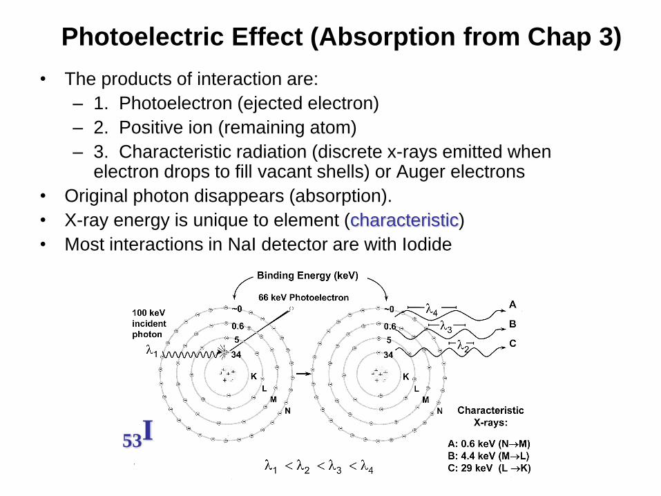
Photoelectric Effect (Absorption from Chap 3)
• The products of interaction are:
– 1. Photoelectron (ejected electron)
– 2. Positive ion (remaining atom)
– 3. Characteristic radiation (discrete x-rays emitted when electron drops to fill vacant shells) or Auger electrons
• Original photon disappears (absorption).
• X-ray energy is unique to element (characteristic)
• Most interactions in NaI detector are with Iodide
53I

• Occurs for loosely bound
electrons with negligible B.E.
• Source of undesirable scatter
radiation that reduces S/N
• Input: photon
Output: photon + electron
• h·inc = h·scat + K.E. e-
• Scattered photon: 0° 180°
• Scattered electron: 0° φ 90°
Compton Scattering
(increase the
background, Chap 3)
φ
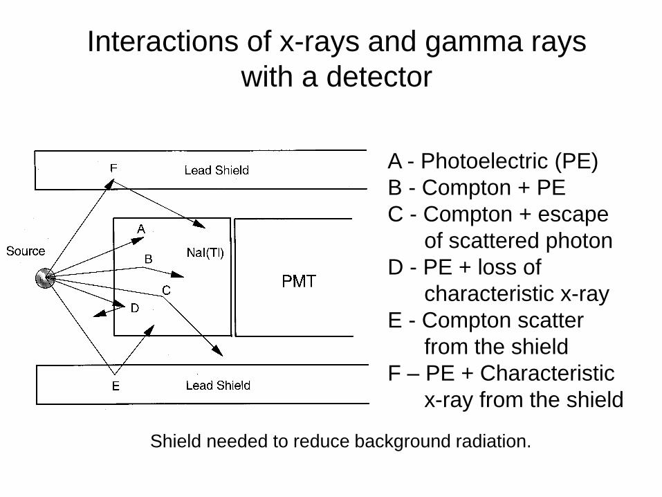
Interactions of x-rays and gamma rays
with a detector
A - Photoelectric (PE)
B - Compton + PE
C - Compton + escape
of scattered photon
D - PE + loss of
characteristic x-ray
E - Compton scatter
from the shield
F – PE + Characteristic
x-ray from the shield
Shield needed to reduce background radiation.

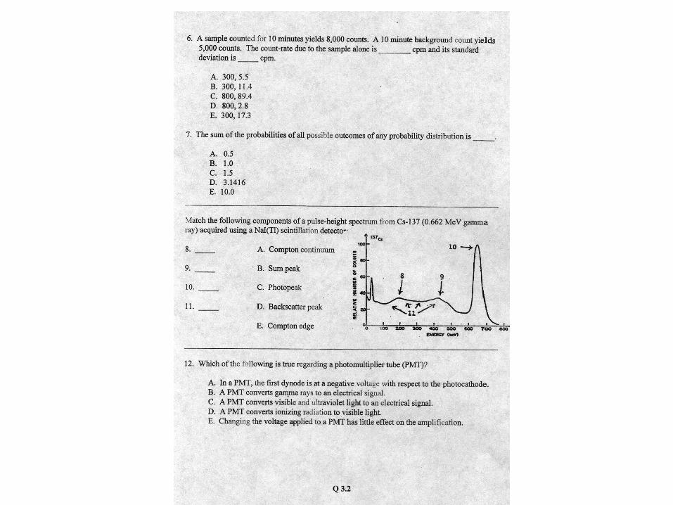
Spectrum of Cesium-137 A – 662 keV PE, total
absorption
B - Compton + escape
C - Compton edge
D – Backscatter from the
shield
E – 32 keV PE, Barium K-
shell x-rays after internal
conversion electron
F – PE with lead shield, K-
shell x-rays from shield 72-88
keV
90% gamma-ray
10% internal conversion
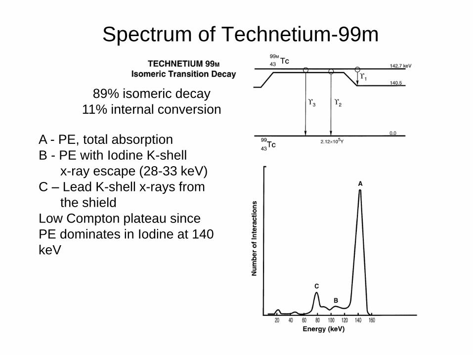
Spectrum of Technetium-99m
A - PE, total absorption
B - PE with Iodine K-shell
x-ray escape (28-33 keV)
C – Lead K-shell x-rays from
the shield
Low Compton plateau since
PE dominates in Iodine at 140
keV
89% isomeric decay
11% internal conversion

Spectrum of Iodine-125—Sum Peak
Sum peak

%100Peak ofCenter at Height Pulse
FWHMResolutionEnergy
Energy Resolution
NaI Energy Resolution of Cs-137 (662 keV) = 7-8%
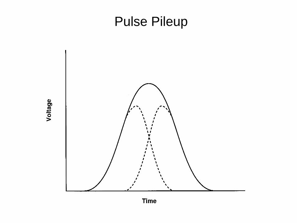
Pulse Pileup
Non-Imaging Detectors

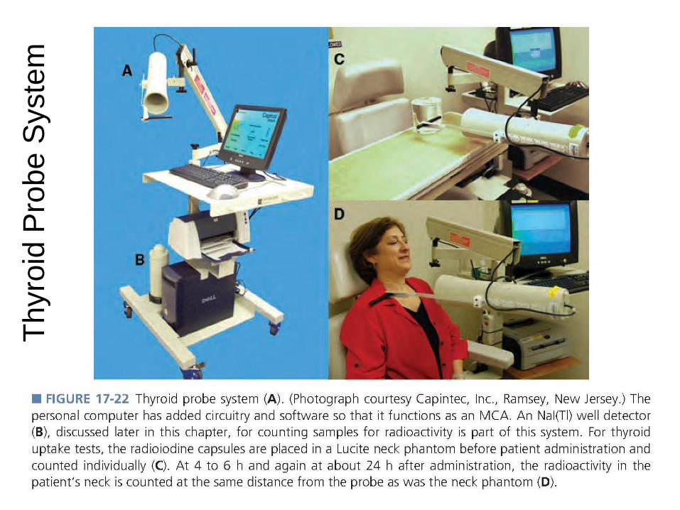
Thyroid Probe System
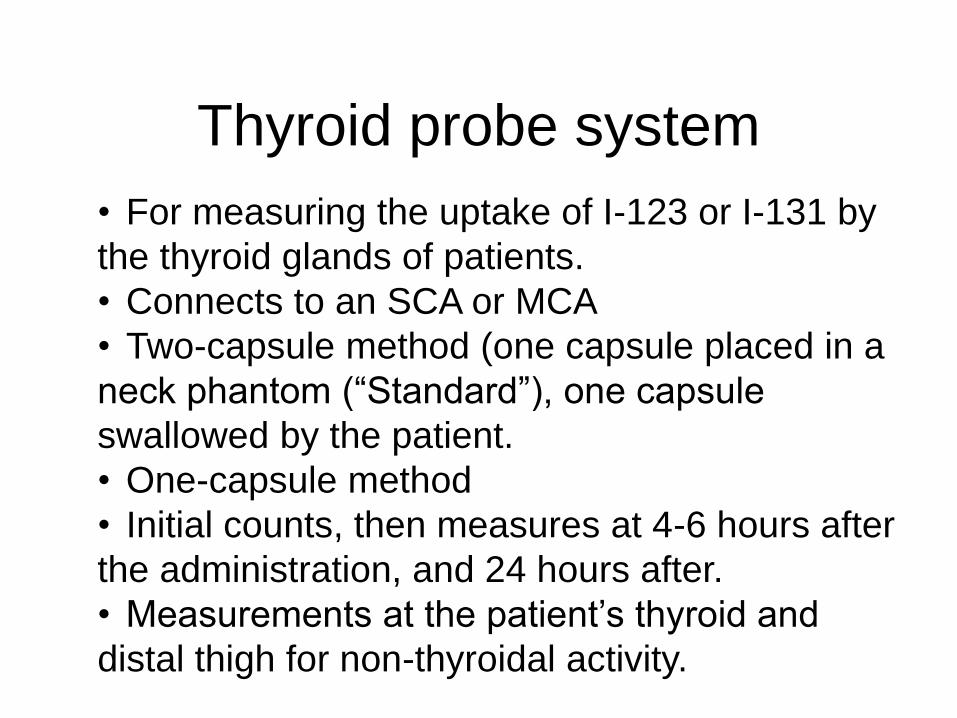
Thyroid probe system
• For measuring the uptake of I-123 or I-131 by
the thyroid glands of patients.
• Connects to an SCA or MCA
• Two-capsule method (one capsule placed in a
neck phantom (“Standard”), one capsule
swallowed by the patient.
• One-capsule method
• Initial counts, then measures at 4-6 hours after
the administration, and 24 hours after.
• Measurements at the patient’s thyroid and
distal thigh for non-thyroidal activity.

Thyroid Uptake Measures
Two-capsule method (accurate, more measures, higher cost):
phantomin capsulepatient ofcount Initial
phantomin standard ofcount Initial
count Backgroundphantomin standard ofCount
countThigh count ThyroidUptake
One-capsule method (fewer measures, lower cost, subject to instability,
technologist error, and dead-time effects):
2/1/693.0
count Backgroundphantomin capsule ofCount (Initial)
countThigh count ThyroidUptake
Tte
Th
yro
id P
robe
Syste
m
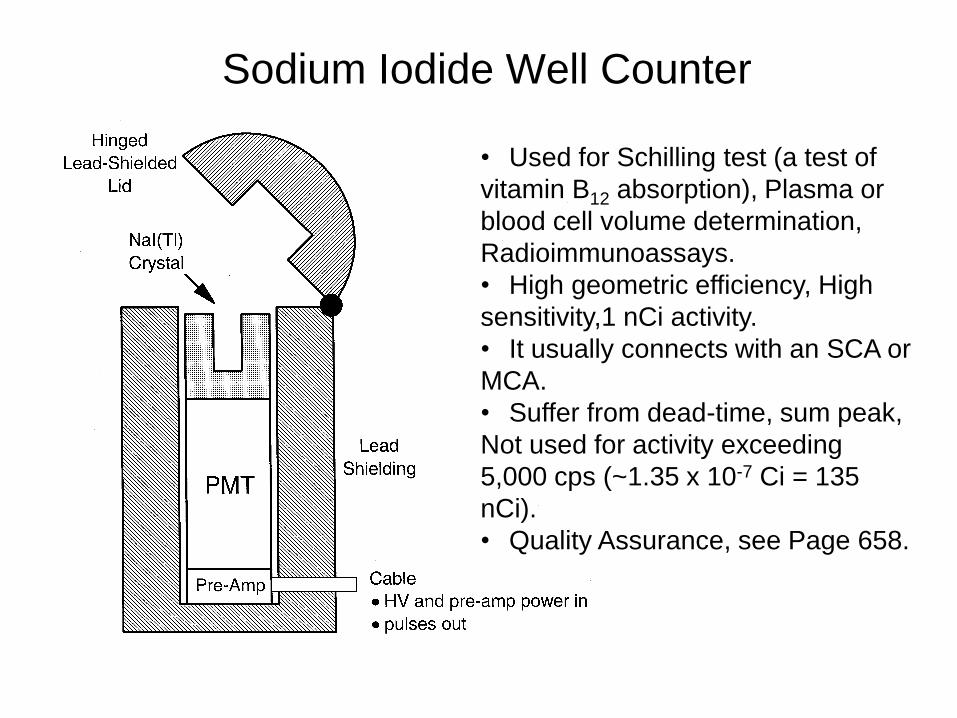
Sodium Iodide Well Counter
• Used for Schilling test (a test of
vitamin B12 absorption), Plasma or
blood cell volume determination,
Radioimmunoassays.
• High geometric efficiency, High
sensitivity,1 nCi activity.
• It usually connects with an SCA or
MCA.
• Suffer from dead-time, sum peak,
Not used for activity exceeding
5,000 cps (~1.35 x 10⁻⁷ Ci = 135
nCi).
• Quality Assurance, see Page 658.
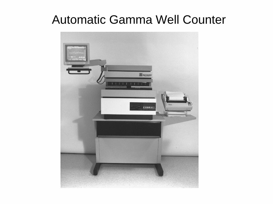
Automatic Gamma Well Counter
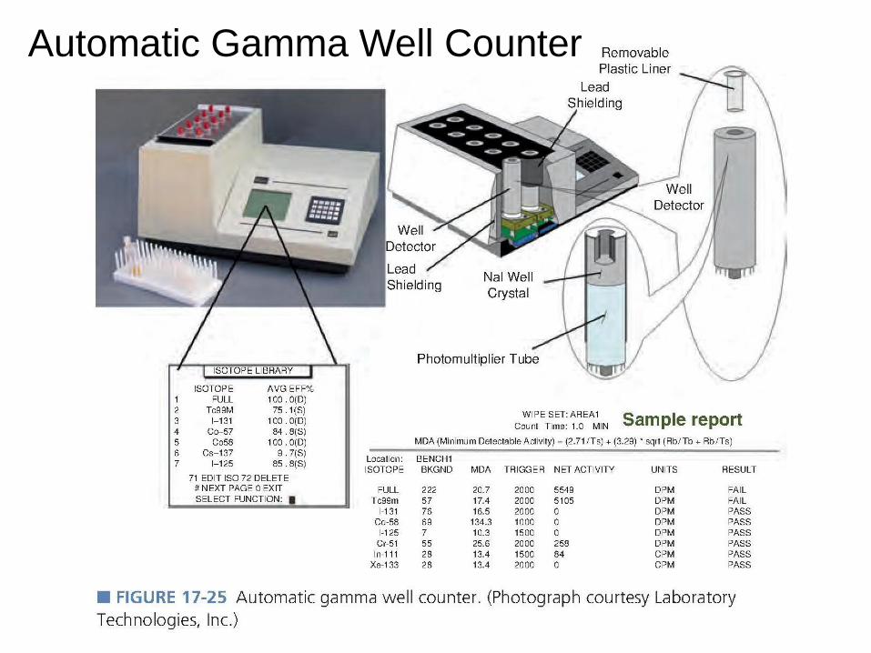
Automatic Gamma Well Counter
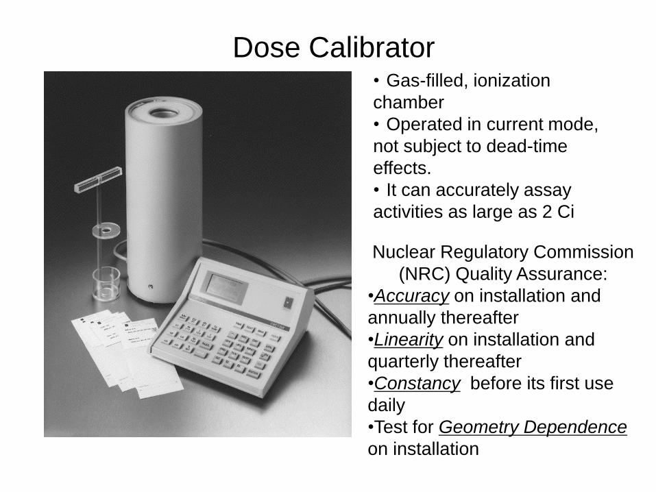
Dose Calibrator
Nuclear Regulatory Commission
(NRC) Quality Assurance:
•Accuracy on installation and
annually thereafter
•Linearity on installation and
quarterly thereafter
•Constancy before its first use
daily
•Test for Geometry Dependence
on installation
• Gas-filled, ionization
chamber
• Operated in current mode,
not subject to dead-time
effects.
• It can accurately assay
activities as large as 2 Ci
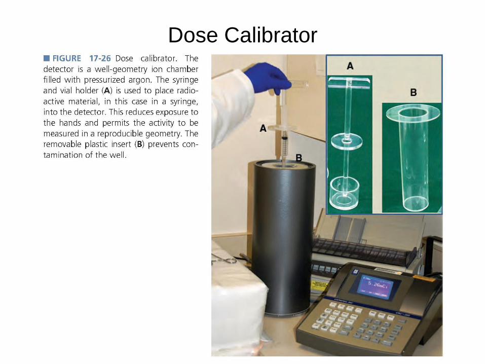
Dose Calibrator
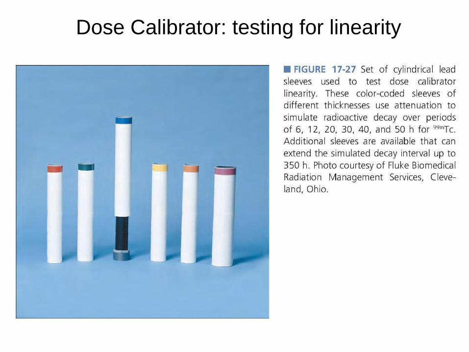
Dose Calibrator: testing for linearity

Molybdenum-99 Concentration Testing
•Mo-99 / Tc-99m beta-minus decay,
•Radiation dose: emitting high energy beta particles with 66-hour
half-life
•High-energy gamma rays will degrade resultant images
•NRC requirement: M0-99 < 0.15 μCi
• The wall of lead container are sufficiently thick to stop all
gamma rays from Tc-99m (140 keV), but thin enough to be
penetrated by many high-energy gamma rays from Mo-99 (740
and 778 keV).
•First, empty lead container is first assayed in the dose calibrator
•Second, the vial of Tc-99m alone is assayed.
K is a correction factor
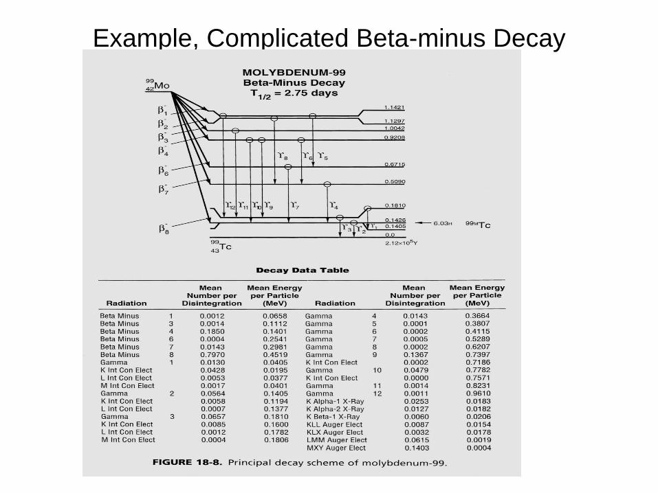
Example, Complicated Beta-minus Decay

Self-reading: Sr-82 and Sr-85
concentration test
Counting Statistics

Binomial Probability Distribution
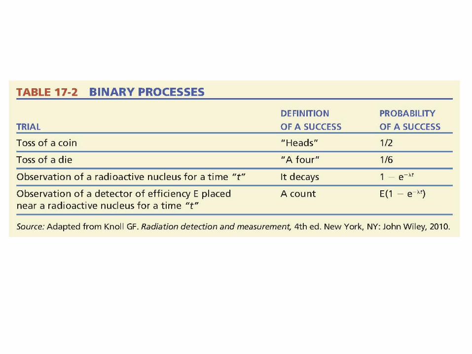
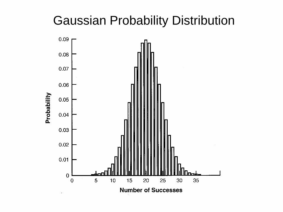
Gaussian Probability Distribution

Counting Statistics
Basic rules:
1. For N counts:
(only true when binominal approaches Gaussian)
2. Fractional error =
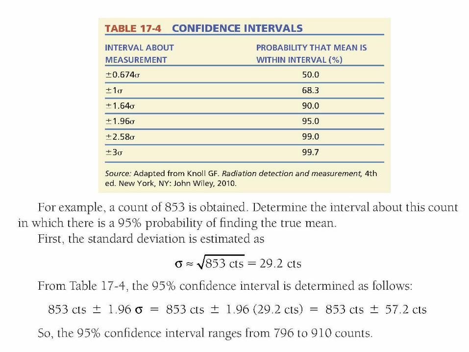
3. Confidence Intervals: Interval about measurements Probability that mean is within interval (%)
±0.674σ 50.0
±1σ 68.3
±1.64σ 90.0
±1.96σ 95.0
±2.58σ 99.0
±3σ 99.7
)( NorN
NN
N
N
1
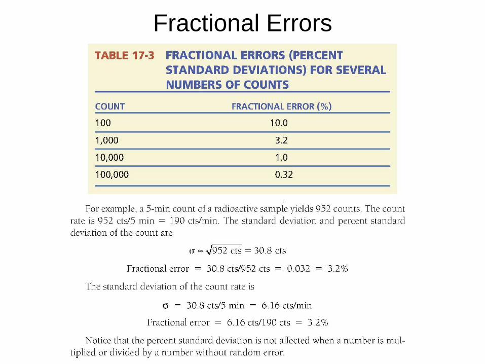
Fractional Errors
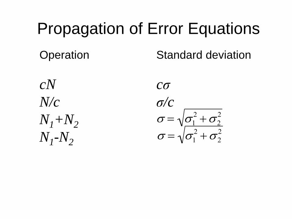
Propagation of Error Equations
Operation Standard deviation
cN cσ
N/c σ/c
N1+N2
N1-N2
12 2
2
12 2
2
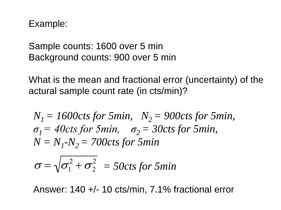
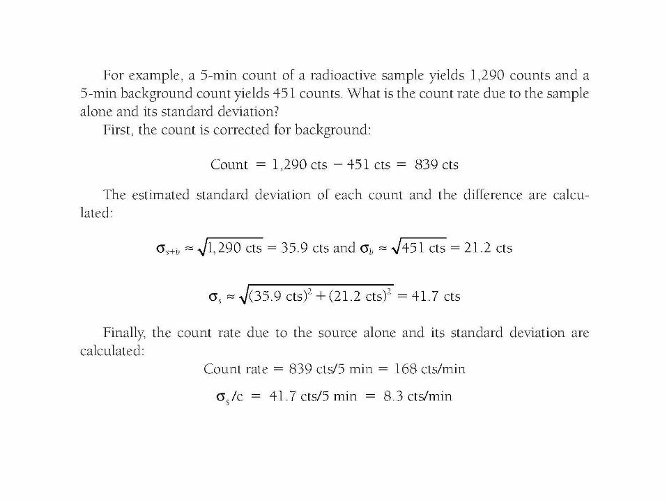
Example:
Sample counts: 1600 over 5 min
Background counts: 900 over 5 min
What is the mean and fractional error (uncertainty) of the
actural sample count rate (in cts/min)?
N1 = 1600cts for 5min, N2 = 900cts for 5min,
σ1 = 40cts for 5min, σ2 = 30cts for 5min,
N = N1-N2 = 700cts for 5min
= 50cts for 5min
Answer: 140 +/- 10 cts/min, 7.1% fractional error
12 2
2