Embed Size (px)
Citation preview

Jour
nal o
f Cel
l Sci
ence
SHORT REPORT
Rab5 is required in metastatic cancer cells for Caveolin-1-enhanced Rac1 activation, migration and invasion
Jorge Dıaz1,2,3, Pablo Mendoza1, Rina Ortiz2,3, Natalia Dıaz2,3, Lisette Leyton3,4, Dwayne Stupack5,Andrew F. G. Quest2,3,4,* and Vicente A. Torres1,3,*
ABSTRACT
Rab5 is a small GTPase that regulates early endosome trafficking
and other cellular processes, including cell adhesion and migration.
Specifically, Rab5 promotes Rac1 activation and cancer cell
migration, but little is known about the upstream regulators of
Rab5. We have previously shown that the scaffolding protein
Caveolin-1 (CAV1) promotes Rac1 activation and migration of
cancer cells. Here, we hypothesized that CAV1 stimulates Rab5
activation, leading to increased Rac1 activity and cell migration.
Expression of CAV1 in B16-F10 mouse melanoma and HT-29(US)
human colon adenocarcinoma cells increased the GTP loading of
Rab5, whereas shRNA-mediated targeting of endogenous CAV1 in
MDA-MB-231 breast cancer cells decreased Rab5–GTP levels.
Accordingly, shRNA-mediated downregulation of Rab5 decreased
CAV1-mediated Rac1 activation, cell migration and invasion in B16-
F10 and HT-29(US) cells. Expression of CAV1 was accompanied
by increased recruitment of Tiam1, a Rac1 guanine nucleotide
exchange factor (GEF), to Rab5-positive early endosomes. Using
the inhibitor NSC23766, Tiam1 was shown to be required for
Rac1 activation and cell migration induced by CAV1 and Rab5.
Mechanistically, we provide evidence implicating p85a (also known
as PIK3R1), a Rab5 GTPase-activating protein (GAP), in CAV1-
dependent effects, by showing that CAV1 recruits p85a, precluding
p85a-mediated Rab5 inactivation and increasing cell migration.
In summary, these studies identify a novel CAV1–Rab5–Rac1
signaling axis, whereby CAV1 prevents Rab5 inactivation, leading
to increased Rac1 activity and enhanced tumor cell migration and
invasion.
KEY WORDS: Rab5, Small GTPases, Metastatic cell migration,
Rac1, Caveolin-1, p85a
INTRODUCTIONRab5 is a central regulator of early endosome dynamics that
controls vesicle formation and fusion, homotypic fusion of early
endosomes, early-to-late endosome maturation and motility along
microtubules (Stenmark, 2009). As a small GTPase, Rab5 cycles
between a GDP- (inactive) and GTP-bound (active) form. In
addition to its canonical role in endocytosis, Rab5 has been
implicated in other cellular processes, such as cell adhesion and
migration (Torres and Stupack, 2011). Rab5 increases the rate of
internalization and recycling of b1 integrins (Pellinen et al., 2006;
Torres et al., 2010), stimulates focal adhesion disassembly
(Mendoza et al., 2013; Palamidessi et al., 2013) and promotes
the activation of Rac1, leading to cell migration (Palamidessi
et al., 2008; Torres et al., 2010). Moreover, Rab5 is implicated in
local actin rearrangement, by recruiting Tiam1, a Rac1 guanine
nucleotide exchange factor (GEF), to early endosomes, leading to
GTP loading of Rac1 and the formation of circular dorsal ruffles
(Lanzetti et al., 2004; Palamidessi et al., 2008). Although the
effectors and downstream signaling of Rab5 have been studied in
detail, little is known about upstream regulators of this GTPase.
In this respect, recent evidence suggests that there is a putative
connection between Rab5-dependent trafficking at early
endosomes and the scaffolding protein Caveolin-1 (CAV1)
(Hagiwara et al., 2009), although the functional relevance of
this relationship has not been addressed.
CAV1 is a 21-kDa protein that regulates numerous signaling
cascades and that, depending on the cell model, acts as either a
non-conventional tumor suppressor or a promoter of metastasis
(Quest et al., 2008; Quest et al., 2013). CAV1 promotes the
migration of both normal and tumor-derived cancer cells by
mechanisms that appear to depend on the cellular context. In
fibroblasts, CAV1-enhanced migration is associated with the
accumulation of CAV1 at the rear of cells and the activation of
RhoA (Grande-Garcia et al., 2007; Sun et al., 2007), whereas in
metastatic cancer cells, migration is not dependent on either of
these events (Urra et al., 2012). Rather, expression of CAV1 in
MDA-MB-231 breast cancer and B16-F10 mouse melanoma
cells is associated with increased turnover of focal adhesions
and augmented Rac1–GTP levels by mechanisms that remain to
be defined (Lobos-Gonzalez et al., 2013; Urra et al., 2012).
Intriguingly, recent evidence has suggested that CAV1
interacts with Rab5 in vitro and that CAV1 promotes Rab5-
dependent endocytosis (Hagiwara et al., 2009). Because
both proteins are implicated in Rac1 activation and cell
migration, we hypothesized that CAV1 promotes Rac1 GTP
loading and migration of cancer cells in vitro by regulating
Rab5. Expression of CAV1 in different metastatic cancer
cells, including B16-F10 (murine melanoma), MDA-MB-231
(human breast adenocarcinoma) and HT-29(US) (human colon
adenocarcinoma) led to increased Rab5 activation. Importantly,
Rab5 was required for CAV1-driven cell migration and invasion,
enhanced Tiam1 recruitment to early endosomes and Rac1
activation, as shown by the results of experiments involving
1Institute for Research in Dental Sciences, Faculty of Dentistry, Universidad deChile, Calle Sergio Livingstone 943, Santiago, Chile. 2Center for MolecularStudies of the Cell, Faculty of Medicine, Universidad de Chile, AvenidaIndependencia 1027, Santiago, Chile. 3Advanced Center for Chronic Diseases(ACCDiS), Faculty of Medicine, Universidad de Chile, Avenida Independencia1027, Santiago, Chile. 4Program of Cell and Molecular Biology, Institute ofBiomedical Sciences, Faculty of Medicine, Universidad de Chile, AvenidaIndependencia 1027, Santiago, Chile. 5Moores UCSD Cancer Center, Universityof California, La Jolla, CA 92093, USA.
*Authors for correspondence ([email protected]; [email protected])
Received 28 August 2013; Accepted 12 March 2014
� 2014. Published by The Company of Biologists Ltd | Journal of Cell Science (2014) 127, 2401–2406 doi:10.1242/jcs.141689
2401
Jour
nal o
f Cel
l Sci
ence
shRNA-targeting of Rab5. Particularly, CAV1-dependentactivation of Rab5 was associated with sequestration of
p85a (also known as PIK3R1), a Rab5 GTPase-activatingprotein (GAP), thereby precluding Rab5 inactivation. Thus,here, we identify a novel CAV1–Rab5–Rac1 signaling axis,which is required for CAV1-enhanced metastatic cancer cell
migration.
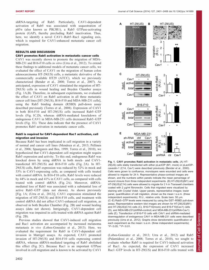
RESULTS AND DISCUSSIONCAV1 promotes Rab5 activation in metastatic cancer cellsCAV1 was recently shown to promote the migration of MDA-MB-231 and B16-F10 cells in vitro (Urra et al., 2012). To extend
these findings to additional models of metastatic cancer cells, weevaluated the effect of CAV1 on the migration of human colonadenocarcinoma HT-29(US) cells, a metastatic derivative of the
commercially available HT29 (ATCC), which we previouslycharacterized (Bender et al., 2000; Torres et al., 2007). Asanticipated, expression of CAV1 stimulated the migration of HT-29(US) cells in wound healing and Boyden Chamber assays
(Fig. 1A,B). Therefore, in subsequent experiments, we evaluatedthe effect of CAV1 on Rab5 activation in all three metastaticcancer cell lines [HT-29(US), B16-F10 and MDA-MB-231 cells],
using the Rab5 binding domain (R5BD) pull-down assaydescribed previously (Torres et al., 2008). Expression of CAV1in both B16-F10 and HT-29(US) cells increased Rab5–GTP
levels (Fig. 1C,D), whereas shRNA-mediated knockdown ofendogenous CAV1 in MDA-MB-231 cells decreased Rab5–GTPlevels (Fig. 1E). These data indicate that the presence of CAV1
promotes Rab5 activation in metastatic cancer cells.
Rab5 is required for CAV1-dependent Rac1 activation, cellmigration and invasionBecause Rab5 has been implicated in cell migration in a varietyof normal and cancer cell lines (Mendoza et al., 2013; Pellinenet al., 2006; Spaargaren and Bos, 1999; Torres et al., 2010), we
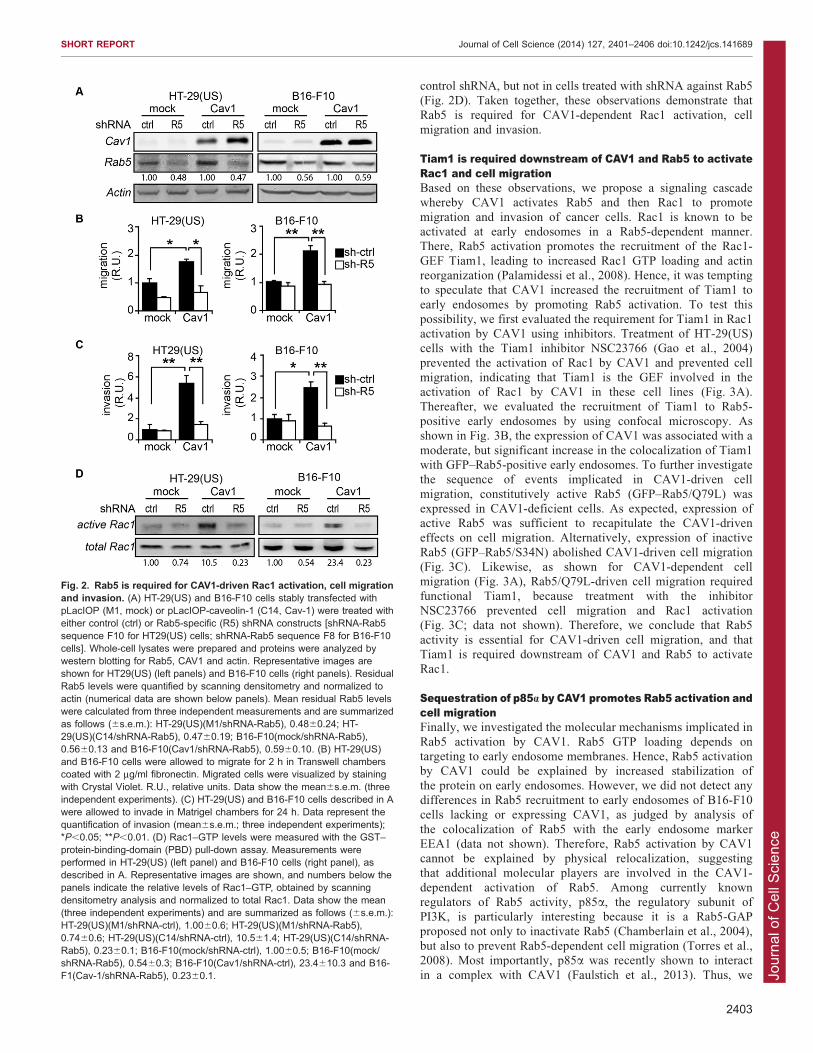
hypothesized that CAV1-dependent cell migration might requireRab5 expression and activity. To this end, endogenous Rab5 wasknocked down by using shRNA in both mock- and CAV1-
transfected HT-29(US) and B16-F10 cells (Fig. 2A). In HT-29(US) cells, Rab5 expression was reduced by 52% in mock and53% in CAV1-expressing cells, as compared with cells treatedwith control shRNA. In B16-F10 cells, Rab5 levels were reduced
by 44% in mock and 41% in CAV1 cells, as compared with cellstreated with control shRNA (Fig. 2A). Moreover, shRNA-mediated loss of Rab5 was associated with a substantial loss of
active Rab5–GTP (data not shown). As shown previously[Fig. 1A; (Urra et al., 2012)], CAV1 expression stimulated themigration of HT-29(US) and B16-F10 cells, and treatment with
control shRNA did not affect CAV1-enhanced cell migration, asobserved in both Boyden Chamber (Fig. 2B) and wound healingassays (data not shown). Intriguingly, CAV1-dependent cell
migration was impaired in cells treated with shRNA against Rab5(Fig. 2B).
Previous studies showed that CAV1-induced cell migrationand Rac1 activation are accompanied by increased melanoma
metastasis in vivo (Lobos-Gonzalez et al., 2013). Here, weevaluated the requirement for Rab5 in CAV1-dependent cellinvasion in Matrigel assays. As expected, CAV1 promoted
invasion of HT-29(US) and B16-F10 cells expressing controlshRNA, whereas shRNA-mediated targeting of Rab5 abolishedthis effect (Fig. 2C). Because Rac1 is an important GTPase
involved in cell migration and is known to be activated by CAV1
(Lobos-Gonzalez et al., 2013; Urra et al., 2012) and Rab5(Palamidessi et al., 2008; Torres et al., 2010), we sought to
evaluate whether Rab5 is required for CAV1-induced activationof Rac1. As expected, the expression of CAV1 increasedRac1–GTP levels in HT-29(US) and B16-F10 cells treated with
Fig. 1. CAV1 promotes Rab5 activation in metastatic cells. (A) HT-29(US) cells stably transfected with either pLacIOP (M1, mock) or pLacIOP-caveolin-1 (C14, Cav1) were described previously (Bender et al., 2000).Cells were grown to confluence, monolayers were wounded and cells wereallowed to migrate for 24 h. Representative phase-contrast images areshown, and the numbers within panels indicate the mean percentage ofwound closure from three independent experiments. (B) HT-29(US)(M1) andHT-29(US)(C14) cells were allowed to migrate for 2 h in Transwell chamberscoated with 2 mg/ml fibronectin. Cells that migrated were visualized bystaining with Crystal Violet. Upper panels, representative images; lowerpanel, quantification of cell migration, shown as the mean6s.e.m. (threeindependent experiments). R.U., relative units. Scale bars: 50 mm.(C–E) Rab5–GTP levels were measured by using the GST–R5BD pull-downassay. Representative western blot images are shown for HT-29(US)(M1)and HT-29(US)(C14) cells (C), B16-F10(mock) and B16-F10(Cav1) cells(D), and MDA-MB-231(shRNA-control) and MDA-MB-231(shRNA-Cav1)cells (E). Transfection of B16-F10 cells with CAV1 and shRNA-mediateddownregulation of endogenous CAV1 in MDA-MB-231 cells were describedpreviously (Urra et al., 2012). Graphs show densitometric quantification ofeach experiment as the mean6s.e.m. (three independent experiments);*P,0.05; **P,0.01.
SHORT REPORT Journal of Cell Science (2014) 127, 2401–2406 doi:10.1242/jcs.141689
2402
Jour
nal o
f Cel
l Sci
ence
control shRNA, but not in cells treated with shRNA against Rab5(Fig. 2D). Taken together, these observations demonstrate that
Rab5 is required for CAV1-dependent Rac1 activation, cellmigration and invasion.
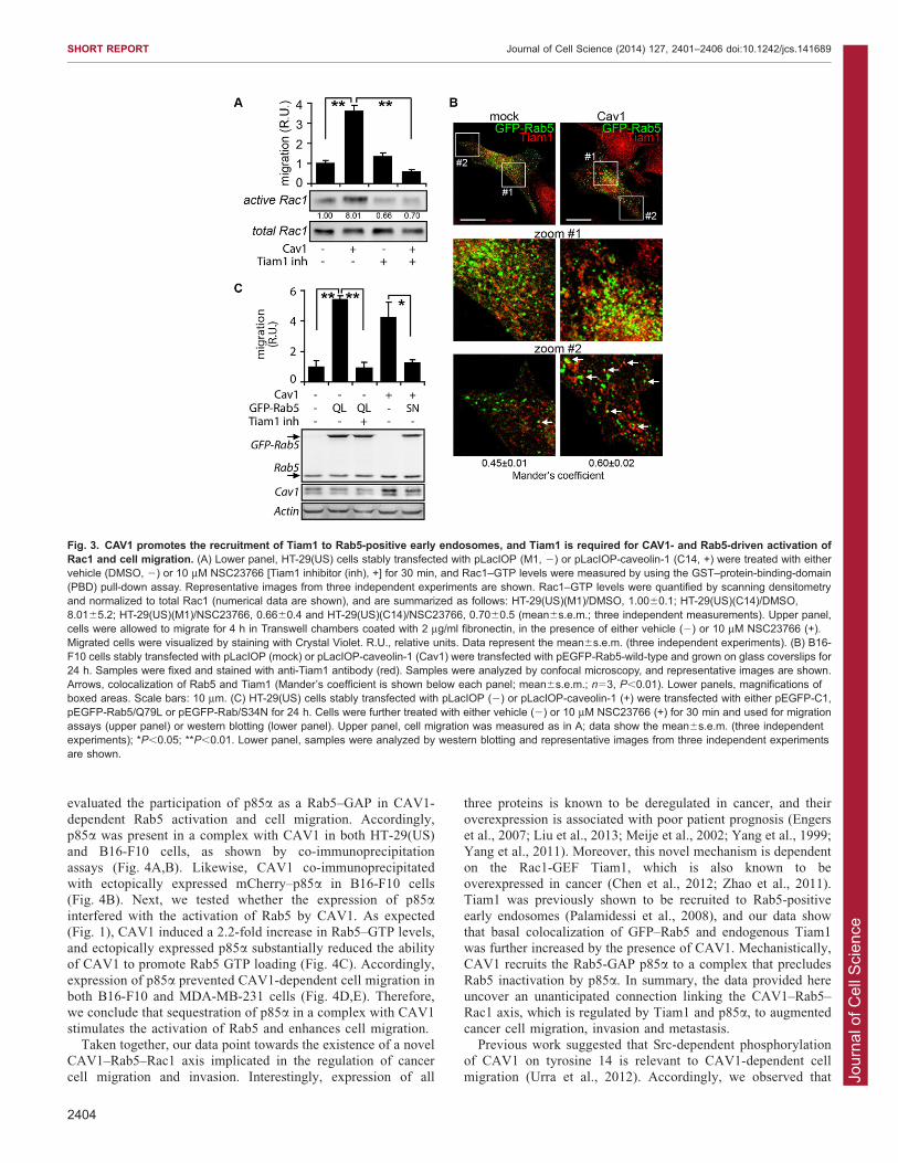
Tiam1 is required downstream of CAV1 and Rab5 to activateRac1 and cell migrationBased on these observations, we propose a signaling cascadewhereby CAV1 activates Rab5 and then Rac1 to promote
migration and invasion of cancer cells. Rac1 is known to beactivated at early endosomes in a Rab5-dependent manner.There, Rab5 activation promotes the recruitment of the Rac1-
GEF Tiam1, leading to increased Rac1 GTP loading and actinreorganization (Palamidessi et al., 2008). Hence, it was temptingto speculate that CAV1 increased the recruitment of Tiam1 to
early endosomes by promoting Rab5 activation. To test thispossibility, we first evaluated the requirement for Tiam1 in Rac1activation by CAV1 using inhibitors. Treatment of HT-29(US)cells with the Tiam1 inhibitor NSC23766 (Gao et al., 2004)
prevented the activation of Rac1 by CAV1 and prevented cellmigration, indicating that Tiam1 is the GEF involved in theactivation of Rac1 by CAV1 in these cell lines (Fig. 3A).
Thereafter, we evaluated the recruitment of Tiam1 to Rab5-positive early endosomes by using confocal microscopy. Asshown in Fig. 3B, the expression of CAV1 was associated with a
moderate, but significant increase in the colocalization of Tiam1with GFP–Rab5-positive early endosomes. To further investigatethe sequence of events implicated in CAV1-driven cell
migration, constitutively active Rab5 (GFP–Rab5/Q79L) wasexpressed in CAV1-deficient cells. As expected, expression ofactive Rab5 was sufficient to recapitulate the CAV1-driveneffects on cell migration. Alternatively, expression of inactive
Rab5 (GFP–Rab5/S34N) abolished CAV1-driven cell migration(Fig. 3C). Likewise, as shown for CAV1-dependent cellmigration (Fig. 3A), Rab5/Q79L-driven cell migration required
functional Tiam1, because treatment with the inhibitorNSC23766 prevented cell migration and Rac1 activation(Fig. 3C; data not shown). Therefore, we conclude that Rab5
activity is essential for CAV1-driven cell migration, and thatTiam1 is required downstream of CAV1 and Rab5 to activateRac1.
Sequestration of p85a by CAV1 promotes Rab5 activation andcell migrationFinally, we investigated the molecular mechanisms implicated in
Rab5 activation by CAV1. Rab5 GTP loading depends ontargeting to early endosome membranes. Hence, Rab5 activationby CAV1 could be explained by increased stabilization of
the protein on early endosomes. However, we did not detect anydifferences in Rab5 recruitment to early endosomes of B16-F10cells lacking or expressing CAV1, as judged by analysis of
the colocalization of Rab5 with the early endosome markerEEA1 (data not shown). Therefore, Rab5 activation by CAV1cannot be explained by physical relocalization, suggestingthat additional molecular players are involved in the CAV1-
dependent activation of Rab5. Among currently knownregulators of Rab5 activity, p85a, the regulatory subunit ofPI3K, is particularly interesting because it is a Rab5-GAP
proposed not only to inactivate Rab5 (Chamberlain et al., 2004),but also to prevent Rab5-dependent cell migration (Torres et al.,2008). Most importantly, p85a was recently shown to interact
in a complex with CAV1 (Faulstich et al., 2013). Thus, we
Fig. 2. Rab5 is required for CAV1-driven Rac1 activation, cell migrationand invasion. (A) HT-29(US) and B16-F10 cells stably transfected withpLacIOP (M1, mock) or pLacIOP-caveolin-1 (C14, Cav-1) were treated witheither control (ctrl) or Rab5-specific (R5) shRNA constructs [shRNA-Rab5sequence F10 for HT29(US) cells; shRNA-Rab5 sequence F8 for B16-F10cells]. Whole-cell lysates were prepared and proteins were analyzed bywestern blotting for Rab5, CAV1 and actin. Representative images areshown for HT29(US) (left panels) and B16-F10 cells (right panels). ResidualRab5 levels were quantified by scanning densitometry and normalized toactin (numerical data are shown below panels). Mean residual Rab5 levelswere calculated from three independent measurements and are summarizedas follows (6s.e.m.): HT-29(US)(M1/shRNA-Rab5), 0.4860.24; HT-29(US)(C14/shRNA-Rab5), 0.4760.19; B16-F10(mock/shRNA-Rab5),0.5660.13 and B16-F10(Cav1/shRNA-Rab5), 0.5960.10. (B) HT-29(US)and B16-F10 cells were allowed to migrate for 2 h in Transwell chamberscoated with 2 mg/ml fibronectin. Migrated cells were visualized by stainingwith Crystal Violet. R.U., relative units. Data show the mean6s.e.m. (threeindependent experiments). (C) HT-29(US) and B16-F10 cells described in Awere allowed to invade in Matrigel chambers for 24 h. Data represent thequantification of invasion (mean6s.e.m.; three independent experiments);*P,0.05; **P,0.01. (D) Rac1–GTP levels were measured with the GST–protein-binding-domain (PBD) pull-down assay. Measurements wereperformed in HT-29(US) (left panel) and B16-F10 cells (right panel), asdescribed in A. Representative images are shown, and numbers below thepanels indicate the relative levels of Rac1–GTP, obtained by scanningdensitometry analysis and normalized to total Rac1. Data show the mean(three independent experiments) and are summarized as follows (6s.e.m.):HT-29(US)(M1/shRNA-ctrl), 1.0060.6; HT-29(US)(M1/shRNA-Rab5),0.7460.6; HT-29(US)(C14/shRNA-ctrl), 10.561.4; HT-29(US)(C14/shRNA-Rab5), 0.2360.1; B16-F10(mock/shRNA-ctrl), 1.0060.5; B16-F10(mock/shRNA-Rab5), 0.5460.3; B16-F10(Cav1/shRNA-ctrl), 23.4610.3 and B16-F1(Cav-1/shRNA-Rab5), 0.2360.1.
SHORT REPORT Journal of Cell Science (2014) 127, 2401–2406 doi:10.1242/jcs.141689
2403
Jour
nal o
f Cel
l Sci
ence
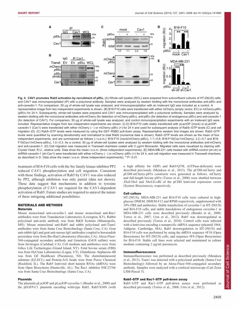
evaluated the participation of p85a as a Rab5–GAP in CAV1-
dependent Rab5 activation and cell migration. Accordingly,p85a was present in a complex with CAV1 in both HT-29(US)and B16-F10 cells, as shown by co-immunoprecipitation
assays (Fig. 4A,B). Likewise, CAV1 co-immunoprecipitatedwith ectopically expressed mCherry–p85a in B16-F10 cells(Fig. 4B). Next, we tested whether the expression of p85ainterfered with the activation of Rab5 by CAV1. As expected(Fig. 1), CAV1 induced a 2.2-fold increase in Rab5–GTP levels,and ectopically expressed p85a substantially reduced the abilityof CAV1 to promote Rab5 GTP loading (Fig. 4C). Accordingly,
expression of p85a prevented CAV1-dependent cell migration inboth B16-F10 and MDA-MB-231 cells (Fig. 4D,E). Therefore,we conclude that sequestration of p85a in a complex with CAV1
stimulates the activation of Rab5 and enhances cell migration.Taken together, our data point towards the existence of a novel
CAV1–Rab5–Rac1 axis implicated in the regulation of cancer
cell migration and invasion. Interestingly, expression of all
three proteins is known to be deregulated in cancer, and their
overexpression is associated with poor patient prognosis (Engerset al., 2007; Liu et al., 2013; Meije et al., 2002; Yang et al., 1999;Yang et al., 2011). Moreover, this novel mechanism is dependent
on the Rac1-GEF Tiam1, which is also known to beoverexpressed in cancer (Chen et al., 2012; Zhao et al., 2011).Tiam1 was previously shown to be recruited to Rab5-positive
early endosomes (Palamidessi et al., 2008), and our data showthat basal colocalization of GFP–Rab5 and endogenous Tiam1was further increased by the presence of CAV1. Mechanistically,CAV1 recruits the Rab5-GAP p85a to a complex that precludes
Rab5 inactivation by p85a. In summary, the data provided hereuncover an unanticipated connection linking the CAV1–Rab5–Rac1 axis, which is regulated by Tiam1 and p85a, to augmented
cancer cell migration, invasion and metastasis.Previous work suggested that Src-dependent phosphorylation
of CAV1 on tyrosine 14 is relevant to CAV1-dependent cell
migration (Urra et al., 2012). Accordingly, we observed that
Fig. 3. CAV1 promotes the recruitment of Tiam1 to Rab5-positive early endosomes, and Tiam1 is required for CAV1- and Rab5-driven activation ofRac1 and cell migration. (A) Lower panel, HT-29(US) cells stably transfected with pLacIOP (M1, 2) or pLacIOP-caveolin-1 (C14, +) were treated with eithervehicle (DMSO, 2) or 10 mM NSC23766 [Tiam1 inhibitor (inh), +] for 30 min, and Rac1–GTP levels were measured by using the GST–protein-binding-domain(PBD) pull-down assay. Representative images from three independent experiments are shown. Rac1–GTP levels were quantified by scanning densitometryand normalized to total Rac1 (numerical data are shown), and are summarized as follows: HT-29(US)(M1)/DMSO, 1.0060.1; HT-29(US)(C14)/DMSO,8.0165.2; HT-29(US)(M1)/NSC23766, 0.6660.4 and HT-29(US)(C14)/NSC23766, 0.7060.5 (mean6s.e.m.; three independent measurements). Upper panel,cells were allowed to migrate for 4 h in Transwell chambers coated with 2 mg/ml fibronectin, in the presence of either vehicle (2) or 10 mM NSC23766 (+).Migrated cells were visualized by staining with Crystal Violet. R.U., relative units. Data represent the mean6s.e.m. (three independent experiments). (B) B16-F10 cells stably transfected with pLacIOP (mock) or pLacIOP-caveolin-1 (Cav1) were transfected with pEGFP-Rab5-wild-type and grown on glass coverslips for24 h. Samples were fixed and stained with anti-Tiam1 antibody (red). Samples were analyzed by confocal microscopy, and representative images are shown.Arrows, colocalization of Rab5 and Tiam1 (Mander’s coefficient is shown below each panel; mean6s.e.m.; n53, P,0.01). Lower panels, magnifications ofboxed areas. Scale bars: 10 mm. (C) HT-29(US) cells stably transfected with pLacIOP (2) or pLacIOP-caveolin-1 (+) were transfected with either pEGFP-C1,pEGFP-Rab5/Q79L or pEGFP-Rab/S34N for 24 h. Cells were further treated with either vehicle (2) or 10 mM NSC23766 (+) for 30 min and used for migrationassays (upper panel) or western blotting (lower panel). Upper panel, cell migration was measured as in A; data show the mean6s.e.m. (three independentexperiments); *P,0.05; **P,0.01. Lower panel, samples were analyzed by western blotting and representative images from three independent experimentsare shown.
SHORT REPORT Journal of Cell Science (2014) 127, 2401–2406 doi:10.1242/jcs.141689
2404
Jour
nal o
f Cel
l Sci
ence
treatment of B16-F10 cells with the Src family kinase inhibitor PP2
reduced CAV1 phosphorylation and cell migration. Consistentwith those findings, activation of Rab5 by CAV1 was also reducedby PP2, although inhibition was only partial (data not shown).
These data suggest that mechanisms in addition to tyrosinephosphorylation of CAV1 are required for the CAV1-dependentactivation of Rab5. Future studies are required to unravel the nature
of these intriguing additional possibilities.
MATERIALS AND METHODSMaterialsMouse monoclonal anti-caveolin-1 and mouse monoclonal anti-Rac1
antibodies were from Transduction Laboratories (Lexington, KY). Rabbit
polyclonal anti-actin antibody was from R&D Systems (Minneapolis,
MN). Mouse monoclonal anti-Rab5 and rabbit polyclonal anti-Tiam1
antibodies were from Santa Cruz Biotechnology (Santa Cruz, CA). Goat
anti-rabbit-IgG and goat anti-mouse-IgG antibodies coupled to horseradish
peroxidase were from Bio-Rad Laboratories (Hercules, CA). Alexa-Fluor-
568-conjugated secondary antibody and Geneticin (G418 sulfate) were
from Invitrogen (Carlsbad, CA). Cell medium and antibiotics were from
Gibco Life Technologies (Grand Island, NY). Fetal bovine serum (FBS)
was from HyClone Laboratories (Logan, UT). Glutathione–Sepharose-4B
was from GE Healthcare (Piscataway, NJ). The chemiluminescent
substrate (EZ-ECL) and Protein-A/G beads were from Pierce Chemical
(Rockford, IL). The Rab5 lentiviral short hairpin RNAs (shRNA) were
from Open Biosystems (Huntsville, AL). The Rac1 inhibitor NSC23766
was from Santa Cruz Biotechnology (Santa Cruz, CA).
PlasmidsThe plasmids pLacIOP and pLacIOP-caveolin-1 (Bender et al., 2000) and
the pEGFP-C1 plasmids encoding wild-type Rab5, Rab5/S34N (with
a high affinity for GDP) and Rab5/Q79L (GTPase-deficient) were
described previously (Mendoza et al., 2013). The pCDH-mCherry and
pCDH-mCherry-p85a constructs were generated as follows. mCherry
and full-length bovine p85a (Torres et al., 2008) were shuttled between
XbaI/NheI and NheI/EcoRI of the pCDH lentiviral expression vector
(System Biosciences), respectively.
Cell cultureHT-29(US), MDA-MB-231 and B16-F10 cells were cultured in high-
glucose DMEM, DMEM-F12 and RPMI respectively, supplemented with
10% FBS and antibiotics. Stable transfection of caveolin-1 in HT-29(US)
and B16-F10 cells, and stable knockdown of endogenous caveolin-1 in
MDA-MB-231 cells were described previously (Bender et al., 2000;
Torres et al., 2007; Urra et al., 2012). Rab5 was downregulated as
described previously (Torres et al., 2010). Control cells were infected
with a lentivirus encoding a nonspecific shRNA sequence (plasmid 1864;
Addgene, Cambridge, MA). Rab5 downregulation in HT-29(US) and
B16-F10 cells was performed by using the shRNA sequence #F10 (Open
Biosystems) for HT-29(US) cells, and sequence #F8 (Open Biosystems)
for B16-F10. Stable cell lines were selected and maintained in culture
medium containing 2 mg/ml puromycin.
ImmunofluorescenceImmunofluorescence was performed as described previously (Mendoza
et al., 2013). Tiam1 was detected with a polyclonal antibody (Santa Cruz
Biotechnology) followed by an Alexa-Fluor-568-conjugated secondary
antibody. Samples were analyzed with a confocal microscope (Carl Zeiss
LSM-Pascal 5).
Rab5–GTP and Rac1–GTP pull-down assayRab5–GTP and Rac1–GTP pull-down assays were performed as
described previously (Torres et al., 2008; Urra et al., 2012).
Fig. 4. CAV1 promotes Rab5 activation by recruitment of p85a. (A) Whole-cell lysates (WCL) were prepared from subconfluent cultures of HT-29(US) cells,and CAV1 was immunoprecipitated (IP) with a polyclonal antibody. Samples were analyzed by western blotting with the monoclonal antibodies anti-p85a andanti-caveolin-1. For comparison, 50 mg of whole-cell lysate was analyzed, and immunoprecipitation with an irrelevant IgG was included as a control. Arepresentative image from two independent experiments is shown. (B) B16-F10 cells were transfected with either mCherry (empty vector, EV) or mCherry-p85a(p85a) for 24 h. Subsequently, whole-cell lysates were prepared and CAV1 was immunoprecipitated with a polyclonal antibody. Samples were analyzed bywestern blotting with the monoclonal antibodies anti-mCherry (for detection of mCherry-p85a), anti-p85a (for detection of endogenous p85a) and anti-caveolin-1(for detection of CAV1). For comparison, 50 mg of whole-cell lysate was analyzed, and control immunoprecipitation experiments with an irrelevant IgG wereincluded. Representative images from two independent experiments are shown. (C,D) B16-F10 cells stably transfected with pLacIOP (mock) or pLacIOP-caveolin-1 (Cav1) were transfected with either mCherry (2) or mCherry–p85a (+) for 24 h and used for subsequent analysis of Rab5–GTP levels (C) and cellmigration (D). (C) Rab5–GTP levels were measured by using the GST–R5BD pull-down assay. Representative western blot images are shown. Rab5–GTPlevels were quantified by scanning densitometry and normalized to total Rab5 (numerical data is shown). Rab5–GTP levels are shown as the mean of fourindependent experiments, and are summarized as follows (6s.e.m.): B16-F10 (mock/mCherry-p85a), 1.160.8; B16-F10(Cav1/mCherry), 2.260.7 and B16-F10(Cav1/mCherry-p85a), 1.060.3. As a control, 50 mg of whole-cell lysates were analyzed by western blotting with the monoclonal antibodies anti-mCherryand anti-caveolin-1. (D) Cell migration was measured in Transwell chambers coated with 2 mg/ml fibronectin. Migrated cells were visualized by staining withCrystal Violet. R.U., relative units. Data show the mean6s.e.m. (three independent experiments). (E) MDA-MB-231 cells treated with shRNA-control (sh-ctr) orshRNA-caveolin-1 (sh-Cav1) were transfected with either mCherry (2) or mCherry–p85a (+) for 24 h, and cell migration was measured in Transwell chambers,as described in D. Data show the mean6s.e.m. (three independent experiments); **P,0.01.
SHORT REPORT Journal of Cell Science (2014) 127, 2401–2406 doi:10.1242/jcs.141689
2405
Jour
nal o
f Cel
l Sci
ence
Migration and invasion assaysCell migration was evaluated in wound healing and Boyden Chamber
assays (Transwell Costar, 6.5-mm diameter, 8-mm pore size), whereas
invasion was evaluated in Matrigel (BD Biosciences, 354480), as
reported previously (Mendoza et al., 2013; Urra et al., 2012).
Image analysisConfocal images were analyzed for colocalization using the ImageJ
software. To this end, the threshold was adjusted in both channels, green
for GFP–Rab5 and red for Tiam1, and colocalization was measured with
the JACoP plugin. Mander’s coefficients were calculated with respect to
GFP–Rab5. At least ten images per experiment were measured.
ImmunoprecipitationImmunoprecipitation was performed as described previously (Torres
et al., 2008). Cell extracts were prepared in a buffer containing 20 mM
Tris-HCl pH 7.4, 150 mM NaCl, 1% NP-40 and protease inhibitors.
Supernatants obtained after centrifugation (at 13,000 g for 2 min at 4 C)
were used for immunoprecipitation assays (500 mg of total protein per
assay) with antibody-coated Protein-A–Sepharose beads. CAV1 was
immunoprecipitated for 2 h with mouse monoclonal anti-CAV1.
Statistical analysisWhere pertinent, results were compared using unpaired t-tests with the
GraphPad Prism 5 software (San Diego, CA). Values averaged from at
least three independent experiments were compared. A P-value of ,0.05
was considered significant.
AcknowledgementsWe acknowledge Francisca Bronfman (Pontificia Universidad Catolica de Chile,Santiago, Chile) for providing the pEGFP-C1 plasmids encoding Rab5 constructs.
Competing interestsThe authors declare no competing interests.
Author contributionsJ.D., A.F.G.Q. and V.A.T. conceived and designed the experiments; J.D., P.M.,R.O., N.D. and V.A.T. performed the experiments; J.D., P.M., L.L., A.F.G.Q. andV.A.T. analyzed the data; L.L., D.S., A.F.G.Q. and V.A.T. contributed reagents/materials/analysis tools; J.D., L.L., A.F.G.Q. and V.A.T. wrote the paper.
FundingThis work was supported by the Fondo Nacional de Desarrollo Cientıfico yTecnologico (FONDECYT) Initiation [grant number 11100287]; and the ComisionNacional de Investigacion Cientıfica y Tecnologica (CONICYT) ‘Insertion of YoungPostdoctoral Researchers in the Academy’ [grant number 79090021] to V.A.T.;FONDECYT [grant numbers 1130250 and ACT 1111]; and CONICYT-FONDAP[grant number 15130011] to A.F.G.Q.; FONDECYT [grant number 1110149]; andIniciativas Cientıficas Milenio [grant number P09-015-F] to L.L.; and CONICYTPhD fellowships to J.D., R.O. and N.D.
ReferencesBender, F. C., Reymond, M. A., Bron, C. and Quest, A. F. (2000). Caveolin-1levels are down-regulated in human colon tumors, and ectopic expression ofcaveolin-1 in colon carcinoma cell lines reduces cell tumorigenicity. Cancer Res.60, 5870-5878.
Chamberlain, M. D., Berry, T. R., Pastor, M. C. and Anderson, D. H. (2004). Thep85alpha subunit of phosphatidylinositol 39-kinase binds to and stimulates theGTPase activity of Rab proteins. J. Biol. Chem. 279, 48607-48614.
Chen, B., Ding, Y., Liu, F., Ruan, J., Guan, J., Huang, J., Ye, X., Wang, S.,Zhang, G., Zhang, X. et al. (2012). Tiam1, overexpressed in mostmalignancies, is a novel tumor biomarker. Mol. Med. Rep. 5, 48-53.
Engers, R., Ziegler, S., Mueller, M., Walter, A., Willers, R. and Gabbert, H. E.(2007). Prognostic relevance of increased Rac GTPase expression in prostatecarcinomas. Endocr. Relat. Cancer 14, 245-256.
Faulstich, M., Bottcher, J. P., Meyer, T. F., Fraunholz, M. and Rudel, T. (2013).Pilus phase variation switches gonococcal adherence to invasion by caveolin-1-dependent host cell signaling. PLoS Pathog. 9, e1003373.
Gao, Y., Dickerson, J. B., Guo, F., Zheng, J. and Zheng, Y. (2004). Rationaldesign and characterization of a Rac GTPase-specific small molecule inhibitor.Proc. Natl. Acad. Sci. USA 101, 7618-7623.
Grande-Garcıa, A., Echarri, A., de Rooij, J., Alderson, N. B., Waterman-Storer,C. M., Valdivielso, J. M. and del Pozo, M. A. (2007). Caveolin-1 regulates cellpolarization and directional migration through Src kinase and Rho GTPases.J. Cell Biol. 177, 683-694.
Hagiwara, M., Shirai, Y., Nomura, R., Sasaki, M., Kobayashi, K., Tadokoro, T.and Yamamoto, Y. (2009). Caveolin-1 activates Rab5 and enhancesendocytosis through direct interaction. Biochem. Biophys. Res. Commun. 378,73-78.
Lanzetti, L., Palamidessi, A., Areces, L., Scita, G. and Di Fiore, P. P. (2004).Rab5 is a signalling GTPase involved in actin remodelling by receptor tyrosinekinases. Nature 429, 309-314.
Liu, L., Xu, H. X., Wang, W. Q., Wu, C. T., Chen, T., Qin, Y., Liu, C., Xu, J., Long,J., Zhang, B. et al. (2013). Cavin-1 is essential for the tumor-promoting effect ofcaveolin-1 and enhances its prognostic potency in pancreatic cancer.Oncogene.
Lobos-Gonzalez, L., Aguilar, L., Diaz, J., Diaz, N., Urra, H., Torres, V. A., Silva,V., Fitzpatrick, C., Lladser, A., Hoek, K. S. et al. (2013). E-cadherin determinesCaveolin-1 tumor suppression or metastasis enhancing function in melanomacells. Pigment Cell Melanoma Res. 26, 555-570.
Meije, C. B., Hakvoort, T. B., Swart, G. W., Westerhof, W., Lamers, W. H.and Das, P. K. (2002). Gene expression patterns in melanocytic cells:candidate markers for early stage and malignant transformation. J. Pathol.196, 51-58.
Mendoza, P., Ortiz, R., Diaz, J., Quest, A. F., Leyton, L., Stupack, D. andTorres, V. A. (2013). Rab5 Activation promotes focal adhesion disassembly,migration and invasiveness of tumor cells. J. Cell Sci. 126, 3835-3847.
Palamidessi, A., Frittoli, E., Garre, M., Faretta, M., Mione, M., Testa, I.,Diaspro, A., Lanzetti, L., Scita, G. and Di Fiore, P. P. (2008). Endocytictrafficking of Rac is required for the spatial restriction of signaling in cellmigration. Cell 134, 135-147.
Palamidessi, A., Frittoli, E., Ducano, N., Offenhauser, N., Sigismund, S.,Kajiho, H., Parazzoli, D., Oldani, A., Gobbi, M., Serini, G. et al. (2013). TheGTPase-activating protein RN-tre controls focal adhesion turnover and cellmigration. Curr. Biol. 23, 2355-2364.
Pellinen, T., Arjonen, A., Vuoriluoto, K., Kallio, K., Fransen, J. A. and Ivaska,J. (2006). Small GTPase Rab21 regulates cell adhesion and controlsendosomal traffic of beta1-integrins. J. Cell Biol. 173, 767-780.
Quest, A. F., Gutierrez-Pajares, J. L. and Torres, V. A. (2008). Caveolin-1: anambiguous partner in cell signalling and cancer. J. Cell. Mol. Med. 12, 1130-1150.
Quest, A. F., Lobos-Gonzalez, L., Nunez, S., Sanhueza, C., Fernandez, J. G.,Aguirre, A., Rodrıguez, D., Leyton, L. and Torres, V. (2013). The caveolin-1connection to cell death and survival. Curr. Mol. Med. 13, 266-281.
Spaargaren, M. and Bos, J. L. (1999). Rab5 induces Rac-independentlamellipodia formation and cell migration. Mol. Biol. Cell 10, 3239-3250.
Stenmark, H. (2009). Rab GTPases as coordinators of vesicle traffic. Nat. Rev.Mol. Cell Biol. 10, 513-525.
Sun, X. H., Flynn, D. C., Castranova, V., Millecchia, L. L., Beardsley, A. R. andLiu, J. (2007). Identification of a novel domain at the N terminus of caveolin-1that controls rear polarization of the protein and caveolae formation. J. Biol.Chem. 282, 7232-7241.
Torres, V. A. and Stupack, D. G. (2011). Rab5 in the regulation of cell motility andinvasion. Curr. Protein Pept. Sci. 12, 43-51.
Torres, V. A., Tapia, J. C., Rodriguez, D. A., Lladser, A., Arredondo, C., Leyton,L. and Quest, A. F. (2007). E-cadherin is required for caveolin-1-mediateddown-regulation of the inhibitor of apoptosis protein survivin via reduced beta-catenin-Tcf/Lef-dependent transcription. Mol. Cell. Biol. 27, 7703-7717.
Torres, V. A., Mielgo, A., Barila, D., Anderson, D. H. and Stupack, D. (2008).Caspase 8 promotes peripheral localization and activation of Rab5. J. Biol.Chem. 283, 36280-36289.
Torres, V. A., Mielgo, A., Barbero, S., Hsiao, R., Wilkins, J. A. and Stupack,D. G. (2010). Rab5 mediates caspase-8-promoted cell motility and metastasis.Mol. Biol. Cell 21, 369-376.
Urra, H., Torres, V. A., Ortiz, R. J., Lobos, L., Dıaz, M. I., Dıaz, N., Hartel, S.,Leyton, L. and Quest, A. F. (2012). Caveolin-1-enhanced motility and focaladhesion turnover require tyrosine-14 but not accumulation to the rear inmetastatic cancer cells. PLoS ONE 7, e33085.
Yang, G., Truong, L. D., Wheeler, T. M. and Thompson, T. C. (1999). Caveolin-1expression in clinically confined human prostate cancer: a novel prognosticmarker. Cancer Res. 59, 5719-5723.
Yang, P. S., Yin, P. H., Tseng, L. M., Yang, C. H., Hsu, C. Y., Lee, M. Y., Horng,C. F. and Chi, C. W. (2011). Rab5A is associated with axillary lymph nodemetastasis in breast cancer patients. Cancer Sci. 102, 2172-2178.
Zhao, L., Liu, Y., Sun, X., He, M. and Ding, Y. (2011). Overexpression of Tlymphoma invasion and metastasis 1 predict renal cell carcinoma metastasisand overall patient survival. J. Cancer Res. Clin. Oncol. 137, 393-398.
SHORT REPORT Journal of Cell Science (2014) 127, 2401–2406 doi:10.1242/jcs.141689
2406