Embed Size (px)
Citation preview
271Edvo-Kit #271
AIDS Kit I: Simulation of HIV Detection by ELISAExperiment Objective:
The objective of the experiment is for students to understand the molecular biology of the human immunodeficiency virus and the pathogenesis of acquired immune deficiency syndrome. The experimental concepts and methodology involved with enzyme linked immunosorbent (ELISA) assays will be introduced in the context of the clinical screening of serum samples for antibodies to the HIV virus.
See page 3 for storage instructions.
&REVISED
UPDATED
271.190612
SAMPLE LITERATURE
Please
refer
to in
cluded
weblin
k for c
orrect
versi
on.

Page
Experiment Components 3Experiment Requirements 3Background Information 4
Experiment Procedures Experiment Overview 7 Student Experimental Procedures 8 Study Questions 11 Instructor's Guidelines Notes to the Instructor 12 Pre-Lab Preparations 13 Avoiding Common Pitfalls 15 Experiment Results and Analysis 15 Answers to Study Questions 16
Safety Data Sheets can be found on our website: www.edvotek.com/safety-data-sheets
Table of Contents
EDVOTEK and The Biotechnology Education Company are registered trademarks of EDVOTEK, Inc.
AIDS Kit I: Simulation of HIV Detection by ELISA EDVO-Kit 271
1.800.EDVOTEK • Fax 202.370.1501 • [email protected] • www.edvotek.com
2
Duplication of any part of this document is permitted for non-profit educational purposes only. Copyright © 2000-2019
EDVOTEK, Inc., all rights reserved. 271.190612
AIDS Kit I: Simulation of HIV Detection by ELISA EDVO-Kit 271

Experiment Components
COMPONENTS Check (√)Store components A-G in the refrigerator.
A 10X ELISA Wash Buffer qB ELISA Dilution Buffer qC Antigen (lyophilized) qD Primary Antibody (lyophilized) qE Secondary Antibody (lyophilized) qF ABTS (lyophilized) qG ABTS Reaction Buffer q
REAGENTS & SUPPLIES Check (√ )Store all components below at room temperature.
• Microtiter plates q• Transfer pipets q• Snap-top microcentrifuge tubes q• 15 mL conical tubes q
Experiment #271is designed for10 lab groups.
• Distilled or deionized water• Beakers• Disposable lab gloves• Safety goggles• Recommended: Automatic micropipettes (50 µL) and tips
Make sure that glassware is clean, dry, and free of soap residue. For convenience, additional disposable transfer pipets can be purchased for liquid removal and washing steps.
All experiment components are intended for educational research only. They are not to be used for diagnostic or drug purposes, nor admin-istered to or consumed by humans or animals.
Requirements (not included with this kit)
None of the components have been prepared from human sources.
AIDS Kit I: Simulation of HIV Detection by ELISAEDVO-Kit 271
3
1.800.EDVOTEK • Fax 202.370.1501 • [email protected] • www.edvotek.com
Duplication of any part of this document is permitted for non-profit educational purposes only. Copyright © 2000-2019
EDVOTEK, Inc., all rights reserved. 271.190612
AIDS Kit I: Simulation of HIV Detection by ELISAEDVO-Kit 271

Acquired immune deficiency syndrome (AIDS) is a disease characterized by the progressive deterioration of an individual’s immune system. The immunological impairment allows infectious agents such as viruses, bacteria, fungi and parasites to invade the body and propagate unchecked. In addition, the incidence of certain cancers dramatically increases in these patients because of their compromised immune system. AIDS is a serious threat to human health and is a global problem. Intensive research is being done to advance methods of detection, clinical treatment and prevention.
About HIV-1
The AIDS etiologic agent (HIV-1) is the human immunodeficiency virus type 1, which is a retrovirus. HIV-1 contains an RNA genome and the RNA dependent DNA polymerase termed reverse tran-scriptase. The structure and replication mechanism of HIV is very similar to other retroviruses. Members of the retrovirus family are involved in the pathogenesis of certain types of leukemia and other sarcomas in humans and animals.
HIV is unique in some of its properties since it specifically targets the immune system, is very immune-evasive, forms significant amounts of progeny virus in vivo during the later stages of the disease, and can be transmitted during sexual activity.
The HIV viral particle is surrounded by a lipid bilayer, derived from the host cell membrane during budding, and a protein capsid (Figure 1). Within the core are two identical RNA molecules 9000 nucleotides in length. Hydrogen bonded to each viral RNA is a cellular tRNA molecule. The core also contains approximately 50 molecules of reverse transcrip-tase. The viral proteins are identified by the pre-fix “gp” (glycoprotein) or “p” (protein) followed by a number indicating the approximate molecu-lar weight in kilodaltons (Table 1). In total, the HIV genome encodes for 19 proteins necessary for the virus’s structure, integration, replication, and disruption of the host cell.
Background Information
HIV Protein Name Category Protein Description
gp41 Envelope Antigens Transmembrane protein
gp120 Envelope Antigens CD4 binding protein
p17 Core Antigens Matrix protein
p24 Core Antigens Capsid protein
p31 Enzymes Integrase
p51 Enzymes Reverse transcriptase
Table 1: Examples of HIV Proteins
Figure 1: The HIV Virus
Glycoprotein
RNA
Reverse transcriptase
Core
Protein coat
Lipid bilayer
Protease
Integrase
Human Immunodeficiency VirusHIV
AIDS Kit I: Simulation of HIV Detection by ELISA EDVO-Kit 271
1.800.EDVOTEK • Fax 202.370.1501 • [email protected] • www.edvotek.com
4
Duplication of any part of this document is permitted for non-profit educational purposes only. Copyright © 2000-2019
EDVOTEK, Inc., all rights reserved. 271.190612
AIDS Kit I: Simulation of HIV Detection by ELISA EDVO-Kit 271

Detecting HIV Infection in Patient Samples
An individual can be infected with HIV through unprotected sex, a blood transfusion, or by intravenous injection with a contaminated needle. Virus or virally infected cells are found in bodily fluids such as semen and blood. Dur-ing the early stages of infection in an immunocompetent person the HIV virus elicits immune responses. However, the viral reverse transcriptase has a high error rate, leading to an extremely high rate of mutations. This allows some of the viral variants to survive and produce progeny having a similar capacity to escape immunosurveillance.
Due to the highly infectious nature of HIV it is essential that patients are identified as quickly as possible following infection. This allows for patients to understand their potential risks, minimize transmission of the virus, and im-mediately begin treatment if necessary. Almost all modern HIV detection tests collect serum or saliva samples from the patient to identify the presence of antibodies against HIV proteins. These antibodies are produced within a few weeks of HIV infection and are incredibly specific, making them ideal for HIV screening.
ANTIBODIES
Antibodies (also called immunoglobulins, or Igs) are specialized proteins that allow the immune system to distin-guish between “self” and “non-self” proteins or polysaccharides. These Y-shaped molecules comprise four linked polypeptide chains: two identical “heavy chains” and two identical “light chains” (Figure 2). The antigen binding sites are located at the ends of the short arms of the Y. The amino acid sequence in this region is variable, allowing for each anti-body to recognize a unique epitope (a particular location within an antigen).
Because of their specificity, researchers can use antibodies to de-tect the presence of specific biomolecules (i.e. peptides, proteins, antigens and hormones) in a complex sample. One technique, called western blotting, identifies a particular protein in a mixed sample. Immunohistochemistry uses antibodies to label specific antigens within a cell or tissue. To quantitatively measure the amount of an antigen within a sample, scientists use the Enzyme Linked ImmunoSorbent Assay (ELISA). The assay produces a signal that is easy to detect and to quantify, even when starting with complex mixtures like cellular lysates.
THE ELISA
ELISAs were originally developed to measure the quantity of antibodies in a solution, but have since been adapted to detect many different types of antigens. Traditional ELISAs require two antibodies. The first antibody, called the “primary antibody”, recognizes the antigen of interest. For example, an ELISA that detects the HIV virus might be designed to use an antibody that recognizes one of the virion’s coat proteins. In a clinical HIV ELISA, the assay will determine if HIV antibodies are present in the patient samples.
The “secondary antibody” recognizes the primary antibody – since our primary antibody is produced by human immune cells we would use a secondary antibody that specifically recognizes human antibodies. The secondary antibody is covalently linked to an enzyme called Horseradish Peroxidase (HRP) that lets us detect the presence of the antibody-antigen complex (Figure 3A). HRP has a high catalytic activity – its substrate turnover rates exceed 106 per second – allowing us to quickly detect even the smallest amount of antigen.
To perform an ELISA, the samples are added to the wells and the antigens are allowed to adsorb to the surface through hydrophobic associations (Figure 3B). In an HIV ELISA, the antigens will be viral proteins prepared ahead of
Figure 2: General Structure of an antibody.
Antigen binding sites
Light chain
Heavy chain
Fab region
Fc region
Antigen 1: binds
Antigen 2:no binding
5
1.800.EDVOTEK • Fax 202.370.1501 • [email protected] • www.edvotek.com
Duplication of any part of this document is permitted for non-profit educational purposes only. Copyright © 2000-2019
EDVOTEK, Inc., all rights reserved. 271.190612
AIDS Kit I: Simulation of HIV Detection by ELISAEDVO-Kit 271

time. ELISAs are often performed in transparent microtiter plates made of polystyrene or polyvinyl chloride plastics. Scientists add antigens to the wells and allow them to non-specifically stick to the plastic through hydrophobic and electrostatic interactions. After washing away any excess fluid, the wells are “blocked” with a protein-containing buffer, which prevents non-specific interactions between the antibody and the plastic wells.
Next, a patient sample, or a control sample, is added to the wells and the mixture is allowed to incubate for a short time. If anti-HIV antibodies are present in the patient sample, they will recognize and bind to the HIV anti-gens. Following the incubation period, the wells are washed to remove any primary antibody that did not bind with the antigen.
After the wash, an enzyme-linked secondary antibody is added to the wells where it recognizes and binds to the primary antibody (if present). Importantly, if there was no primary antibody in the patient sample there will be nothing for the secondary antibody to bind. As before, the excess antibody is removed from the wells by washing with buffer. If the secondary antibody has bound to the primary antibody, it will stay in the well.
Finally, a clear, colorless solution of ABTS (2,2'-azino-bis(3-ethylbenzothiazoline-6-sulphonic acid)) and hydrogen peroxide is added to each well. The HRP enzyme on the secondary antibody oxidizes ABTS in wells where the anti-gen-antibody complex is present, turning the clear substrate solution blue-green. Since each enzyme breaks down many substrate molecules, the ELISA can detect even the smallest amount of antigen. While the color change from clear to blue-green is detectable by eye, measuring the sample’s absorbance at 405 nm provides a quantitative result.
This experiment replicates a clinical screen to detect HIV antibodies in a simulated patient blood sample. Students will incubate prepared antigens in a microtiter plate, wash to remove unadsorbed protein, and then incubate with control and patient samples. If a patient is positive for HIV, their blood will contain anti-HIV antibodies that can bind to the proteins in the well. An HRP-linked secondary is then added to detect primary antibodies, if present. Finally, substrate is added to each well and monitored to determine the status of the assay. At the conclusion of the experiment students will provide an HIV diagnosis for each patient.
Figure 3: Optimized ELISA workflow
Product
Substrate
HRP Enzyme
Secondary Antibody
Primary Antibody
Antigen
ADD SAMPLE ANTIGEN BINDS BIND PRIMARY
BIND SECONDARY ADD SUBSTRATE READ
A. B.
1.800.EDVOTEK • Fax 202.370.1501 • [email protected] • www.edvotek.com
6
Duplication of any part of this document is permitted for non-profit educational purposes only. Copyright © 2000-2019
EDVOTEK, Inc., all rights reserved. 271.190612
AIDS Kit I: Simulation of HIV Detection by ELISA EDVO-Kit 271

EXPERIMENT OBJECTIVE:
The objective of this experiment is to understand the molecular biology of the human immunodeficiency virus and the pathogenesis of acquired immune deficiency syndrome. The experimental concepts and methodology involved with enzyme linked immunosorbent (ELISA) assays will be introduced in the context of the clinical screening of serum samples for antibodies to the virus.
LABORATORY SAFETY
1. Gloves and goggles should be worn routinely as good laboratory practice.2. DO NOT MOUTH PIPET REAGENTS - USE PIPET PUMPS OR BULBS.3. Always wash hands thoroughly with soap and water after handling contaminated
materials.
LABORATORY NOTEBOOKS:
Address and record the following in your laboratory notebook or on a separate worksheet.
Before starting the Experiment:
• Carefully read the introduction and the protocol. Use this information to form a hypothesis for this experiment. • Predict the results of your experiment.
During the Experiment:
• Record (draw) your observations, or photograph the results.
After the Experiment:
• Formulate an explanation from the results.• Determine what could be changed in the experiment if the experiment were repeated.• Write a hypothesis that would reflect this change.
Experiment Overview
Wear gloves and safety goggles
AIDS Kit I: Simulation of HIV Detection by ELISAEDVO-Kit 271
7
1.800.EDVOTEK • Fax 202.370.1501 • [email protected] • www.edvotek.com
Duplication of any part of this document is permitted for non-profit educational purposes only. Copyright © 2000-2019
EDVOTEK, Inc., all rights reserved. 271.190612
AIDS Kit I: Simulation of HIV Detection by ELISAEDVO-Kit 271

Student Experimental Procedures
PERFORMING THE ELISA
1. LABEL the wells of the microtiter plate as shown.
2. LABEL the transfer pipets as outlined in the box below. These 8 pipets will be used to add and remove liquid from the wells.
3. Using the “Ag” transfer pipet or a micropipette, ADD 3 drops or 50 µL of Antigen (Ag) to all 12 wells.
4. INCUBATE the plate at room temperature for 5 minutes.
5. Using the “Ag” pipet REMOVE all of the liquid from the wells.
6. Using the “Wash” transfer pipet WASH each well by adding wash buffer until the wells are almost full (~200 µL). Do not allow the buffer to spill over into adjacent wells.
7. REMOVE all of the wash buffer using the transfer pipet designated for each row.
8. REPEAT steps 6 and 7 to wash the wells once more.
(Wash) 1x PBST Wash Buffer (P1) Patient 1 Sample(Ag) HIV Antigen (P2) Patient 2 Sample(-) Negative Control (2°AB) Secondary Antibody(+) Positive Control (ABTS) ABTS Substrate
Wear gloves and safety goggles
AIDS Kit I: Simulation of HIV Detection by ELISA EDVO-Kit 271
1.800.EDVOTEK • Fax 202.370.1501 • [email protected] • www.edvotek.com
8
Duplication of any part of this document is permitted for non-profit educational purposes only. Copyright © 2000-2019
EDVOTEK, Inc., all rights reserved. 271.190612
AIDS Kit I: Simulation of HIV Detection by ELISA EDVO-Kit 271

Student Experimental Procedures, continued
Wear gloves and safety goggles
9. Using the “-” transfer pipet or a micropipette, ADD 3 drops or 50 µL of the negative con-trol to all three wells in the top row.
10. As in step 9, ADD the "+", "P1", and "P2" samples to all three wells in the appropriate rows, taking care to use the correct pipets or changing tips between each sample.
11. INCUBATE the plate at room temperature for 5 minutes.
12. Using the correct transfer pipet for each row, REMOVE all of the primary antibody from each well.
13. WASH each well twice with fresh wash buffer. Between washes REMOVE all of the wash buffer using the transfer pipet designated for each row.
OPTIONAL STOPPING POINT: For overnight storage, ADD 200 µL of wash buffer to each well. Care-fully cover the samples and place the plate in the refrigerator. The experiment should be resumed during the next lab period. Remove the wash buffer and continue with Step 14.
14. Using the “2°AB” labeled transfer pipet or a micropipette, ADD 3 drops or 50 µL of the secondary antibody to each well.
15. INCUBATE the plate at room temperature for 5 minutes.
16. Using the labeled transfer pipet for each row, REMOVE all of the secondary antibody from each well.
9
1.800.EDVOTEK • Fax 202.370.1501 • [email protected] • www.edvotek.com
Duplication of any part of this document is permitted for non-profit educational purposes only. Copyright © 2000-2019
EDVOTEK, Inc., all rights reserved. 271.190612
AIDS Kit I: Simulation of HIV Detection by ELISAEDVO-Kit 271
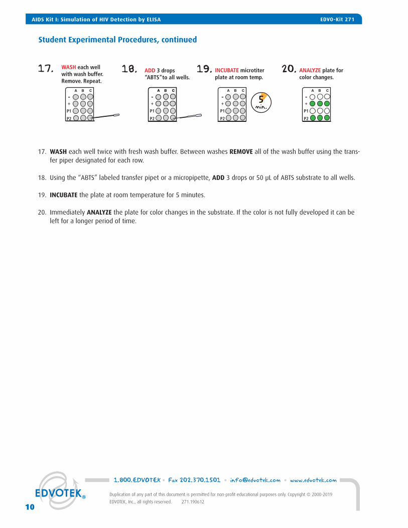
17. WASH each well twice with fresh wash buffer. Between washes REMOVE all of the wash buffer using the trans-
fer piper designated for each row.
18. Using the “ABTS” labeled transfer pipet or a micropipette, ADD 3 drops or 50 µL of ABTS substrate to all wells.
19. INCUBATE the plate at room temperature for 5 minutes.
20. Immediately ANALYZE the plate for color changes in the substrate. If the color is not fully developed it can be left for a longer period of time.
Student Experimental Procedures, continued
1.800.EDVOTEK • Fax 202.370.1501 • [email protected] • www.edvotek.com
10
Duplication of any part of this document is permitted for non-profit educational purposes only. Copyright © 2000-2019
EDVOTEK, Inc., all rights reserved. 271.190612
AIDS Kit I: Simulation of HIV Detection by ELISA EDVO-Kit 271
Study Questions
1. Describe the mechanism of ELISA. Why is ELISA so sensitive? Why is it necessary to block unoccupied binding sites in the microtiter wells? Why is it important to have a positive control?
2. Why is anti-HIV-1 antibody screened instead of the virus itself?
3. Why are there so many immunological variants of HIV?
4. The elimination of several steps in the ELISA could be accomplished if the primary antibody was made into an enzyme conjugate. Why is this generally not done? What can cause a false positive in an ELISA?
AIDS Kit I: Simulation of HIV Detection by ELISAEDVO-Kit 271
11
1.800.EDVOTEK • Fax 202.370.1501 • [email protected] • www.edvotek.com
Duplication of any part of this document is permitted for non-profit educational purposes only. Copyright © 2000-2019
EDVOTEK, Inc., all rights reserved. 271.190612
AIDS Kit I: Simulation of HIV Detection by ELISAEDVO-Kit 271

Instructor's Guide
OVERVIEW OF INSTRUCTOR’S PRELAB PREPARATIONS:
Some of the components can be prepared ahead of time, aliquoted, and stored in the refrigerator (4° C) until needed. See the table below for information on advanced preparation of reagents.
10X ELISA Wash Buffer (A)
Dilute to 1X solution and aliquot
Anytime before the experiment. Cover and store in the refrigerator.
Component: What to do: When:
ELISA Dilution Buffer (B)
Aliquot for negativecontrol and patientsamples
Anytime before the experiment. Store tubes in the refrigerator.
Whey Antigen (C) Rehydrate and aliquotUp to one week before performing the experiment.
Anti-Whey Primary Antibody (D)
Rehydrate and aliquotUp to one week before performing the experiment.
Secondary Antibody (E)
Rehydrate and aliquot Up to one day before performing the experiment.
ABTS Substrate (F) Rehydrate and aliquot Up to one week before performing the experiment.
Red = Prepare immediately before module. Yellow = Prepare shortly before module. Green = Flexible / prepare up to a week before the module.
1.800.EDVOTEK • Fax 202.370.1501 • [email protected] • www.edvotek.com
12
Duplication of any part of this document is permitted for non-profit educational purposes only. Copyright © 2000-2019 EDVOTEK,
Inc., all rights reserved. 271.190612
INSTRUCTOR'S GUIDE AIDS Kit I: Simulation of HIV Detection by ELISA EDVO-Kit 271

Pre-Lab Preparations
Preparation of the Wash Buffer
1. Add all of the 10x ELISA Wash Buffer (A) to 180 mL of distilled water and mix well. Label as “Wash Buffer”.2. Dispense 18 mL into small beakers for each lab group.
Preparation of the Antigen
1. Transfer 7 mL of ELISA Dilution Buffer (B) to a 15 mL conical tube. Label the tube “Antigen”.2. Carefully remove the stopper from the vial of lyophilized Antigen (C) and transfer approximately 0.5 mL of the
ELISA Dilution Buffer from the tube in step 1. Close the stopper and gently shake the vial to mix.3. Transfer the entire contents of reconstituted Antigen back to the 15 mL tube from step 1. Mix well. 4. Label 10 microcentrifuge tubes "Ag" and dispense 650 µL into each tube.
Preparation of the Controls and Patient Samples
1. Label 10 microcentrifuge tubes "-CTRL" and 10 tubes as "P1". Dispense 200 µL ELISA Dilution Buffer (B) into each tube.
2. Transfer 7 mL of ELISA Dilution Buffer (B) to a 15 mL conical tube. Label the tube “1°AB”.3. Carefully remove the stopper from the vial of lyophilized Primary Antibody (D) and transfer approximately 0.5 mL of the ELISA Dilution Buffer from the tube in step 1. Close the stopper and gently shake the vial to mix.4. Transfer the entire contents of reconstituted Primary Antibody back to the 15 mL tube from step 1. Mix well. 5. Label 10 microcentrifuge tubes "+ CTRL" and 10 tubes as "P2". Dispense 200 µL of the primary antibody into
each tube.
Preparation of the Microtiter Plates
1. As shown in the figure (right), orient the microtiter plate so that the numbers 1-12 are at the top and the letters A-H are on the left.
2. Cut each plate on the dotted lines as shown in the figure. Each piece will contain 3 wells on one axis and 4 wells on the other axis. Each lab group will receive one piece.
A
B
C
D
E
F
G
H
1 2 3 4 5 6 7 8 9 10 11 12
Row 1
Row 2
Row 3
Row 4
Row 1
Row 2
Row 3
Row 4
Cutting lines depicted by dashed lines
13
1.800.EDVOTEK • Fax 202.370.1501 • [email protected] • www.edvotek.com
Duplication of any part of this document is permitted for non-profit educational purposes only. Copyright © 2000-2019
EDVOTEK, Inc., all rights reserved. 271.190612
INSTRUCTOR'S GUIDEEDVO-Kit 271 AIDS Kit I: Simulation of HIV Detection by ELISA

Pre-Lab Preparations
Each Lab Student Group Should Receive:
1 Microtiter plate (3 x 4 well)1 Microcentrifuge tube containing 650 µL Antigen1 Microcentrifuge tube containing 650 µL Secondary Antibody1 Microcentrifuge tube containing 650 µL ABTS1 Microcentrifuge tube containing 200 µL - Control1 Microcentrifuge tube containing 200 µL + Control1 Microcentrifuge tube containing 200 µL P11 Microcentrifuge tube containing 200 µL P28 Transfer pipets1 Beaker containing 18 mL Wash Buffer1 Empty beaker for waste
Preparation of Secondary Antibody (NOTE: Prepare on same day as needed
for the experiment.)
1. Transfer 7 mL of ELISA Dilution Buffer (B) to a 15 mL conical tube. Label the tube “2°AB”.
2. Carefully remove the stopper from the vial of lyophilized Secondary Antibody (E) and transfer approximately 0.5 mL of the ELISA Dilution Buffer from the tube in step 1. Close the stopper and gently shake the vial to mix.
3. Transfer the entire contents of reconstituted Secondary Antibody back to the 15 mL tube from step 1. Mix well.
4. Label 10 microcentrifuge tubes "2°AB". Dispense 650 µL per tube.
Preparation of ABTS Substrate
1. Transfer 10 ml ABTS Reaction Buffer (G) into a 15 mL conical tube. Label the tube "ABTS". 2. Carefully remove the stopper from the vial of lyophilized ABTS (F) and transfer approximately 0.5 mL of the
ABTS from the tube in step 1. Close the stopper and gently shake the vial to mix.3. Transfer the entire contents of re-hydrated ABTS to the 15 mL conical tube from step 1. Mix well. 4. Label 10 microcentrifuge tubes “ABTS”. Dispense 650 µL per tube.
1.800.EDVOTEK • Fax 202.370.1501 • [email protected] • www.edvotek.com
14
Duplication of any part of this document is permitted for non-profit educational purposes only. Copyright © 2000-2019
EDVOTEK, Inc., all rights reserved. 271.190612
INSTRUCTOR'S GUIDE AIDS Kit I: Simulation of HIV Detection by ELISA EDVO-Kit 271

The ABTS substrate will change color to dark green in wells con-taining a positive result on the ELISA.
Students should first confirm that the results from the control samples are correct. The wells in the first row (-CTRL) should have no color change, while wells in the second row (+CTRL) should be dark green.
The patient samples should identify Patient 2 as positive for HIV, with all 3 wells showing a dark green color similar to the posi-tive control samples.
- CTRL
+ CTRL
P1
P2
Avoiding Common Pitfalls
1. Students should be advised to be very careful when transferring solutions into and out of the microtiter plate wells.
2. Use only clean or appropriately labeled pipets.3. Do not attempt to empty the microtiter wells by shaking it out. This will often result in contaminating adjacent
wells.4. Wash the wells gently and slowly, without force.
Experiment Results and Analysis
15
1.800.EDVOTEK • Fax 202.370.1501 • [email protected] • www.edvotek.com
Duplication of any part of this document is permitted for non-profit educational purposes only. Copyright © 2000-2019
EDVOTEK, Inc., all rights reserved. 271.190612
INSTRUCTOR'S GUIDEEDVO-Kit 271 AIDS Kit I: Simulation of HIV Detection by ELISA

Please refer to the kit insert for the Answers to
Study Questions