Embed Size (px)
Citation preview
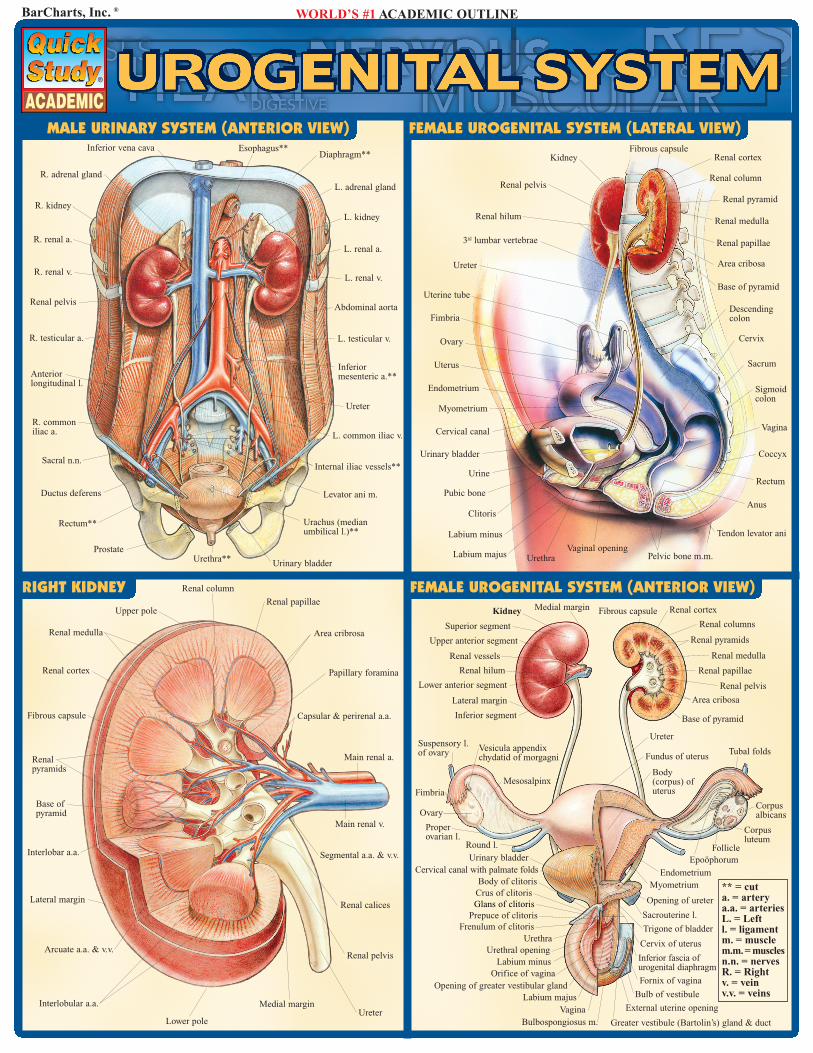
MALE URINARY SYSTEM (ANTERIOR VIEW)
BarCharts, Inc. ® WORLD’S #1 ACADEMIC OUTLINE
Renal medulla
Renal cortex
Fibrous capsule
Renalpyramids
Base ofpyramid
Interlobar a.a.
Lateral margin
Arcuate a.a. & v.v.
Segmental a.a. & v.v.
Main renal a.
Main renal v.
Renal calices
Renal pelvis
Medial marginUreter
Interlobular a.a.
Lower pole
Kidney Medial margin Fibrous capsule Renal cortex
Renal columns
Renal pyramids
Renal medulla
Renal papillae
Renal pelvis
Area cribosa
Base of pyramid
Ureter
Epoöphorum
Tubal foldsFundus of uterus
Body(corpus) ofuterus
Myometrium
Follicle
Corpusluteum
Corpusalbicans
Endometrium
Opening of ureter
Trigone of bladder
Sacrouterine l.
Cervical canal with palmate folds
Cervix of uterus
External uterine opening
Fornix of vagina
Inferior fascia ofurogenital diaphragm
Vagina
Bulb of vestibule
Greater vestibule (Bartolin’s) gland & ductBulbospongiosus m.
Labium majusOpening of greater vestibular gland
Orifice of vaginaLabium minus
Urethral openingUrethra
Frenulum of clitorisPrepuce of clitoris
Body of clitorisCrus of clitoris
Urinary bladderRound l.
Properovarian l.
Ovary
Fimbria
Suspensory l.of ovary Vesicula appendix
chydatid of morgagni
Mesosalpinx
Inferior segment
Lateral margin
Lower anterior segment
Renal hilum
Renal vessels
Upper anterior segment
Superior segment
R. adrenal gland
Urethra**
L. renal v.
L. renal a.
Esophagus**Inferior vena cava
L. kidney
L. adrenal gland
Diaphragm**
Rectum**
Ductus deferens
Sacral n.n.
R. commoniliac a.
Anteriorlongitudinal l.
R. testicular a.
Renal pelvis
R. renal a.
R. kidney
Inferiormesenteric a.**
L. testicular v.
Abdominal aorta
Prostate
Urinary bladder
Urachus (medianumbilical l.)**
Levator ani m.
Internal iliac vessels**
L. common iliac v.
Ureter
Uterine tube
Fimbria
Ovary
Uterus
Endometrium
Myometrium
Cervical canal
Urinary bladder
Urine
Pubic bone
Clitoris
Labium minus
Labium majus UrethraVaginal opening
Pelvic bone m.m.
Tendon levator ani
Anus
Rectum
Coccyx
Vagina
Cervix
Sigmoidcolon
Sacrum
Descendingcolon
R. renal v.Ureter
3rd lumbar vertebrae
Renal pelvis
Renal hilum
KidneyFibrous capsule
Renal cortex
Renal column
Renal pyramid
Renal medulla
Renal papillae
Area cribosa
Base of pyramid
Upper pole
Renal column
Renal papillae
Area cribrosa
Papillary foramina
Capsular & perirenal a.a.
Glans of clitoris
FEMALE UROGENITAL SYSTEM (ANTERIOR VIEW)
** = cuta. = arterya.a. = arteriesL. = Leftl. = ligamentm. = musclem.m.= musclesn.n. = nervesR. = Rightv. = veinv.v. = veins
RIGHT KIDNEY
FEMALE UROGENITAL SYSTEM (LATERAL VIEW)
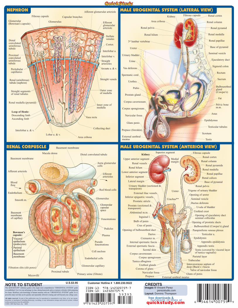
This QUICKSTUDY® reference guide is the single most comprehensive “UROGENITAL SYSTEM” guideever published. Use it to your advantage in class, during homework and as a memory refresher while preparingfor exams. Reinforce your knowledge of human anatomy with our “UROGENITAL SYSTEM” guide. It is apowerful study tool that can be quickly and repeatedly referred to during and well beyond your college years.
All rights reserved. No part of this publication may be reproduced or transmitted in any form, or by any means,electronic or mechanical, including photocopy, recording, or any information storage and retrieval system, withoutwritten permission from the publisher. ©2003, 2005 BarCharts, Inc. 0608
NOTE TO STUDENT Customer Hotline # 1.800.230.9522
ISBN-13: 978-142320759-7ISBN-10: 142320759-9
CREDITSImages ® Vincent Perez
perezstudio.comLayout: Dominic Thompson
NEPHRON
Macula densa
Basement membrane
Distal convoluted tubule
Juxta glomerularcells
Efferentarterioles
Red blood cells
Glomerulus
Fenestrations
Glomerularcapsularspace
Plasma
Pedicles
Pseudofenestrations
Endothelial cells
Cell nucleus
Glomerular capillary
Primary urine (filtrate)Microvilli
Afferent arteriole
Basement membrane
Smooth m.
Basementmembraneof capillary
Filtration slits (slit pores)
Proximal tubule
Endothelium
Visceralepithelium(podocytes)ParietalepitheliumBasementmembrane
Bowman’scapsule:
Bloodflow
Glomerulus
Fibrous capsule Capsular branches
Afferent glomerular arteriole
Efferentglomerulararteriole
Stellatevenules
Glomerular(Bowman’s capsule)
Cortex
Interlobar a.
Interlobar v.
Arcuate a. & v.
Straightarterioles
Straight venule
Distalconvoluteduriniferoustubule
Outer zoneof medulla
Renal medulla (pyramid)Inner zone ofmedulla
Vasa recta
Area cribosaLobar a. & v.
Interlobar a. & v.
Loop of Henle:Descending limbAscending limb
Collecting duct
Proximalconvoluteduriniferoustubule
Renal (uriniferous)tubule (nephron)
Peritubularcapillaries
Straight segmentsof renal tubules
Prostate (sectioned &transparent)
KidneySuperior segment
Upper anterior segment
Renal vessels
Lower anterior segment
Lateral margin
Inferior segment
External iliac vessels
Fibrous capsule
Renal column
Inferior epigastric vessels
Urinary bladder (sectioned &transparent)
Prostatic utricle
Inguinal l.
Bulb of penis
Epididymis
Testicular a.
Renal papillae
Seminal vesicleUrachus**
Opening of ureter
Base of pyramid
Renal medulla
Renal pyramids
Ureter Trigone of urinary bladder
Opening of prostatic ducts
Bulbourethral (Cowper’s) gland
Pampiniform venous plexus
Crus of penis
Dartos
Cremaster m.
Internal spermatic fascia
External spermatic fascia
Scrotal skin
Corona of glans
Prepuce
Medialmargin
Renal cortex
Renal calices
Renal pelvis
Ductus deferens
Urethral crest
Uvula of bladder
Opening of bulbourethral duct
Testis (covered by visceral layerof tunica vaginalis)
Parietal layer
Opening of ejaculatory ductseminal colliculus
Appendix epididymis
Appendix testis
Corpus spongiosum
Urethral glandsTunica albuginea
Corpus cavernosum
Trabeculae
Intercavernous septum ofdeep (Buck’s ) fascia
Valve of navicular fossa
Glans of penisNavicular fossa
External urethral meatus
Abdominal m.m.
Renal hilum
RENAL CORPUSCLE MALE UROGENITAL SYSTEM (ANTERIOR VIEW)
MALE UROGENITAL SYSTEM (LATERAL VIEW)
Urinary bladder
Urine
Urethra
Anus
Sigmoid colon
Sacrum
Ureter
3rd lumbar vertebrae
Renal pelvis
Renal hilum
Kidney Renal cortex
Renal column
Renal pyramid
Renal medulla
Renal papillae
Area cribosa
Base of pyramid
Fibrous capsule
Testis
Scrotum
Testicular tubules
Epididymus
Bulbourethralgland(Cowper’s)
External urethralmeatus
Prepuce (foreskin)
Glans penis
Navicular fossa
Corpus spongiosum
Corpus cavernosum
Pubis
Spermatic cord
Vas deferens
Pelvic bonem.m.
Prostate gland
Ejaculatory duct
Fat
Rectum
Seminal vesicle
U.S.$3.95
free downloads & hundreds of titles at
quickstudy.com