Embed Size (px)
Citation preview
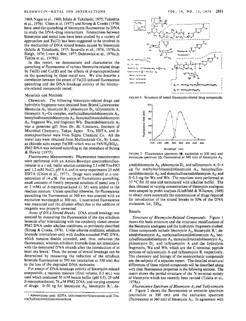
2880 B I O C H E M I S T R Y
Hagen, F. (1979) Anal. Biochem. 93, 299-305. Hartree, E. F. (1972) Anal. Biochem. 48, 422-427. Lacy, E., & Axel, R. (1975) Proc. Natl. Acad. Sci. U.S.A.
Laemmli, U. K. (1971) Nature (London) 227, 680-685. Loening, U. E. (1967) Biochem. J. 102, 251-257. Lutter, L. C. (1978) J. Mol. Biol. 124, 391-420. Lutter, L. C. (1979) Nucleic Acids Res. 6 , 41-56. Maniatis, T., Jeffrey, A., & van de Sande, H. (1975) Bio-
Matsudaira, P. I., & Burgess, D. R. (1978) Anal. Biochem.
Maxam, A. M., & Gilbert, W. (1977) Proc. Natl. Acad. Sci.
Mirzabekov, A. D., Schick, V. V., Belyavisky, A. V., & Bavykin, S. G. (1978) Proc. Natl. Acad. Sci. U.S.A. 75,
Noll, M., & Kornberg, R. D. (1977) J. Mol. Biol. 109,
Oudet, P., Spadafora, C., & Chambon, P. (1978) Cold Spring Harbor Symp. Quant. Biol. 42, 301-312.
Pardon, J. F., Worcester, D. L., Wooley, J. C., Cotter, R. I., Lilley, D. M. J., & Richards, B. M. (1977) Nucleic Acids Res. 4, 3199-3214.
72, 3978-3982.
chemistry 14, 3787-3794.
87, 386-396.
U.S.A. 74, 560-564.
4 1 84-4 1 88.
393-404.
H U A N G , G A L V A N , A N D C R O O K E
Ponder, B. A. J., & Crawford, L. V. (1977) Cell 1 1 , 35-49. Poon, N . H. , & Seligy, V. L. (1978) Exp. Cell. Res. 113,
Roberts, W. K., Dekker, C. A., Rushisky, G. W., & Knight, C. A. (1962) Biochim. Biophys. Acta 55, 664-673.
Shaw, B. R., Herman, T. M., Kovacic, R. T., Beaudreau, G. S., & Van Holde, K. E. (1976) Proc. Natl. Acad. Sci.
95-1 10.
U.S.A. 73, 505-509. Simpson, R. T. (1978) Biochemistry 17, 5524-5531. Simpson, R. T., & Whitlock, J. P., Jr. (1976) Cell 9, 347-353. Sollner-Webb, B., Melchior, W., & Felsenfeld, G. (1978) Cell
Tatchell, K. G., & Van Holde, K. E. (1977) Biochemistry 16,
Tatchell, K. G., & Van Holde, K. E. (1978) Proc. Natl. Acad.
Trifonov, E. N., & Bettecken, T. (1979) Biochemistry 18,
Van Holde, K. E., & Weischet, W. 0. (1978) Biopolymers
Weischet, W. O., Tatchell, K., Van Holde, K. E., & Klump,
Weischet, W. O., Allen, J., Riedel, G., & Van Holde, K. E.
14, 61 1-624.
5295-5303.
Sci. U.S .A. 75, 3583-3587.
454-456.
17, 1387-1403.
H. (1978) Nucleic Acids Res. 5 , 136-160.
(1979) Nucleic Acids Res. (in press).
Quenching of Fluorescence of Bleomycins by Ferrous Ion and Its Correlation with DNA-Breakage Activity? Cheng-Hsiung Huang,* Louis Galvan, and Stanley T. Crooke
ABSTRACT: The quenching of fluorescence of various bleo- mycin-related compounds by Fe(I1) and Cu(I1) was dem- onstrated and used to study DNA-metal ion interactions. The quenching by, both Fe(I1) and Cu(I1) was stoichiometric, sensitive to (ethylenedinitri1o)tetraacetic acid but insensitive to NaCl or urea. Fe(II1) and Mg(I1) failed to induce quenching. The Fe( 11)-induced fluorescence quenching was extensive for drugs shown to be active in DNA breakage: 70-80% for bleomycin A2, deamidobleomycin A2, and bleo- mycin B1 and 50-60% for tallysomycin A and phleomycin D,. Quenching kinetics of these drugs showed a rapid phase followed by a slow phase. Subsequent addition of Cu(I1) caused no further change if maximal quenching by Fe(I1) had been obtained. P-Mercaptoethanol accelerated the slow phase with little change in the extent of maximal quenching. The Fe(I1)-induced quenching was less significant for drugs with less activity in DNA breakage: 20% for the C-terminal peptide fragments Wa and W b and 30% for three bleomycin Az
T h e bleomycins are a family of glycopeptide antibiotics which show various antitumor activities and have been clinically used in chemotherapy of certain human tumors (Blum et al., 1972; Umezawa, 1973-1976; Crooke & Bradner, 1976; Muller &
From the Baylor-Bristol Laboratory, Department of Pharmacology, Baylor College of Medicine, Texas Medical Center, Houston, Texas 77030 (C.-H.H., L.G., and S.T.C.), and Bristol Laboratories, Syracuse, New York 13201 (S.T.C.). Receiued January 11, 1979. This work was supported in part by a grant from Bristol Laboratories and by a grant (CA-10893-10) from the National Cancer Institute.
derivatives with substitutions at a -NH2 of the p-aminoalanine moiety. Subsequently added Cu(I1) readily displaced Fe(I1) from the less active drugs, indicating a weak Fe(I1)-drug interaction. The Cu(I1)-induced quenching varied only slightly with all the tested compounds (30-50%). Subsequent addition of Fe(I1) to the Cu(I1)-quenched drugs caused little change. P-Mercaptoethanol added before, but not after, the addition of Cu(I1) markedly reduced the quenching by Cu(I1). These results indicate a correlation of quenching by Fe(II), but not by Cu(II), with DNA-breakage activity of the drugs, probably relating to the integrity of a - N H 2 at the p-aminoalanine moiety. It is suggested that a proper complex formation of Fe(I1) with active drugs, especially the ligation at the a -NH2 site, may induce specific conformational changes or other chemical events, which can be facilitated by P-mercapto- ethanol, and thus favor the subsequent oxygen ligation and DNA breakage.
Zahn, 1977; Goldberg et al., 1977). The antitumor activity may be related to the ability of bleomycins to cause single- and double-strand breaks of DNA (Suzuki et al., 1969; Haidle, 1971; Muller et al., 1972; Takeshita et al., 1974).
The mechanism of DNA breakage by bleomycins is not yet clearly defined. A variety of effectors have been suggested to be involved in or to affect the DNA-breakage activity of bleomycins. These effectors, among others, include metal ions such as Fe(I1) and Cu(II), reducing agents such as p- mercaptoethanol, and chelators such as EDTA' (Suzuki et al.,
0006-2960/79/0418-2880$01 .OO/O 0 1979 American Chemical Society
B L E O M Y C I N - M E T A L I O N I N T E R A C T I O N S
1969; Nagai et al., 1969; Ishida & Tukahashi, 1975; Takeshita et al., 1976). Chien et al. (1977) and Strong & Crooke (1978) have used the quenching of bleomycin fluorescence by DNA to study the DNA-drug interactions. Interactions between bleomycin and metal ions have been studied by a variety of approaches and Fe(I1) has been suggested to be involved in the mechanism of DNA strand breaks caused by bleomycin (Ishida & Takahashi, 1975; Sausville et al., 1976, 1978a,b; Sleigh, 1976; Lown & Sim, 1977; Dabrowiak et al., 1978a,b; Takita et al., 1978b).
In this report, we demonstrate and characterize the quenching of fluorescence of various bleomycin-related drugs by Fe(I1) and Cu(I1) and the effects of P-mercaptoethanol on the quenching by these metal ions. We also describe a correlation between the extent of Fe(1I)-induced fluorescence quenching and the DNA-breakage activity of the bleomy- cin-related compounds tested.
Materials and Methods Chemicals. The following bleomycin-related drugs and
hydrolytic fragments were obtained from Bristol Laboratories: bleomycin A2, bleomycin B 1', phleomycin Dl, tallysomycin A, bleomycin A2-Cu complex, methylsulfonamidoblemycin A2, benzylsulfonamidobleomycin A2, dansylsulfonamidobleomycin A2, fragment Wa, and fragment Wb. Deamidobleomycin A2 was a generous gift from Dr. H. Umezawa, Institute of Microbial Chemistry, Tokyo, Japan. Tris, EDTA, and 6- mercaptoethanol were from Sigma Chemical Co. All the metal ions were obtained from Mallinckrodt Co., St. Louis, as chloride salts except Fe(II1) which was as FeNH4(S04)2. PM2 DNA was isolated according to the procedure of Strong & Hewitt (1975).
Fluorescence Measurements. Fluorescence measurements were performed with an Amico-Bowman spectrophotofluo- rometer in a 1-mL buffer solution containing 2.5 mM Tris-HC1 and 1.2 mM NaCI, pH 8.4, and in some experiments 25 mM EDTA (Chien et al., 1977). Drugs were studied at a con- centration of 14 pM. For assays of fluorescence quenching, small amounts of freshly prepared solutions of metal ions (1 or 2 mM) or P-mercaptoethanol (1 M) were added to the reaction mixture. Unless specified otherwise, for fluorescence quenching the fluorescence at 360 nm was assayed with an excitation wavelength at 300 nm. Uncorrected fluorescence was measured and the dilution effect due to the addition of reagents was properly corrected.
Assay of DNA Strand Breaks. DNA strand breakage was assayed by measuring the fluorescence of the dye ethidium bromide after intercalating with the covalently closed circular PM2 DNA under alkaline conditions, as previously described (Strong & Crooke, 1978). Under alkaline conditions, ethidium bromide intercalates only with double-stranded PM2 DNA, which remains double stranded, and, thus, enhances the fluorescence, whereas ethidium bromide does not intercalate with the denatured DNA strands after the introduction of a t least one break. Thus, the extent of strand breakage can be determined by measuring the reduction of the ethidium bromide fluorescence at 590 nm (excitation at 530 nm) due to the loss of the degraded DNA molecules.
For assays of DNA-breakage activity of bleomycin-related compounds, a reaction mixture (final volume, 0.5 mL) was used which contained 50 mM borate buffer (pH 9.5), 25 mM P-mercaptoethanol, 76 pM PM2 DNA, and varying amounts of drugs: 0-50 ng for bleomycin AS, bleomycin B1', de-
' Abbreviations used: EDTA, (ethylenedinitri1o)tetraacetic acid; Tris, tris( hydroxymethy1)aminomethane.
V O L . 1 8 , N O . 1 3 , 1 9 7 9 2881
-@
" L U 0
I
I _ _ _ _ _ _ _ _ _ _ _ _ _ _ _ _ _ _ _ _ J ~ W b : R . -NH(CH$,NH(C+,NHI I
FIGURE 1 : Structure of tested bleomycin-related drug compounds.
'"1
2 0 0 240 280 320 360 400 440 480
Wavelength (nm)
FIGURE 2: Fluorescence spectrum (0, excitation at 300 nm) and excitation spectrum (0, fluorescence at 360 nm) of bleomycin A*.
amidobleomycin A2, phleomycin D1, and tallysomycin A; 0-4 pg for methylsulfonamidobleomycin A2, benzylsulfon- amidobleomycin A2, and dansylsulfonamidobleomycin A2; and 0-0.5 mg for Wa and Wb. The reactions were performed at 37 "C for 30 min and terminated with alkaline buffer. The data obtained at varying concentrations of bleomycin analogues were assayed by probit analyses (Litchfield & Wilcoxon, 1949) to obtain more accurately the concentration of drugs required for introduction of the strand breaks in 50% of the DNA molecules, i.e., ED5,.
Results Structure of Bleomycin-Related Compounds. Figure 1
shows the basic structure and the structural modifications of the bleomycin analogues and the hydrolytic fragments studied. These compounds include bleomycin A2, bleomycin B1', de- amidobleomycin A2, methylsulfonamidobleomycin A2, ben- zylsulfonamidobleomycin AZ, dansylsulfonamidoblemcyin A*, phleomycin D1, and tallysomycin A and the hydrolytic fragments, Wa and Wb, which are the C-terminal peptide portions of tallysomycin A and tallysomycin B, respectively. The chemistry and biology of the semisynthetic compounds are the subjects of a separate report. The detailed structural differences of these related compounds will be described along with their fluorescence properties in the following sections. The insert shows the partial structure of the N-terminal moiety of bleomycin which has recently been revised (Takita et al., 1978a).
Fluorescence Spectrum of Bleomycin A2 and Tallysomycin A . Figure 2 shows the fluorescence or emission spectrum (excitation at 300 nm) and the excitation spectrum (fluorescence at 360 nm) of bleomycin A> In agreement with
2882 B l O C H E M I S T R Y H L A N G , G A L V A N , A N D C R O O K E
60 i ’ A
300 340 380 420 460 500
Wavelength (nm)
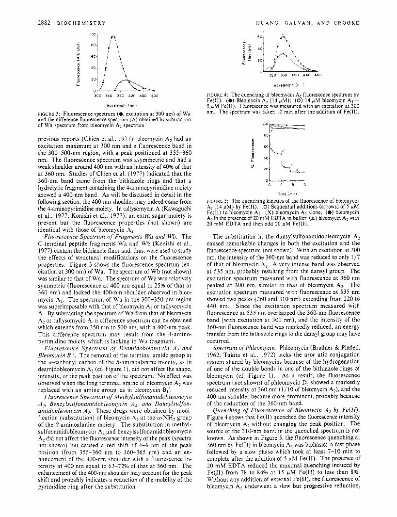
FIGURE 3: Fluorescence spectrum (0, excitation at 300 nm) of Wa and the difference fluorescence spectrum (A) obtained by subtraction of Wa spectrum from bleomycin A2 spectrum.
previous reports (Chien et al., 1977), bleomycin Az had an excitation maximum a t 300 nm and a fluorescence band in the 300-500-nm region, with a peak positioned a t 355-360 nm. The fluorescence spectrum was asymmetric and had a weak shoulder around 400 nm with an intensity of 40% of that a t 360 nm. Studies of Chien et al. (1977) indicated that the 360-nm band came from the bithiazole rings and that a hydrolytic fragment containing the 4-aminopyrimidine moiety showed a 400-nm band. As will be discussed in detail in the following section, the 400-nm shoulder may indeed come from the 4-aminopyrimidine moiety. In tallysomycin A (Kawaguchi et al., 1977; Konishi et al., 1977), an extra sugar moiety is present but the fluorescence properties (not shown) are identical with those of bleomycin A,.
Fluorescence Spectrum of Fragments Wa and Wb. The C-terminal peptide fragments Wa and W b (Konishi et al., 1977) contain the bithiazole fluor and, thus, were used to study the effects of structural modifications on the fluorescence properties. Figure 3 shows the fluorescence spectrum (ex- citation at 300 nm) of Wa. The spectrum of Wb (not shown) was similar to that of Wa. The spectrum of Wa was relatively symmetric (fluorescence a t 400 nm equal to 25% of that a t 360 nm) and lacked the 400-nm shoulder observed in bleo- mycin A,. The spectrum of Wa in the 300-350-nm region was superimposable with that of bleomycin A, or tallysomycin A. By subtracting the spectrum of Wa from that of bleomycin A, or tallysomycin A, a difference spectrum can be obtained which extends from 350 nm to 500 nm, with a 400-nm peak. This difference spectrum may result from the 4-amino- pyrimidine moiety which is lacking in Wa fragment.
Fluorescence Spectrum of Deamidobleomycin A2 and Bleomycin Bl’. The removal of the terminal amido group at the a-carbonyl carbon of the p-aminoalanine moiety, as in deamidobleomycin A, (cf. Figure l ) , did not affect the shape, intensity, or the peak position of the spectrum. No effect was observed when the long terminal amine of bleomycin A, was replaced with an amine group, as in bleomycin BI’.
Fluorescence Spectrum of Methylsulfonamidobleomcyin A] , Benzylsulfonamidobleomycin A2, and Dansylsulfon- amidobleomycin A > . These drugs were obtained by modi- fication (substitution) of bleomycin A, at the a-NH2 group of the 0-aminoalanine moiety. The substitution in methyl- sulfonamidobleomycin A, and benzylsulfonamidobleomycin A, did not affect the fluorescence intensity of the peak (spectra not shown) but caused a red shift of 4-6 nm of the peak position (from 355-360 nm to 360-365 nm) and an en- hancement of the 400-nm shoulder with a fluorescence in- tensity at 400 nm equal to 63-72% of that a t 360 nm. The enhancement of the 400-nm shoulder may account for the peak shift and probably indicates a reduction of the mobility of the pyrimidine ring after the substitution.
Wavelength (n )
FIGURE 4: The quenching of bleomycin A2 fluorescence spectrum by Fe(I1). (0) Bleomycin A2 (14 pLM); (0) 14 pM bleomycin A2 + 5 pM Fe(I1). Fluorescence was measured with an excitation at 300 nm. The spectrum was taken 10 min after the addition of Fe(I1).
80
0 4 8 1 2
Time (min)
FIGURE 5 : The quenching kinetics of the fluorescence of bleomycin A2 (14 pM) by Fe(I1). (0) Sequential additions (arrows) of 5 pM Fe(I1) to bleomycin A,; (X) bleomycin A2 alone; (0 ) bleomycin A2 in the presence of 20 mM EDTA in buffer; (A) bleomycin A2 with 20 mM EDTA and then add 20 pM Fe(I1).
The substitution in the dansylsulfonamidobleomycin A, caused remarkable changes in both the excitation and the fluorescence spectrum (not shown). With an excitation at 300 nm, the intensity of the 360-nm band was reduced to only 1/7 of that of bleomycin A,. A very intense band was observed at 535 nm, probably resulting from the dansyl group. The excitation spectrum measured with fluorescence at 360 nm peaked a t 300 nm, similar to that of bleomycin A,. The excitation spectrum measured with fluorescence a t 535 nm showed two peaks (260 and 310 nm) extending from 220 to 440 nm. Since the excitation spectrum measured with fluorescence at 535 nm overlapped the 360-nm fluorescence band (with excitation at 300 nm), and the intensity of the 360-nm fluorescence band was markedly reduced, an energy transfer from the bithiazole rings to the dansyl group may have occurred.
Spectrum of Phleomycin. Phleomycin (Bradner & Pindell, 1962; Takita et al., 1972) lacks the aror atic conjugation system shared by bleomycins because of the hydrogenation of one of the double bonds in one of the bithiazole rings of bleomycin (cf. Figure 1). As a result, the fluorescence spectrum (not shown) of phleomycin D1 showed a markedly reduced intensity at 360 nm (1 / 10 of bleomycin A2), and the 400-nm shoulder became more prominent, probably because of the reduction of the 360-nm band.
Quenching of Fluorescence of Bleomycin A2 by Fe(II). Figure 4 shows that Fe(I1) quenched the fluorescence intensity of bleomycin A2 without changing the peak position. The source of the 3 10-nm band in the quenched spectrum is not known. As shown in Figure 5, the fluorescence quenching at 360 nm by Fe(I1) in bleomycin A, was biphasic: a fast phase followed by a slow phase which took a t least 7-10 min to complete after the addition of 5 pM Fe(I1). The presence of 20 m M EDTA reduced the maximal quenching induced by Fe(I1) from 78 to 84% at 15 pM Fe(I1) to less than 8%. Without any addition of external Fe(II), the fluorescence of bleomycin A2 underwent a slow but progressive reduction,
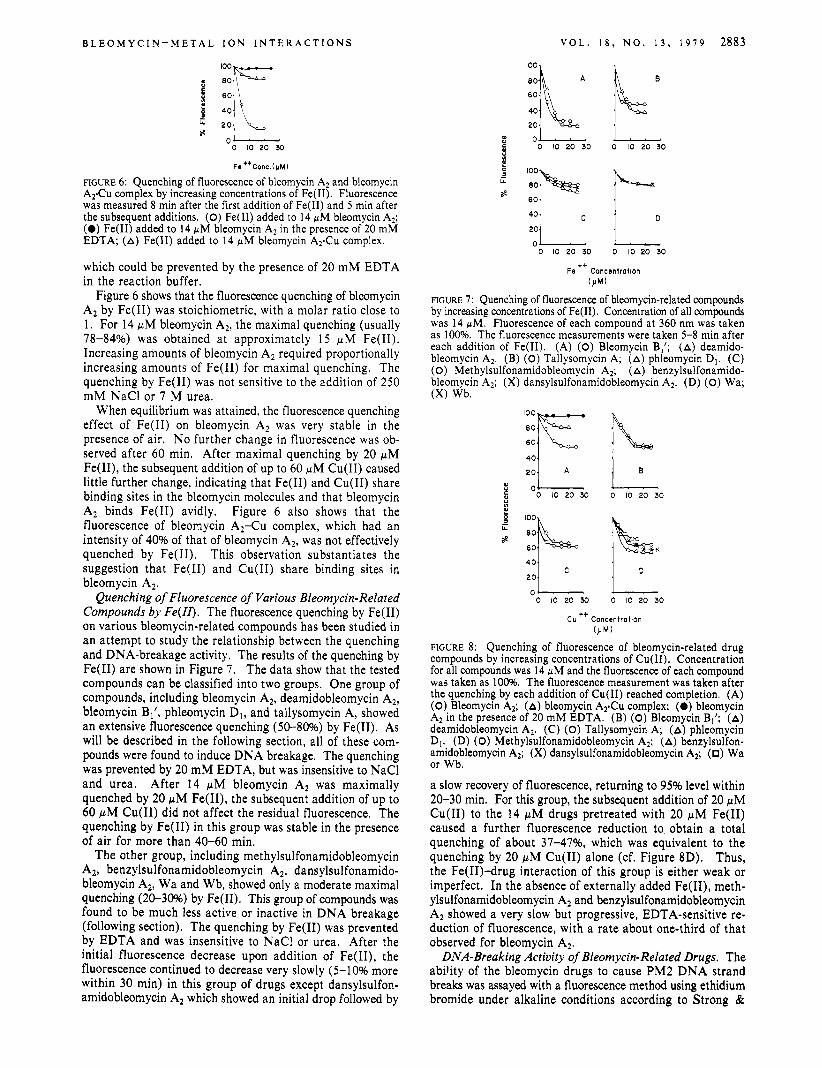
B L E O M Y C I N - M E T A L I O N I N T E R A C T I O N S
3 LL z IZE 20 :.I 0
0 10 20 30
Fe ttConc.(pMl
FIGURE 6: Quenching of fluorescence of bleomycin A2 and bleomycin A2Cu complex by increasing concentrations of Fe( 11). Fluorescence was measured 8 min after the first addition of Fe(I1) and 5 min after the subsequent additions. (0) Fe(I1) added to 14 pM bleomycin A?; (0) Fe(I1) added to 14 pM bleomycin A2 in the presence of 20 mM EDTA; (A) Fe(I1) added to 14 pM bleomycin A 2 C u complex.
which could be prevented by the presence of 20 m M EDTA in the reaction buffer.
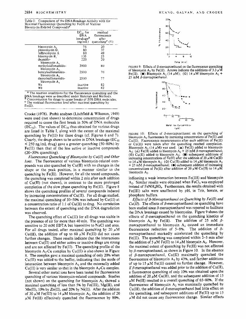
Figure 6 shows that the fluorescence quenching of bleomycin A2 by Fe(I1) was stoichiometric, with a molar ratio close to 1. For 14 p M bleomycin A2, the maximal quenching (usually 78-84%) was obtained a t approximately 15 p M Fe(I1). Increasing amounts of bleomycin A2 required proportionally increasing amounts of Fe(I1) for maximal quenching. The quenching by Fe(I1) was not sensitive to the addition of 250 m M NaCl or 7 M urea.
When equilibrium was attained, the fluorescence quenching effect of Fe(I1) on bleomycin A2 was very stable in the presence of air. No further change in fluorescence was ob- served after 60 min. After maximal quenching by 20 pM Fe(II), the subsequent addition of up to 60 pM Cu(I1) caused little further change, indicating that Fe(1I) and Cu(I1) share binding sites in the bleomycin molecules and that bleomycin A2 binds Fe(I1) avidly. Figure 6 also shows that the fluorescence of bleonycin A2-Cu complex, which had an intensity of 40% of that of bleomycin A*, was not effectively quenched by Fe(I1). This observation substantiates the suggestion that Fe(I1) and Cu(I1) share binding sites in bleomycin AZ.
Quenching of Fluorescence of Various Bleomycin-Related Compounds by Fe(1Z). The fluorescence quenching by Fe(I1) on various bleomycin-related compounds has been studied in an attempt to study the relationship between the quenching and DNA-breakage activity. The results of the quenching by Fe(I1) are shown in Figure 7. The data show that the tested compounds can be classified into two groups. One group of compounds, including bleomycin A2, deamidobleomycin A2, bleomycin B1’, phleomycin D,, and tallysomycin A, showed an extensive fluorescence quenching (SO-80%) by Fe(I1). As will be described in the following section, all of these com- pounds were found to induce DNA breakage. The quenching was prevented by 20 m M EDTA, but was insensitive to NaCl and urea. After 14 p M bleomycin A2 was maximally quenched by 20 p M Fe(II), the subsequent addition of up to 60 pM Cu(I1) did not affect the residual fluorescence. The quenching by Fe(I1) in this group was stable in the presence of air for more than 40-60 min.
The other group, including methylsulfonamidobleomycin Al, benzylsulfonamidobleomycin A*, dansylsulfonamido- bleomycin A2, Wa and Wb, showed only a moderate maximal quenching (20-30%) by Fe(I1). This group of compounds was found to be much less active or inactive in DNA breakage (following section). The quenching by Fe(I1) was prevented by EDTA and was insensitive to NaCl or urea. After the initial fluorescence decrease upon addition of Fe(II), the fluorescence continued to decrease very slowly (5-1 0% more within 30 min) in this group of drugs except dansylsulfon- amidobleomycin A2 which showed an initial drop followed by
V O L . 1 8 , N O . 1 3 , 1979 2883
B
5 100
80
60
G
s ::I , , c, 1 , ,
0 0 10 20 30 0 10 20 30
Fe ’+ Concentration (pM1
FIGURE 7: Quenching of fluorescence of bleomycin-related compounds by increasing concentrations of Fe(I1). Concentration of all compounds was 14 pM. Fluorescence of each compound at 360 nm was taken as 100%. The fluorescence measurements were taken 5-8 min after each addition of Fe(I1). (A) (0) Bleomycin B,’; (A) deamido- bleomycin A2, (B) (0) Tallysomycin A; (A) phleomycin D1. (C) (0) Methylsulfonamidobleomycin A2; (A) benzylsulfonamido- bleomycin A2; (X) dansylsulfonamidobleomycin A2. (D) (0) Wa; (X) Wb.
q , A 1 , 6, 8 0 5 0 10 20 30 0 10 20 30 Ln a,
d 60 x
401 I
0- - 0 10 20 30 0 10 20 30
cu ++ concentration OJM)
FIGURE 8: Quenching of fluorescence of bleomycin-related drug compounds by increasing concentrations of Cu(I1). Concentration for all compounds was 14 pM and the fluorescence of each compound was taken as 100%. The fluorescence measurement was taken after the quenching by each addition of Cu(I1) reached completion. (A) (0) Bleomycin A2; (A) bleomycin A2Cu complex; (0) bleomycin A2 in the presence of 20 mM EDTA. (B) (0) Bleomycin Bl’; (A) deamidobleomycin A2. (C) (0) Tallysomycin A; (A) phleomycin D1. (D) (0) Methylsulfonamidobleomycin A2; (A) benzylsulfon- amidobleomycin A2; (X) dansylsulfonamidobleomycin A2; (0) Wa or Wb.
a slow recovery of fluorescence, returning to 95% level within 20-30 min. For this group, the subsequent addition of 20 p M Cu(I1) to the 14 pM drugs pretreated with 20 pM Fe(I1) caused a further fluorescence reduction to obtain a total quenching of about 37-4796, which was equivalent to the quenching by 20 p M Cu(I1) alone (cf. Figure 8D). Thus, the Fe(I1)-drug interaction of this group is either weak or imperfect. In the absence of externally added Fe(II), meth- ylsulfonamidobleomycin A2 and benzylsulfonamidobleomycin A2 showed a very slow but progressive, EDTA-sensitive re- duction of fluorescence, with a rate about one-third of that observed for bleomycin A2,
DNA-Breaking Activity of Bleomycin-Related Drugs. The ability of the bleomycin drugs to cause PM2 DNA strand breaks was assayed with a fluorescence method using ethidium bromide under alkaline conditions according to Strong &
2884 B I O C H E M I S T R Y H U A N G , G A L V A N , A N D C R O O K E ~~ ~
Table I : Comparison of the DNA-Breakage Activity with the Maximal Fluorescence Quenching by Fe(I1) of Various Bleomycin-Related Compoundsa
EC,, for residual DNA fluorescence
breakageb after (ng/mL) quenching' (%)
bleomycin A, 78 20 phleomycin D, 80 37
bleomycin B , 135 25 deamido- 220 20
methylsulfonamido- 3900 80
benz ylsulfonamido- 2500 70
dansylsulfonamido- 25 00 77
Wa inactive 82 wb inactive 82
tallysomycin A 80 49
bleomycin A,
bleomycin A,
bleomycin A,
bleomycin A,
a The reaction conditions for the fluorescence quenching and the FNA breakage were as described under Materials and Methods.
Concentration for drugs to cause breaks of 50% DNA molecules. The residual fluorescence level after maximal quenching by
rem.
Crooke (1978). Probit analyses (Litchfield & Wilcoxon, 1949) were used (not shown) to determine concentration of drugs required to cause the first break in 50% of DNA molecules (EC50). The values of ECSo thus obtained for various drugs are listed in Table I, along with the extent of the maximal quenching by Fe(I1) for these drugs (cf. Figures 6 and 7) . Clearly, the drugs shown to be active in DNA breakage (EC50 < 250 ng/mL drug) gave a greater quenching (50-80%) by Fe(I1) than that of the less active or inactive compounds (20-30% quenching).
Fluorescence Quenching of Bleomycins by Cu(II) and Other Ions. The fluorescence of various bleomycin-related com- pounds was also quenched by Cu(I1) with no changes in the shape or the peak position, in a manner similar to the quenching by Fe(I1). However, for all the tested compounds, the quenching was completed within 2 min after each addition of Cu(I1) (not shown), in contrast to the more prolonged completion of the slow phase quenching by Fe(I1). Figure 8 shows the quenching profiles of several compounds induced by increasing concentrations of Cu(I1). For all drugs studied, the maximal quenching of 3 0 4 0 % was induced by Cu(I1) at a concentration ratio of 1:l of Cu(I1) to drug. No correlation between the extent of quenching and the DNA degradation was observed.
The quenching effect of Cu(I1) for all drugs was stable in the presence of air for more than 40 min. The quenching was sensitive to 20 mM EDTA but insensitive to NaCl and urea. For all drugs tested, after maximal quenching by 20 pM Cu(II), the addition of up to 60 pM Fe(I1) did not cause further changes. These results indicate that the interactions between Cu(I1) and either active or inactive drugs are strong and are not affected by Fe(I1). The quenching profile of the bleomycin A 2 C u complex by Cu(I1) is also shown in Figure 8. The complex gave a maximal quenching of only 20% when Cu(I1) was added to the buffer, indicating that the mode of interaction between bleomycin A2 and the externally added Cu(I1) is very similar to that in the bleomycin AyCu complex.
Several other metal ions have been tested for fluorescence quenching of various bleomycin-related compounds. Studies (not shown) on the quenching for bleomycin A2 showed a maximal quenching of less than 5% by Fe(III), Mg(II), and Mn(II), 10% by Zn(II), and 20% by Ni(I1). After the additioq of 30 pM Fe(II1) to 14 pM bleomycin A2, the addition of 20 pM Fe(I1) effectively quenched the fluorescence by 80%,
0- 0 4 8 1 2
Time (min )
FIGURE 9: Effects of 0-mercaptoethanol on the fluorescence quenching of bleomycin A2 by Fe(I1). Arrows indicate the additions of 3.3 pM Fe(I1). (0) Blepmycin A2 (14 pM); (0) 14 fiM bleomycin A2 + 25 mM 0-mercaptoethanol.
t 1 \h b
Ok--i??O - 0 IO PO
Metal Ion Concentration (J IM)
FIGURE 10: Effects of 0-mercaptoethanol on the quenching of bleomycin A2 fluorescence by increasing concentrations of Fe(I1) and Cu(I1). Fluorescence measurements after each addition of Fe(I1) or Cu(I1) were taken after the quenching reached completion. Bleomycin A2 (14 fiM) was used. (A) Fe(I1) added to bleomycin A,; (A) Fe(I1) added to bleomycin A2 + 25 mM 0-mercaptoethanol; (0) Cu(I1) added to bleomycin AZ; (W) subsequent additions of increasing concentrations of Fe(I1) after the addition of 20 WM Cu(I1) to 14 fiM bleomycin A2; (0) Cu(I1) added to 14 pM bleomycin A2 + 25 mM 0-mercaptoethanol; (0) subsequent addition of increasing concentrations of Fe(I1) after addition of 20 pM Cu(I1) to 14 pM bleomycin A2
indicating a weak interaction between Fe(II1) and bleomycin A2. Similar results were obtained when FeCl, was employed instead of FeNH,S04. Furthermore, the results obtained with Fe(II1) salts were unaffected by pH, in Tris, borate, or phosphate buffers.
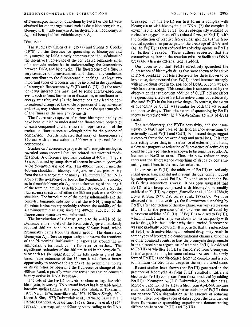
Effects of 0-Mercaptoethanol on Quenching by Fe(II) and Cu(II). The effects of P-mercaptoethanol on quenching have been studied since 0-mercaptoethanol was reported to facilitate the DNA breakage caused by bleomycins. Figure 9 shows the effects of P-mercaptoethanol on the quenching kinetics of bleomycin A2 by Fe(I1). The addition of 25 mM P- mercaptoethanol to bleomycin A2 caused an immediate fluorescence reduction of 5 4 % . The addition of P- mercaptoethanol markedly accelerated the quenching by Fe(I1). The quenching was completed within 2-3 min after the addition of 5 pM Fe(I1) to 14 pM bleomycin A*. However, the maximal extent of quenching by Fe(I1) was not affected by P-mercaptoethanol, as shown in Figure 10. In the absence of 0-mercaptoethanol, Cu(I1) maximally quenched the fluorescence of bleomycin A2 by 43%, and further additions of up to 15 pM Fe(I1) caused no further change. However, if P-mercaptoethanol was added prior to the addition of Cu(II), a fluorescence quenching of only 10% was obtained upon the addition of 20 pM Cu(II), and the subsequent addition of 15 pM Fe(I1) resulted in a overall quenching of 65-8096. If the fluorescence of bleomycin A2 was maximally quenched by Cu(II), the addition of P-mercaptoethanol had little effect on the quenching, and subsequent additions of Fe(I1) up to 15 pM did not cause any fluorescence change. Similar effects
B L E O M Y C I N - M E T A L I O N I N T E R A C T I O N S V O L . 1 8 , N O . 1 3 , 1 9 7 9 2885
breakage: (1) the Fe(I1) ion first forms a complex with bleomycin or with bleomycin plus DNA; (2) the complex is oxygen labile, and the Fe(I1) ion is subsequently oxidized by molecular oxygen, or one of its reduced forms, to Fe(III), with the production of reactive free-radical species; (3) the free- radical species then participate in the breakage of DNA; and (4) the Fe(II1) is then reduced by reducing agents to Fe(I1) for further breakage. These authors suggested that the contaminating iron in the reaction mixture facilitates DNA breakage when no external iron is added.
Our observation that Fe(I1) effectively quenched the fluorescence of bleomycin drugs that were shown to be active in DNA breakage, but less effectively for these shown to be less active, demonstrated that Fe(I1) indeed interacts strongly with active drugs even in the absence of DNA, but only weakly with less active drugs. This conclusion is substantiated by the observation that subsequent addition of Cu(I1) did not affect the quenching effects of Fe(I1) in active drugs but effectively displaced Fe(I1) in the less active drugs. In contrast, the extent of quenching by Cu(I1) was similar for both the active and the inactive drugs. Thus, the extent of quenching by Fe(I1) seems to correlate with the DNA-breakage activity of drugs tested.
The stoichiometry, the EDTA sensitivity, and the insen- sitivity to NaCl and urea of the fluorescence quenching by externally added Fe(I1) and Cu(I1) in all tested drugs suggest a complex formation between drugs and metal ions. It is then interesting to see that, in the absence of external metal ions, a slow but progressive reduction of fluorescence of active drugs could be observed which was shown to be sensitive to EDTA, but not to NaCl or urea. Thus, the slow reduction may represent the fluorescence quenching of drugs by contami- nating metal ions in the reaction mixture.
In contrast to Fe(II), the addition of Fe(II1) caused only slight quenching and did not prevent the quenching induced by subsequently added Fe(I1). This indicates that the Fe- (111)-drug interaction is weak. It has been suggested that Fe(II), after being complexed with bleomycin, is readily oxidized to Fe(II1) by oxygen (Sausville et al., 1976, 1978a,b; Lown & Sim, 1977; Dabrowiak et al., 1978a,b). We have observed that, in active drugs, the fluorescence quenching by Fe(II), after completion of the slow phase, was very stable even after 1 h in the presence of air and was not affected by subsequent addition of Cu(I1). If Fe(I1) is oxidized to Fe(III), which, if added externally, was shown to interact poorly with active drugs, it is then unclear why the quenched fluorescence was not gradually recovered. It is possible that the interaction of Fe(I1) with active bleomycin-related drugs may result in some types of irreversible conformational changes, bindings or other chemical events, so that the bleomycin drugs remain in the altered state regardless of whether Fe(I1) is oxidized to Fe(II1) or whether Fe(II1) is dissociated from bleomycin. It is also possible that, for some unknown reasons, the newly formed Fe(II1) is not dissociated from the complex and is able to maintain the bleomycin drugs in the same altered state.
Recent studies have shown that Fe(II1) generated in the presence of bleomycin Az from Fe(I1) resulted in different bleomycin Fe(II1) complexes from those produced by adding Fe(II1) to bleomycin A2 (J. C. Dabrowiak, unpublished data). Moreover, addition of Fe(I1) to a bleomycin A,-DNA mixture enhances DNA degradation, whereas addition of Fe(II1) does not enhance DNA degradation in the absence of reducing agents. Thus, two other types of data support the data derived from fluorescence quenching experiments demonstrating differences between Fe(I1) and Fe(II1).
of P-mercaptoethanol on quenching by Fe(I1) or Cu(I1) were obtained for other drugs tested such as der.midobleomycin AZ, bleomycin B,’, tallysomycin A, methylsulfonamidobleomycin A2, and benzylsulfonamidobleomycin A2.
Discussion The studies by Chien et al. (1977) and Strong & Crooke
(1978) on the fluorescence quenching of bleomycin and tallysomycin by DNA clearly demonstrated the usefulness of the intensive fluorescence of the conjugated bithiazole rings of bleomycin molecules in understanding the interactions between DNA and bleomycin. The fluorescence of a fluor is very sensitive to its environment, and, thus, many conditions can contribute to the fluorescence quenching. At least two important types of processes may be involved in the quenching of bleomycin fluorescence by Fe(1I) and Cu(I1): (1) the metal ion-drug interactions may lead to some energy-absorbing events such as bond formation or dissocation, or nonfluorescent energy transfer; and (2) the interactions may lead to con- formational changes of the whole or portions of drug molecules and, thus, may reduce the mobility and/or the quantum yield of the fluors in the new environments.
The fluorescence spectra of various bleomycin analogues have been studied to understand the fluorescence properties of each compound and to ensure a proper selection of the excitation-fluorescence wavelength pairs for the purpose of comparison. Results indicated that assay of fluorescence at 360 nm with an excitation at 300 nm was optimal for all compounds.
Studies on fluorescence properties of bleomycin analogues revealed some spectral features related to structural modi- fications. A difference spectrum peaking at 400 nm (Figure 3) was obtained by comparison of spectra between tallysomycin A (or bleomycin A,) and Wa. This 400-nm band formed the 400-nm shoulder in bleomycin A2 and resulted presumably from the 4-aminopyrimidine moiety. The removal of the -NHz group at the a-carbonyl carbon of the P-aminoalanine moiety, as in deamidobleomycin Az, or the shortening of the length of the terminal amine, as in bleomycin Bl’, did not affect the fluorescence spectrum of either the 360-nm peak or the 400-nm shoulder. The introduction of the methylsulfonamido and the benzylsulfonamido substitutions at the a -NHz group of the P-aminoalanine moiety probably reduced the mobility of the 4-aminopyrimidine ring since the 400-nm shoulder of the fluorescence spectrum was enhanced.
The introduction of a dansyl group to the a -NHz of the P-aminoalanine moiety of the bleomycin A2 gave a markedly reduced 360-nm band but a strong 535-nm band, which presumably came from the dansyl group. The dansylated bleomycin A2 offers an opportunity to observe the reactions of the N-terminal half-molecule, especially around the P- aminoalanine terminal, by the fluorescence method. The significant reduction of the 360-nm band in phleomycin D1 substantiates the suggestion of the bithiazole origin of this band. The reduction of the 360-nm band offers a better opportunity to observe the actions of the pyrimidine moiety or its vicinities by observing the fluorescence change of the 400-nm band, especially when one recognizes that phleomycin is very active in DNA breakage.
The role of the Fe(I1) ions, either alone or together with bleomycin, in causing DNA strand breaks has been undergoing intensive studies (Rhaese & Freese, 1968; Ishida & Takahashi, 1975; Nunn, 1976; Sausville et al., 1976, 1978a,b; Sleigh, 1976; Lown & Sim, 1977; Dabrowiak et al., 1978a,b; Takita et al., 1978b; D’Andrea & Haseltine, 1978). Sausville et al. (1976, 1978a,b) have proposed the following steps leading to the DNA
2886 B I O C H E M I S T R Y
As will be demonstrated in a following report, the addition of DNA to the mixture of bleomycin A2 and Fe(I1) caused a further fluorescence quenching, but the same extent of quenching was obtained regardless of whether DNA was added before or after the addition of Fe(I1). The resulting fluorescence quenching upon addition of Fe(I1) and DNA to bleomycin A2 was very stable in the presence of air. No return of fluorescence was seen after 30 min. Therefore, the stability of the quenching effect is not affected by the presence of DNA. Further experiments are underway in an attempt to offer a definite explanation for this observation.
Several observations suggest that Fe(I1) and Cu(I1) may share interacting sites of the bleomycin molecules. Both Fe(I1) and Cu(I1) quenched in fluorescence of bleomycins probably by a stoichiometric complex formation. For the active drugs, the quenching by both metal ions was mutually exclusive. For the less active or inactive drugs, Cu(I1) effectively displaced Fe(I1) to give further quenching of fluorescence. The extent of fluorescence quenching in bleomycin A 2 C u complex by externally added Fe(I1) and Cu(I1) was markedly reduced to a similar degree.
Nevertheless, a detailed comparison of the fluorescence quenching by Fe(I1) and Cu(I1) in various bleomycin ana- logues revealed differences between the two metal ions in their interaction with drugs. The extent of quenching by Fe(I1) varies with structural modifications of drugs. The preservation of the extensive quenching by Fe(I1) in bleomycin B,', de- amidobleomycin A2, phleomycin D1, and tallysomycin A and the loss of such an extensive quenching in the drugs with substitution at the a-NH2 of the (3-aminoalanine moiety of bleomycin A2 indicate that one of the most important groups essential for quenching by Fe(I1) is the free a-NH2 group. The integrity of the amide group of the carbonyl carbon of the (3-aminoalanine and the length of the amine terminal of the bleomycin analogues seem to be less important factors in affecting the quenching affect of Fe(I1). It is possible that the modification or substitution of the a-NH2 may account for the reduction of both the quenching by Fe(I1) and the DNA breakage of the active drugs, although it is still unclear whether or not both parameters bear a cause-effect rela- tionship. In contrast to the quenching effect of Fe(II), all the structural modifications of the bleomycin molecules mentioned above, including those a t the a-NH2 group, had little effect on the quenching by Cu(I1). These differences in mode of quenching may a t least partially explain why Cu(1I) fails to act like Fe(I1) to facilitate the DNA breakage.
The stereochemistry of the metal ion-bleomycin complex has recently been studied. Dabrowiak et al. (1978a) have suggested for Cu(I1)-bleomycin A2 a square-planar complex with four coordinates ligated by the a -NH2 of the P- aminoalanine, N( 1) of the 4-aminopyrimidine residue, N ( 1) of the imidazole group and the sugar carbamoyl function. A slightly different complex structure (square-pyramidal ge- ometry) has recently been proposed by Takita et al. (1978b). In this complex structure, the N" and the deprotonated amide nitrogens of histidine, the N ( l ) of the pyrimidine, and the secondary amine serve the square coordination sites. The a-NH2 of the (3-aminoalanine serves as the apical site and the sixth site may be ligated with the carbamoyl group in the Cu(I1) complex and molecular oxygen in the Fe(I1) complex. Since our studies on the fluorescence quenching showed that the a-NH2 of the (3-aminoalanine moiety was critical for quenching by Fe(I1) but not by Cu(II), it is possible that the proper coordination of this site with Fe(I1) may lead to the formation of a favorable conformation or molecular envi-
H U A Y G , G A L V A N , A N D C R O O K E
ronment which may then facilitate the coordination of oxygen (or the occurrence of other essential events) leading eventually to the breakage of DNA strands.
The addition of P-mercaptoethanol greatly enhanced the DNA-breakage activity of bleomycin (Suzuki et al., 1969; Onishi et a]., 1975). One of the proposed mechanisms is that P-mercaptoethanol serves as a reducing agent to regenerate Fe(I1) from Fe(II1) to ensure further breakage of DNA (Sausville et al., 1978a). We found that 0-mercaptoethanol had different effects on quenching of bleomycin fluorescence by Fe(I1) and Cu(I1). This further demonstrates the dif- ferences between the Fe(I1) and Cu(I1) interactions with drugs. The addition of 25 mM 0-mercaptoethanol to bleomycin A2 accelerated the quenching by Fe(I1) with little alteration in the extent of quenching. The (3-mercaptoethanol added after addition of Cu(I1) had little effect on quenching by Cu(I1). However, if added before Cu(II), P-mercaptoethanol reduced the extent of quenching by Cu(I1) to one-fourth that in the absence of P-mercaptoethanol and the subsequent addition of Fe(I1) showed an effective quenching. For this observation, there may be a t least two possible explanations: (1) Cu(I1) may be reduced by (3-mercaptoethanol to Cu(1) which shows no quenching effect and does not interfere with the subsequent quenching by Fe(I1); or ( 2 ) Cu(1I) may fail to interact with bleomycin in the presence of (3-mercaptoethanol, The former explanation may be unlikely since (5'-mercaptoethanol added after the addition of Cu(I1) showed no effect on quenching, unless the reduction of Cu(I1) fails to occur under this condition. It is also possible that P-mercaptoethanol may cause certain types of conformational changes or other chemical events in the drug molecules so as to facilitate the interaction with Fe(I1) but to make the interaction with Cu(I1) unfa- vorable. Once the Cu(I1) ion has interacted with the drugs, the P-mercaptoethanol becomes ineffective.
The observation that both Fe(I1) and Cu(I1) effectively quench the fluorescence of bleomycin-related drugs in a specific, stoichiometric manner, probably through complex formation, should provide a rapid, sensitive, and useful ap- proach to study the role of these metal ions in the DNA breakage by bleomycins and the reaction mechanisms involved. The correlation between the extent of quenching by Fe(I1) and the DNA-breakage activity, if found to be generally true after testing more bleomycin-related drugs with a wider variety of structural modifications, can be used as a simple and rapid method of screening the activity of new bleomycin-related drugs.
Acknowledgments The authors wish to thank Professor Harris Busch, De-
partment of Pharmacology, Baylor College of Medicine, for his encouragement and guidance. We also wish to thank Dr. A. W. Prestayko for helpful suggestions and Linda Whiteman for excellent typographical assistance.
References Blum, R. H., Carter, S. K., & Agre, K. A. (1972) Cancer 31,
903. Bradner, W. T., & Pindell, M. H . (1962) Nature (London)
196, 682 . Chien, M., Grollman, A. P., & Horwitz, S. B. (1977) Bio-
chemistry 16, 3641. Crooke, S. T., & Bradner, W. T. (1976) J . Med. (Basel) 7 ,
333. Dabrowiak, J. C., Greenaway, F. T., Longo, W . E., Van
Husen, M., & Crooke, S. T. (1978a) Biochim. Biophys. Acta 517, 517.
C O N F O R M A T I O N A L C H A N G E O F T R N A ~ ~ ~
Dabrowiak, J. C., Greenaway, F. T., & Grulich, R. (1978b) Biochemistry 17, 4090.
D’Andrea, A. D., & Haseltine, W. A. (1978) Proc. Natl. Acad. Sci. U.S.A. 75, 3608.
Goldberg, I. H., Beerman, T. A., & Poon, R. (1977) in Cancer (Becker, F. F., Ed,) Vol. 5 , p 427, Plenum Press, New York.
Haidle, C. W. (1971) Mol. Pharmacol. 7 , 645. Ishida, R., & Takahashi, T. (1975) Biochem. Biophys. Res.
Commun. 66, 1432. Kawaguichi, H., Tsukiura, H., Tomita, K., Konishi, M., Saito,
K., Kobaru, S . , Numata, K. I., Fujisawa, K. I., Miyaki, T., Hatori, M., & Koshiyama, H. (1977) J . Antibiot. 30, 779.
Konishi, M., Saito, K. I., Numata, K. I., Tsuno, T., Asama, K., Tsukiura, H., Naito, T., & Kawaguchi, H. (1977) J . Antibiot. 30, 789.
Litchfield, J. T., & Wilcoxon, F. (1949) J . Pharmacol. Exp. Ther. 96, 99.
Lown, J. W., & Sim, S . K. (1977) Biochem. Biophys. Res. Commun. 77, 11 50.
Muller, W. E. G., & Zahn, R. K. (1977) Prog. Nucleic Acid Res. Mol. Biol. 20, 21.
Muller, W. E. G., Yamazaki, Z., Breter, H. J., & Zahn, R. K. (1972) Eur. J . Biochem. 31, 518.
Nagai, K., Yamaki, H., Suzuki, H., Tanaka, N., & Umezawa, H. (1969) Biochim. Biophys. Acta 179, 165.
Nunn, A. D. (1976) J . Antibiot. 29, 1102. Onishi, T., Iwata, H., & Takagi, Y. (1975) J . Antibiot. 77,
Rhaese, H., & Freese, E. (1968) Biochim. Biophys. Acta 155, 745.
476.
V O L . 1 8 , NO. 1 3 , 1 9 7 9 2887
Sausville, E. A., Peisach, J., & Horwitz, S . B. (1976) Biochem.
Sausville, E. A., Peisach, J., & Horwitz, S . B. (1978a)
Sausville, E. A., Stein, R. W., Peisach, J., & Horwitz, S . B.
Sleigh, M. J. (1976) Nucleic Acids Res. 3, 891. Strong, J. E., & Hewitt, R. R. (1975) in Isozymes (Market,
C., Ed.) Vol. 111, p 473, Academic Press, New York. Strong, J. E., & Crooke, S . T. (1978) Cancer Res. (in press). Suzuki, H., Nagai, K., Yamaki, H., Tanaka, N., & Umezawa,
H . (1969) J . Antibiot. 22, 446. Takeshita, M., Horwitz, S . B., & Grollman, A. P. (1974)
Virology 60, 455. Takeshita, M., Grollman, A. P., & Horwitz, S . B. (1976)
Virology 69, 453. Takita, T., Muraoka, Y., & Umezawa, H. (1972) J . Antibiot.
25, 210. Takita, T., Muraoka, Y., Nakatani, T., Fujii, A., Umezawa,
Y., Naganawa, H., & Umezawa, H. (1978a) J . Antibiot. 31, 801.
Takita, T., Muraoka, Y., Nakatani, T., Fujii, A., Iitaka, Y., & Umezawa, H. (1978b) J . Antibiot. 31, 1070.
Umezawa, H. (1973) Biomedicine 18, 549. Umezawa, H. (1974) Fed. Proc., Fed. Am. SOC. Exp. Biol.
33, 2296. Umezawa, H . (1975) in Antibiotics (Corcoran, J. W., &
Hahn, F. E., Eds.) Vol. 111, p 21, Springer-Verlag, Berlin. Umezawa, H. (1976) GANN Monogr. Cancer Res. No. 19,
9.
Biophys. Res. Commun. 73, 814.
Biochemistry 17, 2740.
(1978b) Biochemistry 17, 2746.
Phenylalanyl-tRNA Synthetase Induced Conformational Change of Escherichia coli t RNAphet A. Favre,* J. P. Ballini, and E. Holler
ABSTRACT: Binding of Escherichia coli tRNAPhe to phenyl- alanyl tRNA synthetase induces a broadening of the main positive dichroic band (A,,, 265 nm) of the tRNA, resulting in a 5-nm red shift in the range of 260-280 nm. A second effect is to reduce the rate of photo-cross-linking between residues in positions 8 and 13 by a factor of 2.3. These effects are only detected with the cognate active synthetase but not with other proteins including the methionyl-tRNA synthetase. Formation of the tRNA enzyme complex is required since both effects are lowered by the addition of monovalent cations which are known to decrease the association constant of tRNAs for their synthetases or on addition of a competitor tRNA such as the tRNAPhe from yeast. Detailed analysis of the stoi- chiometry of “protection” against cross-linking revealed that only the strong and not the weak tRNA binding site on the enzyme participates in the tRNA-enzyme rearrangement. The
F a s t kinetics studies of different tRNA, aminoacyl-tRNA synthetase systems, such as the serine system from yeast, the
From the Institut de Recherches en Biologie Molbculaire, Paris, 75005 France (A.F.), the Universitat Regensburg, 84 Regensburg, Federal Republic of Germany (E.H.), and the Institut du Radium, Paris, 75005 France (J.P.B.) Received Junuury 25, 1979. This work was supported by a NATO grant to E. Holler and A. Favre.
0006-2960/79/0418-2887$01 .OO/O
cooperative binding of two magnesium ions to the tRNA- enzyme complex is required for the rearrangement to occur. In a very similar way, the catalytic phenylalanylation depends upon Mg2+. It is likely that the rearrangement is involved in the mechanism of acylation of tRNAPhe. The molecular nature of the rearrangement, as far as tRNAPhc is concerned, was deduced from the photochemical and spectroscopic properties of the tRNA, including the 4-thiouridine luminescence. We propose that the helical structure of tRNAPhe is slightly altered in the course of the rearrangement of the enzyme-tRNA complex. The protection effect against the light-induced cross-linking observed when the enzyme-tRNAPhe complex is formed is shown to be due to a decreased probability for the 8 and 13 residues to assume a highly reactive confor- mational state necessary for the 8-1 3 link formation.
tyrosine system from E. coli (Riesner et al., 1976), and the phenylalanine system from yeast (Krauss et al., 1976; Rigler et al., 1976), have shown that tRNA binding to its cognate ligase proceeds in two steps. The first one, the recombination step, is in first approximation diffusion controlled. The second step involves a rearrangement of the synthetase-tRNA complex and is assumed to be responsible for the recognition
0 1979 American Chemical Society