Embed Size (px)
Citation preview
Abstract— There is no underestimating the importance of modern imaging to the improved detection and management of diseases such as cancer. Ultrasound offers a cost-effective and safe modern imaging modality. A quantitative approach, termed quantitative ultrasound (QUS), offers the capability to examine the anatomic microstructure of tissue, hence opening up opportunities to quantify/diagnose such microstructure. One approach to improve specificity with QUS techniques, a model-based approach, is to develop ultrasonic scattering models that match the anatomic geometry of the tissue type under investigation. To do so, an approach from simple (individual cells) to moderate complexity (groupings of cells imbedded in a supportive structure) to significant complexity (actual tissue/tumors) has merit, especially if the degrees of complexity are with the same cell type. Therefore, an approach for improved imaging capabilities with quantitative ultrasound is that from single cells to biophantoms to tumors, and is discussed herein.
I. INTRODUCTION
There is no underestimating the importance of modern imaging to the improved detection and management of diseases such as cancer. Imaging has played a pivotal role in improving the prognoses for breast cancer. However, these imaging advances have not resulted in a flawless imaging technique and these advances have not come without negative tradeoffs. The advances in imaging have resulted in higher sensitivity to cancer but without significantly increasing specificity. Because these imaging techniques do not offer higher specificity, this has produced the so-called “overdiagnosis” crisis in medical imaging [1, 2]. This overdiagnosis problem has resulted in finding more cancers but the tradeoff is that many more biopsies are conducted with a negative finding for cancer. Improving the specificity will improve overall diagnostic accuracy, translate into a considerable savings in cost and time of physicians and
*Research supported by NIH R01CA111289. W. D. O’Brien, Jr. is the Director of the Bioacoustics Research
Laboratory, is the Donald Biggar Willett Professor of Engineering and is with the Department of Electrical and Computer Engineering at the University of Illinois, 405 N. Mathews, Urbana, IL 61801 USA. (phone: 217-333-2407; e-mail: [email protected]).
A. Han is with the Department of Electrical and Computer Engineering at the University of Illinois, Urbana, IL 61801 USA. (e-mail: [email protected]).
T. Auger is with the l'Ecole Centrale de Lille, Lille, France. (e-mail: [email protected]).
pathologists, and ease the anxiety of patients undergoing these procedures. The main advantage of using quantitative imaging techniques (like quantitative ultrasound, QUS) is the ability to significantly improve specificity. QUS imaging represents one modality with a track record of success for uniquely identifying and classifying disease [3, 4]. Therefore, developing QUS techniques for improving cancer diagnosis, especially the specificity, is medically significant. One approach to improve specificity with QUS techniques, a model-based approach, is to develop ultrasonic scattering models that match the anatomic geometry of the tissue type under investigation. To do so, an approach from simple (individual cells) to moderate complexity (groupings of cells imbedded in a supportive structure) to significant complexity (actual tissue/tumors) has merit, especially if the degrees of complexity are with the same cell type. Therefore, an approach for improved imaging capabilities with quantitative ultrasound is that from single cells to biophantoms to tumors, and is discussed herein.
II. SINGLE CELL QUS
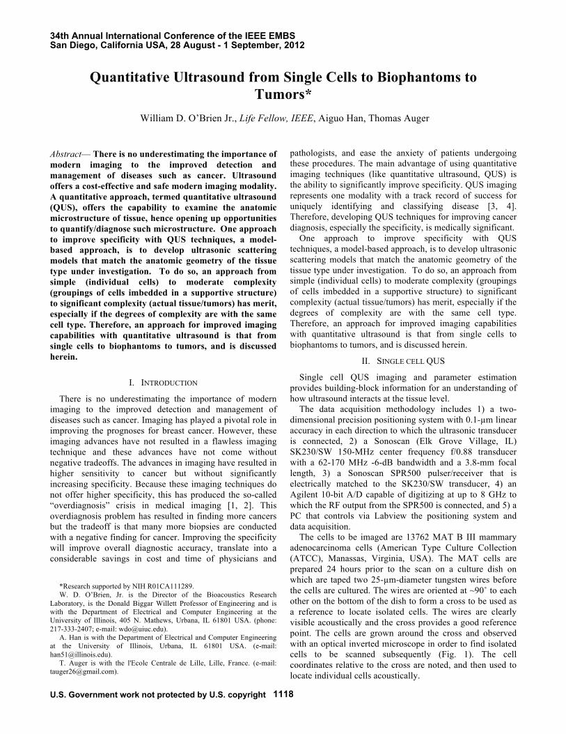
Single cell QUS imaging and parameter estimation provides building-block information for an understanding of how ultrasound interacts at the tissue level. The data acquisition methodology includes 1) a two-dimensional precision positioning system with 0.1-µm linear accuracy in each direction to which the ultrasonic transducer is connected, 2) a Sonoscan (Elk Grove Village, IL) SK230/SW 150-MHz center frequency f/0.88 transducer with a 62-170 MHz -6-dB bandwidth and a 3.8-mm focal length, 3) a Sonoscan SPR500 pulser/receiver that is electrically matched to the SK230/SW transducer, 4) an Agilent 10-bit A/D capable of digitizing at up to 8 GHz to which the RF output from the SPR500 is connected, and 5) a PC that controls via Labview the positioning system and data acquisition. The cells to be imaged are 13762 MAT B III mammary adenocarcinoma cells (American Type Culture Collection (ATCC), Manassas, Virginia, USA). The MAT cells are prepared 24 hours prior to the scan on a culture dish on which are taped two 25-µm-diameter tungsten wires before the cells are cultured. The wires are oriented at ~90˚ to each other on the bottom of the dish to form a cross to be used as a reference to locate isolated cells. The wires are clearly visible acoustically and the cross provides a good reference point. The cells are grown around the cross and observed with an optical inverted microscope in order to find isolated cells to be scanned subsequently (Fig. 1). The cell coordinates relative to the cross are noted, and then used to locate individual cells acoustically.
Quantitative Ultrasound from Single Cells to Biophantoms to Tumors*
William D. O’Brien Jr., Life Fellow, IEEE, Aiguo Han, Thomas Auger
34th Annual International Conference of the IEEE EMBSSan Diego, California USA, 28 August - 1 September, 2012
1118U.S. Government work not protected by U.S. copyright
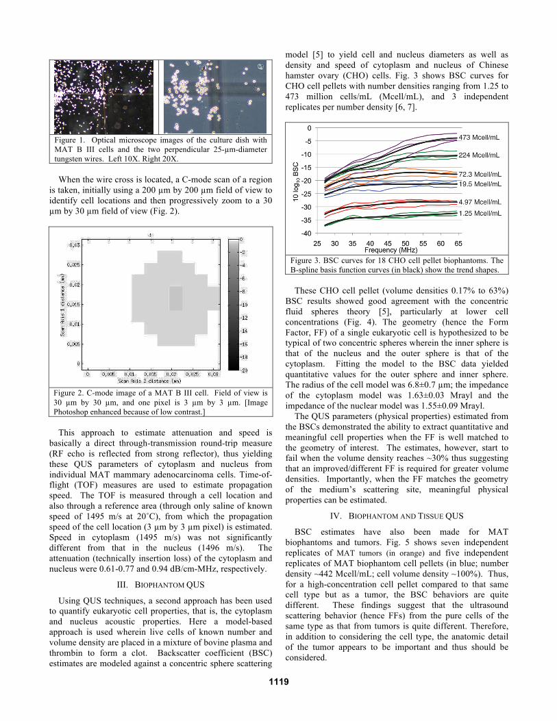
Figure 1. Optical microscope images of the culture dish with MAT B III cells and the two perpendicular 25-µm-diameter tungsten wires. Left 10X. Right 20X.
When the wire cross is located, a C-mode scan of a region is taken, initially using a 200 µm by 200 µm field of view to identify cell locations and then progressively zoom to a 30 µm by 30 µm field of view (Fig. 2).
Figure 2. C-mode image of a MAT B III cell. Field of view is 30 µm by 30 µm, and one pixel is 3 µm by 3 µm. [Image Photoshop enhanced because of low contrast.]
This approach to estimate attenuation and speed is basically a direct through-transmission round-trip measure (RF echo is reflected from strong reflector), thus yielding these QUS parameters of cytoplasm and nucleus from individual MAT mammary adenocarcinoma cells. Time-of-flight (TOF) measures are used to estimate propagation speed. The TOF is measured through a cell location and also through a reference area (through only saline of known speed of 1495 m/s at 20˚C), from which the propagation speed of the cell location (3 µm by 3 µm pixel) is estimated. Speed in cytoplasm (1495 m/s) was not significantly different from that in the nucleus (1496 m/s). The attenuation (technically insertion loss) of the cytoplasm and nucleus were 0.61-0.77 and 0.94 dB/cm-MHz, respectively.
III. BIOPHANTOM QUS
Using QUS techniques, a second approach has been used to quantify eukaryotic cell properties, that is, the cytoplasm and nucleus acoustic properties. Here a model-based approach is used wherein live cells of known number and volume density are placed in a mixture of bovine plasma and thrombin to form a clot. Backscatter coefficient (BSC) estimates are modeled against a concentric sphere scattering
model [5] to yield cell and nucleus diameters as well as density and speed of cytoplasm and nucleus of Chinese hamster ovary (CHO) cells. Fig. 3 shows BSC curves for CHO cell pellets with number densities ranging from 1.25 to 473 million cells/mL (Mcell/mL), and 3 independent replicates per number density [6, 7].
Figure 3. BSC curves for 18 CHO cell pellet biophantoms. The B-spline basis function curves (in black) show the trend shapes.
These CHO cell pellet (volume densities 0.17% to 63%) BSC results showed good agreement with the concentric fluid spheres theory [5], particularly at lower cell concentrations (Fig. 4). The geometry (hence the Form Factor, FF) of a single eukaryotic cell is hypothesized to be typical of two concentric spheres wherein the inner sphere is that of the nucleus and the outer sphere is that of the cytoplasm. Fitting the model to the BSC data yielded quantitative values for the outer sphere and inner sphere. The radius of the cell model was 6.8±0.7 µm; the impedance of the cytoplasm model was 1.63±0.03 Mrayl and the impedance of the nuclear model was 1.55±0.09 Mrayl. The QUS parameters (physical properties) estimated from the BSCs demonstrated the ability to extract quantitative and meaningful cell properties when the FF is well matched to the geometry of interest. The estimates, however, start to fail when the volume density reaches ~30% thus suggesting that an improved/different FF is required for greater volume densities. Importantly, when the FF matches the geometry of the medium’s scattering site, meaningful physical properties can be estimated.
IV. BIOPHANTOM AND TISSUE QUS
BSC estimates have also been made for MAT biophantoms and tumors. Fig. 5 shows seven independent replicates of MAT tumors (in orange) and five independent replicates of MAT biophantom cell pellets (in blue; number density ~442 Mcell/mL; cell volume density ~100%). Thus, for a high-concentration cell pellet compared to that same cell type but as a tumor, the BSC behaviors are quite different. These findings suggest that the ultrasound scattering behavior (hence FFs) from the pure cells of the same type as that from tumors is quite different. Therefore, in addition to considering the cell type, the anatomic detail of the tumor appears to be important and thus should be considered.
1119
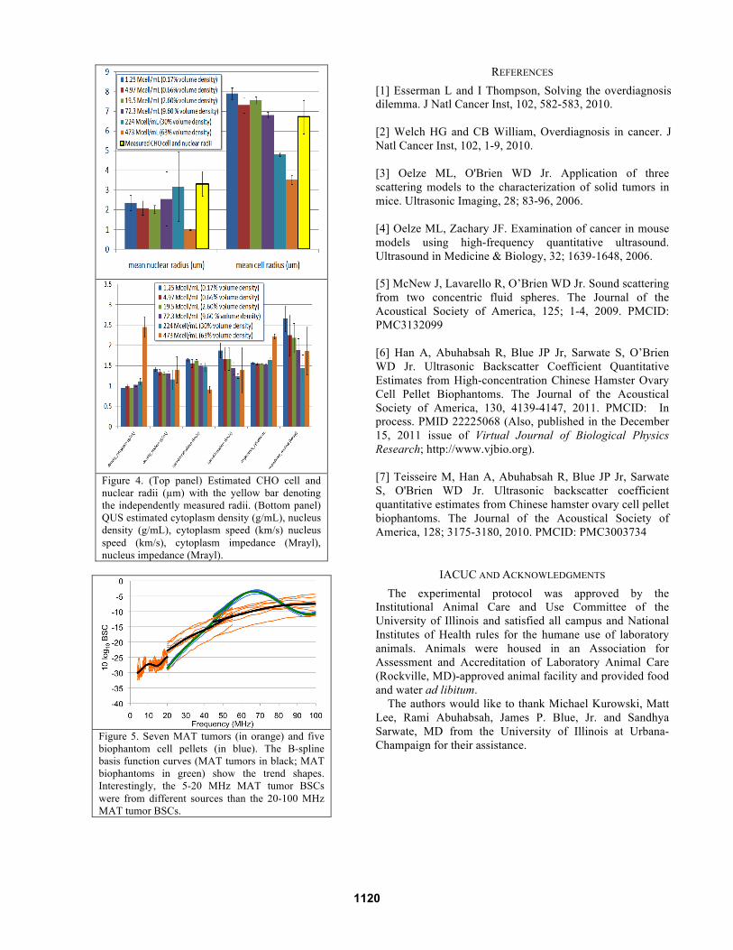
Figure 4. (Top panel) Estimated CHO cell and nuclear radii (µm) with the yellow bar denoting the independently measured radii. (Bottom panel) QUS estimated cytoplasm density (g/mL), nucleus density (g/mL), cytoplasm speed (km/s) nucleus speed (km/s), cytoplasm impedance (Mrayl), nucleus impedance (Mrayl).
Figure 5. Seven MAT tumors (in orange) and five biophantom cell pellets (in blue). The B-spline basis function curves (MAT tumors in black; MAT biophantoms in green) show the trend shapes. Interestingly, the 5-20 MHz MAT tumor BSCs were from different sources than the 20-100 MHz MAT tumor BSCs.
REFERENCES [1] Esserman L and I Thompson, Solving the overdiagnosis dilemma. J Natl Cancer Inst, 102, 582-583, 2010. [2] Welch HG and CB William, Overdiagnosis in cancer. J Natl Cancer Inst, 102, 1-9, 2010. [3] Oelze ML, O'Brien WD Jr. Application of three scattering models to the characterization of solid tumors in mice. Ultrasonic Imaging, 28; 83-96, 2006. [4] Oelze ML, Zachary JF. Examination of cancer in mouse models using high-frequency quantitative ultrasound. Ultrasound in Medicine & Biology, 32; 1639-1648, 2006. [5] McNew J, Lavarello R, O’Brien WD Jr. Sound scattering from two concentric fluid spheres. The Journal of the Acoustical Society of America, 125; 1-4, 2009. PMCID: PMC3132099 [6] Han A, Abuhabsah R, Blue JP Jr, Sarwate S, O’Brien WD Jr. Ultrasonic Backscatter Coefficient Quantitative Estimates from High-concentration Chinese Hamster Ovary Cell Pellet Biophantoms. The Journal of the Acoustical Society of America, 130, 4139-4147, 2011. PMCID: In process. PMID 22225068 (Also, published in the December 15, 2011 issue of Virtual Journal of Biological Physics Research; http://www.vjbio.org). [7] Teisseire M, Han A, Abuhabsah R, Blue JP Jr, Sarwate S, O'Brien WD Jr. Ultrasonic backscatter coefficient quantitative estimates from Chinese hamster ovary cell pellet biophantoms. The Journal of the Acoustical Society of America, 128; 3175-3180, 2010. PMCID: PMC3003734
IACUC AND ACKNOWLEDGMENTS The experimental protocol was approved by the Institutional Animal Care and Use Committee of the University of Illinois and satisfied all campus and National Institutes of Health rules for the humane use of laboratory animals. Animals were housed in an Association for Assessment and Accreditation of Laboratory Animal Care (Rockville, MD)-approved animal facility and provided food and water ad libitum. The authors would like to thank Michael Kurowski, Matt Lee, Rami Abuhabsah, James P. Blue, Jr. and Sandhya Sarwate, MD from the University of Illinois at Urbana-Champaign for their assistance.
1120