Embed Size (px)
Citation preview
Zapparoli et al. BMC Cancer 2013, 13:206http://www.biomedcentral.com/1471-2407/13/206
TECHNICAL ADVANCE Open Access
Quantitative threefold allele-specific PCR(QuanTAS-PCR) for highly sensitive JAK2 V617Fmutant allele detectionGiada V Zapparoli1, Robert N Jorissen2,3, Chelsee A Hewitt1, Michelle McBean1, David A Westerman1,4
and Alexander Dobrovic1,4,5,6,7*
Abstract
Background: The JAK2 V617F mutation is the most frequent somatic change in myeloproliferative neoplasms,making it an important tumour-specific marker for diagnostic purposes and for the detection of minimal residualdisease. Sensitive quantitative assays are required for both applications, particularly for the monitoring of minimalresidual disease, which requires not only high sensitivity but also very high specificity.
Methods: We developed a highly sensitive probe-free quantitative mutant-allele detection method, QuantitativeThreefold Allele-Specific PCR (QuanTAS-PCR), that is performed in a closed-tube system, thus eliminating themanipulation of PCR products. QuantTAS-PCR uses a threefold approach to ensure allele-specific amplification ofthe mutant sequence: (i) a mutant allele-specific primer, (ii) a 3′dideoxy blocker to suppress false-positiveamplification from the wild-type template and (iii) a PCR specificity enhancer, also to suppress false-positiveamplification from the wild-type template. Mutant alleles were quantified relative to exon 9 of JAK2.
Results: We showed that the addition of the 3′dideoxy blocker suppressed but did not eliminate false-positiveamplification from the wild-type template. However, the addition of the PCR specificity enhancer near eliminatedfalse-positive amplification from the wild-type allele. Further discrimination between true and false positives wasenabled by using the quantification cycle (Cq) value of a single mutant template as a cut-off point, thus enablingrobust distinction between true and false positives. As 10,000 JAK2 templates were used per replicate, the assay had asensitivity of 1/10-4 per replicate. Greater sensitivity could be reached by increasing the number of replicates analysed.Variation in replicates when low mutant-allele templates were present necessitated the use of a statistics-basedapproach to estimate the load of mutant JAK2 copies. QuanTAS-PCR showed comparable quantitative results whenvalidated against a commercial assay.
Conclusions: QuanTAS-PCR is a simple, cost-efficient, closed-tube method for JAK2 V617F mutation quantification thatcan detect very low levels of the mutant allele, thus enabling analysis of minimal residual disease. The approach canbe extended to the detection of other recurrent single nucleotide somatic changes in cancer.
Keywords: Mutation detection, Myeloproliferative neoplasms, Minimal residual disease, qPCR, Real time PCR,JAK2 V617F, BRAF V600E, EGFR T790M
* Correspondence: [email protected] of Pathology, Peter MacCallum Cancer Centre, St AndrewsPlace, East Melbourne, Victoria 3002, Australia4Division of Cancer Medicine, Peter MacCallum Cancer Centre, St AndrewsPlace, East Melbourne, Victoria 3002, AustraliaFull list of author information is available at the end of the article
© 2013 Zapparoli et al.; licensee BioMed Central Ltd. This is an Open Access article distributed under the terms of the CreativeCommons Attribution License (http://creativecommons.org/licenses/by/2.0), which permits unrestricted use, distribution, andreproduction in any medium, provided the original work is properly cited.
Zapparoli et al. BMC Cancer 2013, 13:206 Page 2 of 13http://www.biomedcentral.com/1471-2407/13/206
BackgroundMyeloproliferative neoplasms (MPN) are clonal hema-topoietic stem cell malignancies comprising several di-verse pathologies. The 2008 World Health Organization(WHO) classification incorporated JAK2 V617F mutationstatus into the diagnosis of BCR-ABL negative MPN [1].This mutation is the most frequent somatic change inMPN, occurring in over 95% of patients with polycy-themia vera (PV) and in 50% of patients with essentialthrombocythemia (ET) or primary myelofibrosis (PMF)[2]. The valine to phenylalanine substitution at amino acid617 causes a disruption of the auto-inhibitory JH2 domainof JAK2, leading to constitutive activation of the JAK2tyrosine kinase activity and a consequent loss of controlin cell proliferation and growth [3,4]. Small-molecule in-hibitors targeting JAK2-driven cancers have recently en-tered clinical trials [5,6].The JAK2 V617F change results from the c.1849G > T
point mutation in exon 14 of the JAK2 gene (COSMICID: COSM12600). Numerous detection methods for theJAK2 c.1849G > T mutation have been published in-cluding, restriction fragment length polymorphism ana-lysis, Sanger sequencing, pyrosequencing, amplificationrefractory mutation system, allele-competitive blockerPCR, and melt curve analysis or high resolution melting(HRM) [7-20].For the detection of minimal residual disease, highly
sensitive quantitative assays are required. Several quanti-tative PCR methods based on mutation-specific primersor probes or LNA-modified oligonucleotides have beendeveloped [21-27]. These range in sensitivity from me-dium (0.1-1% mutant alleles) to high (< 0.1% mutant al-leles). Less sensitive assays (with a detection limit of 1-3%mutant alleles) are useful for disease diagnosis [28].In the minimal residual disease context, the detection
of low levels of the mutation can be challenging, es-pecially due to the occurrence of false positives. Thus,accurate and sensitive V617F testing of treated MPN pa-tient samples needs to be performed using assays thatminimise or at least enable the recognition of false posi-tives. For these purposes, we developed a new highlysensitive assay, based on allele-specific PCR, which isable to specifically and efficiently suppress amplificationof the wild-type allele.The method that we developed, QuanTAS-PCR, is a
probe-free quantitative PCR method based on a three-fold approach to ensure specific amplification of themutant JAK2 allele: (i) the use of allele-specific primers toamplify the mutant allele (sometimes known as allele-specific PCR or ARMS), combined with (ii) the use ofa non-extendible dideoxy oligonucleotide complementaryto the wild-type allele and known as a blocker, and (iii)the use of a PCR specificity enhancer. By combining thesethree measures, we achieved high analytical sensitivity
(one single mutant allele per well) coupled with high ana-lytical specificity, providing a robust quantitative assay.
MethodsPatients and controlsThis study was covered by an approval from the PeterMacCallum Cancer Centre Ethics Committee (projectnumber 03/90). DNA from 27 peripheral blood samplesand 11 bone marrow aspirates of patients with a suspecteddiagnosis of myeloproliferative neoplasms had originallybeen used for diagnostic testing using our previouslyreported JAK2 assay [17]. Normal controls included thir-teen anonymised blood samples obtained from the Aus-tralian Red Cross Blood Service as well as an additionalblood sample taken under informed consent. The humanerythroblast leukemia cell line HEL, which bears a homo-zygous JAK2 V617F mutation, was used as a source of100% mutant DNA. The human promyelocytic leukemiacell line HL-60 was used as a source of 100% wild-typeDNA.
DNA extractionDNA was extracted using either the DNeasy bloodand tissue kit (Qiagen, Hilden, Germany) or the WizardGenomic DNA Purification kit (Promega, Madison, WI)as per the manufacturer’s instructions. DNA quantifica-tion was performed using the Qubit dsDNA HS Assaykit and the Qubit 2.0 Fluorometer (Life Technologies,Carlsbad, CA). The Qubit readings were used as a guide-line for dilution of the DNA samples.
Dilution series for mutant allele quantificationA set of mutant allele dilutions was prepared by mixingquantification cycle (Cq) normalised HEL DNA (MUT)which harbors the mutant allele only, and HL-60 DNA(WT), which harbors the wild-type allele only. In earlierexperiments, the HEL DNA (MUT) had been normalisedfor their JAK2 copy number, based on their amplification,and mixed with genomic DNA extracted from a healthyblood donor (WT). Each MUT/WT mix was made to afinal DNA concentration of 16.5 ng/μl, which correspondsto a total number of 5,000 JAK2 copies/μl.The number of JAK2 copies/μl was calculated on the
basis that one diploid human cell contains approxi-mately 6.6 pg of DNA and therefore 33 ng human gen-omic DNA contains approximately 10,000 copies of eachdiploid gene. The mixes contained a decreasing propor-tion of the mutant allele relative to the wild-type allele,as follows: 30%, 10%, 3%, 1%, 0.3%, 0.1%, 0.03% and0.01%. The 100% mutant and the 100% wild-type controlDNA samples were included in each PCR. For qualitycontrol, the total JAK2 copy number of each MUT/WTmix was tested by running the JAK2 exon 9 PCR, using
Zapparoli et al. BMC Cancer 2013, 13:206 Page 3 of 13http://www.biomedcentral.com/1471-2407/13/206
LinRegPCR 12.5 software [28-30] which can be downloadedat http://LinRegPCR.nl.
JAK2 exon 9 PCR: normalisation for JAK2 copy numberNormalisation of the JAK2 copy number is required foraccurately creating a dilution series. We amplified an 85bp amplicon from exon 9 of the JAK2 gene (GenBankaccession number EF194100). Reactions were carried outin white 96 well LightCycler 480 Multiwell Plates on theLightCycler 480 (Roche Diagnostics, Penzberg, Germany)in a 10 μl final reaction volume comprising 1X PCR Buffer(Qiagen) containing 1.5 mmol/L of MgCl2, 200 μmol/L ofeach deoxynucleotide triphosphate (Fisher Biotec, Perth,Australia), 400 nmol/L of the forward primer and 200nmol/L of the reverse primer (JAK2_Ex9_F: 5′-TTAACTGCAGATGCACATCATTACCT-3′ and JAK2_Ex9_R: 5′-GGCCATGACAGTTGCTTTGTATATT-3′) (GeneWorks,Adelaide, Australia), 5 μmol/L of SYTO 9 (Invitrogen,Carlsbad, CA), 0.25 units of HotStar Taq DNA polymerase(Qiagen).2 μl of template was used at a concentration of 16.5 ng/
μl, which corresponded to a total of about 10,000 JAK2copies per reaction.The PCR conditions included an initial denaturation
of 15 minutes at 95°C, followed by 60 cycles of 20 sec-onds at 94°C (when run along with the V617F mutationassay; 55 cycles when run by itself for quantification), 40seconds at 63°C, 30 seconds at 72°C; 1 cycle of 1 minuteat 95°C, 1 minute at 45°C, and a high resolution melting(HRM) step from 65°C to 95°C, increasing at 0.2°C persecond. The HRM step was included as a quality controlstep to identify non-specific amplification.Both the 100% mutant and the 100% wild-type con-
trols were normalised for JAK2 copy number in order tobegin with equivalent numbers of amplifiable JAK2 tem-plates. The two cell lines were normalised against DNAobtained from a normal peripheral blood sample whichwas used as Cq reference based on its high quality anddiploidy. LinRegPCR 12.5 software was used to deter-mine the Cq values obtained from the normal peripheralblood DNA and from the HEL and HL-60 DNA controlsamples. The resulting values were used to adjust theconcentration of each DNA, so that each had equivalentnumbers of amplifiable JAK2 templates.
QuanTAS-PCRThe quantitative PCR method developed in this studyconsists of two PCR assays; a mutation-specific assay andan exon 9 copy number normalising reference assay (asalready described above). For the detection of the JAK2exon 14 V617F mutation, the mutant allele-specific com-petitive blocker assay amplifies only the mutant allele.The JAK2 V617F mutation PCR assay was designed com-plementary to the sense strand for the region framing the
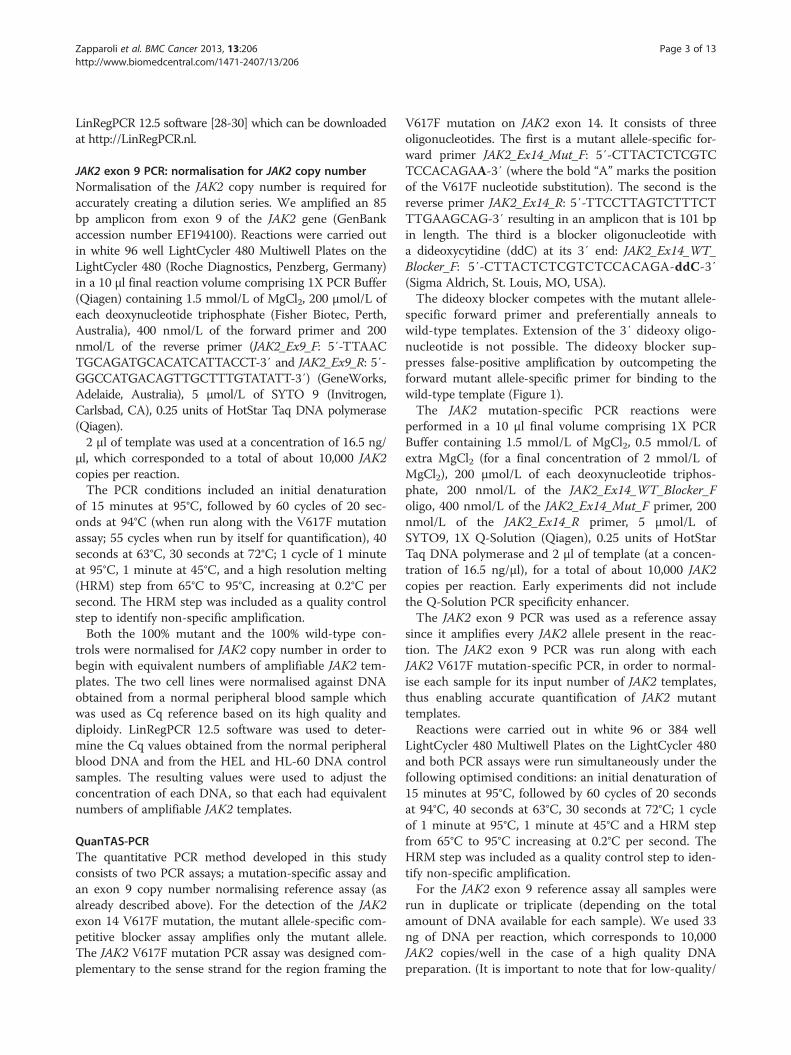
V617F mutation on JAK2 exon 14. It consists of threeoligonucleotides. The first is a mutant allele-specific for-ward primer JAK2_Ex14_Mut_F: 5′-CTTACTCTCGTCTCCACAGAA-3′ (where the bold “A” marks the positionof the V617F nucleotide substitution). The second is thereverse primer JAK2_Ex14_R: 5′-TTCCTTAGTCTTTCTTTGAAGCAG-3′ resulting in an amplicon that is 101 bpin length. The third is a blocker oligonucleotide witha dideoxycytidine (ddC) at its 3′ end: JAK2_Ex14_WT_Blocker_F: 5′-CTTACTCTCGTCTCCACAGA-ddC-3′(Sigma Aldrich, St. Louis, MO, USA).The dideoxy blocker competes with the mutant allele-
specific forward primer and preferentially anneals towild-type templates. Extension of the 3′ dideoxy oligo-nucleotide is not possible. The dideoxy blocker sup-presses false-positive amplification by outcompeting theforward mutant allele-specific primer for binding to thewild-type template (Figure 1).The JAK2 mutation-specific PCR reactions were
performed in a 10 μl final volume comprising 1X PCRBuffer containing 1.5 mmol/L of MgCl2, 0.5 mmol/L ofextra MgCl2 (for a final concentration of 2 mmol/L ofMgCl2), 200 μmol/L of each deoxynucleotide triphos-phate, 200 nmol/L of the JAK2_Ex14_WT_Blocker_Foligo, 400 nmol/L of the JAK2_Ex14_Mut_F primer, 200nmol/L of the JAK2_Ex14_R primer, 5 μmol/L ofSYTO9, 1X Q-Solution (Qiagen), 0.25 units of HotStarTaq DNA polymerase and 2 μl of template (at a concen-tration of 16.5 ng/μl), for a total of about 10,000 JAK2copies per reaction. Early experiments did not includethe Q-Solution PCR specificity enhancer.The JAK2 exon 9 PCR was used as a reference assay
since it amplifies every JAK2 allele present in the reac-tion. The JAK2 exon 9 PCR was run along with eachJAK2 V617F mutation-specific PCR, in order to normal-ise each sample for its input number of JAK2 templates,thus enabling accurate quantification of JAK2 mutanttemplates.Reactions were carried out in white 96 or 384 well
LightCycler 480 Multiwell Plates on the LightCycler 480and both PCR assays were run simultaneously under thefollowing optimised conditions: an initial denaturation of15 minutes at 95°C, followed by 60 cycles of 20 secondsat 94°C, 40 seconds at 63°C, 30 seconds at 72°C; 1 cycleof 1 minute at 95°C, 1 minute at 45°C and a HRM stepfrom 65°C to 95°C increasing at 0.2°C per second. TheHRM step was included as a quality control step to iden-tify non-specific amplification.For the JAK2 exon 9 reference assay all samples were
run in duplicate or triplicate (depending on the totalamount of DNA available for each sample). We used 33ng of DNA per reaction, which corresponds to 10,000JAK2 copies/well in the case of a high quality DNApreparation. (It is important to note that for low-quality/
Figure 1 Representation of the mutant allele-specific PCR and 3′ dideoxy blocker oligonucleotide methodology. Two PCR primers, amutant allele-specific forward primer and a reverse primer, are present in each reaction. In addition, a forward wild-type allele-specific dideoxyblocker (designated WT blocker), which binds to the wild-type allele but is incapable of extension, is present. It will outcompete the forwardmutant allele-specific primer (designated MUT specific) for binding to the wild-type template and, coupled with the already low rate ofamplification of a mutant allele-specific primer from a wild-type template, will further reduce the rate of false-positive amplification.
Zapparoli et al. BMC Cancer 2013, 13:206 Page 4 of 13http://www.biomedcentral.com/1471-2407/13/206
degraded DNA samples, more material would need tobe applied to reach the same total JAK2 copy number, asthe amplifiable templates will be fewer in number. Thiscan be monitored using the JAK2 exon 9 reference assayand comparing the Cq of any high quality diploid gen-omic DNA preparation to the Cq of the low-quality/de-graded samples tested.)Samples were run in triplicate for the JAK2 mutation-
specific PCR. If any of the 3 replicates showed no ampli-fication, we repeated the assay by running 10 replicatesof that specific sample. The low-mutation-level mixes(MUT/WT 0.03% and 0.01%) were always run in repli-cates of 10. Using an increased number of replicates forthe low concentration samples firstly allowed us to havea reliable reference for the Cq value obtainable from onesingle copy of the mutant allele and secondly, producedan increased amount of data which facilitated the useof a statistics-based approach for the estimation of theaverage mutant JAK2 copy number (see below). The nega-tive control samples (genomic DNA from healthy blooddonors and cell lines DNA samples) were run in at least10 replicates.
Real-time data analysisThe real-time PCR data were analysed using LinRegPCR12.5 software. The raw run data (not-baseline-corrected)for both the JAK2 exon 9 PCR (reference assay) and theJAK2 mutation-specific PCR were transferred from theLightCycler 480 to the LinRegPCR 12.5 software usingthe “LC480 Conversion: conversion of raw LC480 data”software (available on the Heart Failure Research Centerwebsite, at http://downloads.hfrc.nl, and treated as twodifferent data sets (amplicon groups).The LinRegPCR program performs sample by sample
baseline correction, and finds a “window-of-linearity” foreach amplicon group. This software uses linear regres-sion to fit a straight line through each set of amplifica-tion curves: the slope of this regression line gives thePCR efficiency for each individual sample. It also esti-mates the “mean PCR efficiency” (E) for each ampliconby calculating the mean value of all sample efficienciesobtained per amplicon group. In performing the mean
efficiency per amplicon calculation, the software wasmanually set to automatically exclude efficiency valuesdeviating more than 5% from the median efficiency andsamples which did not show amplification.The data were first analysed using the automated
LinRegPCR functions and subsequently manually correctedwhere required (as suggested by the LinRegPCR user man-ual, version 12.x). The fluorescence quantification threshold(Nq) was set to “common” for the JAK2 exon 9 and JAK2exon 14 amplicon groups.The LinRegPCR software uses the Nq value and the
fractional cycle number (Cq) (needed for each sample toreach the Nq threshold) to calculate the starting concen-tration (N0) of each target (expressed in arbitrary fluor-escence units), according to the equation N0 = Nq/ECq.The N0 values obtained for each sample and for the100% mutant control (HEL DNA), for both the JAK2exon 9 control assay and the JAK2 exon 14 test assay arethen used to calculate the JAK2 mutant load for each ofthe sample replicates, as described by the equation:
Mut % ¼Ex14ð Þ Individual sample replicates N0 valueEx9ð Þ Average sample replicates N0 value
Ex14ð Þ Average 100%Mutant replicates N0 valueEx9ð Þ Average 100%Mutant replicates N0 value
�100
The numerator in this formula measures the mutant(exon 14) to total (exon 9) JAK2 ratio for a given sample,and the denominator normalises this value by calculatingthe same ratio for the 100% JAK2 mutant control.The formula allowes the monitoring of possible varia-
tions within the resulting mutation burden values of thereplicates of each sample. The mean JAK2 mutant-alleleburden per sample was calculated by averaging the indi-vidual percentage for each replicate of that sample, ex-cluding any obvious outlier values.
Criteria to minimise the scoring of false positivesWe then aimed to develop criteria to minimise the scor-ing of false positives. For this, it was necessary to iden-tify the Cq value given by the amplification of a singlemutant allele. Running 10 replicates of the 0.01% MUT/WT mix in each PCR run allowed us to have a reliable
Zapparoli et al. BMC Cancer 2013, 13:206 Page 5 of 13http://www.biomedcentral.com/1471-2407/13/206
reference for the Cq value obtainable from one singlecopy of the mutant allele, which was then used as aguideline for the identification of rare false-positive oc-currences. Signals that amplify significantly later areconsidered to be false positives, as it is not possible tohave a template number between 1 and 0.
Samples with very low numbers of mutant allelesSamples with very low numbers of mutant alleles presenteda particular challenge for quantification. In these samples,there will be no specific amplification in one or more of
A
B
MUT 100%MUT/WT 10%MUT/WT 1%MUT/WT 0.1%MUT/WT 0.01%WT 100%NTC
MUT 100%MUT/WT 10%MUT/WT 1%MUT/WT 0.1%MUT/WT 0.01%WT 100%NTC
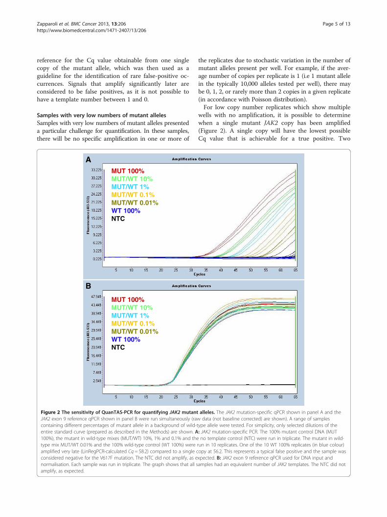
Figure 2 The sensitivity of QuanTAS-PCR for quantifying JAK2 mutantJAK2 exon 9 reference qPCR shown in panel B were run simultaneously (racontaining different percentages of mutant allele in a background of wild-entire standard curve (prepared as described in the Methods) are shown. A100%), the mutant in wild-type mixes (MUT/WT) 10%, 1% and 0.1% and thetype mix MUT/WT 0.01% and the 100% wild-type control (WT 100%) wereamplified very late (LinRegPCR-calculated Cq = 58.2) compared to a single cconsidered negative for the V617F mutation. The NTC did not amplify, as enormalisation. Each sample was run in triplicate. The graph shows that all samplify, as expected.
the replicates due to stochastic variation in the number ofmutant alleles present per well. For example, if the aver-age number of copies per replicate is 1 (i.e 1 mutant allelein the typically 10,000 alleles tested per well), there maybe 0, 1, 2, or rarely more than 2 copies in a given replicate(in accordance with Poisson distribution).For low copy number replicates which show multiple
wells with no amplification, it is possible to determinewhen a single mutant JAK2 copy has been amplified(Figure 2). A single copy will have the lowest possibleCq value that is achievable for a true positive. Two
alleles. The JAK2 mutation-specific qPCR shown in panel A and thew data (not baseline corrected) are shown). A range of samplestype allele were tested. For simplicity, only selected dilutions of the: JAK2 mutation-specific PCR. The 100% mutant control DNA (MUTno template control (NTC) were run in triplicate. The mutant in wild-
run in 10 replicates. One of the 10 WT 100% replicates (in blue colour)opy at 56.2. This represents a typical false positive and the sample wasxpected. B: JAK2 exon 9 reference qPCR used for DNA input andamples had an equivalent number of JAK2 templates. The NTC did not
Zapparoli et al. BMC Cancer 2013, 13:206 Page 6 of 13http://www.biomedcentral.com/1471-2407/13/206
copies will amplify earlier by a Cq value that is propor-tional to the amplification efficiency of the assay. As ourJAK2 mutation-specific assay typically has an amplifica-tion efficiency approximately equal to 1.7, this will beclose to 1 Cq earlier. The probabilities of occurrence ofeach of the counts, y, can be modeled as a modified ver-sion of the Poisson distribution (right-censored Poissondistribution) with mean copy number, λ. The possibleoutcomes for more than two copies are grouped to-gether as a single observation type, as we are unable toresolve these further. The probabilities for the numberof JAK2 V617F copies in a given experiment are calcu-lated as described by the formula:
Pr y; λð Þ ¼λye−λ
y!; y≤2
1−e−λ 1þ λþ λ2
2
� �; y>2
8>><>>:
The value of lambda (λ) represents the average copynumber of mutant JAK2 alleles assessed per replicate(e.g. the standard curve mix MUT/WT 0.03% will con-tain an average copy number λ = 3, when testing 10,000JAK2 copies per tube). The formula consists of two com-ponents. The first component (y ≤ 2) describes thePoisson distribution probability of observing y = 0, 1 or 2copies of mutant JAK2. The second component (y > 2)describes the Poisson distribution probability of observ-ing more than 2 copies of mutant JAK2. The probabilityof observing more than 2 copies is given as one minusthe probabilities of observing 0, 1 and 2 copies.A point estimate of the average number of mutant JAK2
copies was obtained as the value of λ that maximises thelikelihood of this distribution, given the observed number(y) of mutant JAK2 copies in each experimental replicate(see formula above). The associated 95% confidence inter-val was obtained finding the values of λ for which theapplication of the likelihood test would not reject thenull hypothesis of λ ≠ λ0, where λ0 is the value lambdafound by the maximum likelihood-based method de-scribed above. These calculations were performed usingthe R statistical computing software v2.141.0 [31]. Therelevant code is provided (Additional file 1).
ResultsThe use of high resolution melting (HRM) as a qualitycontrol stepThe exon 9 and the JAK2 V617F mutation-specific PCRassays use probe-free real-time amplification with a fluor-escent intercalating dye (SYTO 9) to enable both quantifi-cation and HRM. The dye binds to double stranded DNAspecifically and fluoresces only when intercalated. TheHRM step allows the ready identification of any non-specific amplification, such as primer dimers or non-
targeted sequences, both during optimisation experimentsand routinely as a quality control for all experiments. Useof HRM in preference to gel electrophoresis both im-proves the workflow and eliminates PCR product ma-nipulation as HRM is performed in the same reactionvessel immediately after the real-time amplification (oftendescribed as a closed-tube system).While we did not see primer dimers, non-specific
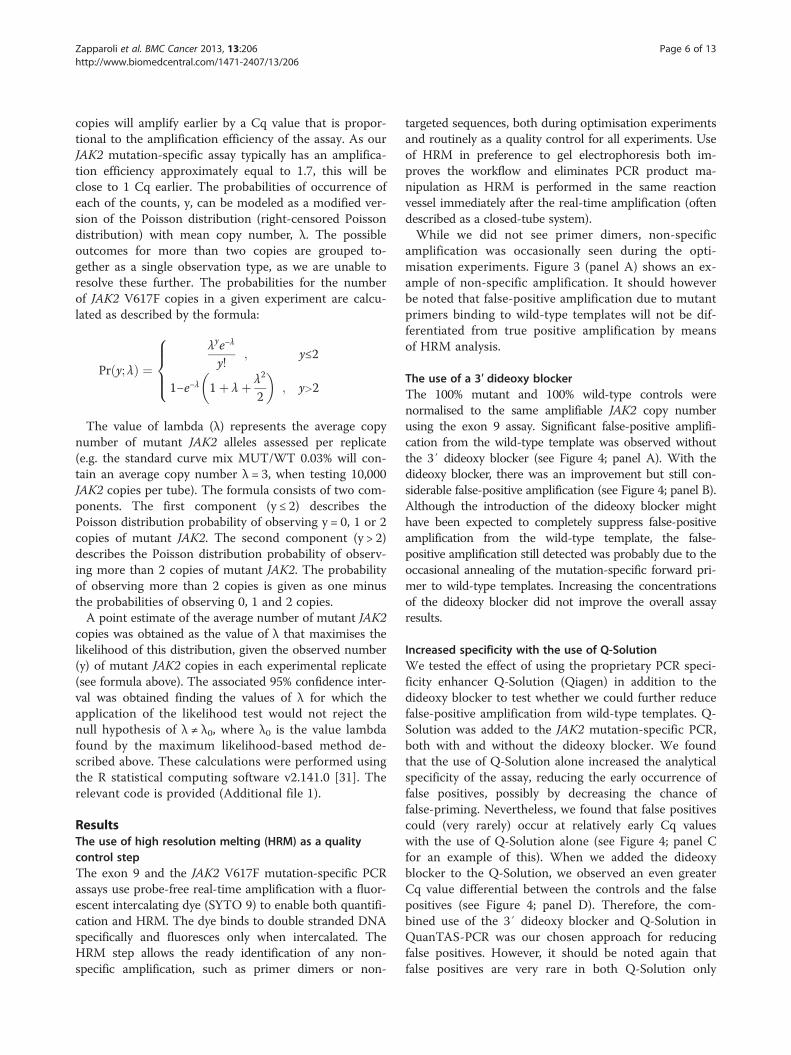
amplification was occasionally seen during the opti-misation experiments. Figure 3 (panel A) shows an ex-ample of non-specific amplification. It should howeverbe noted that false-positive amplification due to mutantprimers binding to wild-type templates will not be dif-ferentiated from true positive amplification by meansof HRM analysis.
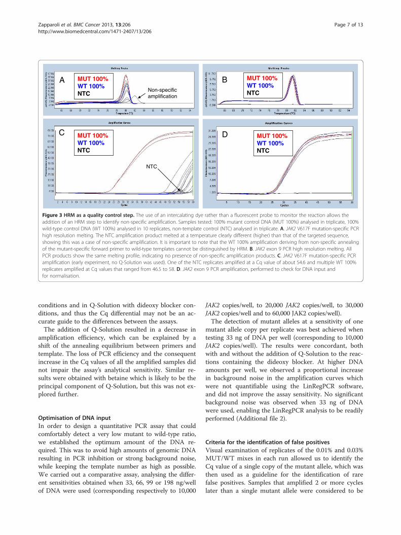
The use of a 3′ dideoxy blockerThe 100% mutant and 100% wild-type controls werenormalised to the same amplifiable JAK2 copy numberusing the exon 9 assay. Significant false-positive amplifi-cation from the wild-type template was observed withoutthe 3′ dideoxy blocker (see Figure 4; panel A). With thedideoxy blocker, there was an improvement but still con-siderable false-positive amplification (see Figure 4; panel B).Although the introduction of the dideoxy blocker mighthave been expected to completely suppress false-positiveamplification from the wild-type template, the false-positive amplification still detected was probably due to theoccasional annealing of the mutation-specific forward pri-mer to wild-type templates. Increasing the concentrationsof the dideoxy blocker did not improve the overall assayresults.
Increased specificity with the use of Q-SolutionWe tested the effect of using the proprietary PCR speci-ficity enhancer Q-Solution (Qiagen) in addition to thedideoxy blocker to test whether we could further reducefalse-positive amplification from wild-type templates. Q-Solution was added to the JAK2 mutation-specific PCR,both with and without the dideoxy blocker. We foundthat the use of Q-Solution alone increased the analyticalspecificity of the assay, reducing the early occurrence offalse positives, possibly by decreasing the chance offalse-priming. Nevertheless, we found that false positivescould (very rarely) occur at relatively early Cq valueswith the use of Q-Solution alone (see Figure 4; panel Cfor an example of this). When we added the dideoxyblocker to the Q-Solution, we observed an even greaterCq value differential between the controls and the falsepositives (see Figure 4; panel D). Therefore, the com-bined use of the 3′ dideoxy blocker and Q-Solution inQuanTAS-PCR was our chosen approach for reducingfalse positives. However, it should be noted again thatfalse positives are very rare in both Q-Solution only
D MUT 100%WT 100%NTC
ANon-specificamplification
MUT 100%WT 100%NTC
B MUT 100%WT 100%NTC
C MUT 100%WT 100%NTC
NTC
Figure 3 HRM as a quality control step. The use of an intercalating dye rather than a fluorescent probe to monitor the reaction allows theaddition of an HRM step to identify non-specific amplification. Samples tested: 100% mutant control DNA (MUT 100%) analysed in triplicate, 100%wild-type control DNA (WT 100%) analysed in 10 replicates, non-template control (NTC) analysed in triplicate. A. JAK2 V617F mutation-specific PCRhigh resolution melting. The NTC amplification product melted at a temperature clearly different (higher) than that of the targeted sequence,showing this was a case of non-specific amplification. It is important to note that the WT 100% amplification deriving from non-specific annealingof the mutant-specific forward primer to wild-type templates cannot be distinguished by HRM. B. JAK2 exon 9 PCR high resolution melting. AllPCR products show the same melting profile, indicating no presence of non-specific amplification products. C. JAK2 V617F mutation-specific PCRamplification (early experiment, no Q-Solution was used). One of the NTC replicates amplified at a Cq value of about 54.6 and multiple WT 100%replicates amplified at Cq values that ranged from 46.5 to 58. D. JAK2 exon 9 PCR amplification, performed to check for DNA input andfor normalisation.
Zapparoli et al. BMC Cancer 2013, 13:206 Page 7 of 13http://www.biomedcentral.com/1471-2407/13/206
conditions and in Q-Solution with dideoxy blocker con-ditions, and thus the Cq differential may not be an ac-curate guide to the differences between the assays.The addition of Q-Solution resulted in a decrease in
amplification efficiency, which can be explained by ashift of the annealing equilibrium between primers andtemplate. The loss of PCR efficiency and the consequentincrease in the Cq values of all the amplified samples didnot impair the assay’s analytical sensitivity. Similar re-sults were obtained with betaine which is likely to be theprincipal component of Q-Solution, but this was not ex-plored further.
Optimisation of DNA inputIn order to design a quantitative PCR assay that couldcomfortably detect a very low mutant to wild-type ratio,we established the optimum amount of the DNA re-quired. This was to avoid high amounts of genomic DNAresulting in PCR inhibition or strong background noise,while keeping the template number as high as possible.We carried out a comparative assay, analysing the differ-ent sensitivities obtained when 33, 66, 99 or 198 ng/wellof DNA were used (corresponding respectively to 10,000
JAK2 copies/well, to 20,000 JAK2 copies/well, to 30,000JAK2 copies/well and to 60,000 JAK2 copies/well).The detection of mutant alleles at a sensitivity of one
mutant allele copy per replicate was best achieved whentesting 33 ng of DNA per well (corresponding to 10,000JAK2 copies/well). The results were concordant, bothwith and without the addition of Q-Solution to the reac-tions containing the dideoxy blocker. At higher DNAamounts per well, we observed a proportional increasein background noise in the amplification curves whichwere not quantifiable using the LinRegPCR software,and did not improve the assay sensitivity. No significantbackground noise was observed when 33 ng of DNAwere used, enabling the LinRegPCR analysis to be readilyperformed (Additional file 2).
Criteria for the identification of false positivesVisual examination of replicates of the 0.01% and 0.03%MUT/WT mixes in each run allowed us to identify theCq value of a single copy of the mutant allele, which wasthen used as a guideline for the identification of rarefalse positives. Samples that amplified 2 or more cycleslater than a single mutant allele were considered to be
JAK2 EX 14(no ddC Blocker;no Q-Solution)
COLOR LEGEND:MUT 100%WT 100%NTC
JAK2 EX 14(with ddC Blocker;with Q-Solution)
COLOR LEGEND:MUT 100%WT 100%NTC
JAK2 EX 14(no ddC Blocker;with Q-Solution)
COLOR LEGEND:MUT 100%WT 100%NTC
JAK2 EX 14(with ddC Blocker;no Q-Solution)
COLOR LEGEND:MUT 100%WT 100%NTC
Cq difference between 100% MUT and first false positive ~ 20 cycles
Cq difference between 100% MUT and first false positive ~ 23 cycles
Cq difference between 100% MUT and first false positive ~ 16 cycles
Cq difference between 100% MUT and first false positive ~ 23 cycles
A
B
C
D
Figure 4 Increasing the assay specificity of the JAK2 V617F mutation-specific PCR. Samples tested: 100% mutant control DNA (MUT 100%)analysed in triplicate, 100% wild-type control DNA (WT 100%) analysed in 10 replicates, non-template control (NTC) analysed in triplicate. A.Mutant allele-specific PCR. The reactions contained two oligonucleotides: the mutant allele-specific forward primer and the reverse primer. Thegraph shows a significant number of false-positive amplifications. We observed a Cq value difference of 20 cycles between the MUT 100% andthe first false-positive amplification. B. Mutant allele-specific PCR with the introduction of the wild-type specific 3′ dideoxy blocker. The reactionscontained three oligonucleotides: the mutant allele-specific forward primer, the wild-type allele specific blocker and the reverse primer. The graphstill shows the presence of a number of false positives. We observed a Cq value difference of 23 cycles between the MUT 100% and the firstfalse-positive amplification. C. Mutant allele-specific PCR with the introduction of 1X Q-Solution. The reactions contained two oligonucleotides:the mutant allele-specific forward and the reverse primers. The graph shows a significant reduction of false-positive amplifications to a single falsepositive. We observed a Cq value difference of 16 cycles between the MUT 100% and the first false-positive amplification. D. Mutant allele-specificPCR with the introduction of both the 3′ dideoxy blocker and 1X Q-Solution. The reactions contained three oligonucleotides: the mutant allele-specific forward primer, the wild-type allele-specific blocker and the reverse primer. One false-positive amplification was observed, at a very lateCq value. We observed a Cq value of 23 cycles difference between the MUT 100% and the first false-positive amplification.
Zapparoli et al. BMC Cancer 2013, 13:206 Page 8 of 13http://www.biomedcentral.com/1471-2407/13/206
Zapparoli et al. BMC Cancer 2013, 13:206 Page 9 of 13http://www.biomedcentral.com/1471-2407/13/206
false positives, as it is not possible to have amplificationof less than one template.
Testing of negative controlsA total number of 13 “negative” control DNA samplesfrom randomly selected, healthy blood donors weretested. Each sample was confirmed to be operationallynegative for the JAK2 V617F mutation by testing 10 rep-licates. Each showed no positives in all replicates whenassessed with QuanTAS-PCR. Occasionally we observedone replicate out of the 10 amplifying at a very late Cqvalue, significantly later than the Cq value for the ampli-fication of a single mutant allele copy.In order to generate a standard series of controls, a
JAK2 V617F mutation-negative cell line was required.We chose the HL-60 cell line, given that this cell line(and the V617F mutant allele positive HEL cell line) arereadily available. We tested 60 replicates of HL-60 DNAusing QuanTAS-PCR at the optimised conditions to ver-ify that it was truly negative for the mutation. Only 1replicate out of 60 showed any amplification. However,this was at a LinRegPCR-calculated Cq value equal to58.2 (Figure 2; panel A), whereas on the same PCR run,one single copy amplified at a LinRegPCR-calculatedCq value of 56.2, and thus could be easily determinedto be an artefactual false positive. These results wereconfirmed by analysing our data using the maximumlikelihood-based method (described in the Methods).According to the statistical analysis, the WT 100% DNAextracted from the HL-60 cell line was estimated to havean average JAK2 copy number of 0.016 (confidenceinterval 0.001 - 0.073) per reaction input. Since thisvalue is considerably less than one, we can confidentlyregard such late amplification as a false positive. ThusHL-60 was confirmed to be a suitable 100% wild-typecontrol.
Analytical sensitivityIn order to assess the analytical sensitivity of theQuanTAS-PCR assay, a set of mutant/wild-type mixeswere prepared by mixing Cq normalised HEL DNA(MUT) and HL-60 DNA (WT). Samples with the JAK2V617F mutant allele present at levels of 100%, 30%, 10%,3%, 1%, 0.3%, 0.1%, 0.03%, 0.01% were tested as well asthe 100% wild-type control. The assay showed an analyt-ical sensitivity of one mutant allele per well (Figure 2;panel A).When testing the 0.03% and 0.01% mixes (ten replicates
each), we also calculated the average JAK2 copy numberfor each replicate. We obtained a value of 2.00 copies(confidence interval 1.20 - 3.10) for the 0.03% mix and avalue of 0.60 (confidence interval 0.24 - 1.22) for the0.01% mix. Both estimations can be considered suffi-ciently accurate, when compared to their corresponding
expected values equal to 3 and 1 allele copies per wellrespectively.
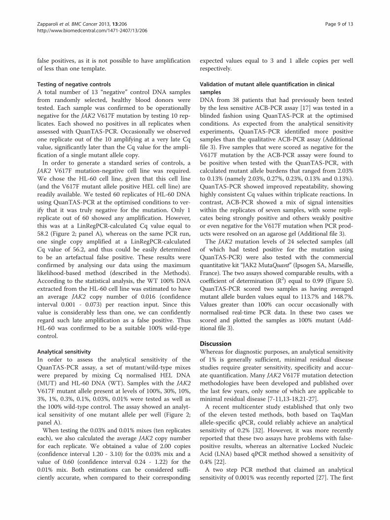
Validation of mutant allele quantification in clinicalsamplesDNA from 38 patients that had previously been testedby the less sensitive ACB-PCR assay [17] was tested in ablinded fashion using QuanTAS-PCR at the optimisedconditions. As expected from the analytical sensitivityexperiments, QuanTAS-PCR identified more positivesamples than the qualitative ACB-PCR assay (Additionalfile 3). Five samples that were scored as negative for theV617F mutation by the ACB-PCR assay were found tobe positive when tested with the QuanTAS-PCR, withcalculated mutant allele burdens that ranged from 2.03%to 0.13% (namely 2.03%, 0.27%, 0.23%, 0.13% and 0.13%).QuanTAS-PCR showed improved repeatability, showinghighly consistent Cq values within triplicate reactions. Incontrast, ACB-PCR showed a mix of signal intensitieswithin the replicates of seven samples, with some repli-cates being strongly positive and others weakly positiveor even negative for the V617F mutation when PCR prod-ucts were resolved on an agarose gel (Additional file 3).The JAK2 mutation levels of 24 selected samples (all
of which had tested positive for the mutation usingQuanTAS-PCR) were also tested with the commercialquantitative kit “JAK2 MutaQuant” (Ipsogen SA, Marseille,France). The two assays showed comparable results, with acoefficient of determination (R2) equal to 0.99 (Figure 5).QuanTAS-PCR scored two samples as having averagedmutant allele burden values equal to 113.7% and 148.7%.Values greater than 100% can occur occasionally withnormalised real-time PCR data. In these two cases wescored and plotted the samples as 100% mutant (Add-itional file 3).
DiscussionWhereas for diagnostic purposes, an analytical sensitivityof 1% is generally sufficient, minimal residual diseasestudies require greater sensitivity, specificity and accur-ate quantification. Many JAK2 V617F mutation detectionmethodologies have been developed and published overthe last few years, only some of which are applicable tominimal residual disease [7-11,13-18,21-27].A recent multicenter study established that only two
of the eleven tested methods, both based on TaqManallele-specific qPCR, could reliably achieve an analyticalsensitivity of 0.2% [32]. However, it was more recentlyreported that these two assays have problems with false-positive results, whereas an alternative Locked NucleicAcid (LNA) based qPCR method showed a sensitivity of0.4% [22].A two step PCR method that claimed an analytical
sensitivity of 0.001% was recently reported [27]. The first
R2 = 0.99
0
10
20
30
40
50
60
70
80
90
100
0 10 20 30 40 50 60 70 80 90 100
JAK2 MutaQuant assay
Qu
anT
AS
-PC
R a
ssay
Figure 5 Regression analysis of the mutant allelic percentagesof 24 clinical samples determined using the “JAK2 MutaQuant”assay (Ipsogen) (on the x axis) and QuanTAS-PCR assay (on they axis).
Zapparoli et al. BMC Cancer 2013, 13:206 Page 10 of 13http://www.biomedcentral.com/1471-2407/13/206
step enriched the mutant allele fraction using a LNA-based wild-type blocking system, whilst the subsequentPCR step was an allele-specific qPCR to quantify the mu-tant allele. The claimed sensitivity is unlikely as the sensi-tivity of any methodology is limited by the number oftemplates initially used in the assay. This sensitivity couldonly be reached beginning with 100,000 templates or 300ng of DNA in the tube. In addition, the first enrichmentstep will prevent this method from being truly quantita-tive. Furthermore, any two step approach (even one whichhas a limited number of amplification cycles in the firststep) increases the risk of cross-contamination betweensamples when performing the second step.We developed a novel single-tube, single-step, mutant
allele-specific quantitative PCR assay: QuanTAS-PCR(Quantitative Threefold Allele-Specific PCR) assay. It usesa combination of established and novel features. We com-bined the basic principles of (i) the use of a 3′mismatchedprimer in allele-specific PCR, [33], (ii) of a wild-typeblocker [34] and (iii) we introduced the use of a PCR spe-cificity enhancer.In allele-specific PCR, the 3′ end of the mutant allele-
specific primer is placed over the mutation site, so thatmutant templates are preferentially amplified [35]. How-ever, as numerous investigators have found, extensionsfrom the wild-type allele do still occur at low frequen-cies. Mutant-specific amplification is enhanced by theaddition of a wild-type blocker to the PCR reaction [34].A wild-type blocker is an oligonucleotide complemen-tary to the wild-type allele with its 3′ end placed overthe mutation site. The blocker also bears a 3′ modifi-cation which is incapable of 3′-5′ extension, therefore“blocking” the wild-type allele amplification. In our as-
say, we used a 3′dideoxycytidine oligonucleotide blockercomplementary to the sense strand where the wild-typesequence terminates at the G at position 1849.Dideoxy blockers are oligonucleotides bearing a dideoxy
nucleotide (ddNTP) at their 3′ ends. Dideoxy nucleo-tides are effective chain terminators used in Sanger sequen-cing [36]. ddNTPs are essentially deoxyribonucleotides(dNTPs) without a 3′-hydroxyl group (-OH) on their de-oxyribose sugar. Therefore 3′ddNTPs are unable to forma phosphodiester bond with any other dNTP and chainelongation is not possible.A combination of allele-specific PCR and dideoxy
oligonucleotide blockers was first reported for the ana-lysis of disease causing mutations in several genes [37]but seems not to have been widely adopted. However,there are major differences between the previous meth-odology and the one reported here, including the use ofqPCR rather than endpoint PCR.We enhanced the analytical specificity of the mutation-
specific assay by adding a proprietary co-solvent, Q-Solution, to the PCR reaction. This increased the PCRspecificity of the JAK2 mutation-specific assay, showinga further decreased occurrence of false-positive resultswhen compared to the same assay run without Q-Solution. To the best of our knowledge, this is the firstapplication of the use of a co-solvent to increase speci-ficity for the detection of minimal residual disease. Im-portantly, the addition of a PCR specificity enhancer tothe PCR reaction is more efficient in suppressing false-positive amplification from wild-type templates than theaddition of a dideoxy blocker. By combining the use ofa dideoxy blocker and a PCR specificity enhancer, weachieved high analytical sensitivity (one single mutant-allele per well) coupled with high analytical specificity,providing a robust quantitative assay.Another unique feature of our methodology is the com-
bined normalisation for the input of DNA and correctionfor copy number variation by calibrating directly to theJAK2 copy number in a control PCR (JAK2 exon 9 PCR),using efficiency-corrected calculation of the starting con-centrations per sample. This reference PCR was used foreach sample including the controls, allowing accuratequantification of the mutant allele burden.The use of real-time PCR technology allowed us to dis-
tinguish false-positive amplifications from true positiveones, by determining the Cq value at which each samplereplicate was amplified with the JAK2 mutation-specificassay. We were able to readily identify false positivesbased on their high Cq values. False-positive samples arethose that clearly amplify later than the reactions startingwith a single copy of the template.The quantitative JAK2 exon 9 and mutation-specific
PCR assays are designed to enable both real-time amplifi-cation analysis and HRM analysis in the one run on a real-
Zapparoli et al. BMC Cancer 2013, 13:206 Page 11 of 13http://www.biomedcentral.com/1471-2407/13/206
time PCR instrument. SYTO-9, a fluorescent intercalatingdye which does not inhibit PCR reactions when used atsaturating conditions [38], binds double stranded DNAspecifically and fluoresces only when intercalated. The useof a closed-tube method like the QuanTAS-PCR is desir-able in a diagnostic setting, being quicker, more reliable,and able to minimise PCR carry-over problems.When testing a sample that is expected to have very
low levels of mutant JAK2 alleles (below 0.03%), werecommend using additional replicates, and analysingthe data using a statistical estimation approach such asthe maximum likelihood-based method described in theMethods section. As a reference, 10 replicates of the0.01% MUT/WT mix should be also run on each PCRrun, in order to have a reliable indication of the Cq valueof one single copy of the mutant allele. This Cq valuecan then be used as a cut-off to distinguish positive am-plifications from any rare false-positive event.Results obtained with QuanTAS-PCR were validated
against those obtained with our previously publishedACB-PCR qualitative assay [17]. The ACB-PCR assay wasable to detect as few as 1% mutant alleles, but oftenpresented some reproducibility problems, i.e. some sam-ple replicates of the ACB-PCR assay showed ambiguousor even contrasting results between replicates. In thesecases the ACB-PCR assay had to be repeated in order toconfirm the positivity of those samples. Five clinical sam-ples that were previously reported as negative for theV617F mutation when tested by ACB-PCR, were clearlypositive when tested with the more sensitive QuanTAS-PCR, scoring mutant-allele burdens which ranged from2.03% to 0.13%. Furthermore, the QuanTAS-PCR gener-ated similar mutant-allele burden results for each of thesample triplicates, thus showing very good reproducibility(Additional file 3).The QuanTAS-PCR’s quantitative results were also vali-
dated against those obtained with the commercially avail-able kit “JAK2 MutaQuant” (Ipsogen) which is based on adual TaqMan probe allelic discrimination approach. All ofthe samples that were determined to have the V617F mu-tation when tested with QuanTAS-PCR, were then testedin a blinded fashion with the Ipsogen kit. The two quanti-tative tests generated comparable mutant-allele burdenvalues and the resulting values were very well correlated(Additional file 3).
ConclusionsIn this study, we have developed a mutant allele-specificquantitative PCR assay, with a unique combination offeatures; a mutation-specific primer, a 3′ dideoxy blocker,a PCR specificity enhancer and real time amplificationanalysis, which is suitable for minimal residual diseasestudies. A recent review concluded that a JAK2 V617F de-tection assay should be both specific and sensitive enough
to detect a mutant allele burden as low as 1–3% [28].Thus QuanTAS-PCR is also suitable as a diagnosticmethod.QuanTAS-PCR shows a very high analytical sensitivity
and the ability to efficiently suppress wild-type alleleamplification. We can consistently and reproducibly de-tect the presence of extremely low levels of JAK2 V617Fin a wild-type allele background, being able to detect aslow as one mutant template per well. When higher num-bers of replicates are tested, higher sensitivities areachievable depending on the total number of templatesassessed. The combination of principles in this assay canthus be used in minimal residual disease monitoring, notjust for JAK2 V617F, but for other common recurrentsingle nucleotide mutations in solid tumours, e.g. BRAFc.1799T > A (V600E) or the acquired resistance mutationEGFR c.2369C > T (T790M).
Additional files
Additional file 1: The R script used in determining average JAK2mutant copy number.
Additional file 2: LinRegPCR analysis: background noise increasesas the DNA input per well increases. The JAK2 mutation-specific PCRamplification curves are shown without baseline correction. A range ofsamples containing different percentages of the mutant allele (MUT)relative to the wild-type allele (WT) were tested (MUT 100%, MUT/WT30%, MUT/WT 10%, MUT/WT 3%, MUT/WT 1%, MUT/WT 0.3%, MUT/WT0.1%, MUT/WT 0.03%, MUT/WT 0.01%). The four panels show how thebackground noise increases as the DNA input per well increases. Panel A:33 ng of DNA per well; Panel B: 66 ng of DNA per well; Panel C: 99 ng ofDNA per well. Panel D: 198 ng of DNA per well. The LinRegPCR softwareindicated that some of the samples could not be analysed due to highbackground noise when 66, 99 or 198 ng of DNA were tested, but thisnever occurred when using 33 ng DNA per well. As discussed in theResults section, we concluded that the optimal amount of DNA to beassessed per well corresponded to 33 ng (Panel A).
Additional file 3: Validation of QuanTAS-PCR JAK2 assay. The resultsfrom the QuanTAS-PCR assay in comparison with the ACB-PCR assay andthe Ipsogen JAK2 MutaQuant assay. Samples in grey were excluded fromcalculating the average score as they were obvious outliers.
AbbreviationsACB-PCR: Allele-specific competitive blocker PCR; ARMS: Amplificationrefractory mutation system; Cq: Quantification cycle; dNTPs: Deoxynucleotidetriphosphate; HRM: High resolution melting; MPN: Myeloproliferativeneoplasm; MUT: Mutant; NTC: No-template control; PCR: Polymerase chainreaction; WT: Wild-type.
Competing interestsThe authors declare that they have no competing interests.
Authors’ contributionsGVZ co-designed the experimental concept, performed and validated theassays and co-wrote the manuscript. RNJ developed the low copy numberanalysis. CAH designed the reference assay. MM performed and provided theACB-PCR results. DAW supervised the development of the assay andprovided the clinical samples. AD co-designed the experimental concept,supervised the development of the assay and co-wrote the manuscript. Allauthors read and approved the final manuscript.
Authors’ informationAlexander Dobrovic and David A Westerman are equal senior authors.
Zapparoli et al. BMC Cancer 2013, 13:206 Page 12 of 13http://www.biomedcentral.com/1471-2407/13/206
AcknowledgementsWe would like to thank our fellow lab members Ida Candiloro, Hongdo Do,Jonathan Weiss and Stephen Wong for critical discussion, Hilda Lau for cellculture. Jan Ruijter provided further comments on the analysis, especiallyregarding the LinRegPCR software. The Australian Red Cross Blood Serviceprovided samples that were used as normal controls. This project wassupported by grants from the Peter MacCallum Cancer Foundation to DAWand Victorian Cancer Agency to AD. Abacus ALS supplied one of theIpsogen kits used for validation in this study, at no cost.
Author details1Department of Pathology, Peter MacCallum Cancer Centre, St AndrewsPlace, East Melbourne, Victoria 3002, Australia. 2Walter and Eliza Hall Instituteof Medical Research, Parkville 3052, Australia. 3Department of MedicalBiology, University of Melbourne, Parkville, Australia. 4Division of CancerMedicine, Peter MacCallum Cancer Centre, St Andrews Place, EastMelbourne, Victoria 3002, Australia. 5Department of Pathology, University ofMelbourne, Parkville, Victoria 3010, Australia. 6Sir Peter MacCallumDepartment of Oncology, University of Melbourne, Parkville, Victoria 3010,Australia. 7Molecular Pathology Research & Development, Peter MacCallumCancer Centre, St Andrews Place, East Melbourne, Victoria 3002, Australia.
Received: 7 February 2013 Accepted: 26 March 2013Published: 24 April 2013
References1. Vardiman JW, Thiele J, Arber DA, Brunning RD, Borowitz MJ, Porwit A, Harris
NL, Le Beau MM, Hellstrom-Lindberg E, Tefferi A, Bloomfield CD: The 2008revision of the world health organization (WHO) classification of myeloidneoplasms and acute leukemia: rationale and important changes. Blood2009, 114:937–951.
2. Tefferi A: Molecular drug targets in myeloproliferative neoplasms: mutantABL1, JAK2, MPL, KIT, PDGFRA, PDGFRB and FGFR1. J Cell Mol Med 2009,13:215–237.
3. James C, Ugo V, Le Couedic J, Staerk J, Delhommeau F, Lacout C, Garcon L,Raslova H, Berger R, Bennaceur-Griscelli A, et al: A unique clonal JAK2mutation leading to constitutive signalling causes polycythaemia vera.Nature 2005, 434:1144–1148.
4. Kaushansky K: On the molecular origins of the chronic myeloproliferativedisorders: it all makes sense. Blood 2005, 105:4187–4190.
5. Pardanani A, Tefferi A: Targeting myeloproliferative neoplasms with JAKinhibitors. Curr Opin Hematol 2011, 18:105–110.
6. Reddy MM, Deshpande A, Sattler M: Targeting JAK2 in the therapy ofmyeloproliferative neoplasms. Expert Opin Ther Targets 2012, 16:313–324.
7. Baxter E, Scott L, Campbell P, East C, Fourouclas N, Swanton S, Vassiliou G,Bench A, Boyd E, Curtin N, et al: Acquired mutation of the tyrosine kinaseJAK2 in human myeloproliferative disorders. Lancet 2005, 365:1054–1061.
8. Er T-K, Lin S-F, Chang J-G, Hsieh L-L, Lin S-K, Wang L-H, Lin C-W, Chang C-S,Liu T-C: Detection of the JAK2 V617F missense mutation by highresolution melting analysis and its validation. Clin Chim Acta 2009,408:39–44.
9. Jelinek J, Oki Y, Gharibyan V, Bueso-Ramos C, Prchal JT, Verstovsek S, BeranM, Estey E, Kantarjian HM, Issa JP: JAK2 mutation 1849G > T is rare in acuteleukemias but can be found in CMML, Philadelphia chromosome-negative CML, and megakaryocytic leukemia. Blood 2005, 106:3370–3373.
10. Jones AV, Kreil S, Zoi K, Waghorn K, Curtis C, Zhang L, Score J, Seear R,Chase AJ, Grand FH, et al: Widespread occurrence of the JAK2 V617Fmutation in chronic myeloproliferative disorders. Blood 2005, 106:2162–2168.
11. Kannim S, Thongnoppakhun W, Auewarakul CU: Two-round allele specific-polymerase chain reaction: a simple and highly sensitive method forJAK2V617F mutation detection. Clin Chim Acta 2009, 401:148–151.
12. Larsen T, Christensen J, Hasselbalch H, Pallisgaard N: The JAK2 V617Fmutation involves B- and T-lymphocyte lineages in a subgroup ofpatients with Philadelphia-chromosome negative chronicmyeloproliferative disorders. Br J Haematol 2007, 136:745–751.
13. Levine R, Wadleigh M, Cools J, Ebert B, Wernig G, Huntly B, Boggon T,Wlodarska I, Clark J, Moore S, et al: Activating mutation in the tyrosinekinase JAK2 in polycythemia vera, essential thrombocythemia, andmyeloid metaplasia with myelofibrosis. Cancer Cell 2005, 7:387–397.
14. Poodt J, Fijnheer R, Walsh IBB, Hermans MHA: A sensitive and reliablesemi-quantitative real-time PCR assay to detect JAK2 V617F in blood.Hematol Oncol 2006, 24:227–233.
15. Qian J, Lin J, Yao D-M, Chen Q, Xiao G-F, Ji R-B, Li Y, Yang J, Qian Z: Rapiddetection of JAK2 V617F mutation using high-resolution melting analysiswith LightScanner platform. Clin Chim Acta 2010, 411:2097–2100.
16. Sutton BC, Allen RA, Zhao ZJ, Dunn ST: Detection of the JAK2V617Fmutation by asymmetric PCR and melt curve analysis. Cancer Biomark2007, 3:315–324.
17. Tan AYC, Westerman DA, Dobrovic A: A simple, rapid, and sensitivemethod for the detection of the JAK2 V617F mutation. Am J Clin Pathol2007, 127:977–981.
18. Ugo V, Tondeur S, Menot ML, Bonnin N, Le Gac G, Tonetti C, Mansat-De MasV, Lecucq L, Kiladjian JJ, Chomienne C, et al: Interlaboratory developmentand validation of a HRM method applied to the detection of JAK2 exon 12mutations in polycythemia vera patients. PLoS One 2010, 5:e8893.
19. Xu X, Zhang Q, Luo J, Xing S, Li Q, Krantz SB, Fu X, Zhao ZJ: JAK2(V617F):Prevalence in a large Chinese hospital population. Blood 2007,109:339–342.
20. Zhao AH, Gao R, Zhao ZJ: Development of a highly sensitive method fordetection of JAK2V617F. J Hematol Oncol 2011, 4:40.
21. Cankovic M, Whiteley L, Hawley RC, Zarbo RJ, Chitale D: Clinicalperformance of JAK2 V617F mutation detection assays in a moleculardiagnostics laboratory. Am J Clin Pathol 2009, 132:713–721.
22. Denys B, El Housni H, Nollet F, Verhasselt B, Philippe J: A real-timepolymerase chain reaction assay for rapid, sensitive, and specificquantification of the JAK2V617F mutation using a locked nucleicacid-modified oligonucleotide. J Mol Diagn 2010, 12:512–519.
23. Hammond E, Shaw K, Carnley B, P’ng S, James I, Herrmann R: Quantitativedetermination of JAK2 V617F by TaqMan: an absolute measure ofaveraged copies per cell that may be associated with the different typesof myeloproliferative disorders. J Mol Diagn 2007, 9:242–248.
24. Kroger N, Badbaran A, Holler E, Hahn J, Kobbe G, Bornhauser M, Reiter A,Zabelina T, Zander AR, Fehse B: Monitoring of the JAK2-V617F mutationby highly sensitive quantitative real-time PCR after allogeneic stem celltransplantation in patients with myelofibrosis. Blood 2007, 109:1316–1321.
25. Lippert E, Boissinot M, Kralovics R, Girodon F, Dobo I, Praloran V, Boiret-Dupre N, Skoda RC, Hermouet S: The JAK2-V617F mutation is frequentlypresent at diagnosis in patients with essential thrombocythemia andpolycythemia vera. Blood 2006, 108:1865–1867.
26. Sidon P, Heimann P, Lambert F, Dessars B, Robin V, El Housni H: Combinedlocked nucleic acid and molecular beacon technologies for sensitivedetection of the JAK2V617F somatic single-base sequence variant. ClinChem 2006, 52:1436–1438.
27. Siebolts U, Lange T, Niederwieser D, Wickenhauser C: Allele-specific wild-typeblocker quantitative PCR for highly sensitive detection of rare JAK2 p.V617F point mutation in primary myelofibrosis as an appropriate tool forthe monitoring of molecular remission following therapy. J Clin Pathol 2010,63:370–372.
28. Bench AJ, White HE, Foroni L, Godfrey AL, Gerrard G, Akiki S, Awan A, CarterI, Goday-Fernandez A, Langabeer SE, et al: Molecular diagnosis of themyeloproliferative neoplasms: UK guidelines for the detection of JAK2V617F and other relevant mutations. Br J Haematol 2013, 160:25–34.
29. Ruijter JM, Ramakers C, Hoogaars WM, Karlen Y, Bakker O, van den Hoff MJ,Moorman AF: Amplification efficiency: linking baseline and bias in theanalysis of quantitative PCR data. Nucleic Acids Res 2009, 37:e45.
30. Tuomi JM, Voorbraak F, Jones DL, Ruijter JM: Bias in the Cq value observedwith hydrolysis probe based quantitative PCR can be corrected with theestimated PCR efficiency value. Methods 2010, 50:313–322.
31. R Development Core Team: R: A language and environment for statisticalcomputing. R Foundation for Statistical Computing. Vienna, Austria; 2011.ISBN 3-900051-07-0, URL http://www.R-project.org/.
32. Lippert E, Girodon F, Hammond E, Jelinek J, Reading NS, Fehse B, Hanlon K,Hermans M, Richard C, Swierczek S, et al: Concordance of assays designedfor the quantification of JAK2V617F: a multicenter study. Haematologica2009, 94:38–45.
33. Okayama H, Curiel DT, Brantly ML, Holmes MD, Crystal RG: Rapid, nonradioactivedetection of mutations in the human genome by allele-specific amplification.J Lab Clin Med 1989, 114:105–113.
Zapparoli et al. BMC Cancer 2013, 13:206 Page 13 of 13http://www.biomedcentral.com/1471-2407/13/206
34. Seyama T, Ito T, Hayashi T, Mizuno T, Nakamura N, Akiyama M: A novelblocker-PCR method for detection of rare mutant alleles in the presence ofan excess amount of normal DNA. Nucleic Acids Res 1992, 20:2493–2496.
35. Newton CR, Graham A, Heptinstall LE, Powell SJ, Summers C, Kalsheker N,Smith JC, Markham AF: Analysis of any point mutation in DNA. Theamplification refractory mutation system (ARMS). Nucleic Acids Res 1989,17:2503–2516.
36. Sanger F, Nicklen S, Coulson AR: DNA sequencing with chain-terminatinginhibitors. Proc Natl Acad Sci U S A 1977, 74:5463–5467.
37. Orou A, Fechner B, Utermann G, Menzel HJ: Allele-specific competitiveblocker PCR: a one-step method with applicability to pool screening.Hum Mutat 1995, 6:163–169.
38. Gudnason H, Dufva M, Bang DD, Wolff A: Comparison of multiple DNAdyes for real-time PCR: effects of dye concentration and sequencecomposition on DNA amplification and melting temperature. NucleicAcids Res 2007, 35:e127.
doi:10.1186/1471-2407-13-206Cite this article as: Zapparoli et al.: Quantitative threefold allele-specificPCR (QuanTAS-PCR) for highly sensitive JAK2 V617F mutant alleledetection. BMC Cancer 2013 13:206.
Submit your next manuscript to BioMed Centraland take full advantage of:
• Convenient online submission
• Thorough peer review
• No space constraints or color figure charges
• Immediate publication on acceptance
• Inclusion in PubMed, CAS, Scopus and Google Scholar
• Research which is freely available for redistribution
Submit your manuscript at www.biomedcentral.com/submit


![BMC Evolutionary Biology BioMed Central...SAR19B5 and SARA3C (Additional file 1). We found that correcting for null allele frequencies [37] did not qualita-tively affect the results](https://img.pdfslide.us/doc/110x75/612eaaa81ecc51586942f501/bmc-evolutionary-biology-biomed-central-sar19b5-and-sara3c-additional-file.jpg)


![BMC Evolutionary Biology BioMed Central · 2016. 5. 27. · SAR19B5 and SARA3C (Additional file 1). We found that correcting for null allele frequencies [37] did not qualita-tively](https://img.pdfslide.us/doc/110x75/612eaa9d1ecc51586942f4f9/bmc-evolutionary-biology-biomed-central-2016-5-27-sar19b5-and-sara3c-additional.jpg)


![Steiner Threefold[1]](https://img.pdfslide.us/doc/110x75/544e7943af7959e91e8b49fc/steiner-threefold1.jpg)