Embed Size (px)
Citation preview
Quantitative proteomics reveals the dynamics ofprotein changes during Drosophila oocyte maturationand the oocyte-to-embryo transitionIva Kronjaa, Zachary J. Whitfielda,b,1, Bingbing Yuana, Kristina Dzeykc, Joanna Kirkpatrickc, Jeroen Krijgsveldc,and Terry L. Orr-Weavera,b,2
aWhitehead Institute, Cambridge, MA 02142; bDepartment of Biology, Massachusetts Institute of Technology, Cambridge, MA 02138; and cEuropeanMolecular Biology Laboratory, Proteomics Core Facility, 69117 Heidelberg, Germany
Contributed by Terry L. Orr-Weaver, October 1, 2014 (sent for review July 30, 2014; reviewed by Thomas U. Mayer and Mariana F. Wolfner)
The onset of development is marked by two major, posttranscrip-tionally controlled, events: oocyte maturation (release of the pro-phase I primary arrest) and egg activation (release from thesecondary meiotic arrest). Using quantitative mass spectrometry,we previously described proteome remodeling during Drosophilaegg activation. Here, we describe our quantitative mass spectrome-try-based analysis of the changes in protein levels during Drosophilaoocyte maturation. This study presents the first quantitative survey,to our knowledge, of proteome changes accompanying oocyte mat-uration in any organism and provides a powerful resource for iden-tifying both key regulators and biological processes driving thiscritical developmental window. We show that Muskelin, found tobe up-regulated during oocyte maturation, is required for timelynurse cell nuclei clearing frommature egg chambers. Other proteinsup-regulated at maturation are factors needed not only for lateoogenesis but also completion of meiosis and early embryogenesis.Interestingly, the down-regulated proteins are predominantly in-volved in RNA processing, translation, and RNAi. Integrating datasetson the proteome changes at oocyte maturation and egg activationuncovers dynamics in proteome remodeling during the change fromoocyte to embryo. Notably, 66 proteins likely act uniquely duringlate oogenesis, because they are up-regulated at maturation anddown-regulated at activation. We find down-regulation of thisclass of proteins to be mediated partially by APC/CCORT, a meio-sis-specific form of the E3 ligase anaphase promoting complex/cyclosome (APC/C).
oocyte | embryo | meiosis | spindle | Muskelin
The change from oocyte to embryo marks the onset of devel-opment. Oocyte maturation is a prerequisite for the oocyte-to-
embryo transition. In most animals, oocytes undergo a prolongedarrest in prophase I to permit oocyte growth, differentiation, andstockpiling of maternal components. This arrest is released atoocyte maturation, commonly in response to hormonal cues. Thenuclear envelope then breaks down, followed by assembly of themeiotic spindle. A secondary meiotic arrest ensues, occurring atmetaphase II in most vertebrates and metaphase I in insects (1).The oocyte-to-embryo transition initiates with egg activation,which causes the release of the secondary arrest and the com-pletion of meiosis. In many organisms, egg activation requiresfertilization, but egg activation is independent of fertilization inDrosophila (1). In all cases, the onset of embryogenesis requiressperm entry, fusion of the male and female pronuclei, and thestart of the mitotic embryonic divisions.How is this complex transition from differentiated oocyte into
totipotent embryo regulated? Both oocyte maturation and eggactivation occur in a transcriptionally silent context (2–4). Theseevents are thus posttranscriptionally controlled, and translationalregulation has been shown to play a crucial role. For example,resumption of meiosis in Xenopus depends on translational ac-tivation of cyclin B and mos mRNA by CPEB-mediated poly-adenylation (4). Further work demonstrated that precisely timed
translation of several other mRNAs is required for progressionthrough the meiotic divisions (5, 6). Recent high-throughputstudies highlight extensive translational changes accompanyingoocyte maturation and egg activation in mice (7, 8). Moreover,genome-wide translational and quantitative proteomic analysesof Drosophila egg activation revealed that in addition to wide-spread translational regulation, posttranslational control, likelychanges in protein stability, serve to remodel the proteome forthe onset of embryonic development (9). At Drosophila egg ac-tivation, in addition to changes in protein levels, hundreds ofproteins alter their phosphorylation status (10).A comprehensive proteome analysis of oocyte maturation is
lacking. Insights into protein changes accompanying Xenopus,murine, bovine, and porcine oocyte maturation have emergedfrom mass spectrometry analyses of 2D gels (11–16). In parallel,proteomic studies were conducted to semiquantitatively comparethe protein composition of murine immature and mature oocytes(17). These approaches uncovered changes in factors as diverse asredox regulators, proteasome components, chaperones, and meta-bolic enzymes occurring during oocyte maturation (18). However,these studies lack the sensitivity, coverage, and quantitative in-formation of currently available quantitative mass spectrometry
Significance
Oogenesis aberrations are an important cause of birth defects.During oocyte development, meiotic divisions are coordinatedwith intense transcription and translation, which produce ma-ternally encoded RNAs and proteins that drive early embryonicdevelopment. Oocyte maturation and egg activation result ineggs competent for fertilization and embryogenesis, respectively,and these events occur in the absence of transcription or RNAdegradation. Therefore, it is likely that changes in protein levelsor activity govern these developmental transitions. By providingthe first proteome-wide overview, to our knowledge, of theproteins whose levels change during oocyte maturation andcombining these data with our previous description of proteomeremodeling at egg activation, we describe critical events andfactors driving the development of immature oocyte into anembryogenesis-competent egg.
Author contributions: I.K., Z.J.W., J. Krijgsveld, and T.L.O.-W. designed research; I.K., Z.J.W.,K.D., and J. Kirkpatrick performed research; J. Krijgsveld contributed new reagents/analytictools; I.K., Z.J.W., B.Y., K.D., J. Kirkpatrick, J. Krijgsveld, and T.L.O.-W. analyzed data; and I.K.and T.L.O.-W. wrote the paper.
Reviewers: T.U.M., University of Konstanz; and M.F.W., Cornell University.
The authors declare no conflict of interest.
Freely available online through the PNAS open access option.1Present Address: Department of Microbiology and Immunology, University of California,San Francisco, CA 94158.
2To whom correspondence should be addressed. Email: [email protected].
This article contains supporting information online at www.pnas.org/lookup/suppl/doi:10.1073/pnas.1418657111/-/DCSupplemental.
www.pnas.org/cgi/doi/10.1073/pnas.1418657111 PNAS | November 11, 2014 | vol. 111 | no. 45 | 16023–16028
DEV
ELOPM
ENTA
LBIOLO
GY
techniques, warranting a comprehensive study, which we decided toconduct in Drosophila.Although much remains to be discovered about oocyte matu-
ration in Drosophila, several regulators have already been re-covered through genetic screens and biochemical approaches. Thetiming of maturation likely is controlled by keeping Cyclin B/CDK1activity at bay through inhibition of Polo kinase by Matrimony(Mtrm) and downstream activation of the PP2A phosphatase in-hibitor Endos (19–21). After a still elusive signal initiates matu-ration, an oocyte-specific cytoplasmic poly(A) polymerase, theproduct of the wispy gene, leads to extension of poly(A) tails (22,23). This cytoplasmic polyadenylation results in increased proteinlevels of Mos and Cyclin B, two maturation regulators conservedin vertebrates, and the Drosophila-specific protein Cortex, an ac-tivator of the APC/C (22–25). It has been shown that thousands ofmRNAs are polyadenylated in a wispy-dependent manner duringDrosophila oogenesis and egg activation (22, 23, 26). However, thescale of consequent induction of translation and the number of up-regulated proteins during oocyte maturation remain unknown.Here, we define the proteome changes accompanying matura-
tion of the Drosophila oocyte by quantitative mass spectrometry.By combining these data with our previous study of egg activation,we define the patterns of proteome remodeling through the de-velopmental window that starts with immature oocytes and endswith embryogenesis-competent activated eggs. This approach un-covered an interesting subset of proteins enriched from oocytematuration until egg activation and likely composed of importantregulators of the completion of oogenesis.
ResultsProteome Remodeling During Drosophila Oocyte Maturation.Oocytematuration is a late event of oogenesis. Drosophila oogenesisunfolds through 14 morphologically distinct stages that can berecovered by dissection (Fig. 1A) (27). Recovered egg chambersare composed of three cell types: Germ-line–derived oocyte andnurse cells and somatic follicle cells. Nurse cells are connected totheir sister oocyte via cytoplasmic bridges, and they supportoocyte development by transcribing maternal mRNAs and syn-thesizing maternal proteins until stage 11 of oogenesis. At stage11, the nurse cells transfer their cytoplasm to the oocyte ina process called dumping, and the nuclei die (28). Follicle cellsencapsulate developing egg chambers and secrete the protectivecoverings of the oocyte (Fig. 1A).To define the proteome changes accompanying Drosophila oo-
cyte maturation, we compared protein levels in prematuration andpostmaturation egg chambers by quantitative mass spectrometry.Because of the lack of consensus on whether oocyte maturationoccurs during stage 12 or 13 (20, 29), we isolated as immature eggchambers, stage 11, which are unequivocally arrested in prophase I.As mature oocytes, we isolated stage 14, which have attained thesecondary arrest point at metaphase I.Peptides obtained after trypsin digestion of three independent
preparations of stage 11 and stage 14 egg chambers were differ-entially labeled with stable isotopes via reductive methylation andfurther processed for mass spectrometry analysis. The peptide ratioswere calculated from MS signal intensities of the differentially la-beled peptide pairs by using MaxQuant software (30). The totalnumber of proteins quantified in the three replicates ranged from3,181 to 3,803. Based on our previous transcriptome analysis ofthis developmental window, which identified ∼6,135 mRNAs asexpressed, we estimate that our mass spectrometry measurementsquantified ∼50% of potentially encoded proteins (9). The ratios ofprotein levels at oocyte maturation between the three replicateswere well correlated (Pearson R = 0.55–0.77) (Fig. 1B).The proteome undergoes substantial remodeling at oocyte
maturation. Approximately 30% of detectable proteins signifi-cantly change in levels: 536 proteins increase and 512 decrease(Dataset S1). Statistical analysis with the Limma package, and
not protein ratios alone, was used to define these significantlychanged proteins (P < 0.05; Fig. 1C). The even distribution ofsummed MS signal intensities for proteins significantly up-regulated(shown in magenta) and down-regulated (shown in blue) indicatesthat proteins across all detected abundances are affected (Fig. 1D).These protein changes were validated for selected candidates byimmunoblots (SI Results and Fig. S1 A and B).We note that some of the protein changes during stages 11–14
of Drosophila oogenesis could occur in the nurse or follicle cells.Because nurse cell contents are dumped into the oocyte, the nursecell and oocyte proteome can be viewed as shared. Examination ofthe transcript levels in follicle cells suggests that the majority ofproteins identified as significantly up-regulated during oocytematuration in our proteome measurements stem from the germline and not follicle cells (SI Results and Fig. S2 A and B).
Functional Classes of Proteins Changed During Oocyte Maturation.We expected that proteins changing in levels during oocytematuration would play important roles in oogenesis and, thus,are likely to be encoded by genes with a developmental patternof maternal expression. Indeed, we observed that the mRNAsencoding the vast majority of proteins up-regulated in levels
�� ��
�
�
�
�
�
�
��
�
��
��
�
�
�
�
�
���
���
�
�
��
�� �
��
��
�
��� � �
��
�
�
��
����
�
�
���
��
�
�
�
�
�
�
�
�
�
�
�
�
�
��
�
�� �
��
�
�
�
�
�
�
�
��
�
��
�
��
�
�
�
� �
� ��
�
�
�
��
�
�
��
�
��
����
��� ��
��
�
��
�
��
�
���
� ��
��
�
��
�
�
�
���
�
�
��
�
� �� ��
�
��
����
��� �� �
�
��
�
��
�
��
�
�� ��
� �
��
�
�
��
��
�
���
�
�
��
�
�
�
�� �
�
��
��
����
���� �
�� �
�
�
���
���
�� �� ��
�
��
�
��
�
���
�� �
��� �
�
� �� �
���
�
��
�
��
�
�
��
��
�
��
�
��
� ��
�� �
�� ��
����
�
�
�
�
��
��
�
�
��
��
�
�
��
���
��
�
��
�
���
�
� ����
�
�
�
�
�
�
�
�
��
� ��
����
��
��
�
�
��
��
��
���
�
���
�
��
�
��
�
��
���
�
���
�
�
��
�
�
�
�
�
�
�
�
��
�
�
�� �� ���
��
��
� �������� ��
���
���
�� �
�
��
�� ��
�
���
�
����
�
� ����
��
��
�
���� ��
����� � ��
��
�
�
�
���
�
�
�
�
�
���
�
�� ��
�
���
�
�
� �
��
��
�
��
�
��
����
�
�
�
����
��
�� ����
��
��
�
�
��
�
�
�
�
� ���
�
��� ��
�
�
��
�
��
��
�
�
���
� ����
�� �
�
�� �
��
�� �
��
� ��
��
� �
�
���� �
�
���
� �
��
�
��
�
�
���
�
�
��
����
��
�����
��
����
�
��
��
� �
�
�
���� � �
�
��
�
��
��
�
�
�
����
��
�
� ��
�
��
�� ��� �
�
�
��
���� ��
��
��
�
�
�
�����
�
�� ��� ����
��
��
����
��
��� �����
��
��� ��
�
�� �
� ��� � �
�
��
�
� ���� ���
���� ���� �
�
��
����
�
��
�
�
�
� ���
�
�
� ��
�
��
�
� �
�� �
�
��
��
�
�
��
��
��
��
��
� �
��
�
�
� �
�
���
��� ���
�
��
�� � �� ��� �
��
��� ���� �� � �
�
�
��
� ��
�
�
��
��
�
�
�
� �
����
��
� �� �
�
�
����
�
�
�
�
�
����
�
��
�
��
��
��
� ����
�
����
�� �
��� � ���
�
�
� �
��
�
�
�
�
� �� �
�
�� �� ���
�� ���
���
�
�
�
�
�
��� �
���
�
���
��
�
�
�
�� ��
�
���
��
�
�� ��
� ��
������
��
�
�� �� �� �
�
��
�� �
�
�
� �� ���� ��
���
�� ��
�
��
� �� �� �
�
�����
��
��� �
��
��
���
����
�� �
��
� �
�
��
�
�
� ��
���
�
�����
������
�
�
��
� ��
�� �
����� ��
�
�
�
������ ��
��
�
�
� ��
�
�
� ���
�� � �
��
�
�
��
�� ��
� ��� ��
���������
��
�
���
��
��
�
�
�� �
� �� �� ����� �
�
���
� �
�
���
����
���
�
�
�
�����
��
��� �
�� ���
� �� �� �
�
��
� ��� �
� �
��
�
�� �
��
�
�� ��
��
� �� �
� �
���
��
�
�����
�
� ��
� �
�
�
�
�� �
��� �
�
�
��
�
�
��� �� ����
���
��
��
��
� � ����
� ��
�
�
� ��
�� �
�
�
���
� ���� �
�
��
�
��
�
��
�
�
�
�� ��
�
�� �
�
�
�
�
�� ���
�
��
��
����
�
�
���
��
�
����
� ��
�
��
�
�
�
���
����
�
� ��
�
� �
��� �
���
�
�� ��
��� �� �� �
����
���
��
���
����
���
��
���
� ����
���
� �
�
��� ��
��
� �
�
� �� �
�
� �� ��
��� �
��
�
�
�
��
�
� �
�����
���
���
��
�
�
�
�
�
�
��
��
�
� ������
�
��
�
��
�� �� �
�����
��
�
�
���
�
�
���
���
���
�
��
�� �
�
�
��
��
�
��
��
�
� ��
����
�
�
���
� ��� ��
�
�
�
� � �� ����
�
�� ��
��� �
��
�� �
��
�� ��� ��
�������
�� �
�
�
�
�
��
��
��
�
�
� ��
��
� ����
�
�����
��
��
�
��� ��
�
��
���
��� �
� ��
��� ��
�
��� �� ��
�
��
���
� �
�
���
��� � ��
�
����
��
�
�� ��
�
��
�
�
�
�
�� ���
��
�
����� �
��
�
�� ��
��� ��
��
�
�
� ��
��� �
� � ��
�
��
�� �
� ��
�
���
�
����� � ���� �
�
� ��
�
�� �� �����
�
�
����� �
����
�
��
� �
��� ��
��
�� �� ��
�� ��
�
�
�� �
�
�
����� �
�
�
�
�� ���
����
��
�����
��
�
�
�
��� �
��
�
��
� ��
�
�
�
�
���
� �� ��
�
�
� �
�
��
��
���
���
�
��
� �
�� �
�
�� ���
��
�
��
����
� ���
�
� �� ����
����
�
�
�����
� ��� �
���
�
�� ���� ���
��
��
�
��
��
�
�
�
�
�
�
� �� ��
��
����� ���
�� ��
��
�
��
��
��
�
���
�
�
�
��
� � ��
��
� ����
��
�
�� ���
�
�� �
����� ��
�
�
�� ��
��
��� ��� ���
��
�
� ���
��
��� ����
��
�
���
��
�
�
�
�
��
�
���
�
���
������ �
��� ��
���
��
�� � ���
�
���
�� ��
����
�� ��
�
��
�� �
��
��
��
���
��
��
�
� �
� �
��
��
��
�
�
�
��
�
�
�� �
�
�
�
�
��
�
��
�� ��� �
��
�
�� ���
�
�
��
���
�
�
�
��
���
�� �
��
�� �
���
�
�
�������� ��
�
��� �� ��
��
�
��� �
��
�
� ��
�
�
�
�
���� ��
�
���
�� �
���� �
�
�
���
�
�
�
�
�
�
�
�
�
�
��
�
�
��� �
�
���
�
��
��� ���
��
�
���� �
�
�
�
���
��
���
���
����
�� �� ��
�
�
�� �� ��
�� �
� � �� �
���
��
�
��
� �
�
�
��
�
�
�
��
���
���
�
�
�
�� ���
� ��
�
��� ��
�
��
��
�
�
� ��
���
������ �
�� � ��
�
���
�
�� �� ����
�
�
�
� ���
�� ��� �
�
�
����
����
�������� �
�� ��
� ���
��� �
� �
�
���
� ���� �
��
�� ��
�
�
�
��
�� ��
�
�
��
�
�
�
���
�
�
�
�
���
�
�
��
��
� �
�
���
�
�
� �
��
��
�� ��
��� �
��
�����
� ��� ��
� �
�
� �
�
�
�
�
�
�
�
�
�
�
�
�
��
�
�
�
�
��
�
�
���
�
�� ��� �
�
�
��
���
�
�
�
�
�
�
��
� �
��
��
�
�
�
�
���
�
�� �
� �� � ���� �� ���� �� � � � �� �� � ��� �� � �� ��� �� � ��� � ����� �� ������ � �� �� �� �� �� � ����� ��� ���� �� ���� �� �� ���� �� � ����� �� ��� �� �� �� � ��� � ��� � ��� ��� �� �� �� �� ������� � �� ��� �� �� �� ��� �� ���� �� �� ������ �� ��� � ���� ����� � �� � ��� ��� ��� ����� � ��� � � ��� �� ���� �� � �� � � ��� ��� ��� �� � ��� �� ��� �� �� �� ��� ���� � � �� ���� ��� �� � � �� � ��� � �� ��� �� �� �� �� ����� � ���� � �� � ��� �� �� �� ��� ���� ���� �� ��� �� �� �� �� ������ � �� �� � ��� �� �� � ��� ���� �� ��� �� ��� � �� ����� � ��� �� �� �� ��� � �� � �� �� �� � ��� � �� � ���� � ����� � ��� �� � �� � � ���� �
�
�
�
�
�
�
�
�
�
��
�
�
� �
�
��
�
� �
�
�
��
�
��
�
���
���
�
��
�
�
��
�
�
�
�
�
�
�
��
���
�
�
�
�
�� �
���
�
�
�
��
�
�
�
���
�
�
�
�
�� �
��
�
�
� �
�
�
�
�� ���
�
���
�
�
�
�
� ���
�
� �� �
�
�
�
�
�
��
��
�
�
�� ����
�
� �
�
���
���
� �
�
�
�
�
�
�
�
�
�
����
�
��
�
�
���
��� ��� �� ��� �� � � � ��� � �� ��� � ��� ��� ���� ��� ��� ��� �� � ��� �� ���� ���� �� �� ��� �� �� ������� �� ��� �� � �� ��� ��� �� �� � �� �� ��� � �� � �� �� ��� ������ �� � ���� ��� �� �� ����
�
�
��� �� �
��
�� � ��
��� ��� ��� ��� ��� ����
�
�
�
�
� ��
�
������
�
��
��
���
�
���
��
�
���
�������
�
��
�
�������
��
�
�
���
�
�
��
��
�
�
�
�
��� �� ���
�
� ��� �� ������ �� �� ���� �� �� � ��� �� ���� �� ��� �� ����� � ��� ���� �� � ���� � ��� �� �� ���� ���� ���� �� �� � ��� ��� � �� ��� ����� �� ��� ����� � �� �� � ��� � ��� ��
��
� � �
�
�
��
��
��� �
�
�
�
��
� � ��� ��� ��� �� �� �� ���� � ��� � ��� � �� ��� �� � � �� �� ���� ��� ��� �� ����� �� ��� ��� ����� ������� �� ��� � ������ � ���� � ��� �� �� �� �� ���
�
��
� ��� � ��� ����� ���� ��� ���� ��� �� �� ��� ��� ���� � ��� �� ��� ���� ����� ��� �� �� �� � �� ��� � � � �� �� ���� ������� ���� � ��� ���� ���� ��� ���� �� ��� � ��� ��� ���� � ���� �� ���� �� �� �� �� �� � ���� ��� ��� � ���� � �� ��� � � ���� ���� �� � ��� �� � �� ���� �� ���� ����� ��� �� ������� ���� �� �� ���� �� � �� ��� ������� ���� � ��� � ��� �� � �� � �� ��� �� �� �� �� �� � �� � ��� � �� �� � ���� �������� �� �� � �� ��� � ��� ���� ��� � � ����� �� �� �� �� �� � �� ��� �� �� �� ���� ���� � �� �� ���� �� �� � ��� ��� � �� �� �� ��� ��� �� �� �� �� �� �� �� � �� ���� ���� ��� ��� ��� ��� �� ���� ������ �� �� � ������ � ��� �� � ����� ��� �� � �� ��� �� �� ��� �� � �� ��� ����� �� � ��� ��� �� � �� � �� �� �� ����� � ���� � ��� �������� �� �� ����� � �� �� � �� ���� �� �� ���� � �� � �� ��� ��� ��� � �� ����� �� �� ���� �� � �� ��� �� ��� ��� � �� ���� � �� ���� �� � �� ���� �� ��� �� �� � � ����� ��� �� �� �� ��� ��� � ���� � ��� ��� � �� � ���� ��� �� ��� �� �� ��� � ����� ���� � �� �� � ��� ��� ��� ��� � �� �� ���� �� ���� � �� � �� ����� �� ��� �� �� � ���� ���� ��� �� ��� ��� ��� ��� ����� ��� �� ��� ��� �� � �� �� ��� �� �� �� � ���� � ���� �� ����� ������ ��� ���� � � ���� � � ����� � �� �� ��� �� ��� ���� ������ �� ���� �� ��� ��� �� �� � � �� ��� � �� �� ���� ��� � ���� ��� �� ��� � � �� ����� �� ��� ���� ��� � �� �� ��� ������ �� ��� ��� ���� �� �� ��� � ��� � ��� �� �� �������� ��� ���� ��� � ��� �� ��� �� �� � �� � �� �� ��� ���� �� ������ � ��� ���� �� ��� � � ��� � ��� �� ������ ����� � �� ��� �� �� � �� �� ��� �� ��� �� � �� �� ��� �� �� �� ���� ���� � �� �� ��� � �� �� �� ��� ��� �� �� �� ���� �� � �� � ��� �� ���� �� ���� ��� � �� �� ��� � �� � �� ��� ��� �� ��� ������ ����� ��� � ������ ������� ��� �� ��� �� ��� ������ ��� ���� ��� ���� ��� � � �� ������ ��� �� �� � ������� ����� ��� ��� ��� ���� � ��� ����� ��� �� �� ��� ���� �� ��� ������ ����� �� �� ��� ��� �� ��� �� ���� � �� � �� ���� �� �� � ��� ��� � ����� �� ���� ��� ���� ��� ���� ��� �� �� ���� ���� ��� �� � �� �� � ��� � �� �� ��� ��� ��� ���� �� � ���� �� � �� ��� �� �� �� �� �� �� �� ��� �� �� ��� � �� �� � � �� �� �� ���� ��� �� � ��� �� � ��� � �� ��� �� � ��� ��� � �� �� �� �� ���� � ���� ��� �� � ���� ����� �� ���� ��� �� ��� � �� �� ���� �� ��� �� �� �� �� �� �� ���� ���� � ���� ��� ���� �� �� � �� ����� �� �� ��� ��� �� ��� �� ������ �� ��� ��� �� ���� ��� �� ���� �� ���� �� �� ���� �� � �� � ����� ��� � ��� �� �� ������ �� � ��� �� ���� � � ���� �� �� � ��� �� �� ��� ������ ��� � �� � ���� �� �� ���� ���� �� � �� ��� � �� � ���� �� ��������� � �� ��� � ��� �� �� �� �� �� ���� ���� � ����� � ��� ������ ��� � � �� �� �� ��� ��� ��� ������� � ��� � ��� �� ��� ��� ���� �� �� � ��� � �� ��� � ���� � ���
��� ��
�
�
��
��
�
�
�
�
�
�
�
�
���
��� � �� ���� �� �� � �� � �� �� � � �� �� ��� ��� ��� � ��� � � �� � ��� ���� ���� ��� ��� �� �� �� �� �� ��� ��� ��� �� � � ��� � �� ��� ��� �� � ��� �� �� �� � ���� �� ���� ��� �� � ��� ��� �� �� � � �� � ��� � ��� � �� �� ���� �� ��� ��� �� � ����� �� ��� ��� �� ���� �� �� � ���� ��� ���
��� ��
�
�
�
�
�
�
�
�
�
��
�
�
� �
�
��
�
� �
�
�
��
�
��
�
�� �
�� �
�
��
�
�
��
�
�
�
�
�
�
�
��
���
�
�
�
�
�� �
���
�
�
�
��
�
�
�
�� �
�
�
�
�
���
� �
�
�
� �
�
�
�
�� �� �
�
� ��
�
�
�
�
����
�
� ���
�
�
�
�
�
����
�
�
����
� �
�
� �
�
���
�� �
��
�
�
�
�
�
�
�
�
�
��� �
�
� �
�
�
���� ��� ���� ������ ��� �� �� � ��� ��� ��� ��� �� ��� ��� � ��� �� ����� �� �� ���� ��� ����� �� �� ���� ���� ��� � �� �� ����� �� ���� ����� �� �� �� ��� � ���� � ���� �� ���� � ����� �� �� ��
�
�
�� �
�� �� �
� � ���
� ��� � �� � �� � � � ��� �� �
�
�
�
�
� ��
�
�����
�
�
��
��
���
�
���
��
�
���
�� ���
��
�
��
�
��� �
�� �
��
�
�
���
�
�
��
��
�
�
�
�
� �������
�
��� ���� �� ����� ��� ��� ���� �� ����� ���� ���� ��� ��� �� � �� ��� �� �� ������� �� �� ���� �� �� ��� � ����� ���� �� ��� � �� �� � �� ��� ���� ���� ��� ����� � � ���� �
��
� ��
�
�
��
��
���� �
�
�
��
� ��� � ��� �� �� ���� ��� ��� �� ����� ��� �� ��� �� �� � �� ���� ��� �� �� ����� � �� �� �� ��� ��� ��� � �� ��� � ���� �� �� �� �� ��� �� �� �� ��� ��� � �
�
��
� ��� ����� �� �� ����� �� ��� �� ���� ��� ������� ��� � ���� ��� �� ���� ��� � �� ��� �� ��� �� ���� ��� ��������� ����� ��� � �� � ��� �� � � ��� �� �� �� ��� �� � ���� ��� � �� �� ��� �� �� � �� ���� � �� ��� �� ��� ��� �� ��� �������� � �� ����������� � ����� �� ��� � �� �� � ���� ������ �� �� ��� �� �� �� �� �� �� � �� � � ��� ��� ��� �� ����� ������ ��� � �� �� ��� �� �� ���� ���� � ���� �� � � �� �� ����� ������ �� ����� �� ��� �� ���� ������ � � � ��� �������� � �� �� �� �� ����� ���� � ���� �� � ����� �� ��� �� �� �� ���� �� ���� ��� ����� ��� �� �� �� ����� �� ����� � ��� �� � ��� �� �� ����� �� �� �� � ������ ��� ��� ��� ���� ����� �� ������ � ������ �� ��� ��� �� �� �� �� �� �� �� �� ����� �� ����� �� ���� ��� ������ � �� �� ���� ��� ����� �� ������� ��� ���� ��� ��� �� � ��� ��� � ���� �� � �� ��� �� ���� � ���� � ��� �� ��� ���� � � ��� �� ����� �� �� ��� �� �� ����� �� �� �� ���� �� �� ����� � ���� �� ��� ��� ��� � �� �� ���� �� � ������ �� �� ��� �� ��� �� � ���� � ��� �� � �� �� �� ���� �� �� ��� �� ��� ��� � �� ��� ���� �� ���� � ��� ��� �� � ���� � �� ��� �� �� �� �� �� ��� ���� ��� �� �� �� ���� ��� �� � �� �� � �� ��� �� ���� � ��� �� �� �� � ��� �� ���� � �� �� ��� �� ����� ����� �� �� � �� �� ���� ��� �� �� �� ��� �� � � �� � ���� ���� � ��� ��� ����� �� �� ������ ���� � ���� ��� �� ���� � ������� ��� ���� ��� �� �� ���� �� �� � � ��� ����� �������� �� �� ���� �� ���� ���� � ��� ���� ��� ����� ��� ����� �� ��� ����� ��� ��� � ���� �� �� �� �� ����� ��� ��� ���� �� � �� �� ���� � ��� ���� � ��� � ��� � �� �� ��� �� ��� �������� ���� �� �� ����� � ���� �� �� ����� � �� ���� ���� ��� ������ ��� �� �� �� �� �� ���� �� �� ���� ���� �� ����� ��� � ������ �� � � ��� �� � �� ���� �� �� ���� � � ���� ��� �� ��� � ���� ����� ��� ���� � ���� �� ���� ���� ����� ��� ���� � ���� � ��������� �� �� �� ��� � ��� ��� ������ � ���� �� �� ��� � ��� ���� � ��� �� ��� ���� � �� ��� ��� �� �� ���� �� ��� ����� ��� ��� �� ��� ��� ����� �� ��� �� �� ���� ��� � �� �� ��� ��� � �� �� ���� �� �� � �������� � ���� ������� �� � ��� ��� ��� ���� ��� �� � ���� � ��� � �� �� ���� ��� �� � �� ��� �� ��� �� �� �� �� �� ������ � ���� �� ��� �� ��� �� ���� � ���� � � ��� �� �� ����� ���� �� �� ����� � �� ��� ���� ���� �� ��� ����� � ��� ��� � ��� �� ���� �� �� �� � �� �� ��� ���� ���� � ���� �� ��� ����� � �� ���� � �� ��� �� ���� ����� ��� ��� ��� ��� ������ � �� �� � ��� � ���� � �� ���� �� ��� ��������� ��� �� ��� � � �� ��� � ����� ��� �� � ����� ���� �� ������� �� � �� �� ����� �� � ��� � ��� ���� ���� �� �� � ���� ���� �� ��� ��� ���� � �� ���� �� ��� ���� �� �� �� ��� ��� � ��� ������ � � ���� �� ���� � �� ��� ������ ����� ��� ����� ��� �� � �� �� � ���
�� ���
�
��
�
��
�
� �
�
���
�
�� ���� � � ����� �� �� ��� ����� ��� ��� � � ��� �� �� � � �� � ��� ��� � �� �� �� � �� � ��� �� ��� �� � �� � ��� �� �� � �� ��� �
�
�
log2 ( )
average log2(normalized ratio st14/st11)
aver
age
log 2
(sum
med
pep
tide
inte
nsity
)
average log2(normalized ratio st14/st11)
-[log
2 (p-
valu
e)]
st11st14
log 2(
st14
/st1
1)re
pl.2
repl.1 log2 ( )st11st14 repl.1 log2 ( )st11
st14 repl.2
log 2(
st14
/st1
1)re
pl.3
log 2(
st14
/st1
1)re
pl.3R=0.55 R=0.77R=0.60
-10 1050-5-10
1050-5
B
C D
0 2 4-4 -2
30252015
0 2 4-4 -2
543210
-10 1050-5 -10 1050-5-10
1050-5
-10
1050-5
stage 10stage 11stage 12stage 13
Egg activation
activated embryo
Embryo-genesisOocyte maturation
stage 14 egg
A
Fig. 1. Protein changes during oocyte maturation. (A) Schematic repre-sentation of Drosophila oocyte maturation, egg activation, and early em-bryonic development. The oocyte is shown in orange, nurse cells in blue, andfollicle cells in green. Gray protrusions in stage 13 and 14 oocytes and em-bryos represent dorsal appendages in the egg shell. DNA (within polar bodyin the activated egg or nuclei in the embryo) is shown in red. The stages usedin the quantitative mass spectrometry experiment are shown in bold. (B)Scatterplots showing the correlation between the log2 of normalized pro-tein ratios for three biological replicates of experiments comparing proteinlevels in stage 11 (st11) versus stage 14 (st14) egg chambers. In B–D, Limmaanalysis (53) was used to define proteins significantly up-regulated (ma-genta), unchanged (gray), and down-regulated (blue) during oocyte matu-ration (P < 0.05). Pearson correlation is displayed on each graph. A total of3,147, 2,879, and 2,759 data points are shown in total for Left, Center, andRight, respectively. (C) Volcano plot showing, for 3,477 proteins, P values(−log2) versus average of normalized protein ratios in stage 14 comparedwith stage 11 egg chambers in three replicate experiments. The colorscheme is the same as for B with 536 proteins shown in magenta and 512shown in blue. (D) Scatterplot showing a log2 of normalized ratios for 3,477proteins (C) in stage 14 compared with stage 11 egg chambers (x axis) versuslog2 of a summed peptide Intensity (y axis). The average of three biologicalreplicates is represented.
16024 | www.pnas.org/cgi/doi/10.1073/pnas.1418657111 Kronja et al.
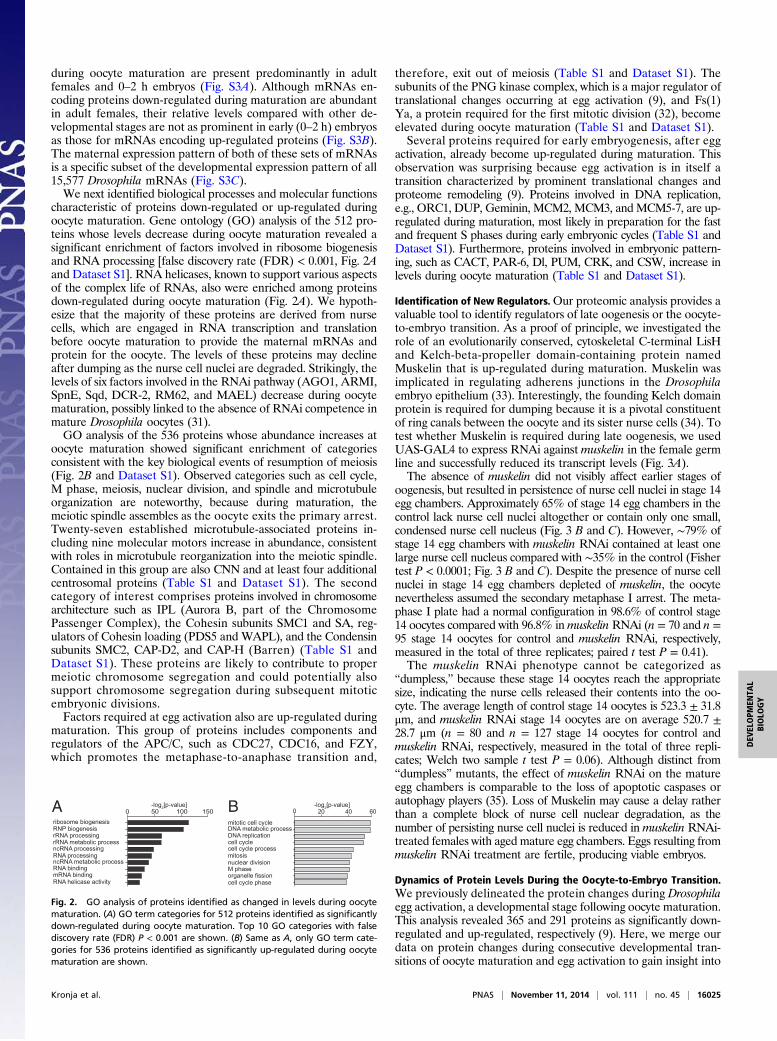
during oocyte maturation are present predominantly in adultfemales and 0–2 h embryos (Fig. S3A). Although mRNAs en-coding proteins down-regulated during maturation are abundantin adult females, their relative levels compared with other de-velopmental stages are not as prominent in early (0–2 h) embryosas those for mRNAs encoding up-regulated proteins (Fig. S3B).The maternal expression pattern of both of these sets of mRNAsis a specific subset of the developmental expression pattern of all15,577 Drosophila mRNAs (Fig. S3C).We next identified biological processes and molecular functions
characteristic of proteins down-regulated or up-regulated duringoocyte maturation. Gene ontology (GO) analysis of the 512 pro-teins whose levels decrease during oocyte maturation revealed asignificant enrichment of factors involved in ribosome biogenesisand RNA processing [false discovery rate (FDR) < 0.001, Fig. 2Aand Dataset S1]. RNA helicases, known to support various aspectsof the complex life of RNAs, also were enriched among proteinsdown-regulated during oocyte maturation (Fig. 2A). We hypoth-esize that the majority of these proteins are derived from nursecells, which are engaged in RNA transcription and translationbefore oocyte maturation to provide the maternal mRNAs andprotein for the oocyte. The levels of these proteins may declineafter dumping as the nurse cell nuclei are degraded. Strikingly, thelevels of six factors involved in the RNAi pathway (AGO1, ARMI,SpnE, Sqd, DCR-2, RM62, and MAEL) decrease during oocytematuration, possibly linked to the absence of RNAi competence inmature Drosophila oocytes (31).GO analysis of the 536 proteins whose abundance increases at
oocyte maturation showed significant enrichment of categoriesconsistent with the key biological events of resumption of meiosis(Fig. 2B and Dataset S1). Observed categories such as cell cycle,M phase, meiosis, nuclear division, and spindle and microtubuleorganization are noteworthy, because during maturation, themeiotic spindle assembles as the oocyte exits the primary arrest.Twenty-seven established microtubule-associated proteins in-cluding nine molecular motors increase in abundance, consistentwith roles in microtubule reorganization into the meiotic spindle.Contained in this group are also CNN and at least four additionalcentrosomal proteins (Table S1 and Dataset S1). The secondcategory of interest comprises proteins involved in chromosomearchitecture such as IPL (Aurora B, part of the ChromosomePassenger Complex), the Cohesin subunits SMC1 and SA, reg-ulators of Cohesin loading (PDS5 and WAPL), and the Condensinsubunits SMC2, CAP-D2, and CAP-H (Barren) (Table S1 andDataset S1). These proteins are likely to contribute to propermeiotic chromosome segregation and could potentially alsosupport chromosome segregation during subsequent mitoticembryonic divisions.Factors required at egg activation also are up-regulated during
maturation. This group of proteins includes components andregulators of the APC/C, such as CDC27, CDC16, and FZY,which promotes the metaphase-to-anaphase transition and,
therefore, exit out of meiosis (Table S1 and Dataset S1). Thesubunits of the PNG kinase complex, which is a major regulator oftranslational changes occurring at egg activation (9), and Fs(1)Ya, a protein required for the first mitotic division (32), becomeelevated during oocyte maturation (Table S1 and Dataset S1).Several proteins required for early embryogenesis, after egg
activation, already become up-regulated during maturation. Thisobservation was surprising because egg activation is in itself atransition characterized by prominent translational changes andproteome remodeling (9). Proteins involved in DNA replication,e.g., ORC1, DUP, Geminin, MCM2, MCM3, andMCM5-7, are up-regulated during maturation, most likely in preparation for the fastand frequent S phases during early embryonic cycles (Table S1 andDataset S1). Furthermore, proteins involved in embryonic pattern-ing, such as CACT, PAR-6, Dl, PUM, CRK, and CSW, increase inlevels during oocyte maturation (Table S1 and Dataset S1).
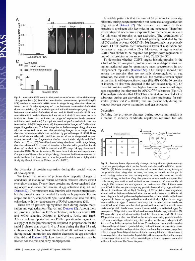
Identification of New Regulators.Our proteomic analysis provides avaluable tool to identify regulators of late oogenesis or the oocyte-to-embryo transition. As a proof of principle, we investigated therole of an evolutionarily conserved, cytoskeletal C-terminal LisHand Kelch-beta-propeller domain-containing protein namedMuskelin that is up-regulated during maturation. Muskelin wasimplicated in regulating adherens junctions in the Drosophilaembryo epithelium (33). Interestingly, the founding Kelch domainprotein is required for dumping because it is a pivotal constituentof ring canals between the oocyte and its sister nurse cells (34). Totest whether Muskelin is required during late oogenesis, we usedUAS-GAL4 to express RNAi against muskelin in the female germline and successfully reduced its transcript levels (Fig. 3A).The absence of muskelin did not visibly affect earlier stages of
oogenesis, but resulted in persistence of nurse cell nuclei in stage 14egg chambers. Approximately 65% of stage 14 egg chambers in thecontrol lack nurse cell nuclei altogether or contain only one small,condensed nurse cell nucleus (Fig. 3 B and C). However, ∼79% ofstage 14 egg chambers with muskelin RNAi contained at least onelarge nurse cell nucleus compared with ∼35% in the control (Fishertest P < 0.0001; Fig. 3 B and C). Despite the presence of nurse cellnuclei in stage 14 egg chambers depleted of muskelin, the oocytenevertheless assumed the secondary metaphase I arrest. The meta-phase I plate had a normal configuration in 98.6% of control stage14 oocytes compared with 96.8% inmuskelin RNAi (n = 70 and n =95 stage 14 oocytes for control and muskelin RNAi, respectively,measured in the total of three replicates; paired t test P = 0.41).The muskelin RNAi phenotype cannot be categorized as
“dumpless,” because these stage 14 oocytes reach the appropriatesize, indicating the nurse cells released their contents into the oo-cyte. The average length of control stage 14 oocytes is 523.3 ± 31.8μm, and muskelin RNAi stage 14 oocytes are on average 520.7 ±28.7 μm (n = 80 and n = 127 stage 14 oocytes for control andmuskelin RNAi, respectively, measured in the total of three repli-cates; Welch two sample t test P = 0.06). Although distinct from“dumpless” mutants, the effect of muskelin RNAi on the matureegg chambers is comparable to the loss of apoptotic caspases orautophagy players (35). Loss of Muskelin may cause a delay ratherthan a complete block of nurse cell nuclear degradation, as thenumber of persisting nurse cell nuclei is reduced in muskelin RNAi-treated females with aged mature egg chambers. Eggs resulting frommuskelin RNAi treatment are fertile, producing viable embryos.
Dynamics of Protein Levels During the Oocyte-to-Embryo Transition.We previously delineated the protein changes during Drosophilaegg activation, a developmental stage following oocyte maturation.This analysis revealed 365 and 291 proteins as significantly down-regulated and up-regulated, respectively (9). Here, we merge ourdata on protein changes during consecutive developmental tran-sitions of oocyte maturation and egg activation to gain insight into
0 20 40 60
mitotic cell cycle
DNA replication
organelle fission
cell cycle process
DNA metabolic process
cell cycle
M phase
mitosisnuclear division
cell cycle phase
0
50 100 150
ribosome biogenesis
rRNA processing
ncRNA processing
RNP biogenesis
rRNA metabolic process
RNA helicase activity
RNA bindingmRNA binding
RNA processingncRNA metabolic process
-log2 gol-]eulav-p[ 2[p-value]A B
Fig. 2. GO analysis of proteins identified as changed in levels during oocytematuration. (A) GO term categories for 512 proteins identified as significantlydown-regulated during oocyte maturation. Top 10 GO categories with falsediscovery rate (FDR) P < 0.001 are shown. (B) Same as A, only GO term cate-gories for 536 proteins identified as significantly up-regulated during oocytematuration are shown.
Kronja et al. PNAS | November 11, 2014 | vol. 111 | no. 45 | 16025
DEV
ELOPM
ENTA
LBIOLO
GY
the dynamics of protein expression during this crucial windowof development.We found that subsets of proteins show opposite changes in
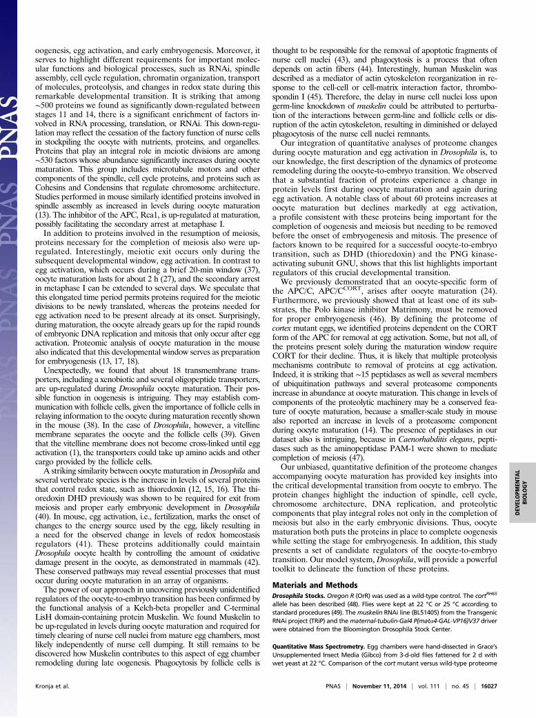
abundance at maturation versus activation, whereas others exhibitsynergistic changes. Twenty-three proteins are down-regulated dur-ing oocyte maturation but increase at egg activation (Fig. 4A andDataset S2). Their functions may interfere with meiotic progression,but the proteins may be needed for early embryogenesis. For ex-ample, the RNAi components Spn-E and RM62 fall into this class,coincident with the reappearance of RNAi competence (31).There are 43 proteins up-regulated both during oocyte matu-
ration and egg activation (Fig. 4A and Dataset S2). Among themare proteins involved in DNA replication, such as several ORCand MCM subunits, DNApol-δ, DNApol-e, RnrL, and RnrS.After a prolonged period without DNA replication during meiosis,a supply of these proteins may be required in preparation for therapid S phases that occur in 4 to 5 min during the first 13 earlyembryonic cycles. In contrast, the levels of 78 proteins decreasedduring oocyte maturation are further decreased at egg activation(Fig. 4A and Dataset S2). Low levels of these proteins may beneeded for meiosis and early embryogenesis.
A notable pattern is that the level of 66 proteins increases sig-nificantly during oocyte maturation but decreases at egg activation(Fig. 4A and Dataset S2). The restricted expression of theseproteins is consistent with key roles in late oogenesis. Therefore,we investigated mechanisms responsible for the decrease in levelsfor this class of proteins at egg activation. The degradation ofproteins at egg activation is, at least partially, mediated by theAPC/C and its activator CORT (24, 36). Interestingly, as previouslyshown, CORT protein itself increases in levels at maturation anddecreases at egg activation (24). Moreover, at egg activation,CORT was shown to be required for proper down-regulation ofone of the proteins in our subset of 66, CycB3 (24, 36).To determine whether CORT targets include proteins in the
subset of 66, we compared protein levels in wild-type versus cortmutant-activated eggs by quantitative mass spectrometry in twoindependent replicates (Dataset S3). Our analysis showed thatamong the proteins that are normally down-regulated at eggactivation, the levels of only about 22% (42 proteins) remain higherin cort than in wild-type–activated eggs (Fig. 4B). Of the 66 proteinsof interest, 44 also were detected in the cort dataset (Fig. 4C). Ofthese 44 proteins, ∼40% have higher levels in cort versus wild-typeeggs, suggesting that they may be APC/CCORT substrates (Fig. 4C).This analysis indicates that CORT has a limited and selected set ofsubstrates during egg activation, with a slight preference for sub-strates (Fisher test P = 0.0008) that are present only during thewindow between oocyte maturation and egg activation.
DiscussionDefining the proteome changes during oocyte maturation isa means to identify candidate regulators required for late
0.0
0.5
1.0
1.5
ctrl muskelin RNAi
Rel
ativ
e m
uske
lin m
RN
A le
vel
0 1 small 1 large 2 3ctrl muskelin RNAi
0
20
40
60
80
100
[%]
ctrl muskelin RNAi
0 nuclei1 small nucleus1 or more large nuclei
A
C
B
Fig. 3. muskelin RNAi leads to the persistence of nurse cell nuclei in stage14 egg chambers. (A) Real time quantitative reverse transcription-PCR (qRT-PCR) analysis of muskelin mRNA levels in stage 14 egg chambers dissectedfrom control females (progeny of cross between maternal-αtubulin-Gal4driver and wild-type) or muskelin germ-line RNAi females (progeny of crossbetween maternal-αtubulin-Gal4 driver and BL51405 muskelin RNAi line).muskelin mRNA levels in the control are set to 1. Actin5c was used for nor-malization. Error bars indicate the range of expression levels measured(minimum and maximum) for triplicate reactions performed within one rep-resentative qRT-PCR experiment. (B) Representative images of DAPI-stainedstage 14 egg chambers. The first image (control) shows stage 14 egg chamberswith no nurse cell nuclei, and the remaining images show stage 14 eggchambers where muskelin is knocked down by germ-line–specific RNAi. Nursecell nuclei are encircled with red lines. Nurse cell nuclei designated as smallappear as small round fragments and often do not stain brightly with DAPI.(Scale bar: 100 μm.) (C) Quantification of categories shown in B in stage 14 eggchambers dissected from control females or females with germ-line knock-down of muskelin (n = 186 in control and 193 stage 14 egg chambers inmuskelin RNAi). Shown is mean ± SD from three independent experiments.Comparison of the number of stage 14 egg chambers that have zero nurse cellnuclei to those that have one or more large cell nuclei shows a highly statis-tically significant difference (Fisher test P < 0.0001).
1021826
Proteins at maturation and at activation
7842152
Proteins at activation Proteins in cort eggs than
wt eggs
Proteins in cort eggs than wt eggs
Pro
tein
s at
mat
urat
ion
Proteins at activation
=
=
43 66 421
23 78 337
117 162 2092
A B
C
Fig. 4. Protein levels dynamically change during the oocyte-to-embryotransition, partly dependent on the female meiosis-specific APC/C activator,CORTEX. (A) Table showing the number of proteins that belong to any ofthe possible nine categories: increase, decrease, or remain unchanged inlevels during maturation and subsequently increase, decrease, or remainconstant at egg activation. Only the proteins whose levels are quantifiedboth during maturation and activation are presented. Consequently, al-though 536 proteins are up-regulated at maturation, 530 of these werequantified in the sample comparing protein levels during egg activation(shown in the three cells at Top). Similarly, of 512 proteins down-regulatedat maturation, 448 were detected at activation and presented in Middle. (B)Venn diagram showing the overlap between the proteins statistically down-regulated in levels at egg activation and statistically higher in cort eggsversus wild-type eggs. Presented are only the proteins whose levels arequantified in all three samples: during maturation, activation, and in com-parison of protein levels in cort mutant versus wild-type activated eggs. Forexample, of 365 proteins quantified as down-regulated at egg activation (9),306 were also detected at maturation (middle column of A), and 194 of these306 proteins were also quantified in the sample comparing protein levels incort versus wild-type activated eggs (left portion of the Venn diagram). (C)Same as B with the exception that shown Venn diagram presents the overlapbetween the proteins up-regulated during maturation and subsequently down-regulated at activation with proteins whose levels are higher in cort eggs thanwild-type eggs. From 66 proteins identified as up-regulated at maturation anddown-regulated at activation (A, Top Middle), 44 were quantified in the samplecomparing protein levels in cort versus wild-type activated eggs and presentedin the left portion of the Venn diagram.
16026 | www.pnas.org/cgi/doi/10.1073/pnas.1418657111 Kronja et al.
oogenesis, egg activation, and early embryogenesis. Moreover, itserves to highlight different requirements for important molec-ular functions and biological processes, such as RNAi, spindleassembly, cell cycle regulation, chromatin organization, transportof molecules, proteolysis, and changes in redox state during thisremarkable developmental transition. It is striking that among∼500 proteins we found as significantly down-regulated betweenstages 11 and 14, there is a significant enrichment of factors in-volved in RNA processing, translation, or RNAi. This down-regu-lation may reflect the cessation of the factory function of nurse cellsin stockpiling the oocyte with nutrients, proteins, and organelles.Proteins that play an integral role in meiotic divisions are among∼530 factors whose abundance significantly increases during oocytematuration. This group includes microtubule motors and othercomponents of the spindle, cell cycle proteins, and proteins such asCohesins and Condensins that regulate chromosome architecture.Studies performed in mouse similarly identified proteins involved inspindle assembly as increased in levels during oocyte maturation(13). The inhibitor of the APC, Rca1, is up-regulated at maturation,possibly facilitating the secondary arrest at metaphase I.In addition to proteins involved in the resumption of meiosis,
proteins necessary for the completion of meiosis also were up-regulated. Interestingly, meiotic exit occurs only during thesubsequent developmental window, egg activation. In contrast toegg activation, which occurs during a brief 20-min window (37),oocyte maturation lasts for about 2 h (27), and the secondary arrestin metaphase I can be extended to several days. We speculate thatthis elongated time period permits proteins required for the meioticdivisions to be newly translated, whereas the proteins needed foregg activation need to be present already at its onset. Surprisingly,during maturation, the oocyte already gears up for the rapid roundsof embryonic DNA replication and mitosis that only occur after eggactivation. Proteomic analysis of oocyte maturation in the mousealso indicated that this developmental window serves as preparationfor embryogenesis (13, 17, 18).Unexpectedly, we found that about 18 transmembrane trans-
porters, including a xenobiotic and several oligopeptide transporters,are up-regulated during Drosophila oocyte maturation. Their pos-sible function in oogenesis is intriguing. They may establish com-munication with follicle cells, given the importance of follicle cells inrelaying information to the oocyte during maturation recently shownin the mouse (38). In the case of Drosophila, however, a vitellinemembrane separates the oocyte and the follicle cells (39). Giventhat the vitelline membrane does not become cross-linked until eggactivation (1), the transporters could take up amino acids and othercargo provided by the follicle cells.A striking similarity between oocyte maturation inDrosophila and
several vertebrate species is the increase in levels of several proteinsthat control redox state, such as thioredoxin (12, 15, 16). The thi-oredoxin DHD previously was shown to be required for exit frommeiosis and proper early embryonic development in Drosophila(40). In mouse, egg activation, i.e., fertilization, marks the onset ofchanges to the energy source used by the egg, likely resulting ina need for the observed change in levels of redox homeostasisregulators (41). These proteins additionally could maintainDrosophila oocyte health by controlling the amount of oxidativedamage present in the oocyte, as demonstrated in mammals (42).These conserved pathways may reveal essential processes that mustoccur during oocyte maturation in an array of organisms.The power of our approach in uncovering previously unidentified
regulators of the oocyte-to-embryo transition has been confirmed bythe functional analysis of a Kelch-beta propeller and C-terminalLisH domain-containing protein Muskelin. We found Muskelin tobe up-regulated in levels during oocyte maturation and required fortimely clearing of nurse cell nuclei from mature egg chambers, mostlikely independently of nurse cell dumping. It still remains to bediscovered how Muskelin contributes to this aspect of egg chamberremodeling during late oogenesis. Phagocytosis by follicle cells is
thought to be responsible for the removal of apoptotic fragments ofnurse cell nuclei (43), and phagocytosis is a process that oftendepends on actin fibers (44). Interestingly, human Muskelin wasdescribed as a mediator of actin cytoskeleton reorganization in re-sponse to the cell-cell or cell-matrix interaction factor, thrombo-spondin I (45). Therefore, the delay in nurse cell nuclei loss upongerm-line knockdown of muskelin could be attributed to perturba-tion of the interactions between germ-line and follicle cells or dis-ruption of the actin cytoskeleton, resulting in diminished or delayedphagocytosis of the nurse cell nuclei remnants.Our integration of quantitative analyses of proteome changes
during oocyte maturation and egg activation in Drosophila is, toour knowledge, the first description of the dynamics of proteomeremodeling during the oocyte-to-embryo transition. We observedthat a substantial fraction of proteins experience a change inprotein levels first during oocyte maturation and again duringegg activation. A notable class of about 60 proteins increases atoocyte maturation but declines markedly at egg activation,a profile consistent with these proteins being important for thecompletion of oogenesis and meiosis but needing to be removedbefore the onset of embryogenesis and mitosis. The presence offactors known to be required for a successful oocyte-to-embryotransition, such as DHD (thioredoxin) and the PNG kinase-activating subunit GNU, shows that this list highlights importantregulators of this crucial developmental transition.We previously demonstrated that an oocyte-specific form of
the APC/C, APC/CCORT, arises after oocyte maturation (24).Furthermore, we previously showed that at least one of its sub-strates, the Polo kinase inhibitor Matrimony, must be removedfor proper embryogenesis (46). By defining the proteome ofcortex mutant eggs, we identified proteins dependent on the CORTform of the APC for removal at egg activation. Some, but not all, ofthe proteins present solely during the maturation window requireCORT for their decline. Thus, it is likely that multiple proteolysismechanisms contribute to removal of proteins at egg activation.Indeed, it is striking that ∼15 peptidases as well as several membersof ubiquitination pathways and several proteasome componentsincrease in abundance at oocyte maturation. This change in levels ofcomponents of the proteolytic machinery may be a conserved fea-ture of oocyte maturation, because a smaller-scale study in mousealso reported an increase in levels of a proteasome componentduring oocyte maturation (14). The presence of peptidases in ourdataset also is intriguing, because in Caenorhabditis elegans, pepti-dases such as the aminopeptidase PAM-1 were shown to mediatecompletion of meiosis (47).Our unbiased, quantitative definition of the proteome changes
accompanying oocyte maturation has provided key insights intothe critical developmental transition from oocyte to embryo. Theprotein changes highlight the induction of spindle, cell cycle,chromosome architecture, DNA replication, and proteolyticcomponents that play integral roles not only in the completion ofmeiosis but also in the early embryonic divisions. Thus, oocytematuration both puts the proteins in place to complete oogenesiswhile setting the stage for embryogenesis. In addition, this studypresents a set of candidate regulators of the oocyte-to-embryotransition. Our model system, Drosophila, will provide a powerfultoolkit to delineate the function of these proteins.
Materials and MethodsDrosophila Stocks. Oregon R (OrR) was used as a wild-type control. The cortRH65
allele has been described (48). Flies were kept at 22 °C or 25 °C according tostandard procedures (49). Themuskelin RNAi line (BL51405) from the TransgenicRNAi project (TRiP) and thematernal-tubulin-Gal4 P{matα4-GAL-VP16}V37 driverwere obtained from the Bloomington Drosophila Stock Center.
Quantitative Mass Spectrometry. Egg chambers were hand-dissected in Grace’sUnsupplemented Insect Media (Gibco) from 3-d-old flies fattened for 2 d withwet yeast at 22 °C. Comparison of the cort mutant versus wild-type proteome
Kronja et al. PNAS | November 11, 2014 | vol. 111 | no. 45 | 16027
DEV
ELOPM
ENTA
LBIOLO
GY
was done by collecting activated eggs from cn cortRH65 bw females (mated tocn cortRH65 bw males and/or OrR males) and from OrR females (mated to steriletwineHB5 males) as in ref. 9. Samples were lysed and extracts prepared as de-scribed (9). Digestion of the proteins and stable isotope labeling of the peptides(peptide dimethylation) were performed as described (50, 51). Mass spectrom-etry and statistical analyses were as detailed in Kronja et al. (9). Severalfollicle cell proteins were removed from the list of identified proteins beforeproceeding with the statistical analysis and are listed in a separate sheet ofDataset S1.
Quantitative PCR. To measure the efficacy of muskelin RNAi, total RNA wasisolated from mature oocytes by homogenizing them in TRIzol (Invitrogen)according to manufacturer’s instructions. These mature oocytes were dis-sected from females that were the progeny of the following crosses: maternal-tubulin-Gal4 driver virgin females mated either to OrR males (control) ormuskelin RNAi (BL51405) males. Synthesis of cDNA and quantitative PCR(qPCR) were performed as described in ref. 9. The sequences of muskelin
and actin5c primers that were used for qPCR normalization are availableupon request.
Immunofluorescence of Egg Chambers.Ovaries were hand-dissected in Grace’sUnsupplemented Insect Media (Gibco) from 3-d-old flies fattened for 2 dwith wet yeast at 25 °C. Then they were fixed and stained with DAPI asdescribed, with the exception that the fixation was done in 4% (vol/vol)formaldehyde in Grace’s unsupplemented insect medium (52).
ACKNOWLEDGMENTS. We thank Sharyn Endow, Axel Imhof, ThomasKaufman, and Anja Nagel for antibodies; TRiP at Harvard Medical School(through NIH/NIGMS Grant R01-GM084947) and the Bloomington StockCenter for stocks; Michelle Carmell, Gregoriy Dokshin, Masatoshi Hara,Mina Kojima, Kara McKinley, Boryana Petrova, and Jessica von Stetinafor helpful comments on the manuscript; George Bell for the help withbioinformatics analysis; and Tom DiCesare for Fig. 1A illustration. Thiswork was supported by the Feodor Lynen Postdoctoral Fellowship by theAlexander von Humboldt Foundation (to I.K.) and NIH Grant GM39341(to T.L.O-W.). T.L.O.-W. is an American Cancer Society Research Professor.
1. Horner VL, Wolfner MF (2008) Transitioning from egg to embryo: Triggers andmechanisms of egg activation. Dev Dyn 237(3):527–544.
2. Telford NA, Watson AJ, Schultz GA (1990) Transition from maternal to embryoniccontrol in early mammalian development: A comparison of several species. Mol Re-prod Dev 26(1):90–100.
3. Walker AK, Boag PR, Blackwell TK (2007) Transcription reactivation steps stimulatedby oocyte maturation in C. elegans. Dev Biol 304(1):382–393.
4. Mendez R, Richter JD (2001) Translational control by CPEB: A means to the end. NatRev Mol Cell Biol 2(7):521–529.
5. Belloc E, Méndez R (2008) A deadenylation negative feedback mechanism governsmeiotic metaphase arrest. Nature 452(7190):1017–1021.
6. Igea A, Méndez R (2010) Meiosis requires a translational positive loop where CPEB1ensues its replacement by CPEB4. EMBO J 29(13):2182–2193.
7. Chen J, et al. (2011) Genome-wide analysis of translation reveals a critical role fordeleted in azoospermia-like (Dazl) at the oocyte-to-zygote transition. Genes Dev25(7):755–766.
8. Potireddy S, Vassena R, Patel BG, Latham KE (2006) Analysis of polysomal mRNApopulations of mouse oocytes and zygotes: Dynamic changes in maternal mRNAutilization and function. Dev Biol 298(1):155–166.
9. Kronja I, et al. (2014) Widespread changes in the posttranscriptional landscape at theDrosophila oocyte-to-embryo transition. Cell Reports 7(5):1495–1508.
10. Krauchunas AR, Horner VL, Wolfner MF (2012) Protein phosphorylation changes re-veal new candidates in the regulation of egg activation and early embryogenesis inD. melanogaster. Dev Biol 370(1):125–134.
11. Marteil G, et al. (2010) EP45 accumulates in growing Xenopus laevis oocytes and hasoocyte-maturation-enhancing activity involved in oocyte quality. J Cell Sci 123(Pt 10):1805–1813.
12. Berger L, Wilde A (2013) Glycolytic metabolites are critical modulators of oocytematuration and viability. PLoS ONE 8(10):e77612.
13. Vitale AM, et al. (2007) Proteomic profiling of murine oocyte maturation.Mol ReprodDev 74(5):608–616.
14. Cao S, Guo X, Zhou Z, Sha J (2012) Comparative proteomic analysis of proteins in-volved in oocyte meiotic maturation in mice. Mol Reprod Dev 79(6):413–422.
15. Berendt FJ, et al. (2009) Highly sensitive saturation labeling reveals changes inabundance of cell cycle-associated proteins and redox enzyme variants during oocytematuration in vitro. Proteomics 9(3):550–564.
16. Kim J, et al. (2011) Identification of maturation and protein synthesis related proteinsfrom porcine oocytes during in vitro maturation. Proteome Sci 9:28.
17. Wang S, et al. (2010) Proteome of mouse oocytes at different developmental stages.Proc Natl Acad Sci USA 107(41):17639–17644.
18. Virant-Klun I, Krijgsveld J (2014) Proteomes of animal oocytes: What can we learn forhuman oocytes in the in vitro fertilization programme? Biomed Res Int 2014:856907.
19. Kim MY, et al. (2012) Bypassing the Greatwall-Endosulfine pathway: Plasticity ofa pivotal cell-cycle regulatory module in Drosophila melanogaster and Caenorhabditiselegans. Genetics 191(4):1181–1197.
20. Von Stetina JR, et al. (2008) alpha-Endosulfine is a conserved protein required foroocyte meiotic maturation in Drosophila. Development 135(22):3697–3706.
21. Xiang Y, et al. (2007) The inhibition of polo kinase by matrimony maintains G2 arrestin the meiotic cell cycle. PLoS Biol 5(12):e323.
22. Benoit P, Papin C, Kwak JE, Wickens M, Simonelig M (2008) PAP- and GLD-2-typepoly(A) polymerases are required sequentially in cytoplasmic polyadenylation andoogenesis in Drosophila. Development 135(11):1969–1979.
23. Cui J, Sackton KL, Horner VL, Kumar KE,WolfnerMF (2008)Wispy, the Drosophila homologof GLD-2, is required during oogenesis and egg activation. Genetics 178(4):2017–2029.
24. Pesin JA, Orr-Weaver TL (2007) Developmental role and regulation of cortex, a meiosis-specific anaphase-promoting complex/cyclosome activator. PLoS Genet 3(11):e202.
25. Vardy L, Orr-Weaver TL (2007) The Drosophila PNG kinase complex regulates thetranslation of cyclin B. Dev Cell 12(1):157–166.
26. Cui J, Sartain CV, Pleiss JA, Wolfner MF (2013) Cytoplasmic polyadenylation is a major mRNAregulator during oogenesis and egg activation in Drosophila. Dev Biol 383(1):121–131.
27. Spradling AC (1993) Developmental genetics of oogenesis. The Development of DrosophilaMelanogaster (Cold Spring Harbor Lab Press, Cold Spring Harbor, NY), Vol 1, pp 1–70.
28. Buszczak M, Cooley L (2000) Eggs to die for: Cell death during Drosophila oogenesis.Cell Death Differ 7(11):1071–1074.
29. Resnick TD, et al. (2009) Mutations in the chromosomal passenger complex and thecondensin complex differentially affect synaptonemal complex disassembly andmetaphase I configuration in Drosophila female meiosis. Genetics 181(3):875–887.
30. Cox J, Mann M (2008) MaxQuant enables high peptide identification rates, in-dividualized p.p.b.-range mass accuracies and proteome-wide protein quantification.Nat Biotechnol 26(12):1367–1372.
31. Kennerdell JR, Yamaguchi S, Carthew RW (2002) RNAi is activated during Drosophilaoocyte maturation in a manner dependent on aubergine and spindle-E. Genes Dev16(15):1884–1889.
32. Lin HF,WolfnerMF (1991) The Drosophilamaternal-effect gene fs(1)Ya encodes a cell cycle-dependent nuclear envelope component required for embryonic mitosis. Cell 64(1):49–62.
33. Shao W, et al. (2010) A modifier screen for Bazooka/PAR-3 interacting genes in the Dro-sophila embryo epithelium. PLoS ONE 5(4):e9938.
34. Xue F, Cooley L (1993) kelch encodes a component of intercellular bridges in Dro-sophila egg chambers. Cell 72(5):681–693.
35. Peterson JS, McCall K (2013) Combined inhibition of autophagy and caspases fails toprevent developmental nurse cell death in the Drosophila melanogaster ovary. PLoSONE 8(9):e76046.
36. Swan A, Schüpbach T (2007) The Cdc20 (Fzy)/Cdh1-related protein, Cort, cooperateswith Fzy in cyclin destruction and anaphase progression in meiosis I and II in Dro-sophila. Development 134(5):891–899.
37. Riparbelli MG, Callaini G (1996) Meiotic spindle organization in fertilized Drosophilaoocyte: Presence of centrosomal components in the meiotic apparatus. J Cell Sci109(Pt 5):911–918.
38. Chen J, et al. (2013) Somatic cells regulate maternal mRNA translation and de-velopmental competence of mouse oocytes. Nat Cell Biol 15(12):1415–1423.
39. Margaritis LH, Kafatos FC, Petri WH (1980) The eggshell of Drosophila melanogaster.I. Fine structure of the layers and regions of the wild-type eggshell. J Cell Sci 43:1–35.
40. Salz HK, et al. (1994) The Drosophila maternal effect locus deadhead encodes a thi-oredoxin homolog required for female meiosis and early embryonic development.Genetics 136(3):1075–1086.
41. Dumollard R, Ward Z, Carroll J, Duchen MR (2007) Regulation of redox metabolism inthe mouse oocyte and embryo. Development 134(3):455–465.
42. Eichenlaub-Ritter U, Wieczorek M, Lüke S, Seidel T (2011) Age related changes inmitochondrial function and new approaches to study redox regulation in mammalianoocytes in response to age or maturation conditions. Mitochondrion 11(5):783–796.
43. Giorgi F, Deri P (1976) Cell death in ovarian chambers of Drosophila melanogaster.J Embryol Exp Morphol 35(3):521–533.
44. May RC, Machesky LM (2001) Phagocytosis and the actin cytoskeleton. J Cell Sci114(Pt 6):1061–1077.
45. Adams JC, Seed B, Lawler J (1998) Muskelin, a novel intracellular mediator of celladhesive and cytoskeletal responses to thrombospondin-1. EMBO J 17(17):4964–4974.
46. Whitfield ZJ, Chisholm J, Hawley RS, Orr-Weaver TL (2013) A meiosis-specific form ofthe APC/C promotes the oocyte-to-embryo transition by decreasing levels of the Polokinase inhibitor matrimony. PLoS Biol 11(9):e1001648.
47. Lyczak R, et al. (2006) The puromycin-sensitive aminopeptidase PAM-1 is required formeiotic exit and anteroposterior polarity in the one-cell Caenorhabditis elegansembryo. Development 133(21):4281–4292.
48. Page AW, Orr-Weaver TL (1996) The Drosophila genes grauzone and cortex arenecessary for proper female meiosis. J Cell Sci 109(Pt 7):1707–1715.
49. Greenspan RJ (1997) Fly Pushing: The Theory and Practice of Drosophila Genetics(Cold Spring Harbor Lab Press, Cold Spring Harbor, NY).
50. Wi�sniewski JR, Zougman A, Nagaraj N, Mann M (2009) Universal sample preparationmethod for proteome analysis. Nat Methods 6(5):359–362.
51. Boersema PJ, Raijmakers R, Lemeer S, Mohammed S, Heck AJ (2009) Multiplex peptidestable isotope dimethyl labeling for quantitative proteomics. Nat Protoc 4(4):484–494.
52. Ivanovska I, Lee E, Kwan KM, Fenger DD, Orr-Weaver TL (2004) The Drosophila MOSortholog is not essential for meiosis. Curr Biol 14(1):75–80.
53. Gentleman RC, et al. (2004) Bioconductor: Open software development for compu-tational biology and bioinformatics. Genome Biol 5(10):R80.
16028 | www.pnas.org/cgi/doi/10.1073/pnas.1418657111 Kronja et al.