Embed Size (px)
Citation preview
Quantitative Imaging Network:Data Sharing and CompetitiveAlgorithmValidationLeveragingThe Cancer Imaging Archive1
Jayashree Kalpathy-Cramer*, John Blake Freymann†,Justin Stephen Kirby†, Paul Eugene Kinahan‡and Fred William Prior§
*Athinoula A. Martinos Center for Biomedical Imaging,Massachusetts General Hospital and Harvard MedicalSchool, Boston, MA; †Clinical Research Directorate/ClinicalMonitoring Research Program (CMRP), Leidos BiomedicalResearch Inc, Frederick National Laboratory for CancerResearch, Frederick, MD; ‡Department of Radiology,University of Washington, Seattle, WA; §MallinckrodtInstitute of Radiology, Washington University Schoolof Medicine, St Louis, MO
AbstractThe Quantitative Imaging Network (QIN), supported by the National Cancer Institute, is designed to promote researchand development of quantitative imaging methods and candidate biomarkers for the measurement of tumor re-sponse in clinical trial settings. An integral aspect of the QIN mission is to facilitate collaborative activities that seekto develop best practices for the analysis of cancer imaging data. The QIN working groups and teams are developingnew algorithms for image analysis and novel biomarkers for the assessment of response to therapy. To validate thesealgorithms and biomarkers and translate them into clinical practice, algorithms need to be compared and evaluated onlarge and diverse data sets. Analysis competitions, or “challenges,” are being conducted within the QIN as ameans toaccomplish this goal. The QIN has demonstrated, through its leveraging of The Cancer Imaging Archive (TCIA), thatdata sharing of clinical images across multiple sites is feasible and that it can enable and support these challenges. Inaddition to Digital Imaging and Communications in Medicine (DICOM) imaging data, many TCIA collections providelinked clinical, pathology, and “ground truth” data generated by readers that could be used for further challenges. TheTCIA-QIN partnership is a successful model that provides resources for multisite sharing of clinical imaging data andthe implementation of challenges to support algorithm and biomarker validation.
Translational Oncology (2014) 7, 147–152
IntroductionThe Quantitative Imaging Network (QIN) [1], supported by theNational Cancer Institute (NCI), is designed to promote researchand development of quantitative imaging methods and candidatebiomarkers for the measurement of tumor response to therapies inclinical trial settings. Current projects focus on development andadaptation of quantitative image analysis algorithms and software,image acquisition protocols, and application of these methodsin current and planned clinical trials. Each QIN team is multi-disciplinary and includes oncologists, clinical and basic imagingscientists, and frequently industrial partners to help promote theadoption of the newly developed quantitative imaging methods.To date, 17 QIN teams have been selected through the NIH peer
review process. Four working groups addressing common, cross-cutting issues have been established, including data collection, image
Address all correspondence to: Jayashree Kalpathy-Cramer, PhD, Athinoula A. MartinosCenter for Biomedical Imaging, Massachusetts General Hospital and Harvard MedicalSchool, 149 13th St, Charlestown, MA 01940. E-mail: [email protected] project has been funded in whole or in part with federal funds from the Na-tional Cancer Institute, National Institutes of Health (NIH), under Contract No.HHSN261200800001E. The content of this publication does not necessarily reflectthe views or policies of the Department of Health and Human Services, nor doesmention of trade names, commercial products, or organizations imply endorsementby the US Government. J.K.-C. is funded in part by the NIH grantsU01CA154602 and R00LM009889 and a contract ST13-4130. P.E.K. is fundedin part by the NIH grant U01CA148131 and Contract 24XS036-004.Received 16 December 2013; Revised 17 March 2014; Accepted 19 March 2014
Copyright © 2014 Neoplasia Press, Inc. All rights reserved 1944-7124/14/$25.00DOI 10.1593/tlo.13862
www.transonc.com
Trans la t iona l Onco logy Volume 7 Number 1 February 2014 pp. 147–152 147
analysis, informatics, and clinical trial design. These workinggroups are staffed by members of the QIN teams and overseenby an Executive Committee comprising NCI staff and QIN sitePrincipal Investigators. A Coordinating Committee that includesthe working group chairs and cochairs as well as NCI staff facili-tates the exchange of information among the working groups andcoordinates tasks that cross the boundaries of the working groups.
An integral aspect of the QIN mission is to facilitate collaborativeactivities that seek to develop best practices for the analysis of cancerimaging data. The working groups and QIN teams are continuallydeveloping new algorithms for image analysis and novel biomarkersfor the assessment of response to therapy in a number of cancers. Tovalidate these algorithms and biomarkers and translate them intoclinical practice, they need to be compared and evaluated on com-mon data sets that are both large and diverse. Appropriate data ofadequate quality and provenance can be difficult to come by, thusdata sharing has become an important component of QIN activities.
To clarify rules of engagement and to encourage meaningful datasharing, QIN adopted a data sharing policy in November 2013. Thespirit of this policy is one of collaboration and flexibility intended to in-troduce a minimal amount of oversight and/or committee work to QINmembers. The QIN is committed to providing commercial and aca-demic investigators an opportunity to access data collected as part ofQIN studies for purposes that are consistent with the missions of theQIN and the NCI. All QIN members, associate/affiliate members, exter-nal collaborators, and companies are made aware of the guiding principlesof the QIN Resource Sharing Policy. The objective of the data sharingpolicy is to help maximize the effectiveness of the QIN by fostering anenvironment of collaboration and sharing while addressing concerns ofdata being used without consent either by a member of the QIN or anexternal collaborator. Concerns about inappropriate data use of theseshared data could hinder the multicenter collaborations within the QIN.
The guiding principles of the QIN data sharing policy are asfollows [2]:
1. Fairness, collegiality, and cooperation in the joint pursuit ofscientific advancement. The QIN encourages use of resourcesgenerated within the QIN consistent with the missions of theQIN and the NCI.
2. The QIN has a responsibility to ensure that the use of QINresources is ethical and scientifically sound.
3. Data will be shared in a manner that allows good use to be madeof them. This includes, for example, proper documentation,indexing, or curation/vetting of data where appropriate.
4. Appropriate attribution and acknowledgement for QIN resourceswill be provided.
5. QIN data and images typically will not be released to individualsor companies before the publication of the project’s primaryaim manuscript.
6. Data sharing will not burden the QIN’s resources such as toimpede its ability to pursue its primary research.
7. Investigators interested in asking research questions of data col-lected as part of QIN projects are encouraged to do so as acollaborative effort within the QIN structure.
8. Investigators interested in using QIN data must agree to adhereto the QIN publication policy.
To facilitate data sharing, theQIN increasingly relies on the facilities ofTheCancer ImagingArchive (TCIA) as a resource, which addressesmany
of the principles set forth in the data sharing policy. Most importantly,the TCIA provides a mechanism for access-controlled data sharing andextensive deidentification services, which comply with Health InsurancePortability and Accountability Act (HIPAA) regulations. This drasticallyreduces the burden on QIN sites when sharing their data.
In December 2010, Washington University was awarded a contractto build and manage a full-featured cancer imaging archive servicethat would support NCI-funded research activities and the cancerresearch community at large. TCIA is a service that provides a publicrepository of cancer images and related clinical data. It has been show-cased as an example of a high-quality big data resource [3] and wascreated for the express purpose of enabling open-science research[4]. Currently, more than 26 million radiologic images and severalthousand pathology images are contained in this repository. TCIAsupports more than 40 active cancer research teams with data andcollaboration resources [2]. In addition, a global research communityuses TCIA data in a wide array of cancer research efforts (e.g., [5–7]).TCIA was selected by the Bioinformatics and Data Sharing workinggroup as the official repository for sharing QIN data. A number ofQIN sites (Vanderbilt University, Oregon Health and Science Uni-versity (OHSU), University of Washington, Moffitt Cancer Center/MAASTRO Clinic, Brigham and Women’s Hospital, University ofIowa, and University of Pittsburgh) currently use TCIA to host andmanage images as both public and private collections.
Analysis competitions or “challenges” are being recognized as a practicalmeans of engaging the community in identifying best algorithms or ap-proaches to solve a given problem [8–10]. Globally, challenges are beingused to develop solutions to issues that range from climate change, cyber-security, self-driving cars, and autonomous robots to developing low-costbiomarkers for tuberculosis [11–13]. The computer science and medicalimaging communities have held series of challenges, typically held inconjunction with conferences such as Medical Image Computing andComputer Assisted Intervention (MICCAI) or Institute of Electricaland Electronics Engineers (IEEE) International Symposium on Bio-medical Imaging (ISBI) [14] to allow algorithm developers of medicalimage analysis software to compete and compare the performance ofalgorithms on common data sets.
Challenges, some of which have spurred major advancement of theirfields, are now being conducted within the QIN as a means to stimulatethe development of image analysis algorithms and novel biomarkers inthe wider community. A key component of such challenges, especiallyin the domain of medical imaging, is access to data. These data needto be shared in a privacy -protected/HIPAA-compliant manner, of asufficiently large sample size to demonstrate statistical significance, en-compass a broad spectrum of presentations of the target disease, andbe of sufficient quality to allow its reuse in retrospective research. Publiclyaccessible archives such as the TCIA that host well-curated clinical orresearch data in a HIPAA-compliant fashion can be a tremendous re-source to the algorithm development community and can be used inchallenges to allow algorithm developers to compare the performanceof their algorithms against other algorithms on the same data.
In this article, we will review the role currently played by TCIA insupport of NCI’s QIN and describe a few of the imaging challengecompetitions being conducted in the QIN that are conducted usingdata from TCIA.
Materials and MethodsTCIA encourages and supports cancer-related open-science commu-nities by deidentifying, hosting, and managing image collections and
148 TCIA Support to QIN Data Sharing and Challenges Kalpathy-Cramer et al. Translational Oncology Vol. 7, No. 1, 2014
providing searchable metadata repositories to facilitate collaborativeresearch [4]. To assure the collections managed by TCIA are of highquality and value to the scientific community, NCI staff work directlywith potential data providers to evaluate new resources. Part of thisprocess, in compliance with Washington University Institutional Re-view Board (IRB) protocols, is to validate that proper informed consentwas obtained or other appropriate steps are taken in compliance withUS and international laws governing human subject research. Once adata set has been accepted by NCI, the TCIA team works with thesubmitter to facilitate information upload to TCIA. The submitteris responsible for identifying appropriate data for submission and de-scribing this data to TCIA staff. The description includes imaging pro-tocol, modality, number of data sets, and information for meaningfulseries descriptions.The TCIA team has defined standard operating procedures for
image data acquisition, deidentification, and curation that adhere tothe HIPAA Privacy Rule and leverage the Digital Imaging and Com-munications in Medicine (DICOM) standards for deidentificationoutlined in the Attribute Confidentiality Profile (DICOM PS 3.15:Appendix E) [15]. TCIA curation focuses primarily on removal of allprotected health information while retaining scientifically meaningfulstandard and private data elements. This process begins with provisionof Radiological Society of North America (RSNA; Oak Brook, IL)Clinical Trials Processor [15,16] software that properly deidentifiesthe information, again in compliance with Washington UniversityIRB protocols. Data submission experts from TCIA staff provide train-ing and support on the use of that software and feedback on the statusof the upload and curation process. Once information has been up-loaded to TCIA’s intake servers, it is curated to assure the anticipatednumber of patients, studies, images, and expected modalities werereceived. TCIA curation includes a review of each image to identifygross artifacts. Data that are questioned are placed into quarantineand reviewed with the image submitter and NCI staff. A final inspec-tion to ensure proper deidentification is performed before the data aremoved to the public TCIA servers for dissemination [17]. The methodsand tools used in this extensive deidentification and curation processare also shared with the wider research community in the form ofa deidentification knowledge base, which is available from the TCIAweb site (http://cancerimagingarchive.net/).TCIA operates as a system of federated software and data reposi-
tories with all information linked using common subject identifiers.This suite of tools includes open-source applications such as theNational Biomedical Imaging Archive [18], Clinical Trials Processor[15], AIME Data Service [19], and Confluence wiki [20] to manageimages and associated image annotations and markup and provide wikifunctionality. Additional software has been created specifically forextending TCIA’s ability to support associated clinical data and toaid in the deidentification and curation processes. TCIA has also de-veloped an open-source private cloud infrastructure with clustereddeployments of these tools for increased performance and reliability [4].Themission of the Image AnalysisWorkingGroup (WG) of theQIN
includes efforts to “provide guidance, coordination, consensus building,and awareness regarding the development of algorithms and methodsfor quantitatively analysis.” The WG consists of Dynamic Contrast-Enhanced Magnetic Resonance Imaging (MRI; DCE-MRI) and thePositron Emission Tomography–Computer Tomography (PET/CT)subgroups. These groups have organized challenges to facilitate thecomparison of algorithms being developed by the differentmember siteson common data sets. An important component of these challenges is
the sharing of a common data set to the participants. The main require-ments for data sets that can be used for these challenges include imagesthat are 1) publicly available and shareable, 2) deidentified (for datathat might contain protected health information), 3) of a sufficientlylarge sample size, and 4) of suitable quality and diversity. As images inthe TCIA have undergone a rigorous deidentification and curationprocess, using them in challenges greatly reduces the burden on theorganizers to select, deidentify, and curate images for use in challenges.Furthermore, images from a set of different collections can be used toensure sufficient sample size and diversity of image appearance andacquisition protocols. The download manager and “shared list” featureof TCIA support the easy dissemination of the images.
A lung nodule segmentation challenge, conducted under theauspices of the PET/CT working group, provided a data set of 52 nod-ules from 41CT studies, all currently available in TCIA. These included10 nodules each from the Lung Image Database Consortium (LIDC)[21] and Reference Image Database to Evaluate Therapy Response(RIDER) [22] collections as well as 10 nodules each from the Stanfordand Moffitt collections that were shared as part the QIN data-sharingplan. Additionally, 12 nodules in a phantom (single volume), scannedat Columbia University (New York, NY) were also shared. Participantsprovided segmentations created using their automatic or semiautomaticsegmentation algorithms. As described in [23], repeatability (and bias inthe case of the phantoms) as well as a variety of performance metricswere calculated for the images submitted by the participants.
In the DCE-MRI challenge, data from two visits of 10 patients eachwere made available by the OHSU QIN team. These data were gener-ated as part of a clinical trial of breast cancer therapy conducted at OHSUand consisted of images from a baseline scan as well as a scan after thefirst round of neoadjuvant chemotherapy. The goal of the challengewas to evaluate the ability of the different software packages for theanalysis of DCE-MRI data to separate responders from nonresponders,as seen in pathology [24]. These data were shared through TCIA.
The TCIA has also supported other image analysis challenges includ-ing the Multimodal Brain Tumor Segmentation (BraTS) challenge[25] held at MICCAI 2013 as well as the prostate segmentation chal-lenge at ISBI (see [26]). The goal of the BraTS challenge was to gaugethe current state of the art in automated brain tumor segmentation andcompare performance between different methods by comparing themto human delineations generated by expert radiologists and neuro-oncologists. The segmentations were to be performed on multimodalMR imaging consisting of T 1 precontrast, T 1 postcontrast, fluid at-tenuated inversion recovery (FLAIR), and T 2 images. For each case,each tumor voxel was labeled using one of four labels (enhancing,necrosis, edema, and nonenhancing tumor), although not all tumorimages have all four classes present. The performance of the algo-rithms was evaluated by comparing the overlap between the algorithm-generated labels and the ground truth consisting of the human-generatedlabels. For the 2013 challenge, for the leaderboard and the on-site chal-lenge phases, cases with all four modalities present were randomlyselected from the TCIA The Cancer Genome Atlas-glioblastoma multi-forme (TCGA-GBM) data set. After preprocessing steps consistingof registration and skull stripping, the images were made available tothe participants.
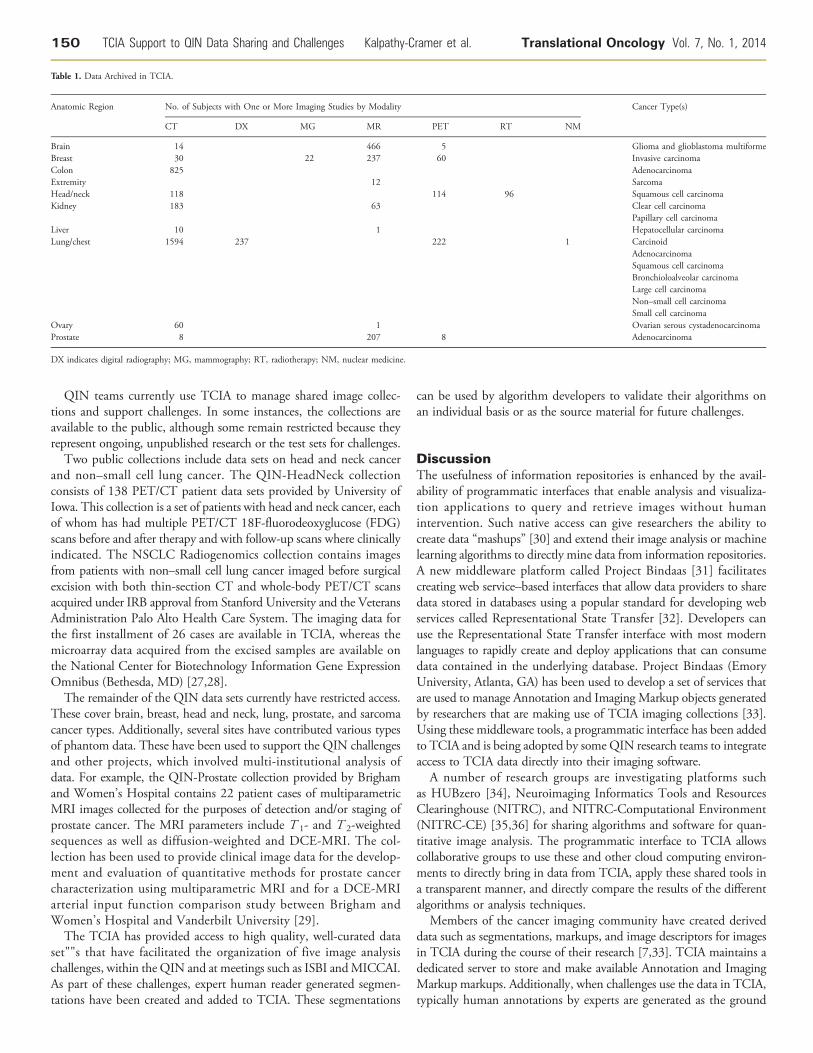
ResultsTCIA supports a large and growing collection of images of a varietyof modalities and anatomic sites as shown in Table 1.
Translational Oncology Vol. 7, No. 1, 2014 TCIA Support to QIN Data Sharing and Challenges Kalpathy-Cramer et al. 149
QIN teams currently use TCIA to manage shared image collec-tions and support challenges. In some instances, the collections areavailable to the public, although some remain restricted because theyrepresent ongoing, unpublished research or the test sets for challenges.
Two public collections include data sets on head and neck cancerand non–small cell lung cancer. The QIN-HeadNeck collectionconsists of 138 PET/CT patient data sets provided by University ofIowa. This collection is a set of patients with head and neck cancer, eachof whom has had multiple PET/CT 18F-fluorodeoxyglucose (FDG)scans before and after therapy and with follow-up scans where clinicallyindicated. The NSCLC Radiogenomics collection contains imagesfrom patients with non–small cell lung cancer imaged before surgicalexcision with both thin-section CT and whole-body PET/CT scansacquired under IRB approval from Stanford University and the VeteransAdministration Palo Alto Health Care System. The imaging data forthe first installment of 26 cases are available in TCIA, whereas themicroarray data acquired from the excised samples are available onthe National Center for Biotechnology Information Gene ExpressionOmnibus (Bethesda, MD) [27,28].
The remainder of the QIN data sets currently have restricted access.These cover brain, breast, head and neck, lung, prostate, and sarcomacancer types. Additionally, several sites have contributed various typesof phantom data. These have been used to support the QIN challengesand other projects, which involved multi-institutional analysis ofdata. For example, the QIN-Prostate collection provided by Brighamand Women’s Hospital contains 22 patient cases of multiparametricMRI images collected for the purposes of detection and/or staging ofprostate cancer. The MRI parameters include T 1- and T 2-weightedsequences as well as diffusion-weighted and DCE-MRI. The col-lection has been used to provide clinical image data for the develop-ment and evaluation of quantitative methods for prostate cancercharacterization using multiparametric MRI and for a DCE-MRIarterial input function comparison study between Brigham andWomen’s Hospital and Vanderbilt University [29].
The TCIA has provided access to high quality, well-curated dataset""s that have facilitated the organization of five image analysischallenges, within the QIN and at meetings such as ISBI andMICCAI.As part of these challenges, expert human reader generated segmen-tations have been created and added to TCIA. These segmentations
can be used by algorithm developers to validate their algorithms onan individual basis or as the source material for future challenges.
DiscussionThe usefulness of information repositories is enhanced by the avail-ability of programmatic interfaces that enable analysis and visualiza-tion applications to query and retrieve images without humanintervention. Such native access can give researchers the ability tocreate data “mashups” [30] and extend their image analysis or machinelearning algorithms to directly mine data from information repositories.A new middleware platform called Project Bindaas [31] facilitatescreating web service–based interfaces that allow data providers to sharedata stored in databases using a popular standard for developing webservices called Representational State Transfer [32]. Developers canuse the Representational State Transfer interface with most modernlanguages to rapidly create and deploy applications that can consumedata contained in the underlying database. Project Bindaas (EmoryUniversity, Atlanta, GA) has been used to develop a set of services thatare used to manage Annotation and Imaging Markup objects generatedby researchers that are making use of TCIA imaging collections [33].Using these middleware tools, a programmatic interface has been addedto TCIA and is being adopted by some QIN research teams to integrateaccess to TCIA data directly into their imaging software.
A number of research groups are investigating platforms suchas HUBzero [34], Neuroimaging Informatics Tools and ResourcesClearinghouse (NITRC), and NITRC-Computational Environment(NITRC-CE) [35,36] for sharing algorithms and software for quan-titative image analysis. The programmatic interface to TCIA allowscollaborative groups to use these and other cloud computing environ-ments to directly bring in data from TCIA, apply these shared tools ina transparent manner, and directly compare the results of the differentalgorithms or analysis techniques.
Members of the cancer imaging community have created deriveddata such as segmentations, markups, and image descriptors for imagesin TCIA during the course of their research [7,33]. TCIA maintains adedicated server to store and make available Annotation and ImagingMarkup markups. Additionally, when challenges use the data in TCIA,typically human annotations by experts are generated as the ground
Table 1. Data Archived in TCIA.
Anatomic Region No. of Subjects with One or More Imaging Studies by Modality Cancer Type(s)
CT DX MG MR PET RT NM
Brain 14 466 5 Glioma and glioblastoma multiformeBreast 30 22 237 60 Invasive carcinomaColon 825 AdenocarcinomaExtremity 12 SarcomaHead/neck 118 114 96 Squamous cell carcinomaKidney 183 63 Clear cell carcinoma
Papillary cell carcinomaLiver 10 1 Hepatocellular carcinomaLung/chest 1594 237 222 1 Carcinoid
AdenocarcinomaSquamous cell carcinomaBronchioloalveolar carcinomaLarge cell carcinomaNon–small cell carcinomaSmall cell carcinoma
Ovary 60 1 Ovarian serous cystadenocarcinomaProstate 8 207 8 Adenocarcinoma
DX indicates digital radiography; MG, mammography; RT, radiotherapy; NM, nuclear medicine.
150 TCIA Support to QIN Data Sharing and Challenges Kalpathy-Cramer et al. Translational Oncology Vol. 7, No. 1, 2014
truth. Such data, e.g., from the ISBI challenges, have been contributedback to TCIA and are stored on the associated challenge wiki pages.They serve as a valuable resource to validate algorithms both withinand outside the context of challenges. Importantly, image segmentationchallenges such as the lung nodule segmentation challenge orga-nized within the QIN and the MICCAI-BRaTS challenge have dem-onstrated the success of automated algorithms in generating suchderived data. In the future, we will explore making use of these validatedalgorithms to generate derived data and add further value to the datain TCIA.As TCIA evolves and grows, its processes must be continually opti-
mized to ensure that new collections of increasing size and complexitycan be brought online in a timely and cost-efficient manner. At somepoint, it becomes inefficient for researchers to download large data setsto their local computing environments due to limitations of networkthroughput and storage requirements. It would then become moreefficient to colocate high-performance computing [37] with large-capacity information resources such as TCIA. Extending TCIA witha user interface that enables researchers to easily and efficiently selectdata and algorithms and launch collocated deep computing jobs thatreturn only the analysis results will greatly enhance the utility of TCIAfor the cancer research community.In summary, the TCIA is a valuable resource for the QIN and the
larger cancer imaging community. TCIA’s rich data sets are generallyhard to obtain for computer scientists. Members of the QIN willcontinue to share high-value data sets in a HIPAA-compliant mannerinto a well-curated and searchable environment. These data sets caninclude high-quality images, imaging metadata, as well as otherclinical data. Such data can facilitate the validation of imaging bio-markers and support reproducible research by providing an avenue toshare the data used in publications or for challenges.The challenges conducted by QIN would not have been pos-
sible without a data-sharing mechanism like TCIA. The TCIA-QINchallenges provide a model and resources for future challenges.In addition to DICOM imaging data, many TCIA collections pro-vide linked clinical, pathology, and even ground truth segmentationdata generated by human readers, which could be used for additionalchallenges. Results from future challenges could readily be made avail-able to serve as benchmarks against which further algorithms couldbe tested.
References[1] Clarke LP, Croft BS, NordstromR, ZhangH, Kellofff G, and Tatum J (2009). Quan-
titative imaging for evaluation of response to cancer therapy.Transl Oncol 2, 195–197.[2] Quantitative Imaging Network Collections. Available at: https://wiki.
cancerimagingarchive.net/x/wwEy. Accessed January 13, 2014.[3] WhiteHouse (2012). Big data fact sheet: Big Data Across the Federal Government.
Available at: http://www.whitehouse.gov/sites/default/files/microsites/ostp/big_data_fact_sheet_final_1.pdf.
[4] Prior F, Clark K, Commean P, Freymann J, Jaffe C, Kirby J, Moore S, Smith K,Tarbox L, Vendt B, et al. (2013). TCIA: an information resource to enable openscience. Conf Proc IEEE Eng Med Biol Soc 2013, 1282–1285.
[5] Hunter L (2013). Radiomics of NSCLC: Quantitative CT Image Feature Characteriza-tion and Tumor Shrinkage Prediction. University of Texas, Houston, TX. Available at:http://digitalcommons.library.tmc.edu/cgi/viewcontent.cgi?article=1365&context=utgsbs_dissertations..
[6] Sivakumar S and Chandrasekar C (2013). Lung nodule detection using fuzzyclustering and support vector machines. Int J Eng Technol 5, 179–185.
[7] Zinn PO, Majadan B, Sathyan P, Singh SK, Majumder S, Jolesz FA, and ColenRR (2011). Radiogenomic mapping of edema/cellular invasion MRI-phenotypesin glioblastoma multiforme. PLoS One 6, e25451.
[8] Stine DD (2009). Federally Funded Innovation Inducement Prizes. DIANE Publishing.Available at: http://www.fas.org/sgp/crs/misc/R40677.pdf.
[9] General Services Administration. A partnership between the public and the govern-ment to solve important challenges. 2014. Available at: https://challenge.gov/.Accessed January 13, 2014.
[10] Kaggle. Kaggle, the leading platform for predictive modeling competitions. 2014.Available at: http://www.kaggle.com/competitions. The DARPA robotics challenge.Available at: http://www.theroboticschallenge.org/. Accessed March 30, 2014.
[11] The DARPA robotics challenge. Available at: http://www.theroboticschallenge.org/,Accessed March 30, 2014.
[12] Ozguner U, Stiller C, and Redmill K (2007). Systems for safety and autonomousbehavior in cars: theDARPAGrandChallenge experience. Proc IEEE 95, 397–412.
[13] Parida SK and Kaufmann SH (2010). The quest for biomarkers in tuberculosis.Drug Discov Today 15, 148–157.
[14] Lakhani KR, Boudreau KJ, Loh PR, Backstrom L, Baldwin C, Lonstein E,Lydon M, MacCormack A, Arnaout RA, and Guinan EC (2013). Prize-basedcontests can provide solutions to computational biology problems. Nat Biotechnol31, 108–111.
[15] Freymann JB, Kirby JS, Perry JH, Clunie DA, and Jaffe CC (2012). Image datasharing for biomedical research—meetingHIPAA requirements for de-identification.J Digit Imaging 25, 14–24.
[16] Radiological Society of North America. The RSNA Clinical Trial Processor.2012. Available at: http://mircwiki.rsna.org/index.php?title=CTP-The_RSNA_Clinical_Trial_Processor: Radiological Society of North America. AccessedJanuary 13, 2014.
[17] Clark K, Vendt B, Smith K, Freymann J, Kirby J, Koppel P, Moore S, Phillips S,Maffitt D, and Pringle M (2013). The Cancer Imaging Archive (TCIA): maintain-ing and operating a public information repository. J Digit Imaging 26, 1045–1057.
[18] National Cancer Institute’s Center for Biomedical Informatics and InformationTechnology. National Biomedical Imaging Archive. 2011. Available at: https://imaging.nci.nih.gov/ncia/login.jsf.
[19] Channin DS, Mongkolwat P, Kleper V, Sepukar K, and Rubin DL (2010). ThecaBIG Annotation and Image Markup project. J Digit Imaging 23, 217–225.
[20] Atlassian. Confluence. 2014. Available at: http://www.atlassian.com/software/confluence. Accessed January 13, 2014.
[21] McNitt-Gray MF, Armato SG III, Meyer CR, Reeves AP, McLennan G, PaisRC, Freymann J, Brown MS, Engelmann RM, and Bland PH (2007). TheLung Image Database Consortium (LIDC) data collection process for noduledetection and annotation. Acad Radiol 14, 1464.
[22] Armato SG III, Meyer CR, Mcnitt-Gray MF, McLennan G, Reeves A, CroftBY, and Clarke LP (2008).RIDER Research Group The Reference ImageDatabase to Evaluate Response to therapy in lung cancer (RIDER) project: aresource for the development of change-analysis software. Clin Pharmacol Ther84, 448–456.
[23] Kalpathy-Cramer J, Zhao B, Goldgof D, Gu Y, Wang X, Gillies R, Yang H, Tan Y,and Napel S (2013). A platform for the comparison of lung nodule segmentationalgorithms: methods and preliminary results. In Radiological Society of North America(RSNA) 99th Scientific Assembly and Annual Meeting, Chicago, IL, December, 2013.
[24] Huang W, Li X, Chen Y, Li X, Chang M-C, Oborski MJ, Malyarenko DI,Muzi M, Jajamovich GH, Fedorov A, et al. (2014). Variations of dynamiccontrast-enhanced magnetic resonance imaging in evaluation of breast cancertherapy response: a multicenter data analysis challenge. Transl Oncol 7, 153–166,
[25] Menze B, ReyesM, Jakab A, Gerstner E, Kirby J, and Farahani K (2013).MICCAIChallenge onMultimodal Brain Tumor Image Segmentation (BRATS). InProceed-ings of the MICCAI Challenge on Multimodal Brain Tumor Image Segmentation(BRATS) 2013. Available at: http://martinos.org/qtim/miccai2013/proc_brats_2013.pdf. Accessed January 31, 2014.
[26] The Cancer Imaging Archive. NCI-ISBI 2013 Challenge - Automated Segmen-tation of Prostate Structures. 2014. Available at: https://wiki.cancerimagingarchive.net/display/Public/NCI-ISBI+2013+Challenge+-+Automated+Segmentation+of+Prostate+Structures. Accessed January 13, 2014.
[27] The Cancer Imaging Archive. NSCLC Radiogenomics. Available at: https://wiki.cancerimagingarchive.net/display/Public/NSCLC+Radiogenomics. AccessedJanuary 13, 2014.
[28] Gevaert O, Xu J, Hoang CD, Leung AN, Xu Y, Quon A, Rubin DL, Napel S, andPlevritis SK (2012). Non–small cell lung cancer: identifying prognostic imagingbiomarkers by leveraging public gene expression microarray data—methods andpreliminary results. Radiology 264, 387–396.
[29] Fedorov A, Fluckiger J, Ayers GD, Li X, Gupta SN, Tempany C, Mulkern R,Yankeelov TE, and Fennessy FM (2014). A comparison of two methods for
Translational Oncology Vol. 7, No. 1, 2014 TCIA Support to QIN Data Sharing and Challenges Kalpathy-Cramer et al. 151

estimating DCE-MRI parameters via individual and cohort based AIFs in prostatecancer: a step towards practical implementation. Magn Reson Imaging 32(4),321–329.
[30] Makki SK and Sangtani J (2008). Data mashups & their applications in enter-prises. In SK Makki, and J Sangtan (Eds.), Internet and Web Applications andServices, 2008. ICIW’08. Third International Conference on: IEEE pp. 445–450.
[31] Sharma A and Saghar YN (2013). Project Bindaas. Available at: http://imaging.cci.emory.edu/wiki/display/BDS/Downloads. Accessed January 31, 2014.
[32] Fielding RT (2000). Architectural Styles and the Design of Network-BasedSoftware Architectures. University of California, Irvine.
[33] Gutman DA, Cooper LA, Hwang SN, Holder CA, Gao J, Aurora TD, DunnWD, Scarpace L, Mikkelsen T, and Jain R (2013). MR imaging predictors of
molecular profile and survival: multi-institutional study of the TCGA glio-blastoma data set. Radiology 267, 560–569.
[34] McLennan M and Kennell R (2010). HUBzero: a platform for dissemination andcollaboration in computational science and engineering. Comput Sci Eng 12, 48–53.
[35] Luo XZ, Kennedy DN, and Cohen Z (2009). Neuroimaging informatics tools andresources clearinghouse (NITRC) resource announcement. Neuroinformatics 7, 55–56.
[36] Kennedy DN and Haselgrove C. The three NITRC’s: software, data and cloudcomputing for brain science and cancer imaging research. Front Neuroinform.Conference Abstract: Neuroinformatics 2013, Stockholm, Sweden, 27 Aug–29 Aug, 2013. DOI:10.3389/conf.fninf.2013.09.00024
[37] Bell G, Gray J, and Szalay A (2006). Petascale computational systems. Computer39, 110–112.
152 TCIA Support to QIN Data Sharing and Challenges Kalpathy-Cramer et al. Translational Oncology Vol. 7, No. 1, 2014


![Quantitative Magnetic Particle Imaging Monitors the ...Magnetic particle imaging (MPI) [16–21], an imaging modality distinct from MRI, produces line-arly quantitative images of iron](https://img.pdfslide.us/doc/110x75/6124fca4732f0c68d25dc95d/quantitative-magnetic-particle-imaging-monitors-the-magnetic-particle-imaging.jpg)