Embed Size (px)
Citation preview
rev bras hematol hemoter. 2 0 1 7;3 9(3):252–258
www.rbhh.org
Revista Brasileira de Hematologia e HemoterapiaBrazilian Journal of Hematology and Hemotherapy
Original article
Quantitative flow cytometric evaluation of CD200,CD123, CD43 and CD52 as a tool for the differentialdiagnosis of mature B-cell neoplasms
Elissandra Machado Arlindoa,∗, Natália Aydos Marcondesa,b, Flavo Beno Fernandesb,Gustavo Adolpho Moreira Faulhabera,b
a Universidade Federal do Rio Grande do Sul (UFRGS), Porto Alegre, RS, Brazilb Laboratório Zanol, Porto Alegre, RS, Brazil
a r t i c l e i n f o
Article history:
Received 16 February 2017
Accepted 2 May 2017
Available online 29 May 2017
Keywords:
Mature B-cell neoplasms
CD200
CD52
CD43
CD123
a b s t r a c t
Background: Distinction between mature B-cell neoplasms can be difficult due to overlap-
ping of immunologic features and clinical manifestations. This study investigated whether
quantifying mean fluorescence intensity of four monoclonal antibodies in a flow cytometry
panel is useful for the differential diagnosis and characterization of these disorders.
Methods: The expressions of CD52, CD200, CD123 and CD43 were analyzed in samples from
124 patients with mature B-cell neoplasms. The quantitative estimation of these antigens
was assessed by mean fluorescence intensity.
Results: The cases included were 78 chronic lymphocytic leukemias, three atypical chronic
lymphocytic leukemias, six marginal zone lymphomas, 11 splenic marginal zone lym-
phomas, nine lymphoplasmacytic lymphomas, six mantle cell lymphomas, two hairy cell
leukemias, two hairy cell leukemias variant, five follicular lymphomas, one Burkitt lym-
phoma and one diffuse large B-cell lymphoma. The mean fluorescence intensity of CD200
was higher in atypical chronic lymphocytic leukemia, chronic lymphocytic leukemia and
hairy cell leukemia cases. CD123 showed higher mean fluorescence intensities in hairy cell
leukemia cells. Chronic lymphocytic leukemia, atypical chronic lymphocytic leukemia and
mantle cell lymphoma had higher expression of CD43 and all follicular lymphoma cases
had very low mean fluorescence intensity values. CD52 expression was consistently positive
among all cases.
Conclusion: Quantitative evaluation of these markers can be a useful additional tool to better
identify some types of mature B-cell neoplasms.
o Bra
© 2017 Associacaby Elsevier Editora Lt
∗ Corresponding author at: Rua Ramiro Barcelos, 2350, 90035-903 Porto
E-mail address: [email protected] (E.M. Arlindo).http://dx.doi.org/10.1016/j.bjhh.2017.05.0021516-8484/© 2017 Associacao Brasileira de Hematologia, Hemoterapiaopen access article under the CC BY-NC-ND license (http://creativecom
sileira de Hematologia, Hemoterapia e Terapia Celular. Published
da. This is an open access article under the CC BY-NC-ND license
(http://creativecommons.org/licenses/by-nc-nd/4.0/).
Alegre, RS, Brazil.
e Terapia Celular. Published by Elsevier Editora Ltda. This is anmons.org/licenses/by-nc-nd/4.0/).

er. 2 0
I
Moogmt(tgfi
eueMtfeiaaiBfCLtilp(ic
sMblhosbt
e
M
AstwCsb
rev bras hematol hemot
ntroduction
ature B-cell neoplasms (MBCN) account for around 80%f all lymphoid neoplasms and comprise a broad spectrumf disorders with different morphologies, clinical aspects,enetics and immunophenotypes. However, they have a moreature lymphoid progenitor in common compared to imma-
ure neoplasms. According to the World Health OrganizationWHO), immunophenotypic similarities of these cells at a cer-ain stage of maturation in conjunction with morphological,enetic and clinical findings allow these diseases to be classi-ed and diagnosed.1
Immunophenotyping by flow cytometry is a fast and cost-ffective technique widely used for the diagnosis and followp of hematological disorders. Multiparametric flow cytom-try (MFC) has become more complex in the diagnosis ofBCN due to the availability of several markers, in addition
o the high number of entities. Usually, the analysis is per-ormed separately for each sample aliquot labeled with four toight monoclonal antibodies (MoAbs); an expert interprets themmunophenotypic profile of neoplastic B-cells and specifies
diagnosis. Preferred markers are those able to differenti-te B-cells from other cells, define the maturation stage anddentify phenotypic aberrations. Among these are the pan-cell markers (CD19 and CD20) and those used for the dif-erential diagnosis (CD10, CD5, CD103, CD43, CD23, CD49d,D81, CD200, CXCR5, immunoglobulin (Ig)M and Kappa andambda chains). A differential diagnosis can be difficult, dueo overlapping immunophenotypic features and similar clin-cal manifestations, such as cases of differentiation betweenymphoplasmacytic lymphoma (LPL) and marginal zone lym-homa (MZL), or between diffuse large B-cell lymphoma
DLBCL) and follicular lymphoma. For the final diagnosis, its important to integrate results of MFC with morphological,linical and cytogenetic analysis.1,2
Some markers are known for their expression in MBCN,uch as the positivity of CD52,3 and negativity of CD43 inZL,4 the usefulness of CD200 in the differential diagnosis
etween chronic lymphocytic leukemia (CLL) and mantle cellymphoma (MCL),5 and the role of CD123 in the diagnosis ofairy cell leukemia (HCL).6 However, the quantitative analysisf these markers in MCBN is seldom discussed, and its expres-ion may be important not only for the differential diagnosis,ut also for prognosis and potential therapeutic factors, givenhe existence of target drugs against some of these markers.
The aim of this study was to evaluate quantitatively thexpression of CD200, CD123, CD43 and CD52 in MBCN.
ethods
ll MCBN cases diagnosed in a reference laboratory in theouth of Brazil from October 2014 to June 2015 were consecu-ively included in the study. Specimens sent for reassessment
ere not included. The study was approved by the local Ethicsommittee; written informed consent was deemed unneces-ary after the signature of a commitment term for the use ofiological material and associated information by researchers.1 7;3 9(3):252–258 253
The panel with the four MoAbs under study was dis-tributed using the following fluorescences: CD123 FITC (clone7G3), CD52 PE (clone 4C8), CD43 APC (clone 1G10) and CD200PerCP-Cy5.5 (clone MRC OX-104). All MoAbs were purchasedfrom BD Biosciences (San Diego, USA). About 1,000,000 cellsfrom whole blood or bone marrow samples anticoagulatedwith EDTA were incubated for 20 min in the dark at roomtemperature with each MoAb. Red blood cells were lysed byincubation with Excellyse I (EXBIO, Praha, CZ), followed byincubation with distilled water. Samples were washed andresuspended in phosphate buffered saline (PBS). All reagentswere used according to the manufacturer’s instructions. Allsamples were processed within 48 h of collection.7
Immediately after preparation, samples were analyzed ona FACSCalibur flow cytometer using CellQuestTM Pro software(BD Biosciences, San Diego, CA, USA). About 100,000 events persample were obtained. InfinicityTM Flow Cytometry version 1.7software (Cytognos, SL, ES) was used for data analysis. Forthe gating strategy, debris were removed, based on forward-scatter (FSC) and side-scatter (SSC) distribution. Neoplasticcells were initially identified by positivity of the CD19 and CD20and then in accordance with the expression of other panelmarkers for MBCN. The mean fluorescence intensities (MFIs)of neoplastic cells were recorded in arbitrary units from 0 to104.
Reproducibility of the fluorescence intensities was pre-served by calibration and daily quality control procedures.Calibrite beads (BD Biosciences, Sao Diego, USA) were usedin order to ensure standardization. All diagnosis were estab-lished in accordance with the WHO criteria.1
Medians and interquartile ranges for each MFI of MoAbwere calculated. The Mann–Whitney test was used to calcu-late the statistical significance of differences between groups.Burkitt lymphoma and DLBCL were not included in the sta-tistical analysis because there was only one case of eachdisease. Differences were considered significant when thep-value <0.05. All statistical analyses were performed usingthe Statistical Package for the Social Sciences v. 18.0 software(SPSS, Chicago, USA).
Results
Immunophenotypic analysis was performed in 124 samples– 67 of peripheral blood (54%) and 57 of bone marrow (46%)– from patients diagnosed with MBCN. Patient mean age was69.3 ± 12.1 years old and 51.6% were men. The distribution ofdifferential diagnoses of MBCN is described in Table 1. Themost common diagnosis, as expected, was CLL due to itshigh prevalence, and the rarest were Burkitt lymphoma andDLBCL. The rate of neoplastic B-cells among total cells persample ranged from 3.7% to 97.1%, with a mean of 52.3 ± 25.4%.Median MFIs for each MoAb of the different groups are shownin Table 2.
The MFI of CD200 was higher in atypical chronic lym-phocytic leukemias (aCLL), CLL and HCL as can be seen in
Figure 1. The MFI of CD200 was higher in aCLL comparedto MCL (p-value = 0.020), follicular lymphoma (p-value = 0.034),MZL (p-value = 0.020) and splenic marginal zone lymphomas(SMZL) (p-value = 0.036). The MFI of CD200 was higher in CLL
254 rev bras hematol hemoter. 2 0 1 7;3 9(3):252–258
400,0
300,0
200,0
100,0
70,0
50,0
30,0
10,04,0
,0aCLL BL CLL DLBCL
CD
200
MF
I
FL HCL HCLv LPL MCL MZL SMZL
Figure 1 – Boxplot showing MFI of CD200 in 124 cases of mature B-cell neoplasms. aCLL: atypical chronic lymphocyticleukemia; BL: Burkitt lymphoma; CLL: chronic lymphocytic leukemia; DLBCL: diffuse large B-cell lymphoma; FL: follicularlymphoma; HCL: hairy cell leukemia; HCLv: hairy cell leukemia variant; LPL: lymphoplasmacytic lymphoma; MCL: mantlecell lymphoma; MZL: marginal zone lymphoma; SMZL: splenic m
Table 1 – Distribution of mature B-cell neoplasms.
Diagnosis Numberof cases
(%)
Atypical chronic lymphocytic leukemia 3 2.4Burkitt lymphoma 1 0.8Chronic lymphocytic leukemia 78 62.9Diffuse large B-cell lymphoma 1 0.8Follicular lymphoma 5 4.0Hairy cell leukemia 2 1.6Hairy cell leukemia variant 2 1.6Lymphoplasmacytic lymphoma 9 7.3Mantle cell lymphoma 6 4.8
Marginal zone lymphoma 6 4.8Splenic marginal zone lymphoma 11 8.9compared to LPL (p-value <0.001), MCL (p-value <0.001), follicu-lar lymphoma (p-value = 0.001), MZL (p-value <0.001) and SMZL(p-value <0.001). The MFI of CD200 was higher in LPL compared
Table 2 – Median MFI values of evaluated markers per disease
Diagnosis CD200
Atypical chronic lymphocytic leukemia 113.7 (70.4/122.2) 7Burkitt lymphoma 4.5 (4.5/4.5) 5Chronic lymphocytic leukemia 61.1 (41.4/89.2) 8Diffuse large B-cell lymphoma 3.8 (3.8/3.8) 6Follicular lymphoma 2.6 (1.8/11.4) 6Hairy cell leukemia 220.3 (163.1/277.5) 83Hairy cell leukemia variant 36.1 (22.1/50.1) 22Lymphoplasmacytic lymphoma 14.0 (12.30/40.2) 9Mantle cell lymphoma 3.5 (2.1/4.1) 13Marginal zone lymphoma 8.3 (4.5/13.2) 11Splenic marginal zone lymphoma 7.6 (4.0/22.9) 12
Data are shown as median (percentile 25/75).
arginal zone lymphoma.
to MCL (p-value = 0.003). The MFI of CD200 was higher in HCLcompared to MCL (p-value = 0.046), MZL (p-value = 0.046) andSMZL (p-value = 0.030). The MFI of CD200 of HCL was about6-fold higher when compared to hairy cell leukemia variant(HCLv), however due to the small number of cases includedit was not possible to show statistical difference in CD200expression between these two neoplasms (p-value = 0.121).
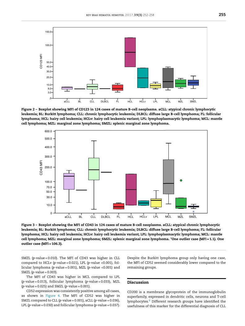
CD123 had higher MFIs in HCL cells as shown in Figure 2.The MFI of CD123 was higher in HCL compared to CLL (p-value = 0.016), LPL (p-value = 0.034), MCL (p-value = 0.046), MZL(p-value = 0.046) and SMZL (p-value = 0.030). The MFI of CD123was higher in SMZL compared to CLL (p-value = 0.003), LPL (p-value = 0.037) and follicular lymphoma (p-value = 0.050). MFIof CD123 had a trend to be higher in HCL compared to HCLv(p-value = 0.121).
Figure 3 illustrates the expression of CD43. All follicularlymphoma cases had very low MFI values. The MFI of CD43was higher in aCLL compared to MZL (p-value = 0.034) and
category.
CD123 CD43 CD52
.5 (5.7/7.6) 133.7 (74.5/154.3) 765.2 (546.5/814.7)
.9 (5.9/5.9) 57.0 (57.0/57.0) 143.0 (143.0/143.0)
.0 (5.6/10.7) 179.8 (112.8/278.7) 777.9 (469.3/1101.0)
.1 (6.1/6.1) 160.9 (160.9/160.9) 549.5 (549.5/549.5)
.4 (4.5/10.0) 6.8 (3.6/13.1) 686.1 (455.9/820.2)
.2 (61.3/105.0) 79.0 (44.4/113.7) 956.4 (911.3/1001.6)
.0 (14.4/29.5) 14.6 (11.9/17.2) 990.3 (705.9/1274.8)
.0 (6.7/11.1) 20.7 (6.5/25.3) 697.1 (447.7/1150.8)
.9 (8.6/30.9) 90.1 (48.6/226.7) 704.5 (538.7/1375.5)
.7 (8.1/16.0) 7.8 (5.0/23.6) 1326.1 (737.5/1847.0)
.3 (9.4/16.1) 7.1 (6.5/8.6) 1297.6 (1096.9/1861.8)
rev bras hematol hemoter. 2 0 1 7;3 9(3):252–258 255
150,0
100,0
50,0
40,0
30,0
20,0
10,0
6,0
3,0
,0aCLL BL CLL DLBCL
CD
123
MF
I
FL HCL HCLv LPL MCL MZL SMZL
Figure 2 – Boxplot showing MFI of CD123 in 124 cases of mature B-cell neoplasms. aCLL: atypical chronic lymphocyticleukemia; BL: Burkitt lymphoma; CLL: chronic lymphocytic leukemia; DLBCL: diffuse large B-cell lymphoma; FL: follicularlymphoma; HCL: hairy cell leukemia; HCLv: hairy cell leukemia variant; LPL: lymphoplasmacytic lymphoma; MCL: mantlecell lymphoma; MZL: marginal zone lymphoma; SMZL: splenic marginal zone lymphoma.
600.0
500.0
400.0
300.0
200.0
100.0
70.0
50.0
30.0
10.0
0aCLL BL CLL DLBCL
CD
43 M
FI
FL HCL HCLv LPL MCL MZL SMZL
Figure 3 – Boxplot showing the MFI of CD43 in 124 cases of mature B-cell neoplasms. aCLL: atypical chronic lymphocyticleukemia; BL: Burkitt lymphoma; CLL: chronic lymphocytic leukemia; DLBCL: diffuse large B-cell lymphoma; FL: follicularlymphoma; HCL: hairy cell leukemia; HCLv: hairy cell leukemia variant; LPL: lymphoplasmacytic lymphoma; MCL: mantlecell lymphoma; MZL: marginal zone lymphoma; SMZL: splenic marginal zone lymphoma. *One outlier case (MFI = 1.1). Oneo
SclS
((
aSL
utlier case (MFI = 106.3).
MZL (p-value = 0.010). The MFI of CD43 was higher in CLLompared to HCLv (p-value = 0.021), LPL (p-value <0.001), fol-icular lymphoma (p-value = 0.001), MZL (p-value <0.001) andMZL (p-value = 0.003).
The MFI of CD43 was higher in MCL compared to LPLp-value = 0.013), follicular lymphoma (p-value = 0.033), MZLp-value = 0.025) and SMZL (p-value = 0.001).
CD52 expression was consistently positive among all cases,s shown in Figure 4. The MFI of CD52 was higher inMZL compared to CLL (p-value = 0.001), aCLL (p-value = 0.036),PL (p-value = 0.030) and follicular lymphoma (p-value = 0.037).
Despite the Burkitt lymphoma group only having one case,the MFI of CD52 seemed considerably lower compared to theremaining groups.
Discussion
CD200 is a membrane glycoprotein of the immunoglobulinsuperfamily, expressed in dendritic cells, neurons and T-celllymphocytes.8 Different research groups have identified theusefulness of this marker for the differential diagnosis of CLL

256 rev bras hematol hemoter. 2 0 1 7;3 9(3):252–258
6000
5000
4000
3000
2000
1000
700500
300
100
30
0aCLL BL CLL DLBCL
CD
52 M
FI
FL HCL HCLv LPL MCL MZL SMZL
Figure 4 – Boxplot showing the MFI of CD52 in 124 cases of mature B-cell neoplasms. aCLL: atypical chronic lymphocyticleukemia; BL: Burkitt lymphoma; CLL: chronic lymphocytic leukemia; DLBCL: diffuse large B-cell lymphoma; FL: follicularlymphoma; HCL: hairy cell leukemia; HCLv: hairy cell leukemia variant; LPL: lymphoplasmacytic lymphoma; MCL: mantle
nic m
cell lymphoma; MZL: marginal zone lymphoma; SMZL: spleversus MCL, including one analyzing a Brazilian population.9
The current study had similar results, and, although one CLLcase showed low expression of this marker, 100% of MCL caseshad significantly lower expressions compared to CLL. In arecent study of CLL patients, cases with low MFI of CD200had a shorter time-to-treatment compared to patients withhigher expression of the marker.10 As previously describedin the immunohistochemical analysis by Pillai et al., CD200can be extremely helpful in the differential diagnosis betweenHCL and HCLv11; in the present study, HCL cases showed atrend to have higher expressions than its variant form. Therewas no difference in CD200 expression between CLL and aCLL(CD5-negative) cases.
CD200 appears to be useful in the differential diagnosisbetween CD5-positive LPL and CLL, as it was lower in theformer compared to the latter. However, if analyzed qualita-tively, this difference could not have been noted, since 30% ofcases showed intermediate positivity, which is in agreementwith what was described in a previous study analyzing theexpression of this marker by immunohistochemistry.12 CD200has also been described as useful to differentiate between CLLand MZL; the results of the present study confirm the findingsdescribed by immunohistochemistry with a lower expressionof the marker in MZL and SMZL.8
CD123 is a subunit of the interleukin-3 receptor. It has beendescribed in several hematological neoplasms.13 Recently,CD123 expression was studied quantitatively using MFI byGarcia-Dabrio et al. who correlated its implication as a prog-nostic factor in de novo acute myeloid leukemia (AML),14 andSL-101, an anti-CD123 antibody-conjugate, is under study for
15
the treatment of CD123-positive AML. The results of thecurrent study are in agreement with previous studies investi-gating the higher expression of this marker in HCL,16,17 sincehigher MFI values were identified in HCL cases and a trend ofarginal zone lymphoma.
HCL to have higher CD123 expression compared to HCLv caseswas also observed. This study identified a higher expression ofCD123 in SMZL compared to CLL, LPL and follicular lymphomacases.
According to a previous study, CD43 expression is simi-lar among CD10-positive MBCN (follicular lymphoma, Burkittlymphoma and DLBCL), one of the most complicated differ-ential diagnoses.18 All five cases of follicular lymphoma hadextremely low expressions when compared to aCLL, CLL andMCL, and the only case of DLBCL expressed this marker atan intermediate intensity as described by previous studies,19
however, analysis of this group was impaired by the low fre-quency of cases.
During the ontogeny of B-cells, CD43 is expressed in earlystages and is lost in intermediate stages, but it is expressedagain in plasma cells and activated mature B-cells.20 Inagreement with the literature, these results identified higherexpression in aCLL, CLL and MCL, and lower expression in MZLand SMZL, probably due to clonality in the intermediate stageof the disease. Despite CD43 not being a relevant marker forthe differential diagnosis of MCL and MZL, caution should betaken in the interpretation of these data with respect to thedifferential diagnosis of MCL versus CD5-positive MZL. OneMCL case (16.0%) had a low expression of this marker similar toMZL and one MZL case (5.8%) showed intermediate expressionsimilar to that seen in MCL.
CD52 is a cell surface glycoprotein whose function is poorlyunderstood and is expressed in lymphocytes, monocytes,macrophages and a few dendritic cells.21 Its expression hasbeen described in several MBCN and positivity for this markerinvolves specific treatments for diseases such as CLL and
LPL.3,22,23 Furthermore, soluble CD52 has been identified as amarker of disease activity in CLL.24 This study identified theexpression of CD52 in 100% of analyzed cases. Although these
er. 2 0
rsdMBcCn
mrcfisst
ttlcdcttgaouia
itfD
C
IofudScb
C
T
A
WHp
r
1
1
1
1
1
1
rev bras hematol hemot
esults seem to be in contrast with those described by othertudies, such as Rodig et al. who reported negativity for someiseases such as Burkitt lymphoma and DLBCL,3 the lowestFIs in the samples of the present study were exactly for
urkitt lymphoma and DLBCL. Chuang et al. analyzed MCLases and identified that only 60% of cases were positive forD52, but the difference in the immunohistochemical tech-ique used may explain the distinct results.25
Since fluorescence intensity measures are important deter-inants in the analysis of leukemias and lymphomas,26
eliable methods for measuring MFI are important for theorrect data interpretation, and, consequently, correct classi-cation of hematological malignancies. For this reason, termsuch as “weak” and “strong” are useful, but, as Henderson et al.uggested, perhaps quantitative values could be used in ordero further explore the information provided.27
There are numerous variables involved in the quantita-ive determination of fluorescence intensity, some related tohe specificity in the chosen MoAb, sample type, anticoagu-ant employed, autofluorescence, type of fixation, cytometerompensation and unit of measurement used to report theata, among others. Even when all these variables are wellontrolled, some caution in interpreting the data should beaken, but given the increasing universal standardization inhe field of immunophenotyping, this type of analysis mayain ground, allowing for comparative figures with greater reli-bility over time. Until then, each center can develop the usef data in MFI according to their case series, in addition tosing the data available in the literature, as long as it is crit-
cally interpreted and the limitations to this type of analysisre understood.
This study has some limitations. The main one is that onlymmunophenotyping and biopsy results as complementaryests for the disease entity definition were accessible. Besides,ew cases of non-CLL cases, such as Burkitt lymphoma andLBCL, were available.
onclusion
n conclusion, the results of this study show the usefulnessf already known markers in MBCN such as CD200 in the dif-erential diagnosis of CLL and MCL. These data suggest thesefulness of more complex analyses quantifying MFI in theifferential diagnosis of MBCN, such as CD52 expression inMZL versus LPL, and CD43 expression in MZL, LPL and SMZLompared to MCL and CLL. Nevertheless, these results shoulde further explored in analyses using a larger sample size.
onflicts of interest
he authors declare no conflicts of interest.
cknowledgments
e acknowledge the Fundo de Incentivo a Pesquisa (FIPE) ofospital de Clínicas de Porto Alegre (HCPA) for financial sup-ort.
1 7;3 9(3):252–258 257
e f e r e n c e s
1. Swerdlow SH, International Agency for Research on CancerWorld Health Organization. WHO classification of tumours ofhaematopoietic and lymphoid tissues. 4th ed. Lyon, France:International Agency for Research on Cancer; 2008.
2. van Dongen JJ, Lhermitte L, Böttcher S, Almeida J,van der Velden VH, Flores-Montero J, et al. EuroFlow antibodypanels for standardized n-dimensional flow cytometricimmunophenotyping of normal, reactive and malignantleukocytes. Leukemia. 2012;26(9):1908–75.
3. Rodig SJ, Abramson JS, Pinkus GS, Treon SP, Dorfman DM,Dong HY, et al. Heterogeneous CD52 expression amonghematologic neoplasms: implications for the use ofalemtuzumab (CAMPATH-1H). Clin Cancer Res.2006;12(23):7174–9.
4. Berger F, Felman P, Thieblemont C, Pradier T, Baseggio L,Bryon PA, et al. Non-MALT marginal zone B-cell lymphomas:a description of clinical presentation and outcome in 124patients. Blood. 2000;95(6):1950–6.
5. Palumbo GA, Parrinello N, Fargione G, Cardillo K, Chiarenza A,Berretta S, et al. CD200 expression may help in differentialdiagnosis between mantle cell lymphoma and B-cell chroniclymphocytic leukemia. Leuk Res. 2009;33(9):1212–6.
6. Del Giudice I, Matutes E, Morilla R, Morilla A,Owusu-Ankomah K, Rafiq F, et al. The diagnostic value ofCD123 in B-cell disorders with hairy or villous lymphocytes.Haematologica. 2004;89(3):303–8.
7. Davis BH, Dasgupta A, Kussick S, Han JY, Estrellado A.Validation of cell-based fluorescence assays: practiceguidelines from the ICSH and ICCS – part II – preanalyticalissues. Cytometry B Clin Cytom. 2013;84(5):286–90.
8. Dorfman DM, Shahsafaei ACD200. (OX-2 membraneglycoprotein) expression in b cell-derived neoplasms. Am JClin Pathol. 2010;134(5):726–33.
9. Sandes AF, de Lourdes Chauffaille M, Oliveira CR, Maekawa Y,Tamashiro N, Takao TT, et al. CD200 has an important role inthe differential diagnosis of mature B-cell neoplasms bymultiparameter flow cytometry. Cytometry B Clin Cytom.2014;86(2):98–105.
0. Miao Y, Fan L, Wu YJ, Xia Y, Qiao C, Wang Y, et al. Lowexpression of CD200 predicts shorter time-to-treatment inchronic lymphocytic leukemia. Oncotarget.2016;7(12):13551–62.
1. Pillai V, Pozdnyakova O, Charest K, Li B, Shahsafaei A,Dorfman DM. CD200 flow cytometric assessment andsemiquantitative immunohistochemical stainingdistinguishes hairy cell leukemia from hairy cellleukemia-variant and other B-cell lymphoproliferativedisorders. Am J Clin Pathol. 2013;140(4):536–43.
2. Alapat D, Coviello-Malle J, Owens R, Qu P, Barlogie B,Shaughnessy JD, et al. Diagnostic usefulness and prognosticimpact of CD200 expression in lymphoid malignancies andplasma cell myeloma. Am J Clin Pathol. 2012;137(1):93–100.
3. Moretti S, Lanza F, Dabusti M, Tieghi A, Campioni D,Dominici M, et al. CD123 (interleukin 3 receptor alpha chain).J Biol Regul Homeost Agents. 2001;15(1):98–100.
4. García-Dabrio MC, Hoyos M, Brunet S, Tormo M, Ribera JM,Esteve J, et al. Complex measurements may be required toestablish the prognostic impact of immunophenotypicmarkers in AML. Am J Clin Pathol. 2015;144(3):484–92.
5. Han L, Jorgensen JL, Brooks C, Shi C, Zhang Q, Nogueras
González GM, et al. Anti-leukemia efficacy and mechanismsof action of SL-101, a novel anti-CD123 antibody-conjugate, inacute myeloid leukemia. Clin Cancer Res. 2017;23(13):3385–95.
oter.
1
1
1
1
2
2
2
2
2
2
2
258 rev bras hematol hem
6. Stetler-Stevenson M, Tembhare PR. Diagnosis of hairy cellleukemia by flow cytometry. Leuk Lymphoma. 2011;52 Suppl.2:11–3.
7. Venkataraman G, Aguhar C, Kreitman RJ, Yuan CM,Stetler-Stevenson M. Characteristic C.D103, CD123 expressionpattern defines hairy cell leukemia: usefulness of CD123 andCD103 in the diagnosis of mature B-cell lymphoproliferativedisorders. Am J Clin Pathol. 2011;136(4):625–30.
8. Schniederjan SD, Li S, Saxe DF, Lechowicz MJ, Lee KL,Terry PD, et al. A novel flow cytometric antibody panel fordistinguishing Burkitt lymphoma from CD10+ diffuse largeB-cell lymphoma. Am J Clin Pathol. 2010;133(5):718–26.
9. Ma XB, Zheng Y, Yuan HP, Jiang J, Wang YP. CD43 expressionin diffuse large B-cell lymphoma, not otherwise specified:CD43 is a marker of adverse prognosis. Hum Pathol.2015;46(4):593–9.
0. Wikén M, Björck P, Axelsson B, Perlmann P. Induction of CD43expression during activation and terminal differentiation ofhuman B cells. Scand J Immunol. 1988;28(4):457–64.
1. Hale G, Xia MQ, Tighe HP, Dyer MJ, The Waldmann H.
CAMPATH-1 antigen (CDw52). Tissue Antigens.1990;35(3):118–27.2. Rossmann ED, Lundin J, Lenkei R, Mellstedt H, Osterborg A.Variability in B-cell antigen expression: implications for the
2
2 0 1 7;3 9(3):252–258
treatment of B-cell lymphomas and leukemias withmonoclonal antibodies. Hematol J. 2001;2(5):300–6.
3. Owen RG, Hillmen P, Rawstron AC. CD52 expression inWaldenstrom’s macroglobulinemia: implications foralemtuzumab therapy and response assessment. ClinLymphoma. 2005;5(4):278–81.
4. Vojdeman FJ, Herman SE, Kirkby N, Wiestner A,van T’ Veer MB, Tjønnfjord GE, et al. Soluble CD52 is anindicator of disease activity in chronic lymphocytic leukemia.Leuk Lymphoma. 2017:1–7.
5. Chuang SS, Huang WT, Hsieh PP, Tseng HH, Campo E,Colomer D, et al. Mantle cell lymphoma in Taiwan:clinicopathological and molecular study of 21 cases includingone cyclin D1-negative tumor expressing cyclin D2. Pathol Int.2006;56(8):440–8.
6. Borowitz MJ, Bray R, Gascoyne R, Melnick S, Parker JW,Picker L, et al. U.S.-Canadian Consensus recommendations onthe immunophenotypic analysis of hematologic neoplasia byflow cytometry: data analysis and interpretation. Cytometry.1997;30(5):236–44.
7. Henderson LO, Marti GE, Gaigalas A, Hannon WH, Vogt RF.Terminology and nomenclature for standardization inquantitative fluorescence cytometry. Cytometry.1998;33(2):97–105.


![Digital Still Camera - Sony · model name1[MVC-CD200] masterpage:Right filename[D:\WORKS\CD\3067951111\3067951111MVCCD200UC\01COV-MVCCD200UC\010cov.fm] 3-067-951-11(1) Digital Still](https://img.pdfslide.us/doc/110x75/5b096b0e7f8b9a404d8dcc79/digital-still-camera-sony-name1mvc-cd200-masterpageright-filenamedworkscd30679511113067951111mvccd200uc01cov-mvccd200uc010covfm.jpg)