Embed Size (px)
Citation preview
E-Mail [email protected]
Original Paper
Audiol Neurotol 2015;20:39–50 DOI: 10.1159/000362780
Quantifying the Vestibulo-Ocular Reflex with Video-Oculography: Nature and Frequency of Artifacts
Georgios Mantokoudis a Ali S. Saber Tehrani a Jorge C. Kattah c Karin Eibenberger b Cynthia I. Guede c David S. Zee a David E. Newman-Toker a, b
Departments of a Neurology and b Otolaryngology – Head and Neck Surgery, Johns Hopkins University School of Medicine, Baltimore, Md. , and c University of Illinois College of Medicine, Peoria, Ill. , USA
Introduction
The head impulse test (HIT), first described by Hal-magyi and Curthoys in 1988 [Halmagyi and Curthoys, 1988], is a critical component of bedside assessment of vestibular function. The technique leverages a high-accel-eration (1,000–6,000°/s 2 ), rapid (100–200°/s), low-ampli-tude (10–20°) head rotation to assess the integrity of the vestibulo-ocular reflex (VOR). The patient visually fix-ates on a stationary target. If the clinician detects a cor-rective eye movement immediately after the head rota-tion (‘refixation saccade’), a deficient VOR response is indirectly inferred. The HIT can be used to assess the function of each semicircular canal individually [Halma-gyi et al., 2001].
In specialty practice, the horizontal HIT (h-HIT) is now widely used to assist in clinical diagnosis of periph-eral vestibular disorders [Curthoys, 2012; Strupp and Brandt, 2013] such as vestibular neuritis [Blödow et al., 2013], unilateral vestibular loss [Weber et al., 2008], and bilateral vestibulopathy [Petersen et al., 2013]. Impulsive testing of the vertical semicircular canals is useful for di-agnosing inferior vestibular neuritis [Kim and Kim, 2012], postsurgical follow-up of superior canal dehis-
Key Words
Eye movement measurements · Artifacts · Vertigo · Vestibular neuritis · Stroke
Abstract
Video-oculography devices are now used to quantify the vestibulo-ocular reflex (VOR) at the bedside using the head impulse test (HIT). Little is known about the impact of disrup-tive phenomena (e.g. corrective saccades, nystagmus, fixa-tion losses, eye-blink artifacts) on quantitative VOR assess-ment in acute vertigo. This study systematically character-ized the frequency, nature, and impact of artifacts on HIT VOR measures. From a prospective study of 26 patients with acute vestibular syndrome (16 vestibular neuritis, 10 stroke), we classified findings using a structured coding manual. Of 1,358 individual HIT traces, 72% had abnormal disruptive saccades, 44% had at least one artifact, and 42% were unin-terpretable. Physicians using quantitative recording devices to measure head impulse VOR responses for clinical diagno-sis should be aware of the potential impact of disruptive eye movements and measurement artifacts.
© 2014 S. Karger AG, Basel
Received: October 7, 2013 Accepted after revision: April 9, 2014 Published online: December 9, 2014
NeurotologyAudiology
David E. Newman-Toker, MD, PhD, Associate Professor Department of Neurology, The Johns Hopkins University School of MedicineThe Johns Hopkins Hospital , Meyer Building 8-154, 600 North Wolfe Street Baltimore, MD 21287 (USA) E-Mail toker @ jhu.edu
© 2014 S. Karger AG, Basel1420–3030/14/0201–0039$39.50/0
www.karger.com/aud
Dow
nloa
ded
by:
Uni
vers
itäts
bibl
ioth
ek B
ern
19
8.14
3.58
.1 -
3/2
/201
6 2:
35:4
6 P
M
Mantokoudis et al. Audiol Neurotol 2015;20:39–50DOI: 10.1159/000362780
40
cence syndrome repair [Janky et al., 2012], or to assess the success of vestibular neurectomy [Lehnen et al., 2004].
In the emergency department, the h-HIT is also the single most sensitive test for the detection of posterior fossa strokes in patients with acute, continuous vertigo presentations [Newman-Toker et al., 2008; Kattah et al., 2009; Tarnutzer et al., 2011; Newman-Toker et al., 2013a; Newman-Toker et al., 2013b]. In these patients, experts qualitatively assessing three bedside oculomotor tests known as ‘H.I.N.T.S.’ (Head Impulse, Nystagmus, Test of Skew) can diagnose stroke more accurately at the bedside than early MRI of the brain [Kattah et al., 2009; Newman-Toker et al., 2013a].
A noninvasive video-oculography (VOG) device is now available that measures the HIT VOR directly (i.e. without relying on the refixation saccade) and objectively [MacDougall et al., 2009]. Its measurement accuracy has been validated [MacDougall et al., 2009; Weber et al., 2009; Macdougall et al., 2013] against the research labora-tory gold standard for oculomotor recordings (magnetic scleral search coils [Robinson, 1963]). The device was re-cently approved by the US Food and Drug Administra-tion as safe and effective for vestibular testing [GN Oto-metrics]. This VOG device, sometimes referred to as a ‘video HIT’ device, consists of a pair of lightweight gog-gles, similar in appearance to swimming goggles, with an embedded high-speed ( ≥ 250 frames/s) infrared video camera to track eye movements and inertial accelerome-ter in the frame to track head movements [GN Otomet-rics, 2013b].
The VOG HIT approach ensures accurate assessment of VOR function, including avoiding the pitfall of being fooled by ‘covert saccades’ (refixation saccades occurring during the HIT head rotation, making them invisible even to experts when performed clinically at the bedside without quantification) [Weber et al., 2008]. Quantifica-tion offers the added benefit of monitoring progression or recovery through serial testing and follow-up. The im-pulse device has already been used in the outpatient set-ting to help diagnose and monitor peripheral vestibular disorders [MacDougall et al., 2009; Weber et al., 2009; Manzari et al., 2011; Manzari et al., 2013]. Since VOG HIT provides complementary information to routine ca-loric testing of vestibular function [Park et al., 2005], it is likely to become a routine component of vestibular func-tion test batteries in otolaryngology practice [Curthoys, 2012].
The VOG device also offers the potential of broad dis-semination of these approaches to frontline healthcare
settings by assisting nonspecialists (e.g. emergency physi-cians) with HIT testing and interpretation, which is cru-cial for accurate diagnosis of patients with acute dizziness or vertigo [Kattah et al., 2009; Tarnutzer et al., 2011; New-man-Toker, 2012; Newman-Toker et al., 2013a]. We re-cently showed that the device can accurately discriminate central (stroke) from peripheral (vestibular neuritis) causes of the acute vestibular syndrome in the emergency department [Newman-Toker et al., 2013b].
Despite major advances in eye movement recording technology from the 1970s [Young and Sheena, 1975] to the present [Eggert, 2007], however, VOG recordings re-main imperfect. VOG HIT results are subject to measure-ment error for three main reasons: (1) it is an inherently ‘noisier’ technique than scleral search coil recordings be-cause of goggle slippage, imperfect pupil-tracking algo-rithms, and lower sampling rates, (2) application to acute vestibular patients with intrusive eye movement disor-ders that affect normal eye position and tracking (e.g. spontaneous nystagmus in acute vestibular neuritis), and (3) use in less-well controlled clinical settings (e.g. emer-gency department) where environmental conditions (e.g. patient position or room lighting) may vary across trials or patients. As a result, expert assessment and interpreta-tion of HIT results is still required.
Thus far, there has been little study of artifacts in quantitative VOG HIT testing, including how they affect VOR measurement accuracy or clinical interpretation. We sought to characterize the nature of artifacts in VOG HIT recordings obtained from patients with acute ver-tigo in the emergency department. To accomplish this, we first developed a structured coding manual of known artifacts. In this article, we describe frequency and types of artifacts in patients with both normal and abnormal VOR. In a separate article, we will further detail the im-pact of these artifacts on the reliability of gain ratio mea-surements.
Material and Methods
Study Population In this prospective cross-sectional study, we enrolled patients
with acute vestibular syndrome between August 2011 and December 2012 from the emergency departments of two aca-demic tertiary referral centers (OSF Saint Francis Medical Cen-ter, Peoria, Ill., USA, and Johns Hopkins Hospital, Baltimore, Md., USA). Patients visiting the emergency department during recruitment hours (convenience sampling) were systematically screened for acute vestibular syndrome, defined as >24 h of acute continuous dizziness/vertigo, nausea/vomiting, head motion in-tolerance, gait unsteadiness, and nystagmus. Patients with lim-
Dow
nloa
ded
by:
Uni
vers
itäts
bibl
ioth
ek B
ern
19
8.14
3.58
.1 -
3/2
/201
6 2:
35:4
6 P
M

Quantifying the VOR with VOG Audiol Neurotol 2015;20:39–50DOI: 10.1159/000362780
41
ited vision or known prior vestibulopathy or oculomotor disor-der were excluded.
We included here all acute vestibular syndrome patients with a definitive diagnosis for the cause of dizziness-stroke, or vestibular neuritis/labyrinthitis. Strokes were all confirmed by MRI brain with diffusion-weighted imaging in the first 10 days after symptom onset. Vestibular neuritis/labyrinthitis was diagnosed based on (1) appropriate clinical history and exam features [including either unilateral caloric weakness (>25%) or abnormal HIT (low gain <0.68 or gain asymmetry >20%), no skew deviation, unilateral di-rection of nystagmus beating opposite the VOR deficit], (2) nega-tive MRI-diffusion-weighted imaging (i.e. no acute stroke), and (3) 3 months of follow-up without evidence of subsequent posterior-fossa stroke based on case history and exam findings. Enrolled pa-tients who did not undergo MRI brain for clinical care purposes (i.e. those without a definitive final diagnosis) were not included. The study was approved by the institutional review boards of both institutions. All patients provided written informed consent. VOG diagnostic results (but not artifact analysis) from a subset of these patients (n = 12) have been presented in a prior publication [New-man-Toker et al., 2013b].
h-HIT VOG Recordings Patients underwent a structured bedside neuro-otologic exam,
generally in the emergency department [Johns Hopkins, by trained research fellows (G.M. or A.S.S.T.)] or vestibular clinic [OSF Saint Francis, by a trained nurse (C.I.G.)]. The three examiners had sim-ilar training and experience, and their accuracy in performing h-HIT VOR measures was monitored for consistency by assessing the rate of failed HIT maneuvers (i.e. those automatically rejected by the device software). We captured h-HIT measurements using a lightweight, portable VOG device (ICS Impulse; GN Otometrics, Taastrup, Denmark). Patients were either seated in bed or in a chair, and were asked to fix their gaze on a distant target (>1.5 m) on the opposite wall or dividing curtain of a room in the emer-gency department. Eye position was calibrated using laser targets projected forward from the goggles. After calibration, the examin-ers performed a series of inward HITs (i.e. centripetal, lateral-to-center head rotations) toward each ear with the examiner posi-tioned either in front of the patient, placing his/her hand on the patient’s jaw [Halmagyi and Curthoys, 1988] (Johns Hopkins), or behind the patient, holding the patient’s head on the top [Weber et al., 2008] (OSF Saint Francis). At the Johns Hopkins site, we ex-amined from the front because many emergency department exam beds do not readily permit the examiner to be located behind the patient; to avoid obscuring the patient’s line of sight, the examiner was displaced slightly to the right or left (generally at the side of the patient’s bed), and the patient looked around or over the shoul-der of the examiner at the fixation target on the far wall or curtain.
Target head velocity was between 100 and 200°/s and head dis-placement ranged between 5 and 20°. The target number of HITs was prespecified to a range (10–50) instead of a number because (1) not all correctly performed HITs captured by the device are ac-cepted during the final automated processing step (e.g. rejected for eye tracking loss), (2) prior to the study, we did not know the op-timal number of HITs to perform, and (3) prior to the study, we did not know the number of HITs that would be tolerated by acute vestibular patients. Thus, after 10 HITs we continued testing with-in the range as tolerated. If spontaneous nystagmus was present, device software was operated in the nystagmus-adjusted interpre-
tation mode, which alters filtering algorithms for determining in-adequate impulses and calculates compensated VOR gain mea-sures by accounting for the spontaneous, slow-phase drift of the eye.
Data Processing All h-HITs collected and accepted by the algorithm of the de-
vice software were stored and assessed for artifacts by a single, masked trained rater (G.M.) ‘offline’ by processing raw quantita-tive data exported from the ICS Impulse device using Matlab R2012b (Mathworks, Natick, Mass., USA) (online suppl. Appen-dix A; for all online suppl. material, see www.karger.com/doi/10.1159/000362780). A second, independent rater (A.S.S.T.) recoded a random 10% subsample to assess interrater reliability. Each individual head and eye velocity trace (i.e. a single HIT ma-neuver) was deidentified and displayed in random order using a specially designed Matlab script. This allowed raters to remain masked to the patient’s results and diagnosis. The random-order presentation also served to avoid the problem of order effects in coding – specifically being influenced by a pattern emerging from batch analysis of HIT results from the same patient. This decon-textualized interpretation approach was conservative with respect to determining a correct, interpretable trace without artifacts (i.e. increased the chances of counting a trace as an artifact, creating a high standard for an acceptable HIT).
Outcome Measures We created a coding manual (online suppl. Appendix B) for the
classification of HIT results based on physiologic trace morphol-ogy and associated corrective saccades ( fig. 1 ). Raters assessed three distinct classes of eye movement findings in each HIT phys-iologic trace: (1) slow-phase VOR, (2) fast-phase eye movements (i.e. saccades), and (3) artifacts (online suppl. Appendix B). Figure 2 shows exemplar research subject traces demonstrating typical normal and abnormal physiologic responses, without significant artifacts. Figure 3 shows exemplar traces from a normal subject demonstrating different types of deliberately induced artifacts. Note that artifacts shown in figure 3 were created and reliably re-produced under laboratory conditions on healthy subjects, so their origins are definitively known. Artifacts assessed in HIT tracings from patients with acute vestibular syndrome were identified post hoc based on morphologic similarity to these known artifacts, without specific foreknowledge of their true cause.
We coded eight different types of artifacts ( fig. 3 ): phase shift, inappropriately high gain, pseudo-saccades, multiple VOR peaks (i.e. non-bell-shaped curve), excessive post-HIT bounce, eye moves opposite the expected slow phase VOR direction (i.e. with rather than in opposition to the head), trace oscillations (noisy baseline), and unclassifiable artifacts (i.e. multiple different artifact morphol-ogies or unrecognizable morphologies that were clearly nonphysi-ologic). For analyses of disruptive artifacts (i.e. artifacts occurring during the HIT and likely to affect VOR gain measures by distort-ing key landmarks), we do not include excessive bounce since this particular artifact does not influence VOR gain interpretation.
We further classified traces as interpretable or uninterpreta-ble with respect to estimating VOR gain. We used morphological rules that considered ‘uninterpretable’ any trace with a disrup-tive physiologic eye movement (saccade or nystagmus) or dis-ruptive artifact substantially distorting the bell-shaped curve or obscuring key landmarks required for correct VOR calculation,
Dow
nloa
ded
by:
Uni
vers
itäts
bibl
ioth
ek B
ern
19
8.14
3.58
.1 -
3/2
/201
6 2:
35:4
6 P
M
Mantokoudis et al. Audiol Neurotol 2015;20:39–50DOI: 10.1159/000362780
42
such as the peak VOR eye velocity (online suppl. Appendix B). Neither saccades nor nystagmus were considered artifacts: how-ever, these disruptive eye movements sometimes rendered VOR traces uninterpretable if they occurred prior to the peak head velocity (i.e. near the top of the bell-shaped curve). Fast-phase eye movements (nystagmus or saccades) or artifacts were con-sidered ‘nondisruptive’ if they did not occur during the slow-phase VOR, did not substantially distort VOR morphology (bell-shaped curve), or obscure measurement landmarks. For ‘inter-pretable’ HITs, we further classified each as having normal or abnormal gain. Further details about the classification of eye movements and artifacts are described in online supplementary Appendix B.
Statistics We used descriptive statistics, reporting the frequency of normal
or abnormal HIT graphs and specific artifacts. For interrater agree-ment, Cohen’s kappa was calculated using SPSS software version 17
(SPSS Inc., Chicago, Ill., USA). We considered kappas <0.4 poor agreement, 0.4–0.75 fair-to-good agreement, and >0.75 excellent agreement [Fleiss, 1981]. A two-sided χ 2 test was used to compare proportions of HIT traces with artifacts across diagnostic groups.
Results
We enrolled 30 acute vestibular syndrome patients and include 26 patients here who had a definitive diag-nosis according to structured definitions described above. There were 19 men and 7 women, with a mean age of 60.3 years (range: 31–83, interquartile range: 55–68). Of these, 16 patients had a definite diagnosis of vestibular neuritis, and 10 had MRI-confirmed strokes [7
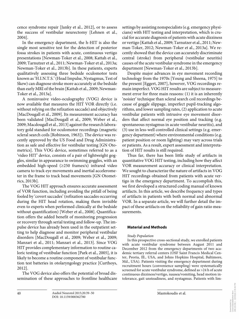
Fig. 1. Physiologic attributes and parameter definitions for a sin-gle, typical, abnormal h-HIT trace. Head velocity traces are shown in red, eye velocity in black. Note that eye movements are in the opposite direction to head movements, but are displayed graphi-cally as superimposed to make visual assessment of VOR gain (eye movements relative to head movements) clearer. H0 = Head veloc-ity onset; E0 = eye velocity onset; H peak = peak head velocity; E peak = peak eye velocity; H bounce = head velocity crosses baseline with head reversal following deceleration (bounce); H stop = head move-
ment stops; E stop = eye movement stops; CCS = covert corrective saccade (during head movement); OCS = overt corrective saccade (after head movement) with dotted line (slope = saccade accelera-tion) to identify E1 saccade onset; VOR latency = E0 – H0; VOR gain = eye velocity divided by head velocity at a specific time dur-ing the HIT (generally E peak /H peak ) or across a range of times (E times /H times ) (generally the ratio of the areas under the two curves over the entire HIT duration); saccade latency = E1 – E0.
Colo
r ver
sion
avail
able
onlin
e
Eye velocity
Head velocityHpeak
E0H0
Epeak
Estop
Hstop
HbounceE1
CCS
OCSReduced VOR gain
0 0.1 0.2 0.3 0.4 0.5 0.6 0.7
0
50
100
150
200
Time (s)
Velo
city
(°/s
)
Dow
nloa
ded
by:
Uni
vers
itäts
bibl
ioth
ek B
ern
19
8.14
3.58
.1 -
3/2
/201
6 2:
35:4
6 P
M

Quantifying the VOR with VOG Audiol Neurotol 2015;20:39–50DOI: 10.1159/000362780
43
posterior inferior cerebellar artery (PICA) territory, 3 anterior inferior cerebellar artery (AICA) territory]. One of the AICA stroke patients had bilateral cerebellar ischemic lesions. All enrolled patients tolerated the h-HITs and accomplished the full examination according to the study protocol without complications.
Primary study results are shown in tables 1–3 . We ana-lyzed 1,358 h-HIT graphs from the 26 patients. We found 72% of HIT traces had abnormal (but pathophysiologi-cally appropriate) disruptive fast eye movements, 44% had at least one artifact, and 42% of traces were deemed unin-terpretable by visual inspection (despite having been ac-cepted by the device software’s internal filtering algo-rithm). Only 46% of the traces were both artifact free and without disruptive fast eye movements. Detailed results with interrater agreements are presented in table 1 .
Slow-Phase VOR Of the 1,358 HIT traces, 58% (n = 794/1,358, 26
patients) had interpretable VOR results ( fig. 2 a–d). Of the interpretable results, 61% (n = 485/794, 25 patients) had clearly normal, 31% (n = 245/794, 23 patients) clearly abnormal h-HIT gain, and 8% (n = 64/794, 10 patients) an abrupt decline of gain ( fig. 2 d). Mean HIT velocity was 148°/s (SD 39, range: 52–348). Nondisrup-tive artifacts were present in 22% of interpretable HITs.
Fast-Phase Eye Movements Of all fast phase eye movements, one third could be
identified either as a corrective saccade associated with an abnormal HIT or spontaneous nystagmus; however, two thirds could not be readily classified ( table 1 ). We found
Fig. 2. Exemplar individual HIT traces showing the range of typical normal and abnormal physiologic VOR and saccade findings un-der various clinical circumstances. Slow-phase VOR gain can be either normal ( a , b ) or deficient ( c , d ). HITs can be performed through a wide range of head velocities (two examples shown in a and b ). The morphology of fast eye movements is the same re-gardless of underlying physiology, but their location, timing, pat-tern, and association with normal or abnormal VOR may allow differentiation of saccades ( e , f , h ) from nystagmus ( g , possibly h ). e A typical, overt (late) refixation saccade after a deficient VOR.
f , h Covert (early) refixation saccades after a deficient VOR re-sponse. g A rhythmic run of nystagmus beats after a normal VOR response (note that the nystagmus beats might be confused for refixation saccades, both covert and overt). h ‘Wrong-way’ sac-cades, probably representing nystagmus towards the VOR deficit. Minor, nondisruptive fixation losses or mini-blinks are present in panels d (far left of trace) and g (far right of trace). There is also a small degree of ‘noisy baseline’ artifact in some of the eye traces, most obvious in panels d and f .
Colo
r ver
sion
avail
able
onlin
e
Fast-
phas
e ey
e m
ovem
ents
Slow
-pha
se V
OR
0 0.1 0.2 0.3 0.4 0.5 0.6 0.7
0
50
100
150
200
250
Spee
d (°/
s)
0
50
100
150
200
250
Spee
d (°/
s)
0 0.1 0.2 0.3 0.4 0.5 0.6 0.7Time (s)
Time (s)
Abnormal HITwith low gain
Normal HIT plus nystagmus(‘pseudo’ corrective saccades)g
c
0 0.1 0.2 0.3 0.4 0.5 0.6 0.7Time (s)
Time (s)0 0.1 0.2 0.3 0.4 0.5 0.6 0.7
0
50
100
150
200
250
Spee
d (°/
s)
0
50
100
150
Spee
d (°/
s)
Abnormal HITwith abrupt decline of gain
‘Wrong-way’ saccadestowards a deficient VORh
d
0 0.1 0.2 0.3 0.4 0.5 0.6 0.7Time (s)
Time (s)
0
50
100
150
Spee
d (°/
s)
0
0
50
100
150
200
250
0.1 0.2 0.3 0.4 0.5 0.6 0.7
Spee
d (°/
s)Normal HIT
at lower head velocity
Abnormal HITcovert saccadef
b
0 0.1 0.2 0.3 0.4 0.5 0.6 0.7
0
50
100
150
200
250
Time (s)
Spee
d (°/
s)
0 0.1 0.2 0.3 0.4 0.5 0.6 0.7
0
50
100
150
200
250
Time (s)
Spee
d (°/
s)EyeHead
Normal HIT
Abnormal HITovert saccade
a
e
Dow
nloa
ded
by:
Uni
vers
itäts
bibl
ioth
ek B
ern
19
8.14
3.58
.1 -
3/2
/201
6 2:
35:4
6 P
M

Mantokoudis et al. Audiol Neurotol 2015;20:39–50DOI: 10.1159/000362780
44
Fig. 3. Exemplar HIT traces demonstrating the range of typical ar-tifacts. a–h depict seven artifacts intentionally generated and re-produced under laboratory conditions in a single normal subject: phase shift ( a ), inappropriately high gain ( b ), pseudo-saccades ( c , d ), multiple VOR peaks (i.e. non-bell-shaped curve) ( e ), excessive
post-HIT bounce ( f ), eye moves opposite the expected slow phase VOR direction (i.e. with rather than in opposition to the head) ( g ), and trace oscillations (noisy baseline) ( h ). These artifacts of known underlying cause served as the morphologic template for deter-mining the nature of artifacts post hoc in patient traces.
Table 1. Morphology of 1,358 h-HIT traces from 26 patients with acute vestibular syndrome
Morphologic attributes of video h-HIT tracing1 Number of h-HITs, n (%)
Kappa2
Slow-phase VOR normal VOR 485 (36) 0.782abnormal VOR without abrupt decline 245 (18) 0.548abnormal VOR with abrupt decline 64 (5) 0.2703
incorrect morphology (uninterpretable traces)4 564 (42) 0.666
Fast-phase eyemovements
corrective saccades 202 (15) 0.526spontaneous nystagmus 110 (8) 0.9053
unclassifiable (either or both) 667 (49) 0.728none 379 (28) 0.798
Artifacts5 one or more artifacts (not counting ‘bounce’) 599 (44) 0.635no artifacts 759 (56) 0.635
1 All within-category classifications in this table are mutually exclusive and jointly exhaustive (one and only one per h-HIT). Thus, all percentages in each category sum to 100%.
2 Cohen’s kappa reports the interrater agreement above that expected by chance alone.3 Note that in the random 10% subsample coded by both raters, these events were rare events (<10 total). Thus, the kappa values may
not represent stable estimates of anticipated agreement in a larger sample.4 Includes disruptive artifacts as well as disruptive physiologic fast phase eye movements.5 Includes all artifacts except ‘bounce’ (see Methods). Disruptive fast-phase eye movements (i.e. physiologic nystagmus and saccades)
were not considered artifacts.
Colo
r ver
sion
avail
able
onlin
e
EyeHead
Spee
d (°/
s)
Spee
d (°/
s)
Spee
d (°/
s)
Spee
d (°/
s)
Time (s)
Time (s) Time (s) Time (s) Time (s)
Time (s) Time (s) Time (s)
Spee
d (°/
s)
Spee
d (°/
s)
Spee
d (°/
s)
Delay/phase shift(loose strap)
High gain (wrong calibration)
Pseudo-saccade (mini-blink)
Pseudo-saccade (blink)
Two peaks (touchinggoggles or miniblink)
Head overshoot (bounce)(investigator induced)
Eye trace goes wrongdirection (patient inattention)
Trace oscillations(pupil tracking loss)
Spee
d (°/
s)
a
e f gg h
b c d
Dow
nloa
ded
by:
Uni
vers
itäts
bibl
ioth
ek B
ern
19
8.14
3.58
.1 -
3/2
/201
6 2:
35:4
6 P
M
Quantifying the VOR with VOG Audiol Neurotol 2015;20:39–50DOI: 10.1159/000362780
45
45% of all detected refixation saccades were covert ( fig. 2 f). All 26 acute vestibular syndrome patients had either spontaneous or gaze-evoked nystagmus evident ei-ther with or without fixation. However, 5 of 26 patients showed no evidence of nystagmus during the HIT record-ings (either due to suppression by visual fixation or eye-position-dependent nystagmus).
We identified nystagmus in 8% (n = 110/1,358, 15 pa-tients) as defined in figure 2 g. In 2% (n = 28/1,358, 9 pa-
tients) of all HITs, nystagmus rendered VOR gain diffi-cult to interpret. Multiple wrong-way saccades ( fig. 2 h) were found in 1% (n = 15/1,358, 6 patients).
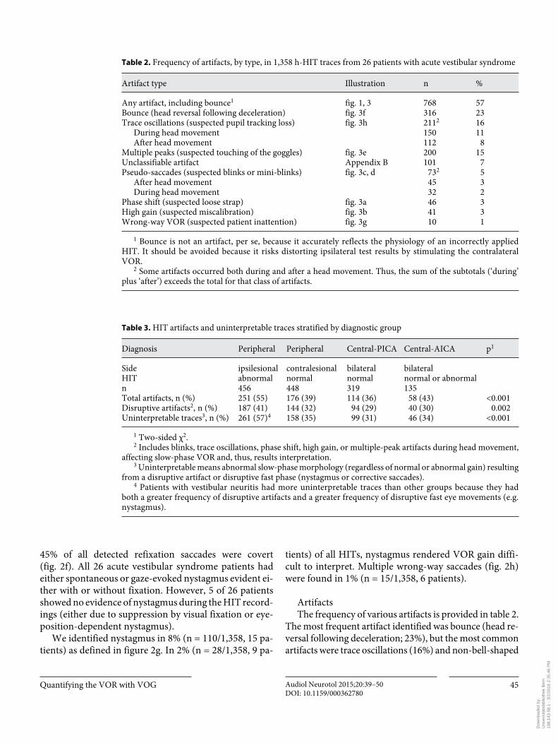
Artifacts The frequency of various artifacts is provided in table 2 .
The most frequent artifact identified was bounce (head re-versal following deceleration; 23%), but the most common artifacts were trace oscillations (16%) and non-bell-shaped
Table 2. Frequency of artifacts, by type, in 1,358 h-HIT traces from 26 patients with acute vestibular syndrome
Artifact type Illustration n %
Any artifact, including bounce1 fig. 1, 3 768 57Bounce (head reversal following deceleration) fig. 3f 316 23Trace oscillations (suspected pupil tracking loss) fig. 3h 2112 16
During head movement 150 11After head movement 112 8
Multiple peaks (suspected touching of the goggles) fig. 3e 200 15Unclassifiable artifact Appendix B 101 7Pseudo-saccades (suspected blinks or mini-blinks) fig. 3c, d 732 5
After head movement 45 3During head movement 32 2
Phase shift (suspected loose strap) fig. 3a 46 3High gain (suspected miscalibration) fig. 3b 41 3Wrong-way VOR (suspected patient inattention) fig. 3g 10 1
1 Bounce is not an artifact, per se, because it accurately reflects the physiology of an incorrectly applied HIT. It should be avoided because it risks distorting ipsilateral test results by stimulating the contralateral VOR.
2 Some artifacts occurred both during and after a head movement. Thus, the sum of the subtotals (‘during’ plus ‘after’) exceeds the total for that class of artifacts.
Table 3. HIT artifacts and uninterpretable traces stratified by diagnostic group
Diagnosis Peripheral Peripheral Central-PICA Central-AICA p1
Side ipsilesional contralesional bilateral bilateralHIT abnormal normal normal normal or abnormaln 456 448 319 135Total artifacts, n (%) 251 (55) 176 (39) 114 (36) 58 (43) <0.001Disruptive artifacts2, n (%) 187 (41) 144 (32) 94 (29) 40 (30) 0.002Uninterpretable traces3, n (%) 261 (57)4 158 (35) 99 (31) 46 (34) <0.001
1 Two-sided χ2.2 Includes blinks, trace oscillations, phase shift, high gain, or multiple-peak artifacts during head movement,
affecting slow-phase VOR and, thus, results interpretation.3 Uninterpretable means abnormal slow-phase morphology (regardless of normal or abnormal gain) resulting
from a disruptive artifact or disruptive fast phase (nystagmus or corrective saccades).4 Patients with vestibular neuritis had more uninterpretable traces than other groups because they had
both a greater frequency of disruptive artifacts and a greater frequency of disruptive fast eye movements (e.g. nystagmus).
Dow
nloa
ded
by:
Uni
vers
itäts
bibl
ioth
ek B
ern
19
8.14
3.58
.1 -
3/2
/201
6 2:
35:4
6 P
M

Mantokoudis et al. Audiol Neurotol 2015;20:39–50DOI: 10.1159/000362780
46
VOR curves (multiple peaks) (15%). Unclassifiable arti-facts (7%) and pseudo-saccades from suspected blinks (5%) were the next most frequent, with other artifacts occurring in only 3% or fewer of all HITs. The head impulse technique holding the jaw of the patient was associated with more fre-quent artifacts than when standing behind the patient and holding the top of the head (52 vs. 42%, p = 0.005).
We sporadically saw inappropriately high gains (>1.2) in 41 traces from 13 patients; 2 of these patients had sys-tematically higher gains (majority of HIT traces). Figure 3 b illustrates the higher amplitude of eye velocity after a miscalibration under laboratory conditions.
Artifacts were more frequent in patients with an ab-normal VOR ( table 3 ). Patients with PICA strokes (all with bilaterally normal head impulses) had more inter-pretable traces compared to patients with vestibular neu-ritis (unilaterally abnormal; 69 vs. 43%, p < 0.001). Pa-tients with AICA strokes who displayed variable h-HIT VOR patterns with some normal and others abnormal had intermediate results, but were overall more similar to PICA strokes than to neuritis cases (66% interpretable).
Figure 4 illustrates the impact of artifacts on compos-ite results, showing two ‘runs’ of multiple HIT maneu-vers in a single patient that happened to include a large number of individual HIT traces without artifacts and with artifacts.
Discussion
To our knowledge, this is the first systematic analysis of video HIT morphology and artifacts. We coded and classified 1,358 HITs in 26 acute vestibular patients. We noted eight different types of artifacts. We found a high frequency of h-HIT traces with at least one artifact, par-ticularly in patients with unilateral VOR gain deficits. Some artifacts occurred during the critical phase of head movement, reducing our ability to interpret the VOR gain one third of the time. Since these head impulses were still classified and accepted as a valid head impulse trace by the device algorithm, some of these artifacts might contribute to false gain calculations. We found that spontaneous nys-tagmus was often difficult to distinguish from corrective saccades in patients with abnormal VOR gains. Coding of completely normal HIT trace morphology had extremely high interrater reliability. Our findings are important be-cause they raise critical technical considerations for ob-taining and interpreting VOR gain results from VOG HIT testing, which are used frequently in clinical practice.
Slow-Phase VOR The interrater agreement for the interpretation of nor-
mal slow phases was excellent (kappa 0.78). The ability to train frontline providers and neurology or otolaryngolo-
Fig. 4. Back-to-back HIT sessions in a single patient’s ear with a normal VOR. Shown are complete, unfiltered device results from two HIT ‘runs’ from the same ear in the same patient (PICA stroke) obtained during a single visit. Each run shows individual HIT trac-es temporally superimposed to demonstrate the aggregate result. a Shows a ‘clean’ run, essentially free of artifacts (without any post-processing or ‘clean up’). The few eye deviations present are either
small saccades (small fixation losses or nystagmus beats) or non-disruptive blink-related artifacts. b A relatively ‘noisy’ run, with more substantial artifacts. Here most of the eye deviations appear to be from recording artifacts, which might be further character-ized if the HIT morphology were assessed for each individual HIT (rather than aggregated and superimposed).
Colo
r ver
sion
avail
able
onlin
e
a b0 0.1 0.2 0.3 0.4 0.5 0.6 0.7
0
50
100
150
200
250
Time (s)
Velo
city
(°/s
)
EyeHead
0 0.1 0.2 0.3 0.4 0.5 0.6 0.7
0
50
100
150
200
Time (s)
Velo
city
(°/s
)
EyeHead
Dow
nloa
ded
by:
Uni
vers
itäts
bibl
ioth
ek B
ern
19
8.14
3.58
.1 -
3/2
/201
6 2:
35:4
6 P
M
Quantifying the VOR with VOG Audiol Neurotol 2015;20:39–50DOI: 10.1159/000362780
47
gy consultants to reliably recognize a normal result is im-portant for acute diagnosis since a bilaterally normal VOR response in a patient with acute vestibular syn-drome is highly suggestive of a central nervous system cause (usually stroke) [Newman-Toker et al., 2008; Kat-tah et al., 2009; Newman-Toker et al., 2013a; Newman-Toker et al., 2013b].
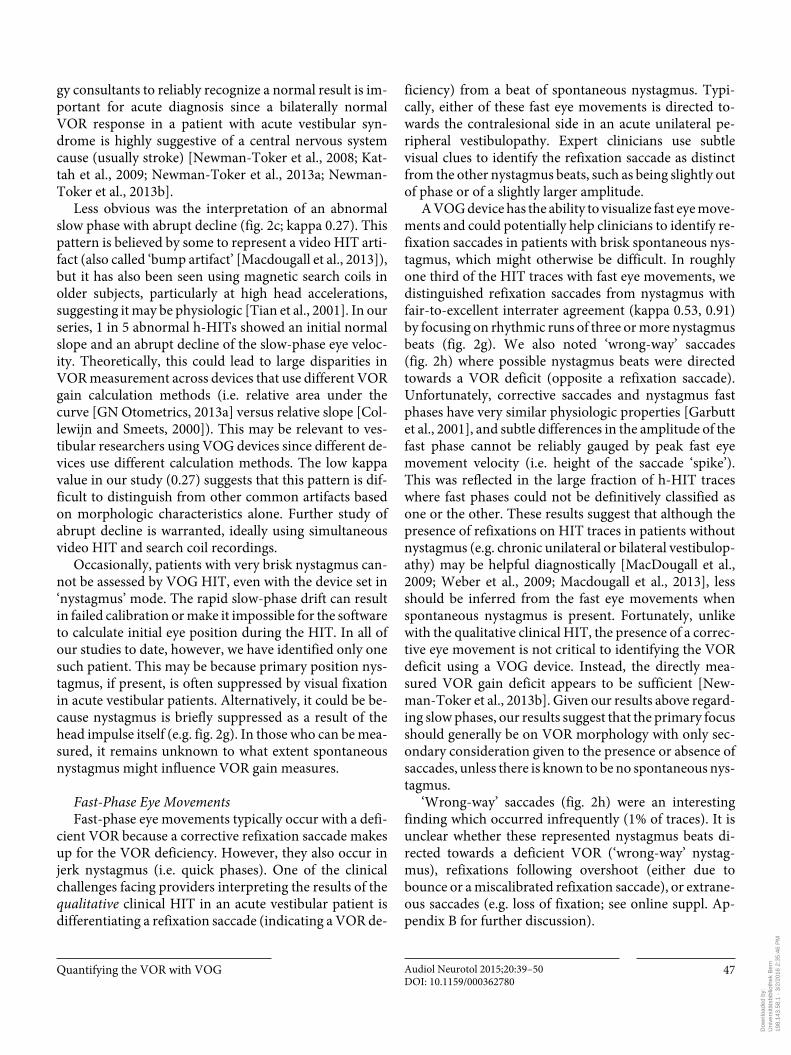
Less obvious was the interpretation of an abnormal slow phase with abrupt decline ( fig. 2 c; kappa 0.27). This pattern is believed by some to represent a video HIT arti-fact (also called ‘bump artifact’ [Macdougall et al., 2013]), but it has also been seen using magnetic search coils in older subjects, particularly at high head accelerations, suggesting it may be physiologic [Tian et al., 2001]. In our series, 1 in 5 abnormal h-HITs showed an initial normal slope and an abrupt decline of the slow-phase eye veloc-ity. Theoretically, this could lead to large disparities in VOR measurement across devices that use different VOR gain calculation methods (i.e. relative area under the curve [GN Otometrics, 2013a] versus relative slope [Col-lewijn and Smeets, 2000]). This may be relevant to ves-tibular researchers using VOG devices since different de-vices use different calculation methods. The low kappa value in our study (0.27) suggests that this pattern is dif-ficult to distinguish from other common artifacts based on morphologic characteristics alone. Further study of abrupt decline is warranted, ideally using simultaneous video HIT and search coil recordings.
Occasionally, patients with very brisk nystagmus can-not be assessed by VOG HIT, even with the device set in ‘nystagmus’ mode. The rapid slow-phase drift can result in failed calibration or make it impossible for the software to calculate initial eye position during the HIT. In all of our studies to date, however, we have identified only one such patient. This may be because primary position nys-tagmus, if present, is often suppressed by visual fixation in acute vestibular patients. Alternatively, it could be be-cause nystagmus is briefly suppressed as a result of the head impulse itself (e.g. fig. 2 g). In those who can be mea-sured, it remains unknown to what extent spontaneous nystagmus might influence VOR gain measures.
Fast-Phase Eye Movements Fast-phase eye movements typically occur with a defi-
cient VOR because a corrective refixation saccade makes up for the VOR deficiency. However, they also occur in jerk nystagmus (i.e. quick phases). One of the clinical challenges facing providers interpreting the results of the qualitative clinical HIT in an acute vestibular patient is differentiating a refixation saccade (indicating a VOR de-
ficiency) from a beat of spontaneous nystagmus. Typi-cally, either of these fast eye movements is directed to-wards the contralesional side in an acute unilateral pe-ripheral vestibulopathy. Expert clinicians use subtle visual clues to identify the refixation saccade as distinct from the other nystagmus beats, such as being slightly out of phase or of a slightly larger amplitude.
A VOG device has the ability to visualize fast eye move-ments and could potentially help clinicians to identify re-fixation saccades in patients with brisk spontaneous nys-tagmus, which might otherwise be difficult. In roughly one third of the HIT traces with fast eye movements, we distinguished refixation saccades from nystagmus with fair-to-excellent interrater agreement (kappa 0.53, 0.91) by focusing on rhythmic runs of three or more nystagmus beats ( fig. 2 g). We also noted ‘wrong-way’ saccades ( fig. 2 h) where possible nystagmus beats were directed towards a VOR deficit (opposite a refixation saccade). Unfortunately, corrective saccades and nystagmus fast phases have very similar physiologic properties [Garbutt et al., 2001], and subtle differences in the amplitude of the fast phase cannot be reliably gauged by peak fast eye movement velocity (i.e. height of the saccade ‘spike’). This was reflected in the large fraction of h-HIT traces where fast phases could not be definitively classified as one or the other. These results suggest that although the presence of refixations on HIT traces in patients without nystagmus (e.g. chronic unilateral or bilateral vestibulop-athy) may be helpful diagnostically [MacDougall et al., 2009; Weber et al., 2009; Macdougall et al., 2013], less should be inferred from the fast eye movements when spontaneous nystagmus is present. Fortunately, unlike with the qualitative clinical HIT, the presence of a correc-tive eye movement is not critical to identifying the VOR deficit using a VOG device. Instead, the directly mea-sured VOR gain deficit appears to be sufficient [New-man-Toker et al., 2013b]. Given our results above regard-ing slow phases, our results suggest that the primary focus should generally be on VOR morphology with only sec-ondary consideration given to the presence or absence of saccades, unless there is known to be no spontaneous nys-tagmus.
‘Wrong-way’ saccades ( fig. 2 h) were an interesting finding which occurred infrequently (1% of traces). It is unclear whether these represented nystagmus beats di-rected towards a deficient VOR (‘wrong-way’ nystag-mus), refixations following overshoot (either due to bounce or a miscalibrated refixation saccade), or extrane-ous saccades (e.g. loss of fixation; see online suppl. Ap-pendix B for further discussion).
Dow
nloa
ded
by:
Uni
vers
itäts
bibl
ioth
ek B
ern
19
8.14
3.58
.1 -
3/2
/201
6 2:
35:4
6 P
M

Mantokoudis et al. Audiol Neurotol 2015;20:39–50DOI: 10.1159/000362780
48
HIT Trace Artifacts Artifacts were surprisingly common, with one or more
artifacts seen in nearly half of the collected impulses and disruptive artifacts seen in more than a third. Many of the artifacts were probably induced by the examiners them-selves (e.g. ‘bounce’ in delivering impulse, touching gog-gles), despite their extensive training and experience. Technique also matters – holding the top of the head in-stead of the jaw has a slightly lower risk for creating iat-rogenic artifacts, although this difference could also have been attributable to specific examiner skill since each site consistently applied only one technique. Interestingly, ar-tifacts occurred 10–20% more often (absolute increase) in patients with abnormal h-HITs, a difference that was highly statistically significant ( table 3 ). Whether this re-flects misclassification (i.e. physiologic disruptive eye movements erroneously called artifacts) or a true effect of the impaired VOR (e.g. due to greater risk of pupil track-ing loss with more refixation saccades) merits additional study.
The most common ‘artifact’ (technique error) was head ‘bounce’ ( table 2 ). This artifact results from the ex-aminer inadvertently rotating the head back in the oppo-site direction (rather than coming to a full stop) at the end of the impulse, following deceleration. Bounce was gener-ally minor, and only adversely affected trace interpreta-tion if severe. In our experience, bounce only affects data quality with novice examiners (first hour of training).
The second most common artifact was noisy eye trac-es with multiple VOR peaks (i.e. non-bell-shaped mor-phology; fig. 3 e). This trace morphology could be easily reproduced under laboratory conditions by touching the goggles directly or indirectly (e.g. by touching the strap). However, multiple peaks could also be created by a mini-blink without pupil tracking loss or by nystagmus with a quick phase in the opposite direction. Regardless of the cause, this artifact was the most common reason for an uninterpretable trace.
Another major source of artifacts was pupil tracking loss resulting in oscillations on the trace or even loss of the recording trace ( fig. 3 h). This was the second most com-mon cause of an uninterpretable trace. Pupil tracking can be affected by environmental lighting conditions via mio-sis or reflected light off the cornea, both of which reduce pupil-tracking efficiency. Optimal lighting conditions ap-peared to be soft, even relatively low light, although re-cordings were generally robust to the typical emergency department or clinic room lighting. Pupil tracking can also be affected by the position of the mirroring infrared LEDs [Eggert, 2007], narrow palpebral fissures (eyelid
ptosis, brow ptosis, congenital/racial, etc.), or even light-reflecting makeup (e.g. eyelash mascara or eyelid liner). Independent of the cause, we found that pupil tracking could usually be improved by adjusting software settings (e.g. pupil detection thresholds). The algorithm of our VOG system was efficient and rejected 98% of h-HITs af-ter detection of a pupil tracking loss.
Other artifact types were uncommon in our series ( ≤ 5%). Goggle slippage during head acceleration oc-curred if the goggles were not attached firmly enough. This problem can cause an artifact with an undershooting of the eye velocity curve at the beginning of a head move-ment [Bartl et al., 2009]. In addition, the eye graph might lead the head graph (phase shift; fig. 3 a) or vice versa. La-tency between the start of a head movement and the com-pensatory eye movement has a physiological range of 7–15 ms, which should be taken into account [Leigh and Zee, 2006]. Video goggle slippage due to a high mass in-ertia [Bartl et al., 2009; Weber et al., 2009] has been de-scribed previously and a model for slippage compensation proposed [Bartl et al., 2009]. A similar phenomenon can be seen with the search coil technique with a coil slippage on the conjunctiva [Jorns-Haderli et al., 2007]. In our se-ries, only 3% of HITs showed a significant phase lag (>20 ms) between eye and head trace due to goggle slippage.
VOR Gain Distortions due to Artifacts A complete analysis of the effect of artifacts on VOR
gain and diagnostic classification is beyond the scope of this article and will be presented elsewhere. We note, however, that a particular error, incorrect calibration, consistently resulted in false gain elevations. We were able to reproduce traces with gains above 1.2 in normal subjects under laboratory conditions by performing re-peated HITs with an inaccurate calibration ( fig. 3 b). This might occur naturally in patient who was inattentive dur-ing calibration. Some of our patients were drowsy or dis-tracted and did not look accurately at the projected laser dot; others had brisk nystagmus that probably affected the quality of fixation and, hence, calibration. In some cases, recalibration was required to obtain valid results during our testing protocols.
Apparently physiologic high gains (at least up to 1.2) can be observed for HITs [MacDougall et al., 2009; Agrawal et al., 2014] with fixation distance near rather than far or in patients with spectacle corrections [Cannon et al., 1985]. They can also occur in patients with cerebel-lar disease due to disinhibition of the vestibular nuclei, although this has only been shown in patients with bilat-eral chronic lesions, rather than those with acute unilat-
Dow
nloa
ded
by:
Uni
vers
itäts
bibl
ioth
ek B
ern
19
8.14
3.58
.1 -
3/2
/201
6 2:
35:4
6 P
M
Quantifying the VOR with VOG Audiol Neurotol 2015;20:39–50DOI: 10.1159/000362780
49
eral ones [Walker and Zee, 2005]. We found 2 out of our 26 patients with mean VOR gains above 1.2. One had a stroke in the left lateral medulla and bilateral elevated gain values; the other had a left vestibular neuritis with elevated gains on the contralesional side. Both showed major artifacts on most of the traces, rendering most of the measurements obtained uninterpretable. We con-cluded the high mean gains were likely artifactual.
Although these results might seem concerning for VOG interpretive validity, additional recent analyses sug-gest that artifacts primarily introduce random noise, and overall diagnostic classification is not meaningfully af-fected [Mantokoudis, unpubl.]. The high frequency of ar-tifacts does decrease precision, so relying on fewer than 10 unfiltered HITs for mean VOR gain measures is not advisable. Manual filtering of artifacts is also recom-mended.
Limitations
We did not validate our impulse findings using scleral search coil recordings. Artifacts were determined based on morphology alone, so we cannot be sure all were arti-facts. Our methods likely overstate the frequency of arti-facts somewhat because some of the traces might have been physiologic rather than artifactual, per se. Non-real-time analysis of eye traces did not allow strong inferences to be drawn about the underlying causes for artifacts in each specific case. Coding reliability was low for some types of artifacts. Exam technique differences might have influenced results. We tested only one VOG head impulse device, and it remains unknown whether our results gen-eralize to other similar recording systems. Finally, there was no healthy, asymptomatic control group for compar-ison of artifact frequency.
Potential Implications
Since video HIT has gained popularity among ENT physicians and neurologists, knowledge about the cor-rect interpretation of quantitative h-HITs is essential. Our results indicate that use of video goggles in patients with acute vertigo should currently be limited to provid-ers familiar with the range of possible artifacts and their diagnostic implications. Some artifacts from blinks or inattention are probably unavoidable, while others are technique dependent (e.g. bounce). Familiarity with a coding manual ( fig. 1–3 ; online suppl. Appendix B)
might prove valuable to practitioners adopting this tech-nology. Our findings also suggest it may be easier to train physicians or technicians to reliably identify truly nor-mal traces than to differentiate abnormal traces from ar-tifacts. The ability for frontline providers to do so might offer the possibility to speed identification of acute stroke in acute vestibular syndrome patients, including in the emergency department or even prehospital care setting [Newman-Toker et al., 2013b]. In most cases it would also help correctly select the most appropriate clinical consultant to further evaluate and treat a patient’s acute dizziness or vertigo-stroke: neurologist for bilaterally normal VOR results, or a vestibular otologist to distin-guish artifacts from true unilateral or bilateral abnormal VOR results.
Conclusions
As video h-HIT testing appears to be associated with a high frequency of artifacts, training in interpretation is essential. Awareness of the most common artifacts can help practitioners avoid creating them (for those which are iatrogenic) or misinterpreting the results when they occur. Further study is essential to determine the clinical diagnostic implications of artifacts frequently encoun-tered during video HIT testing as the use of this technique increases.
Acknowledgements
This study was supported by the Swiss National Science Foun-dation (PBBEP2 136573).
Disclosure Statement
The authors report no conflicts of interest.
References Agrawal Y, Schubert M, Migliaccio A, Schneider E, Zee D, Carey J: Evaluation of quantitative head impulse testing using search coils vs. video-oculography in older individuals. Otol Neurotol 2014; 35: 283–288.
Bartl K, Lehnen N, Kohlbecher S, Schneider E: Head impulse testing using video-oculogra-phy. Ann NY Acad Sci 2009; 1164: 331–333.
Blödow A, Helbig R, Bloching M, Walther LE: Isolated functional loss of the lateral semicir-cular canal in vestibular neuritis (in German). HNO 2013; 61: 46–51.
Dow
nloa
ded
by:
Uni
vers
itäts
bibl
ioth
ek B
ern
19
8.14
3.58
.1 -
3/2
/201
6 2:
35:4
6 P
M
Mantokoudis et al. Audiol Neurotol 2015;20:39–50DOI: 10.1159/000362780
50
Cannon SC, Leigh RJ, Zee DS, Abel LA: The effect of the rotational magnification of corrective spectacles on the quantitative evaluation of the VOR. Acta Otolaryngol 1985; 100: 81–88.
Collewijn H, Smeets JB: Early components of the human vestibulo-ocular response to head ro-tation: latency and gain. J Neurophysiol 2000; 84: 376–389.
Curthoys IS: The interpretation of clinical tests of peripheral vestibular function. Laryngoscope 2012; 122: 1342–1352.
Eggert T: Eye movement recordings: methods. Dev Ophthalmol 2007; 40: 15–34.
Fleiss JL: Statistical Methods for Rates and Pro-portions. Hoboken, Wiley, 1981.
Garbutt S, Harwood MR, Harris CM: Compari-son of the main sequence of reflexive saccades and the quick phases of optokinetic nystag-mus. Br J Ophthalmol 2001; 85: 1477–1483.
GN Otometrics: ICS Impulse. http://www.icsim-pulse.com/.
GN Otometrics: ICS Impulse FAQ. 2013a. http://www.otometrics.com/ ∼ /media/Download-Library/Otometrics/PDFs/ICS%20Impulse/7-26-8801-EN_04_STD.pdf.
GN Otometrics: Video head impulse test – data-sheet. 2013b http://www.otometrics.com/ ∼ /media/DownloadLibrary/Otometrics/PDFs/ICS%20Impulse/7-26-9899-EN_02_STD.pdf.
Halmagyi GM, Aw ST, Cremer PD, Curthoys IS, Todd MJ: Impulsive testing of individual semicircular canal function. Ann NY Acad Sci 2001; 942: 192–200.
Halmagyi GM, Curthoys IS: A clinical sign of ca-nal paresis. Arch Neurol 1988; 45: 737–739.
Janky KL, Zuniga MG, Carey JP, Schubert M: Bal-ance dysfunction and recovery after surgery for superior canal dehiscence syndrome. Arch OtolaryngolHead Neck Surg 2012; 138: 723–730.
Jorns-Haderli M, Straumann D, Palla A: Accura-cy of the bedside head impulse test in detect-ing vestibular hypofunction. J Neurol Neuro-surg Psychiatry 2007; 78: 1113–1118.
Kattah JC, Talkad AV, Wang DZ, Hsieh YH, New-man-Toker DE: HINTS to diagnose stroke in the acute vestibular syndrome: three-step bed-side oculomotor examination more sensitive than early MRI diffusion-weighted imaging. Stroke 2009; 40: 3504–3510.
Kim JS, Kim HJ: Inferior vestibular neuritis. J Neurol 2012; 259: 1553–1560.
Lehnen N, Aw ST, Todd MJ, Halmagyi GM: Head impulse test reveals residual semicircular ca-nal function after vestibular neurectomy. Neurology 2004; 62: 2294–2296.
Leigh RJ, Zee DS: The vestibular-optokinetic sys-tem; in Leigh RJ, Zee DS, (eds): The Neurol-ogy of Eye Movements. New York, Oxford University Press, 2006, pp 20–107.
Macdougall HG, McGarvie LA, Halmagyi GM, Curthoys IS, Weber KP: The video Head Im-pulse Test (vHIT) detects vertical semicircu-lar canal dysfunction. PloS One 2013; 8: e61488.
MacDougall HG, Weber KP, McGarvie LA, Hal-magyi GM, Curthoys IS: The video head im-pulse test: diagnostic accuracy in peripheral vestibulopathy. Neurology 2009; 73: 1134–1141.
Manzari L, Burgess AM, MacDougall HG, Brad-shaw AP, Curthoys IS: Rapid fluctuations in dynamic semicircular canal function in early Meniere’s disease. Eur Arch Otorhinolaryn-gol 2011; 268: 637–639.
Manzari L, MacDougall HG, Burgess AM, Cur-thoys IS: New, fast, clinical vestibular tests identify whether a vertigo attack is due to ear-ly Meniere’s disease or vestibular neuritis. La-ryngoscope 2013; 123: 507–511.
Newman-Toker DE: Symptoms and signs of neu-ro-otologic disorders. Continuum 2012; 18: 1016–1040.
Newman-Toker DE, Kattah JC, Alvernia JE, Wang DZ: Normal head impulse test differen-tiates acute cerebellar strokes from vestibular neuritis. Neurology 2008; 70: 2378–2385.
Newman-Toker DE, Kerber KA, Hsieh YH, Pula JH, Omron R, Saber Tehrani AS, Mantok-oudis G, Hanley DF, Zee DS, Kattah JC: HINTS outperforms ABCD2 to screen for stroke in acute continuous vertigo and dizzi-ness. Acad Emerg Med 2013a;20: 986–996.
Newman-Toker DE, Saber Tehrani AS, Mantok-oudis G, Pula JH, Guede CI, Kerber KA, Blitz A, Ying SH, Hsieh YH, Rothman RE, Hanley DF, Zee DS, Kattah JC: Quantitative video-oculography to help diagnose stroke in acute vertigo and dizziness: toward an ECG for the eyes. Stroke 2013b;44: 1158–1161.
Park HJ, Migliaccio AA, Della Santina CC, Minor LB, Carey JP: Search-coil head-thrust and ca-loric tests in Meniere’s disease. Acta Otolar-yngol 2005; 125: 852–857.
Petersen JA, Straumann D, Weber KP: Clinical di-agnosis of bilateral vestibular loss: three sim-ple bedside tests. Ther Adv Neurol Disord 2013; 6: 41–45.
Robinson DA: A method of measuring eye move-ment using a scleral search coil in a magnetic field. IEEE Trans Biomed Eng 1963; 10: 137–145.
Strupp M, Brandt T: Peripheral vestibular disor-ders. Curr Opin Neurol 2013; 26: 81–89.
Tarnutzer AA, Berkowitz AL, Robinson KA, Hsieh YH, Newman-Toker DE: Does my diz-zy patient have a stroke? A systematic review of bedside diagnosis in acute vestibular syn-drome. CMAJ 2011; 183:E571–E592.
Tian JR, Shubayev I, Baloh RW, Demer JL: Im-pairments in the initial horizontal vestibulo-ocular reflex of older humans. Exp Brain Res 2001; 137: 309–322.
Walker MF, Zee DS: Asymmetry of the pitch ves-tibulo-ocular reflex in patients with cerebellar disease. Ann NY Acad Sci 2005; 1039: 349–358.
Weber KP, Aw ST, Todd MJ, McGarvie LA, Cur-thoys IS, Halmagyi GM: Head impulse test in unilateral vestibular loss: vestibulo-ocular re-flex and catch-up saccades. Neurology 2008; 70: 454–463.
Weber KP, MacDougall HG, Halmagyi GM, Cur-thoys IS: Impulsive testing of semicircular-canal function using video-oculography. Ann NY Acad Sci 2009; 1164: 486–491.
Young LR, Sheena D: Survey of eye movement re-cording methods. Behav Res Methods 1975; 7: 397–429.
Dow
nloa
ded
by:
Uni
vers
itäts
bibl
ioth
ek B
ern
19
8.14
3.58
.1 -
3/2
/201
6 2:
35:4
6 P
M