Embed Size (px)
Citation preview
Quantifying Structural Changes with the Application of Osteopathic Manual Medicine
(OMM) in Tegucigalpa, Honduras
Abraham Billy Hardee III, D.O.
Dissertation submitted to the Faculty of the
Virginia Polytechnical Institute and State University (Virginia Tech)
in partial fulfillment of the requirements for a degree of
Doctor of Philosophy
in
Education, Curriculum and Instruction
H. Dean Sutphin, Chair
Richard Stratton
Kerry Redican
Per Gunnar Brolinson
January 29, 2008
Blacksburg, Virginia
Copyright 2008, Abraham B. Hardee III
Keywords: Osteopathic Manual Medicine, Lesion, Manipulation, & Forceplate
Quantifying Structural Changes with the Application of Osteopathic Manual Medicine
(OMM) in Tegucigalpa, Honduras
Abraham Billy Hardee III
(ABSTRACT)
The primary purpose of this research was to compare quantifiable structural asymmetry
to changes visualized in center of pressure (CoP) and/or postural sway by the use of an
Isobalance forceplate in order to introduce principles of osteopathic medicine to a Latin
America culture. Osteopathic manual medicine (OMM) was used to correct structural
dysfunction found in the study participants at the Baxter Institute in Tegucigalpa,
Honduras. Study participants were students and staff members ranging in age from 18 to
35 and consisting of 24 males and two females. The examination period lasted a total of
five weeks. During weeks one, three, and five, baseline measurements were taken prior to
manipulation using .05 alpha to test significance. Measurements were repeated post
OMM. An educational video was provided during each session. Pretest and posttest
results demonstrated an improvement in understanding of OMM materials provided to
participants. A bivariable chi-square test found that, when manipulated, those with
sacrum dysfunction have an association with a positive improvement in postural sway
(TIC 1 & 2) (p<.05). Also, the multivariable logistic regression model found that
individuals who had no initial change in postural sway (TIC 1 & 2) were more likely to
move to a positive improvement of time in the center of the premeasured diameter
calculated by the Isobalance forceplate, than a decrease in time spent in the center (i.e.
negative improvement) throughout the time of the study (p<.05).

iii
ACKNOWLEDGEMENTS
“There is neither Jew nor Greek, there is neither bond nor free, there is neither male
nor female: for ye are all one in Christ Jesus.”
Galatians 3:28 (King James)
Throughout my medical school and PhD experience, I have traveled half-way around
the world and have visited many people in their own surroundings. However, as much as
I have been blessed to see and be a part of so many medical mission trips in these last
five years, there are two individuals that I need to recognize because without their
encouragement and their testimonies I may not have made it this far.
The late Eddie Adams was my friend and my second biggest fan when I played
football for Virginia Tech, even if he was a Florida boy and loved the Hurricanes. He was
tragically paralyzed at the age of 16 and managed to live long enough to receive his
college degree, write a book, and inspire me to achieve the unachievable. He taught me
that even if you feel what you are going through is not fair, it does not matter because
you will still have to go through it, no matter if you want to or not. Whenever I want to
quit, I think of him and just smile. Thank you for letting me “walk” with you.
My roommate for the last nine years is by far my biggest fan. She has always
been there for me even when I was a child. We have laughed, cried, and grown older
together. She is my number one fan. Even though she has not seen me during these past
nine years, or watched me play football or baseball at Virginia Tech, nor seen my
graduations, or my wedding; she has always listened. My grandmother has been suffering
from macular degeneration for years and the Lord took her sight shortly after I moved in

iv
with her. Thank you Lord for letting me “see” an angel in my lifetime, even if this one is
salty at times.
Thank you to my medical school and Virginia Tech for the opportunity to receive
my PhD. To my chair Dr. Sutphin, and committee members (Drs. Redican, Stratton, and
Brolinson, thank you for your guidance.
A special thanks to Sarah Zalud-Cerrato. This program and its students owe you a
lot of gratitude for all your dedication and commitment.
Baxter Institute, thank you for all you have given me in my quest of finding-out
what I am called to do, Dios Te Bendiga.
Thank you to my family for never giving-up on me but instead, giving their all for
me. As a great thinker once stated “Just because you only have one life to live, you don‟t
have to settle for only one dream.” VDH-Belfon. What I am today is partially what you
have instilled in me. God‟s love and peace be with you all.
To my blessing, my charm, my ministry, and my Deshi girl, thank you for
allowing me the opportunity to be your husband. My most recent journeys in life would
not have been worth the experience without you by my side to make me a better servant
of God. Kasam ki kasam hai kasam se humko pyaar hai sirf tumse.

v
Table of Contents
Abstract ............................................................................................................................... ii
Acknowledgements ............................................................................................................ iii
Table of Contents .................................................................................................................v
Chapter One
Introduction ..........................................................................................................................1
Statement of the Problem .................................................................................................3
Purpose of the Study ........................................................................................................4
Research Questions ..........................................................................................................5
Research Hypotheses ........................................................................................................5
Significance of the Study .................................................................................................6
Assumptions .....................................................................................................................6
Limitations of the Study ...................................................................................................8
Summary ..........................................................................................................................9
Chapter Two
Review of Literature ..........................................................................................................10
Demographics ................................................................................................................10
Osteopathic Medicine .....................................................................................................12
Osteopathic Techniques .................................................................................................17
Anatomy .........................................................................................................................22

vi
Postural Importance ........................................................................................................25
Forceplate .......................................................................................................................28
Summary ........................................................................................................................31
Chapter Three
Methods..............................................................................................................................33
Research Questions ........................................................................................................33
Study Variables ..............................................................................................................33
Inclusion Criteria ........................................................................................................34
Exclusion Criteria .......................................................................................................34
Research Methodology ...................................................................................................35
Procedures ..................................................................................................................35
Population and Sample ...................................................................................................39
Instrumentation ...............................................................................................................39
Statistical Analysis .........................................................................................................41
Summary ........................................................................................................................41
Chapter Four
Results ................................................................................................................................42
Research Question ..........................................................................................................42
Response Rate ................................................................................................................43
Demographic Data ..........................................................................................................43

vii
Finding ...........................................................................................................................43
Osteopathic Manual Medicine ....................................................................................43
Isobalance Forceplate Results ....................................................................................45
Outcomes ..........................................................................................................45
Pretest and Posttest Results ........................................................................................52
Summary ........................................................................................................................55
Charpter Five
Summary ........................................................................................................................57
Conclusion ......................................................................................................................59
Pretest and Posttest Data Analysis .............................................................................59
Osteopathic Manual Medicine ....................................................................................60
Forceplate data analysis ..............................................................................................61
Recommendations ..........................................................................................................62
Recommendations for future research ........................................................................62
Recommendations for Practice ...................................................................................64
References ..........................................................................................................................65
viii
List of Figures
Figure 1
Map of Honduras ........................................................................................................11
Figure 2
Stressors on the musculoskeletal system ....................................................................13
Figure 3
Restrictive Barrier ......................................................................................................16
Figure 4
Treatment options for somatic dysfunction ................................................................18
Figure 5
Direct Technique ........................................................................................................19
Figure 6
Passive Indirect Treatment .........................................................................................20
Figure 7
Muscle Energy ............................................................................................................20
Figure 8
Facilitated positional release ......................................................................................21
Figure 9
Neck Flexion ..............................................................................................................22
Figure 10
Neck extension ...........................................................................................................23

ix
Figure 11
Vertebra ......................................................................................................................23
Figure 12
Thorax.........................................................................................................................24
Figure 13
Vertebral Anatomy .....................................................................................................25
Figure 14
Balance of Posture ......................................................................................................26
Figure 15
Balance of Posture ......................................................................................................26
Figure 16
Spinal curvature ..........................................................................................................27
Figure 17
Osteopathic Landmarks ..............................................................................................28
Figure 18
Foot Position on Forceplate ........................................................................................38
Figure 19
Pretest and Posttest results..........................................................................................53

x
List of Tables
Table 1
Osteopathic Lesions ....................................................................................................44
Table 2
Differences in TIC_Change 1 from week 1-5 .............................................................47
Table 3
TIC_Change 2 no change data ...................................................................................47
Table 4
Significant change in Heel-to-Toe .............................................................................48
Table 5
Significant change in Toe-to-Heel .............................................................................49
Table 6
Lesions in Specific Body Regions .............................................................................50
Table 7
TIC_Change 1 Sacrum ...............................................................................................51
Table 8
Types of questions ......................................................................................................53
Table 9
Defining T.A.R.T .......................................................................................................68
Table 10
Schedule of Project Events .........................................................................................69

xi
Table 11
TIC_Change 1 Uppercervicals ...................................................................................70
Table 12
TIC_Change 1 Lowercervicals ...................................................................................71
Table 13
TIC_Change 1 Thorasic .............................................................................................72
Table 14
TIC_Change 1 Lumbar ...............................................................................................73
Table 15
TIC_Change 1 Innominate .........................................................................................74
Table 16
TIC_Change 1 Leglength ...........................................................................................75
Table 17
TIC_Change 2 Uppercervicals ...................................................................................76
Table 18
TIC_Change 2 Lowercervicals ...................................................................................77
Table 19
TIC_Change 2 Thorasic .............................................................................................78
Table 20
TIC_Change 2 Lumbar ...............................................................................................79
Table 21
TIC_Change 2 Innominate .........................................................................................80
xii
Table 22
TIC_Change 2 Sacrum ...............................................................................................81
Table 23
TIC_Change 2 Leglength ...........................................................................................82
Table 24
Heel-to-Toe Uppercervicals .......................................................................................83
Table 25
Heel-to-Toe Lowercervical ........................................................................................84
Table 26
Heel-to-Toe Thorasic..................................................................................................85
Table 27
Heel-to-Toe Lumbar ...................................................................................................86
Table 28
Heel-to-Toe Innominate ............................................................................................87
Table 29
Heel-to-Toe Sacrum ...................................................................................................88
Table 30
Heel-to-Toe Leglength ...............................................................................................89
Table 31
Toe-to-Heel Uppercervicals .......................................................................................90
Table 32
Toe-to-Heel Lowercervical .......................................................................................91

xiii
Table 33
Toe-to-Heel Thorasic..................................................................................................92
Table 34
Toe-to-Heel Lumbar ...................................................................................................93
Table 35
Toe-to-Heel Innominate .............................................................................................94
Table 36
Toe-to-Heel Sacrum ...................................................................................................95
Table 37
Toe-to-Heel Leglength ...............................................................................................96

xiv
Appendices
Appendix A
Definition of terms .....................................................................................................97
Appendix B
Pre-questionnaire ......................................................................................................101
Appendix C
Post-questionnaire ....................................................................................................108
Appendix D
Participants watching Video .....................................................................................115
Appendix E
Consent form ............................................................................................................116
Appendix F
Examination form .....................................................................................................124
Appendix G
Room Layout ............................................................................................................126
Appendix H
Dual forceplate design ..............................................................................................127
Appendix I
Foot placement/Singe forceplate ..............................................................................128
Appendix J
TIC_Change 2 plot ...................................................................................................129

xv
Appendix K
Heel-to-Change in Quadrant Pressure ......................................................................131
Appendix L
Single best answer questions ....................................................................................133
Appendix M
Questions answered correctly ...................................................................................134
Appendix N
Questions with decrease in response ........................................................................135
Appendix O
IRB approval ............................................................................................................136

Quantifying structural changes 1
Quantifying Structural Changes with the Application of Osteopathic Manual Medicine
(OMM) in Tegucigalpa, Honduras
CHAPTER I
Introduction
The purpose of this study was to assess the impact of osteopathic manual medicine
(OMM), structural change in Center of Pressure (CoP) and postural sway (Time in Center
(TIC) 1 & 2), and the knowledge of OMM among the participants. In addition, this study
led to the development and implementation of an educational packet for participants and
implementation of OMM treatment at the Baxter Institute to introduce osteopathic
medicine to a Latin American culture. Introducing and implementing a new healthcare
option to a subgroup of individuals that has never been exposed to such a treatment could
enhance the use of osteopathic medicine in Honduras.
OMM was implemented in this study to help reduce musculoskeletal discomfort
and/or limitations in the range of motion (ROM). OMM was applied to individuals who
had limitations in their activities of daily living (ADLs) or somatic dysfunction/s that
were found via osteopathic structural evaluation to correct these problems. The change in
CoP and/or postural sway was quantitatively measured by numerical change in the
forceplate readings. The individuals evaluated live in Tegucigalpa, Honduras. In this part
of Central America being free of musculoskeletal pain and having maximal ROM is very
important because many have occupations that require physical labor. The individuals in
this region travel much of the terrain by foot, especially the group studied due to their
missionary responsibilities. Furthermore, the accessibility to automotive travel is limited

Quantifying structural changes 2
due to either economic hardship or poor road conditions in rural locations (Bureau of
Consular Affairs “Honduras,” 2006).
Osteopathic research in developing countries like Honduras is extremely limited.
However, the osteopathic philosophy is well suited for the Honduran culture because of
the necessity of village self-care due to the lack of medicines. Furthermore, medicinal
forms of pain management/relief are limited due to low socio-economic status among
those in this region. There are great opportunities for advancements in healthcare in this
region with the implementation of manual medicine complementing other forms of
treatment of musculoskeletal resulting from structural changes pain and asymmetrical
change due to the stresses caused by activities of daily living (ADL).
Part of the osteopathic philosophy is that the body works at its optimal capacity
when structural disturbances are minimized. OMM can correct certain areas of
discomfort or limited ROM, to most effectively acquire optimum movement of the
musculoskeletal system. The osteopathic belief is that by using bones as levers to relieve
pressure on nerves, veins, and arteries; there can be an alleviation of musculoskeletal
discomfort (Seffinger et al., 2003). Treating the body as a complete unit includes the
fascia and fibrous tissue that intertwines the muscles that unite the body from head to
foot, enabling the body structures to reach a level of biomechanical and physiologic
homeostasis (Martinke, & Dowling, 1997).
The utilization of a forceplate to measure CoP and postural sway has been
previously studied in the U.S. and other countries demonstrating the viability of this
approach to medicine. Furthermore, postural stability measured via a forceplate has
shown trends of decreasing CoP with increasing symmetry of the body (Genthon, and

Quantifying structural changes 3
Rougier, 2005). The forceplate utilized in this study was an Isobalance forceplate,
designed by Isotechnology. This machine calculates stability in two ways. First, the
isobalance machine measures postural sway as a percent weight distribution in a six inch
diameter circle. Second, CoP is measured by pressures placed in different quadrants on
the forceplate. While there have been studies, a review of literature failed to locate
research that measured level of correctable change in postural stability by manual
medicine such as OMM.
Statement of the Problem
There are many reasons that substantiate the need for this research. Osteopathic
manipulative treatment in past studies has been limited to outcomes such as qualitative
pain relief and structural improvement. This study complements previous subjective
research where data such as a pain scale or level of improvement charts are the main
determinates of OMM success. Furthermore, cultural and personal preferences exist in
our society regarding appropriate and inappropriate standards of care provided by a
physician. As such, quantifiable data could be useful to substantiate subjective personal
preference.
Osteopathic medicine is a relatively new profession having been around for about
100 years. As a result, there are fewer doctors and limited geographical and are not as
well known internationally compared with the tradition Medical Doctor (M.D.). Indeed, a
medical doctor, M.D. seems more widely recognized in the U.S, compared with an
osteopathic doctor, D.O. (Doctor of Osteopathic Medicine). In addition to the explanation
this is that the M.D. degree has a much longer history than the D.O.,the number of
colleges awarding M.D. degrees outnumber the colleges awarding the D.O. Currently,

Quantifying structural changes 4
there is a trend for greater acceptance of the D.O. among medical practitioners. Today,
the D.O. and M.D. work alongside one another. However, there still exists a lack of
knowledge, and therefore appreciation and acceptance, of manual medicine as a treatment
option. There is even less research and understanding of OMM in the international
setting. To address lack of understanding and acceptance, research is needed to measure
the effects of OMM treatment objectively.
The specific problem addressed by this research is the lack of measurement effect
of osteopathic manual medicine on structural dysfunction is limiting potential clinical
treatment and acceptance in the medical community. Findings regarding OMM treatment
of structural dysfunction contribute to the literature on osteopathy and have potential to
relieve or reduce complications of postural instability and/or pain among the patient
population in this study.
Purpose of the Study
The purpose of this study was to assess the impact of OMM on structural change
as by measured CoP and postural sway, and thus, increase the level of understanding of
the basic principles of OMM among the participants. Based on findings, the researcher
developed and implemented an educational packet for participants and implemented
OMM treatment at the Baxter Institute. As a result of the data collected in this study, the
information obtained will enhance the scientific literature on OMM, provided an
opportunity for investigator to evaluate the knowledge gained by the participants,
alleviation of their discomforts (through OMM), and their measure of acceptance of
OMM as an effective form of medicine. Additionally, this study enabled the investigator

Quantifying structural changes 5
to quantify postural sway and further evaluate how this measure was correlated to
structural change via manipulation.
Research Questions
1. To what extent will the introduction of OMM lead to reduction of somatic
dysfunction of individuals in the study?
2. To what extent will OMM change the measured indicators of CoP and postural
sway with the utilization of the Isobalance forceplate?
3. To what extent will a particular response (positive, negative, no change,
variable change) be identified by the „outcomes‟ designed in this study?
4. To what extent did participants gain knowledge and accept osteopathic
medicine after viewing the instructional videos presented during the study?
Research Hypotheses
The level of significance test apply to each hypothesis is p<.05.
1. Posttest scores will be higher than pretest scores on knowledge of osteopathic
medicine after participants receive educational materials on OMM.
2. Posttest scores will be higher than pretest on acceptance of OMM among
participants.
3. Posttest forceplate calculations of parameters of balance compared with
pretest following OMM will show decreased postural sway, resulting from a
decrease in body movement on the forceplate.
4. Posttest compared to pretest center of pressure (weight distribution) placed on
the forefoot or hind foot during tandem stance will increase, thus show
improvement of somatic dysfunction following OMM.

Quantifying structural changes 6
5. Somatic dysfunction measurement will decrease between week one to week
five OMM treatment, thus showing improvement.
Significance of the Study
This study is significant because there is a lack of scientific evidence on the
benefit of OMM treatment. Furthermore, the impact of education regarding the
complementary benefit of OMM has not be well documented among populations not
previously exposed to osteopathic medicine. While there is a general lack of research in
the U.S., studies of non-U.S. cultures is even more pronounced. Manipulative treatment
to correct somatic dysfunction and to evaluate the effects OMM has on CoP and postural
sway is very important. Attempting to quantify how much manipulation benefits an
individual is paramount in furthering the teachings of osteopathic medicine. This
approach supplements and adds quantitative scientific measurements to the current
predominate evidence approach to supporting OMM as a viable treatment method.
Furthermore, as demonstrated in this study, educating individuals about why and how
OMM works is a crucial part of evidence-based medicine. Most directly, this study has
merit for relieving somatic dysfunction among the participants. In addition this baseline
pilot has potential application for further study and clinical treatment application.
Assumptions
OMM has not been practiced in Honduras and other countries represented in the
patient population. Therefore, it is assumed that patients included in this study had no
prior experience or knowledge of osteopathic medicine. In addition, it is assumed those
participating in this study have developed an understanding of the ways in which

Quantifying structural changes 7
osteopathic physicians can participate in medical fields other than traditional hospital
medicine as a result of the researcher interventions.
Changes resulting from the treatment include an assessment through numerical
changes on the Isobalance forceplate readings following the application of OMM.
Potential measurement error by multiple raters will be eliminated, as the researcher will
be the exclusive rater.
The Isobalance forceplate was designed for international applications for the
military. This device is a prototype, initially designed to evaluate the effects of blast
exposure on military personnel. This machine measures, but is not limited to, the
following outcomes: changes in body weight distribution (CoP), postural sway, changes
in body weight, and other criterion that is not relevant to this study. To ensure that the
equipment worked properly it was calibrated on a daily basis according to the
manufactures specifications.
The criterion used to analyze the data collected in this study was designed by the
primary researcher and statistician. Current standardizations of criteria for increase or
decrease in the outcomes (listed above) due to blast exposure are still in design phase
according to Isotechnology. However, the use of forceplate to quantify postural stability
is well documented in the literature.
Criteria for the evaluation of changes in CoP and postural sway, as it is related to
OMM, have not yet been investigated for this or any other forceplate. The information
gathered through this study can be used to add to a body of knowledge of how
osteopathic manipulation can alter the structure and stance of an individual and manage
somatic dysfunction.

Quantifying structural changes 8
Limitations of the Study
The language barrier was minimized by use of a translator and conversion of all
relevant materials to the Spanish language. While the sample size of 26 may be
considered small by some standards, it is sufficient for analysis in this case study
approach. This study was conducted at an Institute in Tegucigalpa that has a population
of approximately 75 students who come from at least ten different countries in Central
and South America and the Caribbean Islands. Since the individuals studied were not all
citizens of Honduras, they did not represent the Honduran population. Although the
number of participants is limiting, the heterogeneity of the chosen population has
implications for conclusions included in the study because of the different ethnic and
regional differences of the participants. Individuals included in the study were volunteers
and not randomly selected.
Somatic dysfunctions relationship to CoP and postural sway:
The study is limited by the lack of randomization of the participants. Those studied were
a convenience sample. The measuring device was designed to measure CoP and postural
sway, a variable of interest in this study. Measurements of somatic dysfunctions and
correlates of changes in CoP and postural sway were determined by a researcher-
developed protocol to guide observation and reduce potential subjective measurement
error.
A review of literature failed to identify previous studies on correlations between
somatic dysfunction and numerical changes in percentage weight distribution (CoP) and
postural sway on a forceplate. However, the clinical application, mechanical attributes

Quantifying structural changes 9
and capacity of the device according to manufacture specification hold promise for
measurement in this study and future application if successful in this pilot.
Summary
OMM has the potential to address the problem of somatic dysfunction through
clinical application, thus enhancing the quality of life. Research questions and hypotheses
are based on related literature to consider variables and strategies most promising to
provide yield results useful to osteopathic physicians. While the parameters are limited to
the sample in this study, the findings have implications for further research and clinical
treatment. There is a gap in the literature on comparisons of somatic dysfunction with
changes in CoP and postural sway. Educational modules on basic fundamentals of
osteopathic medicine and OMM will help enhance the understanding of osteopathic
manipulation among participants and serve as an introduction of OMM to the Latin
American culture. The definition of terms describes key concepts in operational terms for
this study. Contributions to literature on osteopathic medicine include the application of
forceplate/s and correlations of changes in structural alignment to change CoP and
postural sway. The next chapter will further discuss previous research that has been
performed with forceplates similar to the one used in this study.

Quantifying structural changes 10
CHAPTER II
Review of Literature
In this chapter the history and background of osteopathic medicine, anatomy of
the body, and prior methods of forceplate usage will be discussed. This review will build
a foundation for the study and will establish the quantitative measurement for evaluating
the effects of OMM on body mechanics.
The first section will explain the demographics of the population in Tegucigalpa,
Honduras. Knowing more about this society will lead to a better understanding of the
need for osteopathic medicine in this area, and in particular OMM for musculoskeletal
pain relief. The second section will contain a discussion of the philosophy that makes
osteopathic medicine a distinct practice of medicine, and an explanation of how and why
manipulation works on the body. The final section of the literature review will review the
types of instrumentation that are needed to measure CoP and postural sway. By
understanding how CoP and postural sway is measured, one can measure structural
change of the body with quantitative data points.
Demographics
In Honduras the terrain consists of mountainous areas and coastal beaches and the
government is a democracy with a developing economy. The estimated population as of
2004 was 7,099,000 (Figure 1) (UNICEF, 2005).

Quantifying structural changes 11
Figure 1. Honduras is located in Central America. UNICEF.org key word Honduras.
Roughly 68% of the families in Honduras are poor and live in the rural and peri-urban
areas. About 16% of the population is unemployed in 2001 and 384,832 children between
the ages of five and eighteen were working (UNICEF, 2002). Many of the individuals in
the rural and peri-urban areas only have a primary education.
The greatest healthcare disparities exist in the rural areas. Moises Leon, in the
article “Perceptions of Healthcare in Central America,” found that the dominant form of
healthcare coverage was the Ministry of Health System and Social Security (2003). The
best form of healthcare available is private care, which is available only to the financially
stable portion of the population. The majority of the Central American population does
not fit in this group. Seventy-five percent of the Ministry of Health Hospitals (MOH) are
in rural areas and they provide some services but the resources are limited (Leon, 2003).
Due to the heavy financial burden that healthcare has on the family; “annual
maintenance” exams are often missed. However, the MOH keeps many of the children
that have access to health facilities up-to-date with pediatric vaccinations.

Quantifying structural changes 12
Osteopathic Medicine
The research available on the state of healthcare in Central America, and
specifically Honduras is limited. However, what does exist points to insecurities that the
people have in their country‟s quality of healthcare (Leon, 2003). These insecurities
consist of lack of education about health maintenance, the physicians‟ need to perform
certain procedures, and facility management. The approach toward patient education
along with the osteopathic principles that will be discussed later are the reasons this
practice of medicine would be beneficial for this population. The field of osteopathic
medicine has existed for over 100 years and is currently growing rapidly with 20 schools
in existence in 2005 and currently 28 osteopathic campuses in the United States,
according to the American Osteopathic Association (AOA, 2008). The founder of this
form of medicine was an allopathic physician (M.D.) by the name of Andrew T. Still.
With any type of structure, mechanical or living organism, the optimal function is
directly related to the balance of its components. This same idea was used by A.T. Still
when he began to practice medicine with an osteopathic approach. Avoidance of alcohol,
drugs, and other toxins is the basis of the holistic approach to medicine (mind, body, and
spirit) (Seffinger et al., 2003).
The College of Osteopathic Medicine in Kirksville, Missouri was the first
osteopathic school, and in 1953 the first four osteopathic principles were developed.
These principles are as follows: the body is a unit; structure and function are interrelated;
the body can self-regulate and self-heal; and the body can defend itself. Rational
treatment by osteopathic physicians comes with the understanding of these basic
principles (DiGiovanna, 1997). There were also two more principles added by Sprafka,

Quantifying structural changes 13
Ward, and Neff (1981) which will be discussed below. The Educational Council on
osteopathic Principles study (Figure 2: as cited in Foundations of osteopathic medicine,
2003) displays the coordination of the activities of five basic body functions that are
integrated by the musculoskeletal system when adapted to stressors.
Figure 2. Foundations for Osteopathic Medicine (FOM) 2003, pp. 11.
The evaluation and treatment of the musculoskeletal system will affect the person‟s
ability to adapt to the internal and external stressors.
The second component of osteopathy is the body‟s ability to self-regulate by
neuronal reflexes and hormonal pathways. The neuronal reflexes are seen best in the
cardiac sinuses in managing pressure placed upon the vasculature. When the
baroreceptors are stimulated they respond with either an increase or decrease in the heart
rate and contractility. The hormonal response is seen in many situations most noticeably
in the sympatric response, which is also known as the “fight or flight response”.
Furthermore, the body has the innate ability to defend and repair itself. The body can

Quantifying structural changes 14
recover from illnesses, wounds, broken bones, skin eruptions, and even regression of
cancers. Healing happens when obstacles are removed that prohibit the body‟s optimal
performance (Seffinger et al., 2003).
Structure and function principles are so important because the musculoskeletal
system is intricately connected to the other systems of the body, i.e. the voluntary and
involuntary nervous system. Therefore, the musculoskeletal system serves as a mirror of
health and disease by demonstrating increased sensitivity and inflammation to the muscle
area close to the problem organ/system (DiGiovanna, 1997). Furthermore, where
mechanical disorders are palpated, manipulative medicine is introduced to the area.
OMM is not just limited to the muscles and bony structures (even though these are the
areas of focus in this project); the nervous system can also be affected, which in turn
affects the visceral organs/structures. This enables the body to work at its optimal level,
which will allow self-healing to take place.
The two additions to the four basic principles of osteopathic medicine that were
drafted by Sprafka et al. (1981) state the following:
- When environmental changes overcome the body‟s self-maintenance
capabilities, disease is more likely to exist. Therefore, if the cause can be
manipulated, self-repair is a great possibility.
- Understanding the concept that the body is one component with many
different parts working in concert is crucial for understanding the approach to
the most effective treatment. It is also important to understand that
manipulation is not the most important part of osteopathic medicine but can
have major benefits in the treatment of somatic dysfunction.

Quantifying structural changes 15
Somatic dysfunction (SD) is a term that is used in Osteopathy to describe a
change in function of the somatic framework (vascular, lymphatic, neural, skeletal,
musculoskeletal, and arthrodial). As is stated by DiGiovanna (1997), the term is too
general and can apply to a wide variety of problems that may not be true somatic lesions.
Not all lesions such as fractures, sprains, degenerative processes, and inflammation
qualify as somatic dysfunction. Dr. Mitchell, Sr. (as stated in the text book: An
Osteopathic Approach to Diagnosis and Treatment) stated “implicit in the term „somatic
dysfunction‟ is the notion that manipulation is appropriate effective and sufficient
treatment for it.” There are specific criteria that must be met in order to have the
diagnosis of a true somatic dysfunction.
The acronym that is used to help osteopathic physicians remember the criteria for
somatic dysfunction is “T-A-R-T”. The “T” stands for tissue texture changes. The
changes denoted here are due to soft tissue (skin, fascia, and muscle) palpable changes. In
tissue texture changes, there are two major classifications that aid in determining the time
period at which the somatic dysfunction has taken place. Soft tissues experience change
differently due to acuteness or chronicity of the dysfunction (Table 1). “A” represents an
asymmetry in the vertebral and other bones in the skeletal system. The “R” is for
restriction in range of motion (ROM). There are three components that make up the
barriers to full range of motion including physiologic, restrictive, and anatomical barriers
(Figure 3).

Quantifying structural changes 16
Figure 3.: The restrictive barrier (RB) is the grey shady area on the left side of the graph. This delineates the lack of movement that an
individual can produce due to restriction in muscle, bone, tendon and/or ligaments structures. Foundations for Osteopathic Medicine
(FOM) 2003, pp. 11
The final “T” is for tenderness experienced by the patient during palpation of the tissue in
that area.
Osteopathic physicians name the restriction in movement (i.e. dysfunction) by the
directions in which the body segment moves most freely. For example, if figure 3 was a
cervical segment and rotation occurred around a vertical axis, the cervical unit would be
rotated left and restricted in right rotation. If trying to rotate the segment opposite of the
directions it is rotated (right) would produce little to no movement. Therefore, the
cervical segment will rotate more freely in the left direction. Thus, the dysfunction would
be called a „left rotation dysfunction‟. There are two major types of classifications of
somatic dysfunctions: Type I and Type II dysfunctions. Type I follows Fryette‟s first
principle of motion. This states that group curves involve one or more vertebral
Quantifying structural changes 17
segments, and rotation and side-bending of the spine are in opposite directions
(DiGiovanna, 1997). Type II mechanics of a single vertebral unit follow Fryette‟s second
principle of motion, that single vertebral units have dysfunctions in which rotation and
side-bending occur on the same side. It is important to understand that Fryette mechanics
only exist in the thorax and lumbar vertebra. The cervical vertebra have different
anatomical structures and muscular attachments.
Some of the basic terminology of the treatment of SD needs to be understood
before proceeding to the actual treatment techniques. Active techniques are those that
involve a patient‟s voluntary muscle movement. Passive techniques call for the patient to
relax and let the physician direct movement. In Osteopathy, motion classification consists
of indirect and direct movements. The indirect movement involves taking the body part
away from the area of restriction, while direct techniques engage the barrier of the
restriction.
The major goal of OMM is to relieve pain, improve motion, and positively change
abnormal conditions in the muscles and skeletal system (DiGiovanna, 1997). OMM is
very important for the treatment of back and neck pain. Takala (as cited in Morken et al.,
2003) found that low back pain accounted for 40% of worldwide work-related health cost
and the associated musculoskeletal discomfort.
Osteopathic Techniques
When the diagnosis of the SD has been made, treatment of the specific area
follows. This can be accomplished by the use of the many forms of osteopathic
treatments available (Figure 4). The techniques listed below will not all be used in the
treatment of individuals in Honduras with musculoskeletal complaints; however, a brief

Quantifying structural changes 18
introduction to each one will allow greater understanding of how treatments are
performed. All descriptions of treatment options are taken from the DiGiovanna text (An
Osteopathic Approach to Diagnosis and Treatment) and figures from the Ward et al. text
(Foundation for Osteopathic Medicine).
Figure 4. This picture lists the six types of osteopathic techniques that are used for the treatment of SD.
Foundations for Osteopathic Medicine 2003, pp. 1157.
Myofascial treatment is used in soft tissue areas in particular the muscle and
fascia. These techniques can be active or passive. In Figure 5, the physician is attempting
to find which direction the fascia moves freely. In this picture, if the fascia moved more
freely to the left and a direct technique was to be applied, the movement of the
physician‟s hand would be in the direction of the arrows (towards the right).

Quantifying structural changes 19
Figure 5.Direct technique. Foundations for Osteopathic Medicine 2003, pp. 956.
Counterstrain is used when tender points are located in the muscles, ligaments,
and tendons. These tender points are treated by shortening the muscle involved, holding it
in this position for 90 seconds and then returning the body part to a neutral position.
These points can be described as small tense edematous areas of tenderness about the size
of a fingertip; typically located near bony attachments of tendons, and ligaments or in the
belly of some muscles. This technique is considered to be a passive indirect treatment. In
Figure 6, the instructor has located a tender point on the spinous process, and he is
shortening the musculature on that side by lifting the shoulder to relieve the discomfort.

Quantifying structural changes 20
Figure 6. The physician is causing retraction of the shoulder blades
and decreasing the muscle length yielding to relaxation of the tender musculature.
Foundations for Osteopathic Medicine 2003, pp. 1010.
Muscle Energy is an active direct technique that engages the restriction (the body
part is moved into the barrier); the patient is instructed to move in the opposite direction
for three to five seconds (against resistance). This technique is repeated three or four
times and the physician moves the body part further into the restricted barrier after each
relaxation (Figure 7).
Figure 7. The physician is treating a posterior sacral torsion by having
the left leg pushed to the floor; the patient then pushes up toward the ceiling
for a count of 3-5 seconds. Each time the instructor pushes further and further
down to get through the restrictive barrier. Foundations for Osteopathic Medicine 2003, pp. 899.

Quantifying structural changes 21
Facilitated Positional Release (FPR) is very similar to that of counterstrain because
the physician is placing the body part in the position of comfort (i.e. shortening the
muscle length). Before the area with SD is manipulated, the practitioner places the area in
question into a neutral position (this is different than counterstrain where placement into
a neutral position is not achieved until after treatment) and a facilitating force is added
before the segment is placed in its ease-of-motion or shortening position. The position is
held for about five seconds and then the part is returned to resting position for
reevaluation. The mechanism behind FPR states that placing an inappropriate amount of
gain on the gamma motor neuron of the muscle spindle would result in characteristic
change of the SD (Figure 8). This treatment modality was initially studied by H.W.
Bailey (Foundations for Osteopathic Medicine, 2003).
Figure 8.Demonstrates the technique of FPR by having the practitioner shorten the
musculature of the trapezium, while also applying a downward pressure on the shoulder
with the left hand. Foundations for Osteopathic Medicine 2003, pp. 1020.
One of the most popular and more quick techniques used by osteopathic
physicians is the high velocity low amplitude trusting maneuver (also known as HVLA).
This technique involves a quick force (high-velocity) over a short distance (low-
amplitude). In this type of manipulation joints are moved.

Quantifying structural changes 22
Anatomy
In the cervical spine there are seven vertebral bones, which are divided into three
regions. The first region has two different names (Cervical 0 {C0} or Occipitoatlantal
Joint {OA}) and is composed of two bones (cranium and the Cervical one {C1}). In this
region the major motion is flexion and extension of the head. The second region is called
the Atlantoaxial Joint (AA) or also called Cervical two (C2), and consists of C1 rotating
on C2. The most interesting part of C2 or the AA joint is the dens, which fits into the OA
joint. The third and final components of the cervical spine are the cervicals of three to
seven (C3-C7). The major motion in this region of the neck is side-bending. The ROM
for flexion is 45 degrees: for extension (backwards binding) 85-90 degrees. The rotation
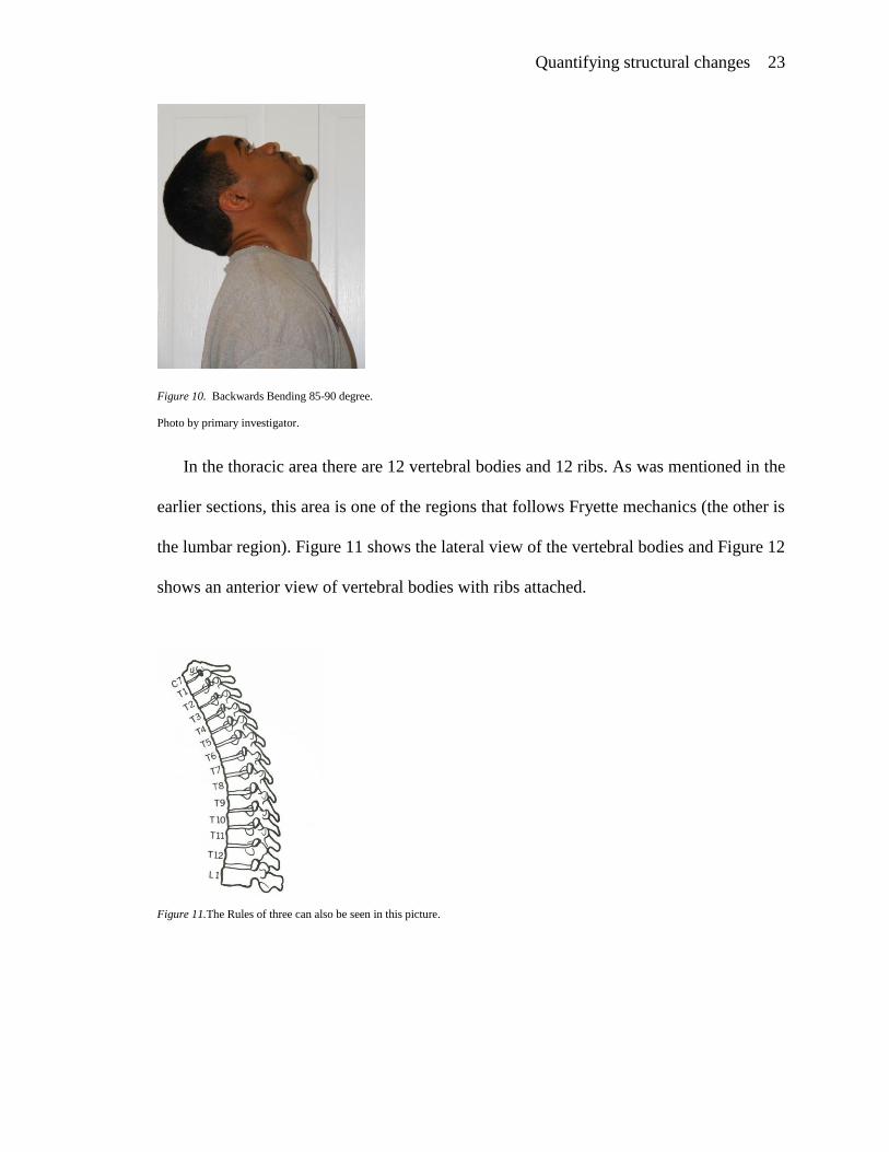
and side-bending components are 85-90 and 40-45 degrees respectively (Figure 9, 10).
Figure 9.Forward Bending 45 degrees. Taken by primary
investigator.
Quantifying structural changes 23
Figure 10. Backwards Bending 85-90 degree.
Photo by primary investigator.
In the thoracic area there are 12 vertebral bodies and 12 ribs. As was mentioned in the
earlier sections, this area is one of the regions that follows Fryette mechanics (the other is
the lumbar region). Figure 11 shows the lateral view of the vertebral bodies and Figure 12
shows an anterior view of vertebral bodies with ribs attached.
Figure 11.The Rules of three can also be seen in this picture.

Quantifying structural changes 24
Figure 12.Thorasic cavity
The thorax can move in flexion, extension, side-bending, and even rotation
(which is its most dominate motion). The rib head (circled) (Figure 13) articulates with
the vertebral body. Having an understanding of the bony landmarks of the spinous and
transverse processes of the spine is needed in order to evaluate and treat SD (Figure 11)
(Fenton, & Phykitt, 1997). The ROM in this area as it relates to side-bending is 20
degrees, and rotation is 40 degrees.
Figure 13. Single vertebrae with rib heads articulating bilaterally.
The lumbar spine (L1-L5) consists of 5 vertebral bodies, and is the site of most
back complaints (L4-L5). The major motion in this section is flexion and extension.

Quantifying structural changes 25
Furthermore, when sacral pathology is present, it is appropriate to treat L5 first and then
the sacrum. The ROM for the lumbar area in flexion is 105 degrees, while extension is 60
degrees. In side-bending and rotation the normal range of motion is 40 degrees.
The pelvis and sacrum unit is composed of three bones: ileum, ischium, and pubic
bones. The sacrum is made of fused bones and a small piece called the coccyx bone. The
pelvis is the major support of the body‟s weight and stance, along with the sacrum. There
can be many dysfunctions found in this area yielding to improper stance and structure.
Problems in this area not only lead to discomfort (muscle tightness, or SD) but can also
lead to neurological problems due to the vast amount of nerves that travel through the
pelvic girdle.
Postural importance
This section will list some of the situations that cause variations in posture, which
can alter forceplate readings even when SD is not found during osteopathic structural
evaluation. Posture is a very complex topic to cover in such a short section; however, the
information below is to further aid in the comprehension of how posture distribution of
the mass of an individual, and the base of support depends on the following three major
conditions (Ward, 2003):
1. Energy requirements of homeostasis
2. Integrity of musculoligamentous structures
3. The compensation of structure at and/or below the skull, which has visual or
balance function on the body
The postural approach to treating pain is very effective because gravity has a
major responsibility in postural homeostasis. Throughout life many conditions can effect

Quantifying structural changes 26
this physiological development of normal curvature such as: anatomic short leg, scoliosis,
small hemipelvis, spondylolisthesis, and wedge vertebrae (Ward, 2003). The alignment
depends on the balance of the curves that are pictured in Figures 14 and 15. When there
are structural changes, the resulting postural compensatory pattern often leads to pain
(Kuchera, 1995).
Figure 14. Postural changes the come with changing in body habitus. An Osteopathic Approach to Diagnosis and
Treatment 1997, pp. 39.
Figure 15. An Osteopathic Approach to Diagnosis and Treatment 1997, pp. 40.

Quantifying structural changes 27
Figure 16 demonstrates the importance of biomechanical principles at work in a
negative response to gravity and poor posture acting on the body. These effects of gravity
on the body cause disruptions in normal physiological group curves leading that can lead
to somatic dysfunction/s. These forces are causing further delineations from normal
group curves of the spine. To properly diagnose dysfunctional areas in the body,
landmarks are used for locating and naming types of lesions found (Figure 17).
. Figure 16. This schematic shows that with rotations in one area, there will be a balanceing rotation in the opposite direction
somewhere else in the body. The end results from a malrotated lower leg can cause upper back complications. Foundations for
Osteopathic Medicine 1997, pp. 605.

Quantifying structural changes 28
Figure 17. These landmarks are used to locate possible asymmetries in posture. An Osteopathic Approach to
Diagnosis and Treatment 1997, pp. 44.
Forcplate
Barbe and Barr, (2006) were able to show that musculoskeletal disorders (MSD)
not only account for a significant proportion of work injuries but also that the key to
controlling the impact of such disorders is prevention or early intervention. Once again,
the latter part of this equation is what osteopathic medicine can do. However, in research,
most osteopathic measurement devices are qualitative. Quantitative measuring tools
provide greater validity. Furthermore, objective data is more widely accepted in the
scientific community.
There have been studies performed in the past that evaluated center of pressure
and ground reaction forces in relationship to posture. One study performed by Takala,
Korhonen, & Viikari-Juntura (1998), showed that postural sway has associations with
low-back symptoms in a working population. The researchers found a small correlation
of sway and low back pain when participants in a quiet stance (not moving and arms

Quantifying structural changes 29
down to the side), were compared with their eyes opened and eyes closed. Due to their
inclusion of the variables eyes open and eyes closed, this study was not included in the
current project, which evaluated center of pressure (CoP) change with eyes open prior to
and after OMM was performed. Also, the relationship of CoP with somatic dysfunction
was evaluated through questionnaires and forceplate readings.
Further studies on ground reaction forces by Jonsson, Seiger, & Hirschfeld
evaluated single-leg stance. Their project was able to demonstrate that difficulties in
maintaining static position are dependent on both an impairment to compensate for the
disturbance of posture (caused by a weight-shifting) and weaknesses in musculature
which support the body. The end result was that the first five seconds of quiet stance is
the most important in maintaining posture on one leg.
More research has been conducted on sway than CoP because of the neuronal
complexities. Zhang, Kiemel, & Jeka (2006) performed a study to evaluate the
engagement of the musculature as it relates to sensory involvement on a forceplate with
in-phase (ankle stability) and anti-phase (hip involvement). This means that the amount
of sway demonstrated by the lower extremities (in-phase) was compared to that of the hip
structures (anti-phase) when the patient would change sensory stimulus (finger to nose
with eyes open/closed and finger to pressure testing). The results showed that increased
sensory involvement (i.e. movement of arms in different directions) would yield to
greater recruitment of the pelvic musculature to aid in maintenance of the upright stance
compared to ankle sway. This means that with bilateral movement of the upper
extremities, pelvic movement would move at great frequencies than the ankles. The study
showed the importance of a longitudinal analysis to alleviate pelvic and sacral

Quantifying structural changes 30
abnormalities. Based on Zhang, Kiemel, & Jeka (2006), the research in Honduras will
address the correlation between structural disturbance and improving functionality.
There is one study that has already evaluated the CoP trajectories between
bilateral and unilateral stance. The conclusion of this study was that the reason that
bilateral stance yields lesser movements as it relates to trajectories is because there is less
surface area when bearing weight on one foot. The incorporation of arm movement
distracts the central nervous system from the balance mechanism of the individual,
yielding to greater instability (Hwang, Huang, Cherng, & Huang, 2006). This was a very
important study because the involvement of the upper extremities decreases the CNS
functional capacity, thus limiting its ability to independently focus on balance. Therefore,
in Honduras the involvement of dual coordination mechanisms of the upper and lower
extremities will not be performed. Direct incorporation of the nervous system will not be
attempted in this project, and quiet stance (standing still) with and without manipulation
was the focus of the Honduras project.
Two studies have direct implication for methodology and design of the research
conducted in Honduras. The first study utilized two forceplates (in contrast to the one
Isobalance forceplate machine used in Honduras) but measured the ground reaction
forces upon the feet. The researchers Jonsson, Seiger, & Hirshfeld (2003) evaluated the
individual in the heel-to-toe position and monitored which foot had the most movement.
The conclusion was that most of the weight was placed on the rear leg and the front leg
was mainly for support. Furthermore, they showed that the tandem stance in the heel-to-
toe position is not the most suitable test when assessing equal weight-bearing capabilities

Quantifying structural changes 31
(Jonsson et al., 2003). This information helped limit this research to tandem stance with
feet shoulder-width apart.
The study by Genthon & Rougier (2004) showed that there is an increase in
energy used in an asymmetric posture, and in a simulated unequal weight distribution. It
also showed that the leg that had a decrease in CoP was causing a great amount of
asymmetry in the body. The study by Genthon & Rougier has direct application in the
Honduran study increasing somatic dysfunction corrects a “correctable” leg length
discrepancy, innominate or postural imbalance.
Summary
Of the studies that were reviewed, none considered correlating manipulative
treatment with changes in postural imbalances and the effect that would have on CoP or
postural discomfort. The review of literature provided important insights into
methodology and background information to construct the present study. Those insights
are incorporated into the methodology section and noted accordingly. Changes in CoP
measured by Isobalance forceplate in concert with OMM is an important line of research.
Future studies with much larger populations and random sampling will be beneficial, yet
the baseline from the present study is a good place to start in Honduras. The
measurements of somatic dysfunction by the use of osteopathic structural evaluation
(physical pathologies that are correctable or manageable with the use of OMM) have
provided data generalizable to other populations as more studies are conducted that
include a wider range of representative groups. Drawing correlations between structural
problems and osteopathic structural evaluation and manipulation through the Isobalance
forceplate can provide fertile information for enhancement of human health with non-

Quantifying structural changes 32
invasive techniques. Using non-invasive and inexpensive techniques is an important
medical advancement in countries with limited access to expensive technologies and
medications. Moreover, it avoids the tendency of modern medicine to rely on prescription
and invasive techniques that are expensive when a more holistic approach is available.
There is a desperate need to quantify manual manipulation as a scientifically-
viable option for structural discomfort, as shown in the review of literature. The
correlations of somatic dysfunction and changes viewed in the CoP will validate that
structural changes present with OMM. Therefore, the effectiveness of the hands-on
approach to medicine as shown in the literature review will be accepted not only by those
in the medical field, but also by those in the scientific community.

Quantifying structural changes 33
CHAPTER III
Methods
This section covers the methodology, design, instrumentation, procedures, and
data analysis. Detailed information about OMM and the Isobalance forceplate used to
measure the participants in the study is included. There are multiple figures and some
tables that will aid in understanding the procedures that took place during the
examination of each individual.
Research Question
1. To what extent will the introduction of OMM lead to reduction of somatic
dysfunction of individuals in the study?
2. To what extent will OMM change the measured indicators of CoP and postural
sway with the utilization of the Isobalance forceplate?
3. To what extent will a particular response (positive, negative, no change,
variable change) be identified by the „outcomes‟ designed in this study?
4. To what extent did participants gain knowledge and accept osteopathic
medicine after viewing the instructional videos presented during the study?
Study Variables
The two major independent variables were 1) OMM procedures, and 2) education.
The dependent variables were 1) level of knowledge, 2) level of acceptance of OMM, 3)
reduction of somatic dysfunctions, and 4) change in CoP and postural sway. Exclusion
and inclusion criteria and positive and negative controls help guide the study. Inclusion
and exclusion factors defined in specific terms how individuals were selected for the
study, thus meeting IRB requirements.

Quantifying structural changes 34
Inclusion Criteria refer to the means of selecting the sample from the population. The
criteria include:
- Status: Baxter Institute student, faculty/staff
- Age: older than eighteen and younger than fifty
Exclusion Criteria clearly define factors that limited participation. The criteria include
potential participants who fit specific conditions:
- Distracting injuries
- Acute traumatic injury
- Inflammatory arthritis
- History of neurological disease
- Osteoporosis risk or history
- Severe atherosclerotic disease
“Patient Treatment” refers to the positive and negative controls in the study
design. The positive controls were the practices performed in the study that were
designed to yield positive results. 1) The participants watched a translated audio-visual
program that explained osteopathic medicine. 2) Each participant received a pretest and
posttest to gauge the level of understanding of musculoskeletal health and osteopathic
manual medicine. 3) All osteopathic manipulations, lectures, and evaluations were
conducted by the primary investigator. 4) All forceplate measurements were conducted
by the secondary investigator. 5) If no somatic dysfunctions were found, the individual
was asked to stand back on the machine for reevaluation and measurements.
Quantifying structural changes 35
The negative controls that were limitations in this study included: 1) Individuals
with chronic dysfunction did not receive optimal benefit because the number of
treatments were not sufficient to correct the many years of structural asymmetry, or
because the defect/s were not correctable (i.e. innominate dysfunction caused by
scoliosis); 2) Healed fractures are resistant to manipulations. 3) Limited comprehension
of the pretest and posttest questions could yield incorrect answers. Comprehension was
limited because of the variations in syntax across a variety of Spanish speaking countries
included in the study. Similarly, some participants were unable to follow commands
either due to language variations in translation of terms or the use of terms that were not
translatable in their purest sense. There was a bilingual translator present for the majority
of the examinations. 4) Many of these individuals participated in soccer games on the
campus of Baxter, and the minor traumas that occurred on the field could reduce the
effectiveness of the OMM they received.
Research Methodology
Procedures followed a case study approach. Protocols were designed to
implement treatment and educational components along with pre and post-measurements.
Instrumentation included equipment in the treatment that provided quantitative indicators.
Additional instrumentation provided attitudinal and knowledge metrics, along with
qualitative opened-ended questions to provide an in-depth assessment with explanatory
power to add meaning to the quantitative indicators.
The pre and posttest (Appendices B & C) were provided via an internet website
called “survey monkey”, and the scores were tabulated by percentage of correct to
incorrect answers. All results from the 20-question pretest and 21-question posttest were

Quantifying structural changes 36
tabulated by comparing the number of correct to incorrect answers. Group scores as
opposed to individual scores were gathered and compared to examine the knowledge
gained from the group as a whole.
The following provides a chronology of events:
1. Upon arrival in Tegucigalpa (January 30th), general preparations were
conducted until the second of February. These preparations consisted of
testing all mechanical devices (Isobalance forceplate) and obtaining a room
and audio-visual equipment (Appendix D) for initiation of the study.
On the fourth and fifth of February an introduction of the project was
conducted in the general assembly area. The project‟s design and
purpose was discussed (Protocol for Consultation). Everyone who
attended received the following: A) the informed consents (Appendix
E) (collected through the six to the seventh of the month), and B) the
exclusion criteria.
2. At the second meeting (the 6th
or 7th
of February) all informed consents were
collected and access codes for the pretest were given. A schedule for baseline
measurements was introduced, with measurement starting as early as that day
(the sixth and ending on the ninth of the month). A manipulation schedule was
given so participants could start receiving their OMM on the 11th
.
The baselines days included: osteopathic evaluation (Appendix F), and
forceplate readings without shoes in the upright position.
Quantifying structural changes 37
3. During the third meeting (on the morning of February 11) those who signed
the informed consent forms and finished the pretest started receiving
manipulation. Also, participants viewed mini lectures via a pre-recorded DVD
that contained the following information:
o Introduction of contents of the study
o Purpose of Study
o Definition of a “Forceplate”
o A list of different physical areas that are treatable through
OMM
o Differences between Osteopathic Physicians, Allopathic
Physicians, Massage Therapists, and Chiropractors
o Osteopathic Philosophy
4. First evaluation and treatment (the week of February 11). Each person‟s visit
consisted of the following:
a. Measuring of CoP and postural sway on the forceplate, osteopathic
structural examination, and then another measuring of CoP and
postural sway.
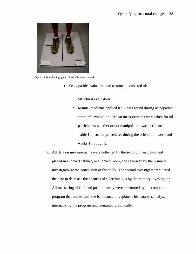
b. The forceplate measurements were conducted in the following manner:
The participants stood erect with arms crossed for 30
seconds on the forceplate (Figure 18).
Quantifying structural changes 38
Figure 18. Isotechnology photo of forceplate used in study.
Osteopathic evaluation and treatment consisted of:
1. Structural evaluation
2. Manual medicine applied if SD was found during osteopathic
structural evaluation. Repeat measurements were taken for all
participants whether or not manipulation was performed.
Table 10 lists the procedures during the orientation week and
weeks 1 through 5.
5. All data on measurements were collected by the second investigator and
placed in a locked cabinet, in a locked room, and reviewed by the primary
investigator at the conclusion of the study. The second investigator tabulated
the date to decrease the chances of selection bias by the primary investigator.
All measuring of CoP and postural sway were performed by the computer
program that comes with the Isobalance forceplate. This data was analyzed
internally by the program and formatted graphically.

Quantifying structural changes 39
Population and Sample
The population included seminary students and staff at the Baxter Institute
Theological Seminary in Tegucigalpa, Honduras and in residence from January 2008 –
February 2008. This group of individuals consisted of 55 males and females on-site at
the time of this study. These individuals are primarily from Central America, South
American, or the Caribbean Islands.
From this population, volunteers were solicited to reach the final sample of 33.
These individuals were selected for the study because of their availability, compliance,
and accessibility. Furthermore, these individuals are educated and valued in their
community because of their higher levels of education and future occupations (pastors).
The participants have the potential to increase knowledge and the literacy rate, which is
very low in many of the areas where they will likely work. The group of participants
were chosen in part for their potential ability to influence the views of their community
about physicians and the healthcare system. The minimum age requirement for
participation in this study is eighteen, the maximum age is fifty. The spoken language is
Spanish.
Instrumentation
There were four interventions that were utilized in this study. When the
participants entered the room to receive the evaluation and/or treatment, an educational
video was played. This video allowed the participants to passively learn about the
osteopathic philosophy. The video was translated by a fellow classmate, Maria Dunn, a
representative of the Virginia College of Osteopathic Medicine (VCOM class of 2009).
This video played continuously in classroom 5 at the Baxter Institute, from the 3:00 until

Quantifying structural changes 40
5:30 p.m. Monday through Thursday of weeks one, three, and five. The video lasted 30
minutes and ran continuously during the manipulation period.
All manipulations were performed by one individual (primary investigator).
During the initial stages of this project (baseline/first week of evaluation and treatment)
Doctor Evelyn Parrish D.O. was present to make sure all osteopathic structural
evaluations and manipulations were performed correctly. Once the initial measurements
were received from the forceplate, the individual was ready for evaluation. The following
photograph (Appendix G) illustrates how the room was set up for the procedures. The
hands-on approach to correcting somatic dysfunction was performed in two possible
ways, depending on the problem found: 1) tenderness of the area, and 2) comfort level of
the individuals to receive certain treatments. The manipulations that were performed on
the individuals consisted of muscle energy and/or HVLA.
The Isobalance forceplate is a machine designed to evaluate many different
functions of the human dynamics of motion. The numerical information collected from
the forceplate evaluated CoP and postural sway. The data was then statistically
manipulated for comparison and possible relationship with OMM. This single forceplate
device analyzed percent weight distribution for a given time and the amount of postural
sway within a six-inch radius. CoP is the specific measurement utilized while individuals
are standing in tandem stance (sometimes on two different forceplates [Appendix H] or
on a single forceplate [Appendix I]).
A general knowledge pretest of what healthcare means to each participant was
administered prior to the treatment and then a similar test was given at the conclusion of
the study to measure the same things. Furthermore, participants were asked prior to and

Quantifying structural changes 41
after completion of the study to select, from multiple choice questions, the functions of an
osteopathic physician. After participating in manipulation and attending a pre-recorded
mini lecture, a posttest was used to determine new knowledge gained. There was no
cronbach alpha performed in this study. The „outcomes‟ designed for the evaluation of
change in the alignment of those that participated in the study, were designed specifically
for this study and have not been tested before.
Statistical Analysis
The pre and posttest answers were analyzed by comparing the percentage of
questions answered correctly.
The forceplate data was analyzed via Chi-Square Goodness of Fit test to see if
participants moved into any of the four outcome categories randomly or if the movement
into outcomes was a result of manipulation. Bivariable Chi-Square tests and multivariable
logistic regression modeling were used to evaluate the association between presence of
lesion in a specific body region (ie, upper cervicals, lower cervicals, thoracic, etc.) and
any of the outcomes one through four.
Summary
The methodology provided a detailed description of the procedures and related
areas. This is important for those who read this study to understand the context for the
investigation and for future researchers who may wish to replicate or build on the
methodology in this study. The figures and tables help visualize the different positions of
the participants during collection of data via the Isobalance forceplate. The proceeding
chapters consist of the results (Chapter four), and a summary, conclusion, discussion, and
recommendations (Chapter five).

Quantifying structural changes 42
CHAPTER IV
Results
The results of this study will be separated into three major categories. The first
category which answers the first research question, provides the number of lesions found
during the osteopathic structural evaluation. The purpose of the osteopathic structural
evaluation, was to find trends in lesions found and body regions most frequently affected.
The second category (addressing research questions two and three) makes the connection
between OMM performed on an individual and structural change. This was done by
monitoring CoP and postural sway via the use of the Isobalance forceplate (figure 18).
The final category, „knowledge gain,‟ examines the amount of material that was retained
during the course of the study and answers the fourth research question. Knowledge gain
was measured by comparing scores on a pretest and posttest distributed to participants in
the study. The tests measured individuals‟ gain in knowledge of the principles of OMM
and the basic importance of being actively involved with one‟s health. Listed below are
the research questions:
Research Questions
1. To what extent will the introduction of OMM lead to reduction of somatic
dysfunction of individuals in the study?
2. To what extent will OMM change the measured indicators of CoP and postural
sway with the utilization of the Isobalance forceplate?
3. To what extent will a particular response (positive, negative, no change,
variable change) be identified by the „outcomes‟ designed in this study?

Quantifying structural changes 43
4. To what extent did participants gain knowledge and accept osteopathic
medicine after viewing the instructional videos presented during the study?
Response Rate
At the initiation of the study surveys were distributed to 32 students and faculty
eligible for the study. By the conclusion of the study at the fifth week, 26 surveys were
distributed to the remaining individuals in the study. While 26 post tests were distributed,
one participant who completed the study did not return a pretest or posttest and the
TIC_Change 1 & 2 data was not taken for this individual. This identified individual
missed part of the measuring on the forceplate settings and also left one session early
without investigators‟ knowledge. This was found only at the conclusion of the study,
when primary investigator reviewed the data (delay in data review was instituted in order
to decrease selection bias). However, while pre and post test calculations and the
TIC_Change 1 & 2 could not be made on this participant, the participant‟s data was
included in the heel-to-toe and toe-to-heel data, along with the total count for SD lesion
found per body region.
Demographic Data
Participants in the study included 24 males and 2 females. The average age of the
participants was 25 – 26 years old. All participants were from Spanish-speaking countries
and 77% of the individuals were from Central America. The participants were composed
of first- through third-year students and two faculty members.
Findings
Osteopathic Manual Manipulation. Not all somatic dysfunctions (SD) result from a
finding on the body that is amenable to manual therapy. Therefore, the particular areas of

Quantifying structural changes 44
the body that can exhibit SD and can also have OMM applied are called structural
lesions. In order to tabulate these lesions correctly, a standard osteopathic structural
evaluation was performed. Once found, lesions were tabulated in table 5 listed below.
The average number of lesions found per manipulation setting decreased from week one
to week five.
Table 1
Osteopathic Lesion
Manipulation
weeks
Total # of SD found
throughout the
study
Average #
of SD per
person
Standard
deviation
Week 1 37 2.74 0.90
Week 3 19 1.41 0.72
Week 5 23 1.70 0.59
The total number of SDs found throughout the study also decreased from week one to
week five, as did the average number of SDs per person.
Of the 80 total somatic dysfunctions that were found during the course of this
study from the 26 participants, 68% of these lesions were found in the thoracic vertebrae.
The most common rotational dysfunction was a neutral component. In the thoracic
vertebrae, the most common type of neutral lesion was a Right on Left (R on L) neutral
lesion. The second most common rotational dysfunction that occurred in the thoracic
region was Right on Right non-neutral lesion. The sacrum body region had the next
highest number of SD lesions, at a total of 12%. The rotational component that occurred

Quantifying structural changes 45
most often at the level of the sacrum was Left on Left (L on L). The following body
regions comprised the remaining somatic dysfunctions found: upper cervicals 8.7%,
lower cervicals 6.2%, leg length 5.0%, and innominate with only 2.5%. There were no
recorded lumbar lesions in this study.
Isobalance Forceplate results. The statistical analysis for each participant‟s percentage of
time in the center and pressure on each of the four quadrants (left heel, left toe, right toe,
and right heel; see figure 17) of the Isobalance Forceplate were plotted against a
combination of weeks and sessions (ie, week 1 before OMM, week 1 after OMM, week 2
before OMM, etc.). A set of criteria called study „outcomes‟ were designed and used to
answer each research question three through five, and they are listed below:
1. Time in center change 1 (TIC_Change 1): comparing pre-manipulation (week
one) with post-manipulation (week five) and looking for the following
responses: no change (NC), negative change (NEG), positive change (POS),
and variable change (V) in the time spent in the center of the six-inch diameter
circle on the forceplate (i.e. postural sway).
2. Time in center change 2 (TIC_Change 2): at least two positive/negative/or no
change data points on the forceplate readings during two manipulation
sessions (example of three positive improvements post-manipulation:
Appendix J).
3. Heel-to-toe change in quadrant pressure: a change in heel-to-toe pressure from
start to finish of the study equaling CoP (example of right foot heel-to-toe
change and left foot toe-to-heel change: Appendix K).

Quantifying structural changes 46
4. Toe-to-heel change in quadrant pressure: a change in toe-to-heel pressure
from the first week to the final week of the study.
For the four outcomes listed above, two null hypotheses were stated and statistically
tested.
1. Movement of the participants into any of the outcome categories was a totally
random process. The statistical analysis used for this hypothesis was a Chi-Square
Goodness of Fit test.
2. There was no association between presence of a lesion in a specific body region
and any of the outcomes. This lack of association was tested using bivariable chi-
square tests and multivariable logistic regression modeling. The no change (NC)
category was used as the baseline of all outcomes.
Throughout the proceeding explanation of data, the above null hypotheses will be
referred to as null hypothesis 1 (NH1) and null hypothesis 2 (NH2). NH1 was testing the
response groups (positive, negative, no change, or variable) as they compared to the
number of participants. Therefore, the results of NH1 answered the third research
question. The next four tables demonstrate this by comparing response groups to the
forceplate data collected from Time in the Center I and II (TIC_1 & TIC_II), heel-to-toe
and toe-to-heel.
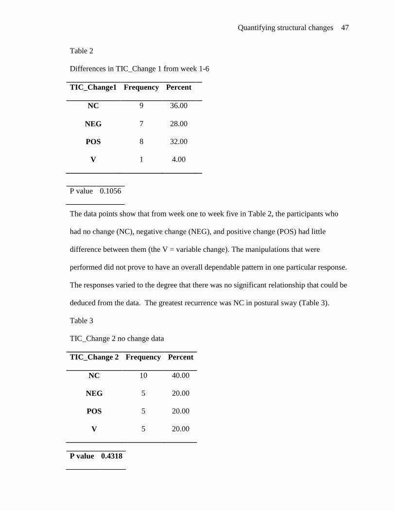
Quantifying structural changes 47
Table 2
Differences in TIC_Change 1 from week 1-6
TIC_Change1 Frequency Percent
NC 9 36.00
NEG 7 28.00
POS 8 32.00
V 1 4.00
The data points show that from week one to week five in Table 2, the participants who
had no change (NC), negative change (NEG), and positive change (POS) had little
difference between them (the V = variable change). The manipulations that were
performed did not prove to have an overall dependable pattern in one particular response.
The responses varied to the degree that there was no significant relationship that could be
deduced from the data. The greatest recurrence was NC in postural sway (Table 3).
Table 3
TIC_Change 2 no change data
TIC_Change 2 Frequency Percent
NC 10 40.00
NEG 5 20.00
POS 5 20.00
V 5 20.00
P value 0.1056
P value 0.4318

Quantifying structural changes 48
The same conclusion can be drawn from the data of TIC_Change 2, even though there
was a greater difference amongst NC and the other three responses, there was still no
significance between the response data. Looking for two of any type of consistent
significant change in the forceplate data as it relates to manipulation could be performed
(outcome 2). The data in Table 4, demonstrates the results of change from pressure
placed on the heel in the first week to pressure placed on the toe by the fifth week. Even
though there was a change in pressure during the five total weeks of the study, the
frequency was not enough to elicit any significant change.
Table 4
Significant change in Heel-to-Toe
Heel-to-Toe Frequency Percent
Changed 16 61.54
No Change 10 38.46
The data in Table 5, shows that no change was appreciated from toe-to-heel criteria. The
NH1 was not disproved and is true with respect to using the Chi-Square Goodness of Fit
test to show complete randomization of the persons who were manipulated and moving
into any of the categories almost equally.
P value 0.3269

Quantifying structural changes 49
Table 5
Significant change in Toe-to-Heel
Toe-to-Heel Frequency Percent
Changed 13 50.00
No Change 13 50.00
The data in Table 6, evaluated the number of participants who presented with or without
SD lesions in particular body regions at least once during the course of the study. The
participants had to have a palpable SD lesion that could have been manipulated via
muscular energy or HVLA technique. Then the total number of individuals who had an
upper cervical lesion (for example) would be placed in the lesion category, and those who
did not were placed in the no lesion group. The percentage of those that had a specific
lesion in a particular body region is also listed under the frequency category. The p values
listed in the table are demonstrating the significance of an individual having a lesion
verses not having a lesion. Some body regions had no lesions found during (ie. lumbar
region) the study and other regions had lesions throughout the study (ie. thoracic region).
P value 1.00

Quantifying structural changes 50
Table 6
Lesion in specific body regions
Body region Frequency Percent P value
1.Upper cervicals Lesion 6 23.08 0.01
No lesion 20 76.92
2.Lower cervicals Lesion 4 15.38 5.33E-04
No lesion 22 84.62
3.Thoracic Lesion 26 100 1.00
No lesion 0
4.Lumbar Lesion 0 1.00
No lesion 26 100
5.Innominate Lesion 2 7.69 1.049E-05
No lesion 24 92.31
6.Sacrum Lesion 9 34.62 0.17
No lesion 17 65.38
7.Leg length Lesion 2 7.69 1.049E-05
No lesion 24 92.31

Quantifying structural changes 51
Table 7, addresses research question two and NH2 with the utilization of a
bivariable chi-square tests and multivariable logistic regression modeling. The bivariable
analyses compared for example a specific body region to change noted by the forceplate.
The bivariable tests measured each body region individually and compared it to each of
the four outcome methods in order to determine change in indicators of postural sway
measured by the Isobalance forceplate during the weeks involved in the study (i.e. if a
sacral lesion was found then there would be a corresponding change in TIC_Change 1).
Table 7
TIC_Change 1 Sacrum
Body Region Lesion vs
No lesion
NC NEG POS V Total P-value
Sacrum
Lesion
2 1 6 0 9
0.03
No Lesion 7 6 2 1 16
There was a positive correlation between finding a sacral lesion and having a positive
change in TIC_Change 1 (an increased amount of time spent in the 6 in. circle). Those
who had sacral lesions at some point within the study had an improvement in postural
sway. The table above is the only bivariable table listed here because out of the 27
bivariable data tables, the sacrum was the only body region that had a correlation with
change in postural sway. The other bivariable data tables are located in the Table sections
9 – 36.
The results from the bivariable chi-squared analysis demonstrated that there was a
correlation between change of postural sway and sacral dysfunctional lesions. Therefore,

Quantifying structural changes 52
the reference point used for the multivariable logistic regression modeling was the no
change (NC) category. The sacral change was evaluated against all of the outcomes listed
on pg 45. The NC was the baseline and was compared to POS and NEG change of
TIC_Change1 & 2, and change in heel-to-toe and toe-to-heel. The TIC_Change 1
demonstrated that for those who had a sacral lesion there would be a movement of NC to
the direction of POS change (p ≤ .04). The odds ratios for these results showed 10.5 times
likelihood if one had a sacral lesion to change from a NC to a POS in postural sway
readings on the forceplate. There was no other statistically significant relationship with
the other outcomes.
Pretest and posttest data. There were 26 participants in this study; however, only 25
participants responded to the pre and posttests. The participants answered a 20 question
pretest before being exposed to the principles of OMM. The 20 questions included
qualitative questions that dealt with the individuals‟ exposure to healthcare professionals
and questions on how the participants manage health-related issues. After the exam, all
participants were then eligible for participation in the study. During the following five
weeks, the subjects were structurally evaluated and OMM was applied to the appropriate
area. The informational video that covered basic fundamentals of osteopathic medicine
(topics such as those mentioned in the review of literature) was presented while
participants were manipulated. The video ran continuously during these sessions. At the
conclusion of the study, a posttest was distributed to all participants to evaluate
knowledge gained and changed ideals of healthcare.

Quantifying structural changes 53
Figure 19. The above graph presents the number of questions on the pretest and posttest (x-axis). On the y-axis is the number of
participants, along with the number of correct answers for those single best answer questions. The total number of person that
answered the experience questions (view table 4 below) can also be found on the y-axis. The blue bars represent the pretest (before the
presentation of the educational materials) and the red bars are indicative of the posttest results.
Figure 19, depicts all the answers from the pre and posttest. The questions utilized in this
pretest and posttest were placed into six different categories as listed in Table 8.
Table 8
Types of questions
Type of questions Question number Total number
questions
1) Single best answer 1, 5, 6, 7, 10, 11, 12, 13,
14, 15, 16, 17, 18, 19, 20
15
2) Questions showing a gain in
knowledge
5, 6, 10, 12, 13, 14, 15, 16,
20
9
3) Questions answered incorrectly on
the pretest or answered correctly on
the pretest and incorrectly on the
posttest
1,11, 17 3
4) Multi-step questions 1,4 2
5) Qualitative (experience) questions 2, 3, 4, 8, 9 5
6) Questions showing no change from
pretest to posttest
7, 18, 19 3

Quantifying structural changes 54
Analyzing each category:
2) Single best answer:
Of the 20 total questions on the pre and posttest exam, there were a total of 15
questions that had only one correct answer. The pretest had an average of
14.5±8.15 persons answering these questions correctly. After the presentation of
the educational video and at the conclusion of the study, the average number of
persons answering the single best answer questions correctly on the posttest was
16.9±7.90. This increase demonstrates an overall gain of knowledge. (Appendix
L).
3) Questions showing a gain in knowledge:
There were a total of nine questions that demonstrated an improvement in correct
answers from pretest to posttest. Question 12 , “One of the osteopathic
philosophies is that the body has the innate ability to heal itself given the body is
free from dysfunction (homeostasis),” showed the most improvement among
these nine questions (Appendix M)..
4) Questions answered incorrectly on the pretest or answered correctly on the pretest
and subsequently answered incorrectly on the posttest:
There were only 3 questions that fell into this category. Question one was a multi-
step question, “Which of these are components of holistic healthcare?” There was
some gain in knowledge for mental health; however, there were fewer correct
responses from pretest to posttest concerning spiritual health and there was no
change regarding physical health. For the other two questions in this category (11,
& 17), there was a slight decrease in correct responses given from pre to posttest.

Quantifying structural changes 55
For each question, 11 & 17 there was a decrease in one response from the start of
the study to its conclusion (Appendix N).
5) Multi-step:
There were two multistep questions. As mentioned above, question one has only
one single best answer. Question four is the other multi-step question and it falls
within the experience group of questions.
6) Qualitative (experience) questions:
This group of questions could also be called the experience group of question.
This category had the most variations in results. The reasoning behind these
variations will be further discussed in chapter five. There were no true single best
answers to any of these questions.
7) No change from pretest to posttest:
This last category of questions were all single best answer questions that
demonstrated no change from the start of the program to its conclusion in results.
Summary
The overall designs of the data collection tools enabled the study to address the
research questions and null hypotheses. Information gathered from the pre and posttests
demonstrated that there was knowledge gained, improvement of overall understanding of
appropriate personal healthcare, and acceptance of OMM by the individuals tested. The
osteopathic manipulation techniques demonstrated there was a decrease in SD lesions
collectively among those in the study. Utilizing the selected outcomes in the study, the
NH2 was not accepted because of the sacral rotational dysfunction and its correlation
with postural sway change. However, the NH1 was be accepted. There are many
Quantifying structural changes 56
possibilities that may explain why this study did not have the improvement in
understanding of material anticipated by the primary investigator, as well as greater
OMM correlations with postural change. Chapter five will give a summary of this pilot
study and appropriately address the problems in the design and execution of the project.
Quantifying structural changes 57
CHAPTER V
Summary, Conclusion, & Recommendations
Summary
The purpose of this study was to assess the impact of educating an unexposed
population to basic principles of osteopathic medicine, and then treating SD with the use
of OMM and measuring balance changes on an Isobalance forceplate. The Project
Investigator decided to attempt to increase the knowledge of this form of medical practice
among a subpopulation in Central America because of the need for an alternative form of
treatment of musculoskeletal pain. Furthermore, addressing the cause of pain, such as
asymmetrical change due to the stresses cause by ADLs in such a mountainous territory,
yielded the perfect opportunity to not only evaluate but also measure structural change
following OMM on this population.
The Isobalance forceplate measures CoP and postural sway. The utilization of this
particular machine for the measure of structural changes that occur during a manipulative
session has not been performed before. However, the machine is built to measure the
most minute changes in balance and pressure placed on the toes or heels of the feet. If
there is to be a structural change made by OMM, the Isobalance forceplate will read this
discrepancy as an imbalance and compare measurements to a preset reference point. The
changes that were induced in each individual by manipulation and recorded by the
forceplate help lead to the quantification of structural change.
Osteopathic medical practitioners take special interest in effecting change and
encouraging healthier lifestyles of rural population and underserved areas in the primary
care. The population chosen for this study fits into the group assessed in this project.

Quantifying structural changes 58
There was no bias in this study due to previous exposure to OMM as the population was
unfamiliar with this practice. Not all abnormalities or deviations from symmetry found on
the body are correctable by osteopathic manipulation. We used the term lesion to describe
the structural abnormalities that could be treated by OMM. As discussed in the
introduction we used the acronym T-A-R-T to help identify dysfunctions that could be
manipulated and were found during the osteopathic structural examination. There are two
types of dysfunctions, Type I and Type II, and these lesions were addressed with either
HVLA and/or muscle energy manipulative techniques. Once the appropriate technique(s)
were applied, a reassessment was performed to evaluate the structural improvement.
Previous studies of postural imbalance have been performed utilizing different
types of single and dual panel forceplate machines; however, there has not been a
correlation made between quantifiable asymmetrical change and correction with OMM.
Of all the following areas - cervical, thoracic, lumbar, innominate, sacrum, and leg length
- individuals who had sacrum lesions demonstrated improvements in postural sway (time
spent in the 6 inch circle) on the forceplate (p≤ .04).
The individuals in this study were their own controls, and on each follow-up
session before manipulation, a measurement was taken on the forceplate. This was done
to test for normalization of the body to previous dysfunction prior to OMM, as well as
duration of manipulation performed. Once measurements were taken, the person was
manipulated and measured again for comparison of forceplate change. It was found that
those who initially had no change in postural sway would develop improvements in
postural sway if they had a sacral lesion (p≤ .04).

Quantifying structural changes 59
The education component was a video that taught basic osteopathic principles and
techniques. A gain in knowledge from the video was tested by pretest and posttest
answers. There was an overall increase in the number of questions answered correctly,
indicating a gain in knowledge throughout the course of the study.
Conclusion
Pretest and Posttest. Of the 20 questions in the pre and posttest, 15 questions had a single
best answer. The increase in correct responses to nine of these single-best-answer
questions can be attributed to exposure to osteopathic principles via the video, as well as
participation in the project itself. Furthermore, the range of question styles - including
multiple choice, multiple answer, and true or false questions - limited the inclination to
guess on each question, thereby ensuring the validity of results. In the following
qualitative/experience-based questions listed below, which measure perceptions of
doctor-patient relationships, there was a 38% increase in yes responses.
3. Has the doctor you visited (if you have a regular doctor) ever asked you how
things are going at home or at school?
a. Yes
b. No
8. Do you feel your doctor communicates well with you and makes sure you
understand what you can do to help yourself?
a. Yes
b. No
9. Does your doctor or one you have visited before explained to you what to do so
you can protect yourself from future related problems?
a. Yes
b. No

Quantifying structural changes 60
Furthermore, by the conclusion of the study there was 96% agreement that
OMM would be accepted in the participant‟s communities if available. There was an
increase in the number of questions answered correctly on the posttest as compared to the
responses on the pretest. However, since the instruments/records were not coded to track
individuals and comparing individual improvement. Thus, the significance was
inconclusive because instrument coding was not sufficient to track and determine results.
Therefore, the following hypothesis one and two (listed below) cannot be determined
with the appropriate level of statistical verification:
Hypothesis 1:
Posttest scores will be higher than pretest scores on knowledge of osteopathic
medicine after participants receive educational materials on OMM.
Hypothesis 2:
Posttest scores will be higher than pretest on acceptance of OMM among
participants.
Osteopathic manipulation. Regular corrections in manipulative lesions with education on
what to do to decrease one‟s chances of injuring him or herself will lead to fewer lesions
over time in the population studied. When exposing a new population to manipulation,
the tendency for the body to want to revert back to its former disposition without
continual conscious awareness of posturing will lead to reoccurring lesions or
development of new compensatory SD.

Quantifying structural changes 61
Forceplate data analysis. Of the four different outcomes as mentioned in Chapter IV
under „Isobalance Forceplate Results‟ the TIC data (postural sway) is the only outcome
that proves to have statistical significance. Only those individuals who have sacral lesions
will demonstrate improvements in the amount of time spent in the six-inch circle.
Therefore, we can infer that the most important segment that needs to be manipulated to
enhance balance is the sacral pelvic component to be correct in the sacral region. As a
result, Hypothesis 3 is accepted.
Hypothesis 3:
Posttest forceplate calculations of parameters of balance compared with pretest
following OMM will show decreased postural sway, yielding to a decrease in
body movement on the forceplate is proven to be correct in the segment of the
sacral region.
Hypothesis number four (stated below) could not be accepted in this study because there
was no relationship in tandem stance improvement in CoP as it relates to certain areas of
pressure on the feet.
Hypothesis 4
Posttest compared to pretest center of pressure (weight distribution) placed on the
forefoot or hind foot during tandem stance will increase, thus show improvement
of somatic dysfunction following OMM.
Research hypothesis number five (listed below) was accepted because there was a
significant decrease noted in the number of somatic dysfunctions found from week one to
week five.

Quantifying structural changes 62
Hypothesis 5:
Somatic dysfunction measurement will decrease between week one to week five
OMM treatment, thus showing improvement.
Recommendations
Recommendation for Future Research. Designing a project that examines the
effectiveness of OMM on the body is not very complicated and in fact has been
performed multiple times in past studies. However, this study was unique in that it
recorded the qualitative component and applied an additional quantitative component
instead. There are four major parts to the project that make it revolutionary. The four
components are: 1) presenting and testing foreign information to an unexposed audience,
2) drawing correlations between postural sway and CoP to an individual‟s post-
manipulative state, 3) charting somatic dysfunctions that have manipulative components
and looking for trends throughout a course of time, and lastly 4) evaluating acceptance of
manipulation as a practice of medicine by an unexposed population.
The translation process of osteopathic terminology was the first major struggle
that was encountered in preparation of educating the participant‟s involved in the study.
Some words that are used in osteopathic medicine do not translate word for word in the
Spanish language; therefore, learning new theories and terminology was required to fully
understand the science behind manipulation. In future studies of foreign populations it is
recommended that education occur not only verbally but visually as well. Learning the
anatomical parts of the body, along with terminology and hands-on medicine was a new
experience for the study participants.
Quantifying structural changes 63
Retrospectively evaluating the design of this study, there are areas that could have
increased the effectiveness and validity of the results obtained in the study. The pretest
and posttest did not have personal identifiers. This presented a major problem when
trying to see if any of the information obtained from the pre and posttest was statistically
significant. Without having personal identifiers in the study there were no tests that could
be used to yield a p-value. The only data that could be deduced from the answers of the
test were improvement in selected correct answers leading to a percentage of overall
increase in selected answers, answered correctly by the group. This is a limitation
because comparing a single individual‟s pretest with his or her posttest could not be
performed. Furthermore, there was one individual who was a part of the study who did
not take the pretest or posttest. There were a total of 26 persons in the study but only 25
participated in the written exams. The same individual also was missing some data from
the forceplate measurements. The postural sway (TIC) data was missing from one
individual yielding to a total of only 25 individuals, while the CoP data had all 26
participants involved in the study.
Listed below are additional recommendations that could help the next researcher
who attempts a similar study.
1. Include a larger sample size and a population that is not just a convenient
population.
2. Examine a population with more chronic issues but within the same age
ranges.
3. Allow for longer courses of treatment that span months as opposed to weeks.

Quantifying structural changes 64
4. Develop more pretest and posttest questions that are worded differently to
better test knowledge gained and understanding of new terms used.
5. Educate individuals who are native to the region about manipulation so that
communication is not compromised by language barriers, limiting the chances
of missing information.
6. Make a clear distinction between manipulation, stretching, and learned
procedure in future studies. A clear delineation of these three will help further
validate osteopathic medicine as an accepted science among non-osteopathic
physicians (those that do not practice manipulation due to a more definite
science to the manual manipulation with numbers, instead statement just as „I
feel much better‟.
Recommendation for Practice. As this particular study was one of the first of its kind
there are further modifications of the design of the current study that will lead to a more
reliable study in the future. The outcomes that were used to interpret a successful
treatment are criteria that were formed specifically for this project. There are no current
non-clinical guidelines for quantifying manipulations as successes or failures. Qualitative
clinical measurements are the accepted norm for OMM. However, a more objective and
reproducible measurement is needed to quantify the effect of OMM for inclusion in the
literature of evidence-based medicine.
1. Have multiple physicians with their own subpopulations and see if results are
similar with the utilization of the same measuring device.
2. Indentify a device that best evaluates the changes witnessed by examiner that
will yield quantitative results.

Quantifying structural changes 65
References
Ahang, Y., Kiemel, T., & Jeka, J., (2006). The influence of sensory information on
two-component coordination during quiet stance. Gait & Posture, 26:267-271.
Bureau of Consular Affairs. (2006, October 11). Honduras. U.S Department of State,
from http://travel.state.gov/travel/cis_pa_tw/cis/cis_1135.html
Baily, H.W., (1976). Some problems in making osteopathic spinal manipulative
therapy appropriate and specific. Journal of American Osteopath Association,
75:486-499.
Barbe, M.F., & Barr, A.E., (2006). Inflammation and the path physiology of work-
related musculoskeletal disorders. National Institute of Health, 20(5): 423-429.
Centers, S., Morelli, M.A., Vallad-Mix, C., & Seffinger, M.A., (2003). General
Pediatrics. In Robert C. Ward et al., Foundations for Osteopathic Medicine:
(pp.305-325). Philadelphia: Lippincott Williams & Wilkins.
DiGiovanna L.E. (1997) Introduction to Osteopathic Medicine. In E.L. Digiovanna
ands. Schiowitz (Eds.), An Osteopathic Approach to Diagnosis and Treatment
(pp.4-5). Philadelphia: Lippincott-Raven Publishers.
Fenton, J.F. & Phykitt, D.E., (1997). Evaluation of the thoracic spine. In Eileen L.
DiGiovanna & S. Schiowitz (Eds.), An Osteopathic Approach to Diagnosis and
Treatment: (pp. 4-5). Philadelphia:
Genthon, N., & Rougier, P., (2004). Influence of an asymmetrical body weight
distribution on the control of undisturbed upright stance. Journal of
Biomechanics, 38:2037-2049.

Quantifying structural changes 66
Hwang, I.S., Huang, C.T., Cherng, R.J., & Huang, C.C., (2006). Postural fluctuations
during pointing from a unilateral or bilateral stance. Human Movement Science,
25:275-291.
Kuchera M.L., (1995). Gravitational stress, musculoskeletal strain, and postural
alignment. Spine. Stats of the Art Reviews. 9(2): 463-490
Genthon, N., & Rougier, P., (2005). Influence of an asymmetrical body weight
distribution on the control of undisturbed upright stance. Journal of
Biomechanics, 38, 2037-2049.
Jonsson, E., Seiger, A., & Hirschfeld, H., (2003). Postural steadiness and weight
distribution during tandem stance in healthy young and elderly adults. Clinical
Biomechanics, 20:202-208.
Jonsson, E., Seiger, A., & Hirschfeld, H., (2004). One-leg stance in healthy young
and elderly adults: a measure of postural steadiness. Clinical Biomechanics,
19:688-694.
Kuchera, M.L. & Kappler, R.E., (2003). Consideration of posture and group curves.
In Robert C. Ward et al. (Eds), Foundations For Osteopathic Medicine: (pp. 580).
Philadelphia: Lippincott Williams & Wilkins.
Leon, M. (2003). Perceptions of health care quality in Central America. International
Journal for Quality in Health Care, 14, pp.67-71.
Martinke, D.J. & Dowling, D.J. (1997). The philosophy of osteopathic medicine. In
Eileen L. DiGiovanna and S. Schiowitz (Eds.), An Osteopathic Approach to
Diagnosis and Treatment: (pp. 4-5). Philadelphia: Lippincott Williams & Wilkins.
Quantifying structural changes 67
Morken, T., Riise, T., Moen, B., Hauge, S.H., Holien, S., Langedrag, A., et al. (2003).
Low back pain and widespread pain predict sickness absence among industrial
workers. BioMed Central, 4:21
Sprafka, S, Ward R.C., & Neff, D., (1981). What characterizes a (sic) osteopathic
principle. Journal of American Osteopathic Association, 81:81-85
Seffinger, M.A., et al. (2003). Osteopathic Philosophy. Foundations For Osteopathic
Medicine (2nd
ed). (pp. 3-18). Philadelphia: Lippincott Williams & Wilkins.
Takala, J., (1999). Introductory report of the international labor office. International
Occupational Safety and Health Information Center. Geneva: International
Labour (sic) Office.
Takala, E.P., Korhonen, I, & Viikari-Juntura, E., (1998). Postural sway and stepping
response among working population: reproducibility, long-term stability, and
associations with symptoms of low back. Clinical Biomechanics, 12:429-437.
Unicef (2006). Honduras: from
http://www.unicef.org/infobycountry/honduras_2026.html
United States Agency of International Development (2005). Health Profile:
Honduras. Retrieved March 2002, from
http://www.usaid.gov/our_work/global_health/aids/Countries/lac/honduras_05.pd
f
Ward, et al. eds. (2003). Foundations For Osteopathic Medicine (2nd
ed).
Philadelphia: Lippincott Williams & Wilkins.

Quantifying structural changes 68
TABLES
Table 9
Defining T.A.R.T
Note: Gives the findings that are present on palpation of someone with an acute or
chronic injury. An Osteopathic Approach to Diagnosis and Treatment 2003, pp. 8.

Quantifying structural changes 69
Table 10
Schedule of project events
Sunday Monday Tuesday Wednesday Thursday Friday Saturday
January 30 31 February 1 2
3 4 5 6 7 8 9
10 11 12 13 14 15 16
17 18 19 20 21 22 23
24 25 26 27 28 29 March 1
2 3 4 5 6 7 8
9 10 11 12 13 14 15
Note: The table represents the schedule followed during the research in Tegucigalpa, Honduras.
Blue-grey coloring = represents the times that participants were not receiving OMM or being evaluated on the Isobalance forceplate.
Yellow coloring = represents the introduction of the study, distribution and collection of informed consents, distribution of access
codes for the pretest.
Green coloring = indicates the scheduled forceplate measurements, osteopathic structural evaluation and treatment, and repeat
measurements after OMM has been performed.
White coloring = these dates represent off weeks from any obligation to the study.

Quantifying structural changes 70
Table 11
TIC_Change 1 Uppercervicals
Body Region Lesion vs
No lesion
NC NEG POS V Total P-value
Uppercervicals
Lesion
3 1 2 0 6
0.88
No Lesion 6 6 6 1 19
Note: NC = no change in postural sway on the forceplate. NEG = Negative change in postural sway on the forceplate. POS = Positive
change in postural sway on the forceplate. V = Variable change in postural sway on the forceplate.
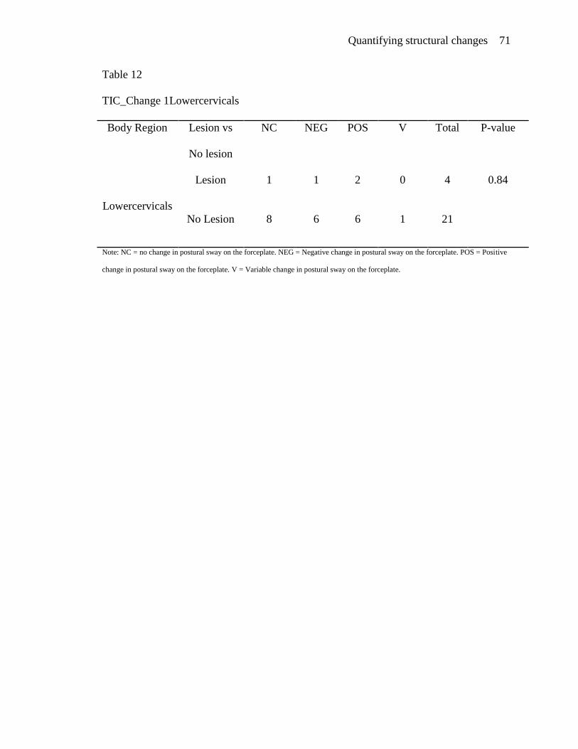
Quantifying structural changes 71
Table 12
TIC_Change 1Lowercervicals
Body Region Lesion vs
No lesion
NC NEG POS V Total P-value
Lowercervicals
Lesion
1 1 2 0 4 0.84
No Lesion 8 6 6 1 21
Note: NC = no change in postural sway on the forceplate. NEG = Negative change in postural sway on the forceplate. POS = Positive
change in postural sway on the forceplate. V = Variable change in postural sway on the forceplate.

Quantifying structural changes 72
Table 13
TIC_Change1 Thorasic
Body Region Lesion vs
No lesion
NC NEG POS V Total P-value
Thorasic
Lesion
9 7 8 1 25
None
No Lesion
Note: NC = no change in postural sway on the forceplate. NEG = Negative change in postural sway on the forceplate. POS = Positive
change in postural sway on the forceplate. V = Variable change in postural sway on the forceplate.
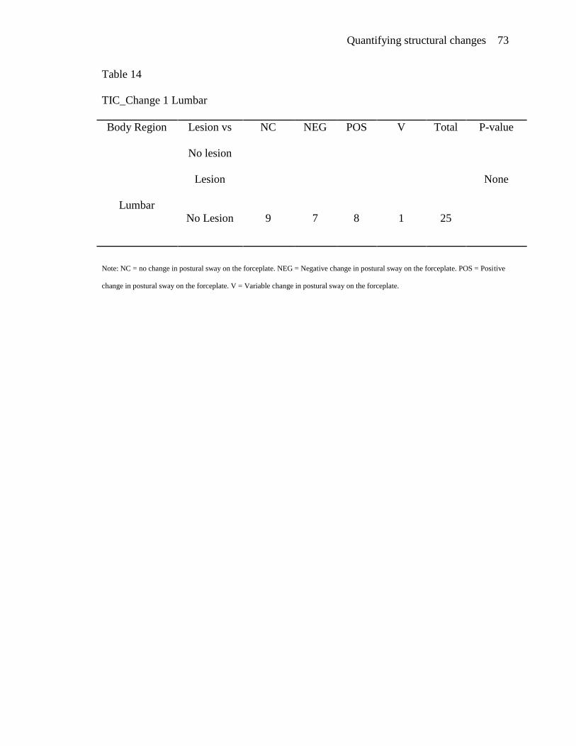
Quantifying structural changes 73
Table 14
TIC_Change 1 Lumbar
Body Region Lesion vs
No lesion
NC NEG POS V Total P-value
Lumbar
Lesion
None
No Lesion 9 7 8 1 25
Note: NC = no change in postural sway on the forceplate. NEG = Negative change in postural sway on the forceplate. POS = Positive
change in postural sway on the forceplate. V = Variable change in postural sway on the forceplate.

Quantifying structural changes 74
Table 15
TIC_Change 1 Innominate
Body Region Lesion vs
No lesion
NC NEG POS V Total P-value
Innominate
Lesion
1 1 0 0 2
0.76
No Lesion 8 6 8 1 23
Note: NC = no change in postural sway on the forceplate. NEG = Negative change in postural sway on the forceplate. POS = Positive
change in postural sway on the forceplate. V = Variable change in postural sway on the forceplate.

Quantifying structural changes 75
Table 16
TIC_Change 1 Leglength
Body Region Lesion vs
No lesion
NC NEG POS V Total P-value
Leglength
Lesion
1 1 0 0 2
0.76
No Lesion 8 6 8 1 23
Note: NC = no change in postural sway on the forceplate. NEG = Negative change in postural sway on the forceplate. POS = Positive
change in postural sway on the forceplate. V = Variable change in postural sway on the forceplate.

Quantifying structural changes 76
Table 17
TIC_Change 2 Uppercervicals
Body Region Lesion vs
No lesion
NC NEG POS V Total P-value
Uppercervicals
Lesion
2 1 2 1 6
0.92
No Lesion 8 4 3 4 19
Note: NC = no change in postural sway on the forceplate. NEG = Negative change in postural sway on the forceplate. POS = Positive
change in postural sway on the forceplate. V = Variable change in postural sway on the forceplate.
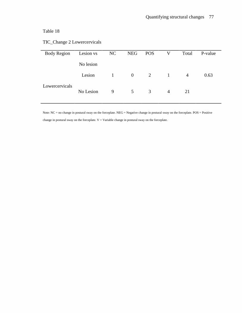
Quantifying structural changes 77
Table 18
TIC_Change 2 Lowercervicals
Body Region Lesion vs
No lesion
NC NEG POS V Total P-value
Lowercervicals
Lesion
1 0 2 1 4
0.63
No Lesion 9 5 3 4 21
Note: NC = no change in postural sway on the forceplate. NEG = Negative change in postural sway on the forceplate. POS = Positive
change in postural sway on the forceplate. V = Variable change in postural sway on the forceplate.

Quantifying structural changes 78
Table 19
TIC_Change 2 Thoracic
Body Region Lesion vs
No lesion
NC NEG POS V Total P-value
Thoracic
Lesion
10 5 5 5 25
None
No Lesion
Note: NC = no change in postural sway on the forceplate. NEG = Negative change in postural sway on the forceplate. POS = Positive
change in postural sway on the forceplate. V = Variable change in postural sway on the forceplate.

Quantifying structural changes 79
Table 20
TIC_Change 2 Lumbar
Body Region Lesion vs
No lesion
NC NEG POS V Total P-value
Lumbar
Lesion
None
No Lesion 10 5 5 5 25
Note: NC = no change in postural sway on the forceplate. NEG = Negative change in postural sway on the forceplate. POS = Positive
change in postural sway on the forceplate. V = Variable change in postural sway on the forceplate.
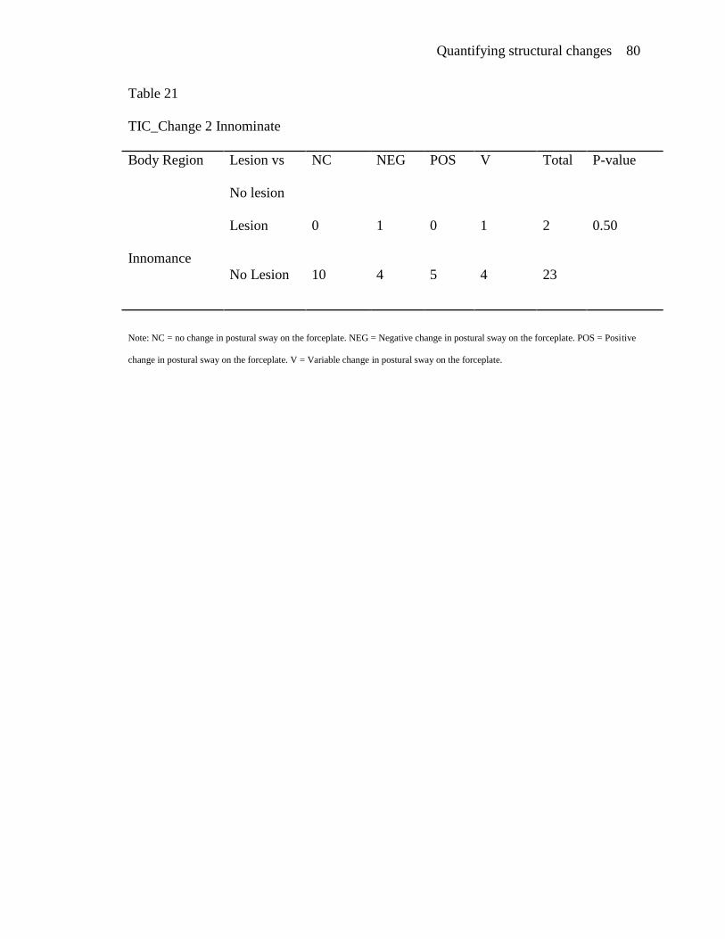
Quantifying structural changes 80
Table 21
TIC_Change 2 Innominate
Body Region Lesion vs
No lesion
NC NEG POS V Total P-value
Innomance
Lesion
0 1 0 1 2
0.50
No Lesion 10 4 5 4 23
Note: NC = no change in postural sway on the forceplate. NEG = Negative change in postural sway on the forceplate. POS = Positive
change in postural sway on the forceplate. V = Variable change in postural sway on the forceplate.

Quantifying structural changes 81
Table 22
TIC_Change 2 Sacrum
Body Region Lesion vs
No lesion
NC NEG POS V Total P-value
Sacrum
Lesion
2 1 3 3 9
0.34
No Lesion 8 4 2 2 16
Note: NC = no change in postural sway on the forceplate. NEG = Negative change in postural sway on the forceplate. POS = Positive
change in postural sway on the forceplate. V = Variable change in postural sway on the forceplate.

Quantifying structural changes 82
Table 23
TIC_Change 2 Leglength
Body Region Lesion vs
No lesion
NC NEG POS V Total P-value
Leglength
Lesion
0 1 0 1 2
0.50
No Lesion 10 4 5 4 23
Note: NC = no change in postural sway on the forceplate. NEG = Negative change in postural sway on the forceplate. POS = Positive
change in postural sway on the forceplate. V = Variable change in postural sway on the forceplate.

Quantifying structural changes 83
Table 24
Heel-to-Toe Uppercervicals
Body Region Lesion vs
No lesion
Change No
Change
Total P-value
Uppercervicals
Lesion
4 2 6
1.00
No Lesion 12 8 20
Note: Change = change seen from CoP on heel-to-toe. No change = no change seen from CoP on heel-to-toe. Data collected via
outcome criteria.

Quantifying structural changes 84
Table 25
Heel-t-Toe Lowercervicals
Body Region Lesion vs
No lesion
Change No
Change
Total P-value
Lowercervicals
Lesion
3 1 4
0.36
No Lesion 13 9 22
Note: Change = change seen from CoP on heel-to-toe. No change = no change seen from CoP on heel-to-toe. Data collected via
outcome criteria.

Quantifying structural changes 85
Table 26
Heel-to-Toe Thorasic
Body Region Lesion vs
No lesion
Change No
Change
Total P-value
Thorasic
Lesion
16 10 26
None
No Lesion
Note: Change = change seen from CoP on heel-to-toe. No change = no change seen from CoP on heel-to-toe. Data collected via
outcome criteria.

Quantifying structural changes 86
Table 27
Heel-to-Toe Lumbar
Body Region Lesion vs
No lesion
Change No
Change
Total P-value
Lumbar
Lesion
None
No Lesion 16 10 26
Note: Change = change seen from CoP on heel-to-toe. No change = no change seen from CoP on heel-to-toe. Data collected via
outcome criteria.

Quantifying structural changes 87
Table 28
Heel-to-Toe
Body Region Lesion vs
No lesion
Change No
Change
Total P-value
Innominate
Lesion
2 0 2
0.51
No Lesion 14 10 24
Note: Change = change seen from CoP on heel-to-toe. No change = no change seen from CoP on heel-to-toe. Data collected via
outcome criteria.

Quantifying structural changes 88
Table 29
Heel-to-Toe Sacrum
Body Region Lesion vs
No lesion
Change No
Change
Total P-value
Sacrum
Lesion
7 2 9
0.40
No Lesion 9 8 17
Note: Change = change seen from CoP on heel-to-toe. No change = no change seen from CoP on heel-to-toe. Data collected via
outcome criteria.

Quantifying structural changes 89
Table 30
Heel-to-Toe Leglength
Body Region Lesion vs
No lesion
Change No
Change
Total P-value
Leglength
Lesion
2 0 2
0.51
No Lesion 14 10 24
Note: Change = change seen from CoP on heel-to-toe. No change = no change seen from CoP on heel-to-toe. Data collected via
outcome criteria.
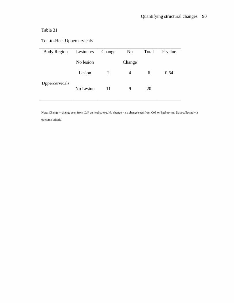
Quantifying structural changes 90
Table 31
Toe-to-Heel Uppercervicals
Body Region Lesion vs
No lesion
Change No
Change
Total P-value
Uppercervicals
Lesion
2 4 6
0.64
No Lesion 11 9 20
Note: Change = change seen from CoP on heel-to-toe. No change = no change seen from CoP on heel-to-toe. Data collected via
outcome criteria.

Quantifying structural changes 91
Table 32
Toe-to-Heel Lowercervicals
Body Region Lesion vs
No lesion
Change No
Change
Total P-value
Lowercervicals
Lesion
1 3 4
0.59
No Lesion 12 10 22
Note: Change = change seen from CoP on heel-to-toe. No change = no change seen from CoP on heel-to-toe. Data collected via
outcome criteria.

Quantifying structural changes 92
Table 33
Toe-to-Heel Thorasic
Body Region Lesion vs
No lesion
Change No
Change
Total P-value
Thorasic
Lesion
13 13 26
None
No Lesion
Note: Change = change seen from CoP on heel-to-toe. No change = no change seen from CoP on heel-to-toe. Data collected via
outcome criteria.

Quantifying structural changes 93
Table 34
Toe-to-Heel Lumbar
Body Region Lesion vs
No lesion
Change No
Change
Total P-value
Lumbar
Lesion
None
No Lesion 13 13 26
Note: Change = change seen from CoP on heel-to-toe. No change = no change seen from CoP on heel-to-toe. Data collected via
outcome criteria.
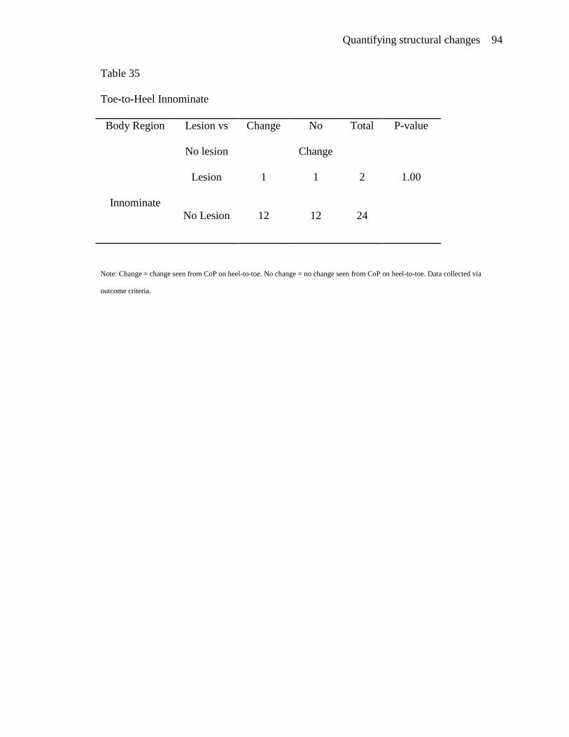
Quantifying structural changes 94
Table 35
Toe-to-Heel Innominate
Body Region Lesion vs
No lesion
Change No
Change
Total P-value
Innominate
Lesion
1 1 2
1.00
No Lesion 12 12 24
Note: Change = change seen from CoP on heel-to-toe. No change = no change seen from CoP on heel-to-toe. Data collected via
outcome criteria.

Quantifying structural changes 95
Table 36
Toe-to-Heel Sacrum
Body Region Lesion vs
No lesion
Change No
Change
Total P-value
Sacrum
Lesion
5 4 9
1.00
No Lesion 8 9 17
Note: Change = change seen from CoP on heel-to-toe. No change = no change seen from CoP on heel-to-toe. Data collected via
outcome criteria.

Quantifying structural changes 96
Table 37
Toe-to Heel Leglength
Body Region Lesion vs
No lesion
Change No
Change
Total P-value
Leg length
Lesion
1 1 2
1.0
No Lesion 12 12 24
Note: Change = change seen from CoP on heel-to-toe. No change = no change seen from CoP on heel-to-toe. Data collected via
outcome criteria.

Quantifying structural changes 97
Appendix A
Definition of Terms
Anatomic barrier: the position to which a joint can be moved without injury: if
moved beyond this point, will lead to disruption of tissue and bony structures
(DiGiovanna & Schiowitz, 1997, p. 86-87).
Baroreceptors: Receptors that detect pressure of blood flow and send messages to
the central nervous system to increase or decrease the peripheral resistance and cardiac
output (Patterson & Wurster, 2003, p.128-129).
Center of Pressure: this term is synonymous with time in center (TIC) and
represents the pressure experienced over a specific amount of time spent in a six inch
radius on the forceplate machine.
Facilitating force: a compression force on a particular area. This compression of
the muscle results in relaxation (Schiowitz et al., 2003, p.1017-1025).
Fight or flight response: the sympathetic nervous system‟s response to an outside
stimulus that frightens or stresses the human body (Tortora & Grabowski, 2003, p.577-
578).
Gamma motor neurons: Neurons that carry messages from the brain to the muscle
spindles. These neurons are located in contractile skeletal muscle fibers (DiGiovanna &
Schiowitz, 1997, p. 23, 25).
Holistic: involving the mind, body, and spirit (Martinke & Dowling, 1997, p. 4-7)
Homeostasis: an open or closed system in the living body in which a constant or
stable condition is maintained (Willard, 2003, p. 150-153).

Quantifying structural changes 98
Involuntary nervous system: A response or responses to a stimulus that are not
under the control of the individual (Willard, 2003, p. 90-119).
Leg length discrepancy: A condition in which one leg is shorter than the other,
whether anatomically or functionally (DiGiovanna & Schiowitz, 1997, p. 210-211).
Mastoid process: the bony structure that is located lateral to the foramen
magnum, to which is attached the sternocleidomastoid muscle (Anderson et al., 2002, p.
1056).
Muscle spindle: A structure that is innervated by sensory and motor neurons. The
function is to send proprioceptive information about the muscle to the central nervous
system, which responds to stress (DiGiovanna & Schiowitz, 1997, p. 24-26).
Musculoskeletal system: the part of the body that consists of bones and the
muscles that attach to them (Wells, 2003, p.75-78).
Nervous system: A system composed of the brain and the spinal column. This
system functions to relay sensory and motor information that comes from the body and
the outside environment (Tortora, & Grabowski, 2003, p. 385-418).
Osteopathic medicine: A field of medicine which recognizes the impact of the
neuromusculoskeletal system on an individual‟s health. Emphasis in placed on the
complex interplay between this system and the viscera under the influence of various
environmental factors and disease processes (DiGiovanna & Schiowitz, 1997, p. 1-2).
Palpation: the practice of touching the body in order to feel changes in the
muscles or skeletal system (DiGiovanna & Schiowitz, 1997, p. 44-46).

Quantifying structural changes 99
Physiologic barrier: the point to which a patient can actively move a joint; its
functional limit. Passive motion to the anatomical barrier is still present (Ward, 2003, p.
1232).
Prolotherapy: injection of a sugar water combination into ligaments that are too
lax, to help strengthen the ligament via remodeling (Scott et el,2003, p.497-498).
Protocol of consultation: formal guideline given in the setting of a meeting
(Kelso, & Rubin, 2003, p. 1171).
Reciprocal inhibition: contraction of agonist muscle that will result in relaxation
of antagonistic musculature (Enrenfeuchter, & Sandhouse, 2003, p. 883, 917).
Restrictive barrier: the limitation of the range of motion that is due to the
dysfunctional process impeding motion of a jount of tissue in a specific direction. This is
the barrier that osteopathic physicians move through in order to restore physiologic ROM
(DiGiovanna & Schiowitz, 1997, p. 8-10).
Somatic dysfunction: impaired or altered function of related components of the
somatic (body framework) system: skeletal, arthrodial, myofascial, vascular, lymphatic,
and neural elements (Jones, 2003, p. 1153-1161).
Spinous process: The bony process that is felt in the midline of the back on the
spine (Hoppenfeld, 2003, p.108-109).
Tandem stance: when an individual stands on both feet at same time (DiGiovanna
& Schiowitz, 1997, p. 239-240).
Tension membranes: these are ligamentous structures in the cranial vault, such as
the falx cerebelli, falx cerebri, and tentorium (DiGiovanna & Schiowitz, 1997, 65-66, 71-
72).

Quantifying structural changes 100
Time in center: analysis of data by comparing +/- or no change, in time spent in
six-inch circle on the Isobalance forceplate.
Visceral organs: organs that are located inside the body. Dysfunctions with these
organs can result in tissue changes, discernable upon palpation (Patterson, & Wurster
,2003, 127-128).
Voluntary nervous system: the part of the nervous system (brain and spinal cord)
over which the individual has full control. This is in contrast to elements of the
involuntary system, such as the heartbeat (Tortora, & Grabowski, 2003, p. 385-418).

Quantifying structural changes 101
Appendix B
Pre questionnaire
1. What components does healthcare cover (check all that apply):
1 Physical health
2 Spiritual Health
3 Mental Health
2. Do you have a doctor that you visit on a regular basis:
1 Yes
2 No
3. Has the doctor you visited (or if you have a regular doctor) ever asked you how
things are going at home or at school?
1 Yes
2 No
4. When you have aches and pains, what form/s of treatment do you use?
1 Pill
2 Surgery
3 Manual Medicine
4 Home remedies
5 No treatment as long as you can still function without too much pain.
5. What is an osteopathic doctor?
1 One who does manipulations only
2 A bone doctor or specialist
3 The same as an Allopathic doctor or M.D.
4 A chiropractor
5 A combination of an M.D. and a chiropractor

Quantifying structural changes 102
6 None of the above
6. Is it necessary to visit your doctor for check-ups if there is nothing wrong?
1 Yes
2 No
7. What is the major difference between an osteopathic physicians and
Chiropractors?
1 Only one can perform manipulations
2 Osteopathic doctors can prescribe medicine like Chiropractors
3 Osteopathic doctors go to medical school
4 Chiropractors can perform surgeries
5 No they are the same
8. Do you feel your doctor communicates well with you and makes sure you
understand what you can do to help yourself?
1 Yes
2 No
9. Does your doctor or one you have visited before explained to you what to do so
you can protect yourself from future related problems?
1 Yes
2 No
10. What are activities of daily living?
1 Running 3 times a week to improve your health
2 Working-out in a gym to build stronger muscles
3 Activities in which you participate in during the day
4 Only the parts of the day that you are walking around
11. Do osteopathic doctors only use manipulations (cracking bones) to treat patients?

Quantifying structural changes 103
1 Yes
2 No
12. Can the body heal itself?
1 Yes
2 No
13. What does the term Somatic Dysfunction mean to you:
1 Tenderness in the muscle along with some swelling
2 Abnormal posture that causes you to have scoliosis, tenderness of the back
3 Having a range of motion that is greater than normal
4 Warm muscle, painful joint, and asymmetry
5 Tenderness, asymmetry, range of motion, and tissue texture changes.
14. Which technique allows you to thrust through the barrier?
1 Direct technique
2 Indirect technique
15. When treating the nervous system can you affect the internal structures?
1 True
2 False
16. What is viserosomatic pain?
1 Pain of the muscles to the internal organs
2 Internal organs causing pain on the external organs
17. Can internal structures cause pain on the skin?
1 Yes
2 No
18. Is stretching important in musculoskeletal health?
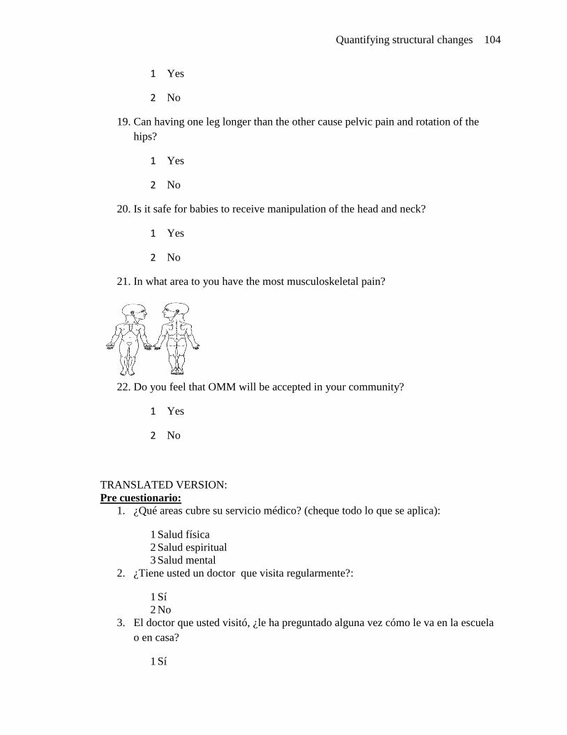
Quantifying structural changes 104
1 Yes
2 No
19. Can having one leg longer than the other cause pelvic pain and rotation of the
hips?
1 Yes
2 No
20. Is it safe for babies to receive manipulation of the head and neck?
1 Yes
2 No
21. In what area to you have the most musculoskeletal pain?
22. Do you feel that OMM will be accepted in your community?
1 Yes
2 No
TRANSLATED VERSION:
Pre cuestionario:
1. ¿Qué areas cubre su servicio médico? (cheque todo lo que se aplica):
1 Salud física
2 Salud espiritual
3 Salud mental
2. ¿Tiene usted un doctor que visita regularmente?:
1 Sí
2 No
3. El doctor que usted visitó, ¿le ha preguntado alguna vez cómo le va en la escuela
o en casa?
1 Sí

Quantifying structural changes 105
2 No
4. Cuando usted tiene dolores, ¿qué trataamiento utiliza?
1 Píldora
2 Cirugía
3 Medicina manual
4 Remedios caseros
5 Ningún tratamiento mientras pueda funcionar sin demasiado dolor.
5. ¿ Qué es doctor osteopathic?
1 Uno quién hace manipulaciones solamente
2 Un doctor o un especialista de hueso
3 Igual que un doctor de Allopathic o un M.D.
4 Un chiropractor
5 Una combinación de un M.D. y un quiropráctico
6 Ninguno de estos
6. ¿Es necesario visitar a su doctor para chequearse si no tiene nada malo?
1 Sí
2 No
7. ¿Cuál es la diferencia principal entre Médicos y quiroprácticos de osteopathic?
1 Solamente uno que puede realizar manipulaciones
2 Los doctores de osteopathic pueden prescribir la medicina como
quiroprácticos.
3 Los doctores de osteopathic van a la escuela médica.
4 Los quiroprácticos pueden realizar cirugías.
5 Ningún son iguales.
8. ¿Le parece que su doctor se comunica bien con usted y se cerciora que usted
entendió lo que usted puede hacer para ayudarse a usted mismo?
1 Sí
2 No
9. ¿Su doctor le ha explicado lo que tiene que hacer para protegerse de su problema
y de futuros problemas?
1 Sí
2 No
10. ¿Qué actividades diariamente hace usted?
1 Corriendo- 3 veces a la semana para mejorar su salud
2 Hacer ejercicios en un gym para hacer los músculos más fuertes
3 Actividades durante todo el día
4 Solamente durante el día cuando hago mi rutina diaria
11. Los doctores de osteopathic son los únicos que usan manipulaciones de huesos
para tratar a los pacientes?

Quantifying structural changes 106
1 Sí
2 No
12. ¿Puede el cuerpo curarse a si mismo?
1 Sí
2 No
13. ¿Qué significa el termino somático de la disfunción ?
1 sensibilidad en el músculo junto con un hinchazón
2 Postura anormal que le hace tener escoliosis, sencibilidad en la espalda
3 Tener una capacidad de movimiento que es mejor de lo normal
4 gusano que penetra el músculo, articulación doloroso, y asimetría
5 sensibilidad, la asimetría, la capacidad de movimiento, y la textura de la piel
cambia.
14. ¿Qué técnica permite que usted empuje a través de la barrera?
1 Técnica directa
2 Técnica indirecta
15. Cuando uno trata el sistema nervioso, ¿ puede usted afectar las estructuras
internas?
1 Verdad
2 Falso
16. ¿Quél es dolor viserosomático?
1 Dolor de los músculos a los órganos internos
2 Órganos internos que causan dolor en los órganos externos
17. ¿Pueden las estructuras internas causar dolor en la piel?
1 Sí
2 No
18. ¿Es el estirarse importante en la salud musculoesquelético?
1 Sí
2 No
19. Tener una pierna más larga que la otra, ¿ puede causar dolor en el área pélvica y
la rotación de las caderas?
1 Sí
2 No
20. ¿Es prudente que los bebés reciban la manipulación de la cabeza y del cuello?
1 Sí
2 No
21. ¿En qué área tiene usted la mayoría de los dolores musculoesquelético?

Quantifying structural changes 107
22. ¿ siente usted que OMM será aceptado en su comunidad?
1 Sí
3 No
Quantifying structural changes 108
Appendix C
Post questionnaire
1. Which one of these are components of holistic healthcare? (check all that apply):
a. Physical health
b. Spiritual Health
c. Mental Health
2. Is it important to visit a doctor regularly?
a. Yes
b. No
3. Is social health an important component to the osteopathic practice of medicine?
a. Yes
b. No
4. Which would be your choice of treatment when dealing with aches and pains that
interrupt your activities of daily living?
a. Pill
b. Surgery
c. Manual Medicine
d. Home remedies
e. No treatment as long as you can still function without too much pain.
5. What is an osteopathic doctor?
a. One who does manipulations only
b. A bone doctor or specialist
c. The same as an Allopathic doctor or M.D.
d. A chiropractor
e. A combination of an M.D. and a chiropractor
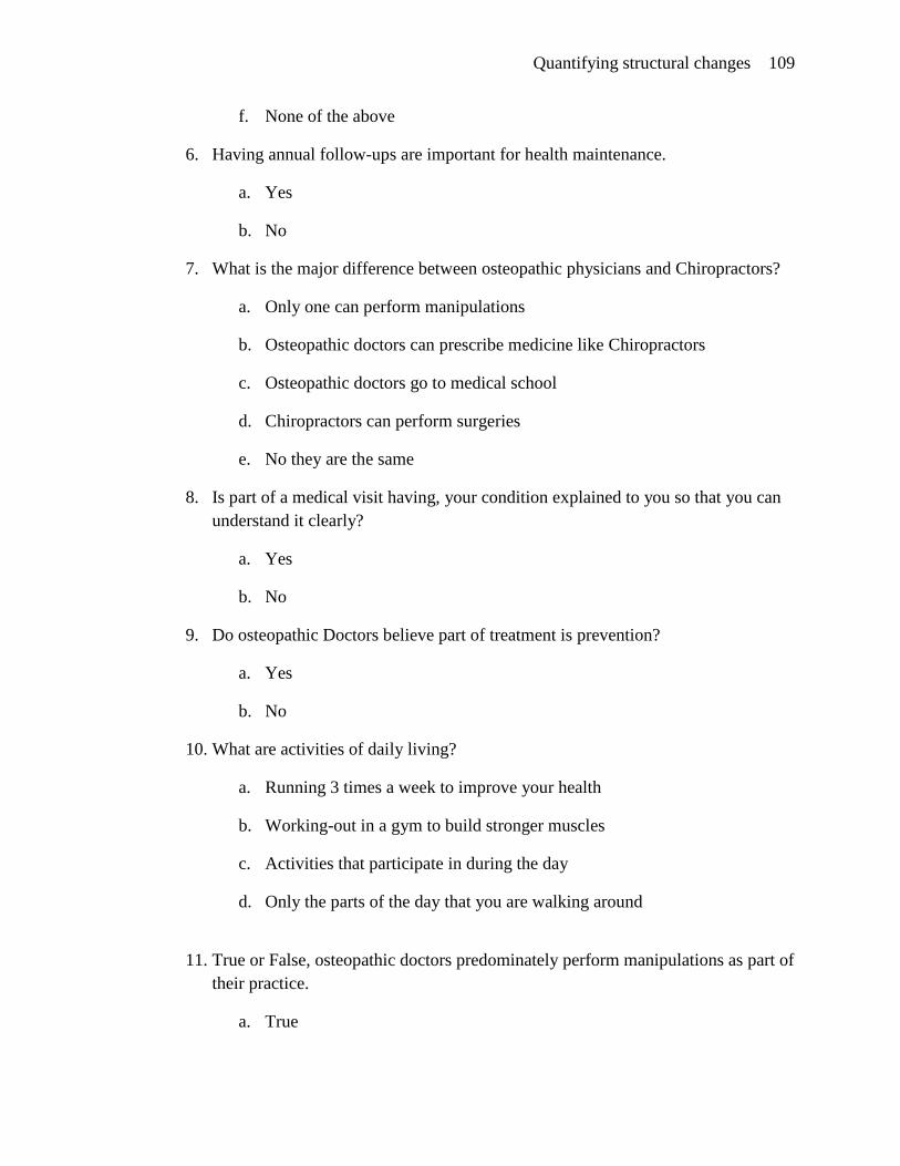
Quantifying structural changes 109
f. None of the above
6. Having annual follow-ups are important for health maintenance.
a. Yes
b. No
7. What is the major difference between osteopathic physicians and Chiropractors?
a. Only one can perform manipulations
b. Osteopathic doctors can prescribe medicine like Chiropractors
c. Osteopathic doctors go to medical school
d. Chiropractors can perform surgeries
e. No they are the same
8. Is part of a medical visit having, your condition explained to you so that you can
understand it clearly?
a. Yes
b. No
9. Do osteopathic Doctors believe part of treatment is prevention?
a. Yes
b. No
10. What are activities of daily living?
a. Running 3 times a week to improve your health
b. Working-out in a gym to build stronger muscles
c. Activities that participate in during the day
d. Only the parts of the day that you are walking around
11. True or False, osteopathic doctors predominately perform manipulations as part of
their practice.
a. True

Quantifying structural changes 110
b. False
12. One of the osteopathic philosophies is that the body has the innate ability to heal
itself given the body is free from dysfunction (homeostasis).
a. True
b. False
13. What does the term Somatic dysfunction mean to you:
a. Tenderness in the muscle along with some swelling
b. Abnormal posture that causes you to have scoliosis, tenderness of the back
c. Having a range of motion that is greater than normal
d. Worm muscle, painful joint, and asymmetry
e. Tenderness, asymmetry, range of motion, and tissue texture changes
14. Which technique allows you to thrust through the barrier?
a. Direct technique
b. Indirect technique
15. When treating the nervous system can you affect the internal structures?
a. True
b. False
16. What is viserosomatic pain?
a. Pain of the muscles to the internal organs
b. Internal organs causing pain on the external organs
17. Can visceral organs cause pain on the skin?
a. Yes
b. No
18. Is stretching important in musculoskeletal health?
a. Yes

Quantifying structural changes 111
b. No
19. Can having a leg length discrepancy cause your pelvis to rotate and cause pain?
a. Yes
b. No
20. Are babies good candidates for manipulation of the head and neck:
a. Yes
b. No
21. Was the problem area that you had in the beginning of the study treated?
a. Yes
b. No
c. Unsure
22. Do you believe OMM as a safe and effect form of treatment options for people in
your community?
a. Yes
b. No
23. Would you recommend this form of pain relief to a:
a. Friend
b. Family
c. Stranger
d. No one
TRANSLATED VERSION:
Pos Cuestionario
1. ¿Cuál de éstos son componentes del cuidado de salud holístico (marque todo lo
que aplique)
1 Salud física
2 Salud espiritual

Quantifying structural changes 112
3 Salud mental
2. ¿Es importante visitar a un doctor regularmente?
1 Sí
2 No
3. ¿Su salud social un componente importante a la Práctica de la medicina
Osteopathic?
1 Sí
2 No
4. ¿Cuál sería su opción de tratamiento cuando tiene que ver con doloresque
interrumpen sus actividades diarias
1 Píldora
2 Cirugía
3 Medicina manual
4 Home remedies
5 Ningún tratamiento mientras usted pueda todavía funcionar sin demasiado
dolor.
5. ¿Qué es un doctor osteopathic?
1 Uno qué hace manipulaciones solamente
2 Un doctor o un especialista del hueso
3 Igual que un doctor de Allopathic o un M.D.
4 Un quiropráctico
5 Una combinación de un M.D. y Un quiropráctico
6 Ninguno de lo dicho
6. Es importante tener chequeos anuales para mantener la salud
1 Sí
2 No
7. ¿Cuál es la diferencia principal entre Médicos y quiroprácticos de osteopathic?
1 Solamente uno puede realizar manipulaciones
2 Los doctores de osteopathic pueden prescribir la medicina como
quiroprácticos
3 doctores osteopathic van a la escuela de medicina
4quiroprácticos pueden hacer cirugias
5 son iguales
8. ¿debería ser parte de una visita médica tener una explicación sobre su condición
de modo que usted pueda entenderla claramente?
1 Sí
2 No
9. ¿los doctores de osteopathic creen que parte de un tratamiento es prevención?
1 Sí

Quantifying structural changes 113
2 No
2 No
11. ¿Qué actividades diariamente hace usted?
1 Corriendo- 3 veces a la semana para mejorar su salud
2 Hacer ejercicios en un gym para hacer los músculos más fuertes
3 Actividades durante todo el día
4 Solamente durante el día cuando hago mi rutina diaria
11. verdadero o falso- ¿los doctores de osteopathic predominatemente hacen las
manipulaciones como parte de su práctica?
1 Verdad
2 Falso
12. Una de las filosofías de osteopathic es que el cuerpo tiene la abilidad de curarse a
si mismo siempre y cuando el cuerpo este libre de disfunción (homeostasis).
1 Verdad
2 Falso
14. ¿Qué significa el termino somático de la disfunción ?
1 sensibilidad en el músculo junto con un hinchazón
2 Postura anormal que le hace tener escoliosis, sencibilidad en la espalda
3 Tener una capacidad de movimiento que es mejor de lo normal
4 gusano que penetra el músculo, articulación doloroso, y asimetría
5 sensibilidad, la asimetría, la capacidad de movimiento, y la textura de la piel
cambia.
15. ¿Qué técnica permite que usted empuje a través de la barrera?
1 Técnica directa
2 Técnica indirecta
16. Cuando uno trata el sistema nervioso, ¿ puede usted afectar las estructuras
internas?
1 Verdad
2 Falso
17. ¿Quél es dolor viserosomático?
1 Dolor de los músculos a los órganos internos
17. Órganos internos pueden causar dolor en los órganos¿Pueden los órganos
viscerales causar dolor en la piel?
1 Sí
2 No
18. ¿es el estirarse importante en la salud musculoesquetal?

Quantifying structural changes 114
1 sí
2 No
20. Tener una pierna más larga que la otra, ¿ puede causar dolor en el área pélvica y
la rotación de las caderas?
1 Sí
2 No
21. ¿Es prudente que los bebés reciban la manipulación de la cabeza y del cuello?
1 Sí
2 No
21. ¿Han sido tratados las áreas problemáticas que usted tenía al principio del
estudio?
1 Sí
2 No
3 Inseguro
22. ¿cree usted que OMM es una forma de tratamiento efectiva y segura para la gente
en su comunidad?
1 Sí
2 No
23. Usted recomendaría esta forma de tratamiento del dolor a :
1 su amigo
2 su familia
3 un desconocido
4 nadie

Quantifying structural changes 115
Appendix D
Students Watching Educational Video
Note: Baxter students watching the educational video

Quantifying structural changes 116
Appendix E
Consent
Explanation of Study:
The purpose of this study is threefold. First, the examination of the current level
of understanding about healthcare and healthcare options in the treatment of
musculoskeletal pain in Honduras (and/or Central America) will be assessed.
Secondly, with the introduction and implementation of Osteopathic Manual
Medicine as a treatment modality, the evaluation of the degree of acceptance and
understanding of the basic philosophies OMM will be studied. The final
component will measure the improvements to the center of pressure (CoP) and
relief of somatic dysfunction following treatment.
o CoP is the measuring of balance on both feet when standing up without
moving.
o SD will be clarified by using the following acronym TART:
o Tissue texture changes (warm or cool skin)
o Asymmetry (when there is unevenness of a part of
the body)
o Range of motion (not being able to move a
particular part of the body in its usual range of
motion)
o Tenderness (sensitive area of muscle which may be
due to misalignment of the skeletal system or the
muscle itself).
During this study you will be asked to participate in one pre and post-test
questionnaire, pre-recorded lectures before treatment of musculoskeletal pain and
measurement of center of pressure.
Participation:

Quantifying structural changes 117
The participation in this study is voluntary and will not be noted in the academic
records. Subjects reserve the right to refuse participation in this study or to
unquestionably withdraw their participation from the study at any moment. The
non-participation in this study will not result in any penalty, will not be noted in
academic records nor will it lead to loss of the subject benefits in any way.
There will be no financial benefit to the subjects in the study as a result of any
medical condition or discomfort following conclusion of the study.
All subjects will be allowed to participate in the educational sessions without
having to receive manipulation.
All participants reserve the rights to disclose their participation in the study to
their primary physician/s if necessary without having any information gathered
during this study reviewed by anyone but the investigation team.
The duration of the study will be from February 2008 through March 2008. A
maximum time requirement of one and half hours once a week, for three weeks
out of the five week test period is needed each day.
Alternative treatments:
There are no alternative procedures that can substitute for osteopathic medicine
and philosophies.
Risk:
There is no risks of physical injury or permanent disability posed by this study.
There is a mild possibility of sore muscles due to manipulation and stretching
techniques.

Quantifying structural changes 118
You cannot receive manipulation if you currently have a history of any
disease or condition listed in the exclusion criteria:
Exclusion Criteria Acute traumatic injury: any type of manipulation on recently
injured areas due to trauma (broken bones, internal surgical
procedures, ligament or muscle ruptures, etc.)
Inflammatory arthritis: manipulating joint spaces in individuals
with uncontrolled arthritis may lead to acute inflammatory
processes
History of neurological disease: working on such areas that may
further deteriorate a person‟s condition is counterintuitive to
helping the structure and function.
Osteoporosis risk or history: osteoporosis and manipulation have
the potential to lead to broken bones.
Cervical disc surgery and herniated cervical disc: the area
should be carefully manipulated be it with muscle energy or
myofacial massage. The reason for this theory is that any further
damage to this are may lead to paralyses.
History of carotid stenosis or vascular stenosis in the thoracic
or lumbar area: with the hardening of the arteries comes
immobility of the vasculature in this area. Therefore, there is a
potential risk of carotid shearing, stroke, vascular compromise to
the head, and or death.
Use of anticoagulative medication: due to the risk of uncontrolled
bleeding.
Severe conditions of cervical spondylitic myeolopathy or those
suffering from Down syndrome
Older than fifty and younger than eighteen
Benefits:
The benefits for participating in this study are an increase in knowledge of
the holistic approach towards medicine. Relief of acute and chronic
musculoskeletal pain.
Increase in flexibility and range of motion; therefore, decreasing chance of injury
during everyday activities (activities of daily living [ADLs]).
Compensations:

Quantifying structural changes 119
There are no compensations that will be given to any of the participants in the
study.
Confidentiality:
The research team will only review information that is obtained during the course
of this study.
Participation or lack thereof will have no effect with any interactions with the
administration at the Baxter Institute.
All information will be placed in a locked cabinet and a locked room in the Baxter
Institute administration office. The information will also be saved on the primary
researcher‟s computer and a back-up disk, which will be accessible, only by the
research team.
All information will be destroyed by December 31, 2010.
Contacts:
If you have any questions please contact Abraham B. Hardee III via
I ______________________ voluntarily agree to participate in this research project.
Signature
Witness
Date
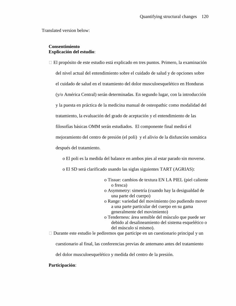
Quantifying structural changes 120
Translated version below:
Consentimiento
Explicación del estudio:
El propósito de este estudio está explicado en tres puntos. Primero, la examinación
del nivel actual del entendimiento sobre el cuidado de salud y de opciones sobre
el cuidado de salud en el tratamiento del dolor musculoesquelético en Honduras
(y/o América Central) serán determinadas. En segundo lugar, con la introducción
y la puesta en práctica de la medicina manual de osteopathic como modalidad del
tratamiento, la evaluación del grado de aceptación y el entendimiento de las
filosofías básicas OMM serán estudiados. El componente final medirá el
mejoramiento del centro de presión (el poli) y el alivio de la disfunción somática
después del tratamiento.
o El poli es la medida del balance en ambos pies al estar parado sin moverse.
o El SD será clarificado usando las siglas siguientes TART (AGRIAS):
o Tissue: cambios de textura EN LA PIEL (piel caliente
o fresca)
o Asymmetry: simetría (cuando hay la desigualdad de
una parte del cuerpo)
o Range: variedad del movimiento (no pudiendo mover
a una parte particular del cuerpo en su gama
generalmente del movimiento)
o Tenderness: área sensible del músculo que puede ser
debido al desalineamiento del sistema esquelético o
del músculo sí mismo).
Durante este estudio le pediremos que participe en un cuestionario principal y un
cuestionario al final, las conferencias previas de antemano antes del tratamiento
del dolor musculoesquelético y medida del centro de la presión.
Participación:

Quantifying structural changes 121
La participación en este estudio es voluntaria y no será observada en expedientes
académicos. Los participantes tienen el derecho de rechazar la participación en
este estudio o de retirar indiscutiblemente su participación en el estudio en
cualquier momento. El no participar en este estudio no dará lugar a ninguna pena,
no será observada en expedientes académicos ni conducirá a la pérdida de las
ventajas sujetas de cualquier manera.
No habrá ventaja financiera/médica a los participantes en el estudio si tienen una
condición médica o un malestar después de la conclusión del estudio.
Los participantes podrán participar en las sesiones educativas sin tener que recibir
la manipulación.
Todos los participantes reservan los derechos de divulgar su participación en el
estudio a su médico/s primario en caso de necesidad sin tener ninguna
información recopilada durante este estudio revisado por cualquier persona con
exepción del equipo de la investigación.
La duración del estudio será de febrero y del marzo de 2008. El requisito de tiempo
para participar en el estudio es de una hora y media cada día.
Tratamientos alternativos:
No hay procedimientos alternativos que pueden substituir la medicina y las
filosofías de osteopathic.
Riesgo:
El riesgo a lesión física participando en este estudio no es ningún riesgo de lesión
física o de la inhabilidad permanente.

Quantifying structural changes 122
Hay posibilidad de que los músculo le duelan debido a la manipulación y las
técnicas de estirar los músculos.
No puede recibir la manipulación si usted actualmente tiene una historia de
los criterios del exculsion:
Criterios de la exclusión Lesión traumática aguda: cualquier tipo de manipulación en las
áreas recientemente dañadas debido al trauma (los huesos rotos, los
procedimientos quirúrgicos internos, el ligamento o el músculo
rotos, etc.)
Artritis inflamatoria: los espacios comunes de manipulación en
individuos con artritis incontrolada pueden conducir a los procesos
inflamatorios agudos
Historia de la enfermedad neurológica: el trabajo en tales áreas
que puedan deteriorar más una condición de la persona no ayudar
a la estructura y a la función.
Riesgo o historia del Osteoporosis: el osteoporosis y la
manipulación tiene el potencial de conducir a los huesos
quebrados.
Cirugía cervical del disco y disco cervical herniated: el área se
debe manipular cuidadosamente sea con energía del músculo o
masaje myofacial. La razón de esta teoría es que son cualquier
daños más otra a esto pueden conducir a las parálisis.
Historia del stenosis carótida o del stenosis vascular en el área
torácica o lumbar: con endurecer de las arterias viene la
inmovilidad del vasculature en esta área. Por lo tanto, hay un
riesgo potencial de esquilar, del movimiento carótidas,
compromiso vascular a la cabeza, y o a la muerte.
Uso de la medicación anticoagulative: debido al riesgo de la
sangría incontrolada.
Condiciones severas de myeolopathy spndylitic cervical o de ésos
que sufren abajo de síndrome
Más viejo de cincuenta y más joven de dieciocho
Ventajas:
Las ventajas para participar en este estudio son un aumento en el conocimiento del
acercamiento holístico hacia la medicina. Alivio al dolor musculoesquelético
agudo y crónico.

Quantifying structural changes 123
Aumentación de flexibilidad y de la gama del movimiento; por lo tanto, ocasión
que disminuye de lesión durante las actividades diarias (actividades de la vida del
diario [ADLs]).
Remuneraciones:
No hay remuneraciones que serán dadas a los participantes en el estudio.
Confidencialidad:
El equipo de investigación serán los únicos que verán toda la información que se
obtenga durante el curso de este estudio.
El no participar en este estudio no tendrá efecto de ninguna manera con la
administración en el Baxter Institute.
Toda la información se pondrá en un gabinete cerrado con llave. Este gabinete
estará en un cuarto cerrado con llave en la ofician de Baxter Institute. La
información también se guardará en la computadora del investigador de este
estudio. Las unicas personas que tiene acceso a esta información son los
miembros del equipo de investigación.
Toda la información será destruída en el año 2010.
Contacts:
Si usted tiene preguntas, por favor comuniquese con Abraham B Hardee III via
Yo, ___________________ voluntariamente doy mi consentimiento para participar en
este proyecto.
Testigo
fecha

Quantifying structural changes 124
Appendix F
Evaluation Form
LOCALIZER: Honduras, Tegucigalpa; Baxter Institute (Location) NOMBRE: ____________________________ APELLIDOS:
_______________________ Project # _____ (First) (Last) SEXO: Hombre Mujer EDAD: ______________ (Gender) (Age)
Previous Injuries: ________________________________
1. Cervicals
CO-C2
a. Neutral R on L L on R 1.
Scoliosis C3-C7
Yes No
b. Flexion L on L R on R
c. Extension L on L R on R 2. Leg
length discrepancy 2. Thoracic
Yes No
a. Neutral R on L L on R
b. Flexion L on L R on R
c. Extension L on L R on R
3. Pelvis – Symphysis Subluxation
a. Left Right
b. Superior Superior
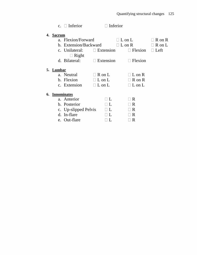
Quantifying structural changes 125
c. Inferior Inferior
4. Sacrum
a. Flexion/Forward L on L R on R
b. Extension/Backward L on R R on L
c. Unilateral: Extension Flexion Left
Right
d. Bilateral: Extension Flexion
5. Lumbar
a. Neutral R on L L on R
b. Flexion L on L R on R
c. Extension L on L L on L
6. Innominates
a. Anterior L R
b. Posterior L R
c. Up-slipped Pelvis L R
d. In-flare L R
e. Out-flare L R

Quantifying structural changes 126
Appendix G
Room Set-up
Note: Second investigator is behind the desk operating the forceplate. One participant is on the forceplate and the other on the table.

Quantifying structural changes 127
Appendix H
Schematic of a Dual Forceplate System

Quantifying structural changes 128
Appendix I
Foot placement on single forceplate
Note: Isotechnology photo of forceplate used in study
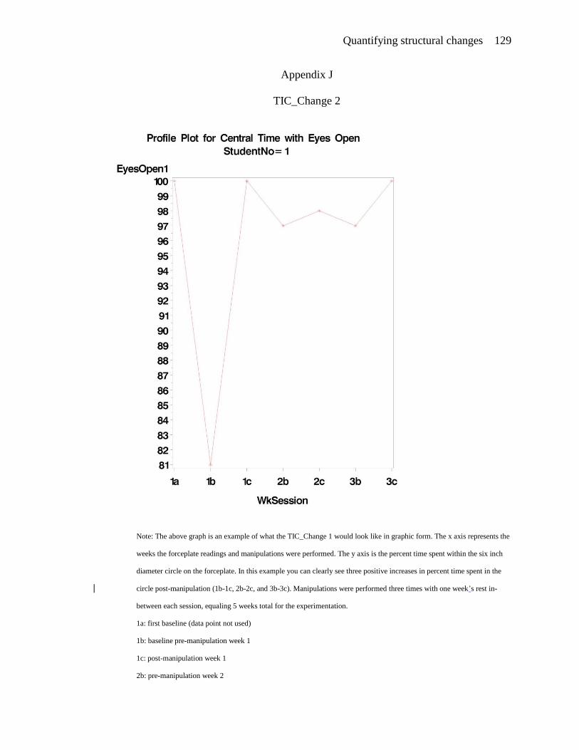
Quantifying structural changes 129
Appendix J
TIC_Change 2
Note: The above graph is an example of what the TIC_Change 1 would look like in graphic form. The x axis represents the
weeks the forceplate readings and manipulations were performed. The y axis is the percent time spent within the six inch
diameter circle on the forceplate. In this example you can clearly see three positive increases in percent time spent in the
circle post-manipulation (1b-1c, 2b-2c, and 3b-3c). Manipulations were performed three times with one week‟s rest in-
between each session, equaling 5 weeks total for the experimentation.
1a: first baseline (data point not used)
1b: baseline pre-manipulation week 1
1c: post-manipulation week 1
2b: pre-manipulation week 2

Quantifying structural changes 130
2c: post-manipulation week 2
3b: pre-manipulation week 3
3c: post-manipulation week 3

Quantifying structural changes 131
Appendix K
Heel-to-Toe Change in Quadrant Pressure
Note: This graph represents the quadrant data. As you can see the right foot has greater amounts of pressure on the heel
during the first baseline week. However, by the conclusion of the study the right toe carries the majority of the weight
when compared to the right heal.
1a: first baseline (data point not used)
1b: baseline pre-manipulation week 1
1c: post-manipulation week 1
2b: pre-manipulation week 2

Quantifying structural changes 132
2c: post-manipulation week 2
3b: pre-manipulation week 3
3c: post-manipulation week 3

Quantifying structural changes 133
Appendix L
Single Best Answer Questions

Quantifying structural changes 134
Appendix M
Questions Answered Correctly

Quantifying structural changes 135
Appendix N
Question with Decrease in Response

Quantifying structural changes 136
Appendix O
IRB Approval Letter