Embed Size (px)
Citation preview
Copyright © 2008 John Wiley & Sons, Ltd. Biomed. Chromatogr. 22: 601–605 (2008)DOI: 10.1002/bmc
Quantification of myrislignan in rat plasma 601ORIGINAL RESEARCHORIGINAL RESEARCH
Copyright © 2008 John Wiley & Sons, Ltd.
BIOMEDICAL CHROMATOGRAPHYBiomed. Chromatogr. 22: 601– 605 (2008)Published online 6 February 2008 in Wiley InterScience(www.interscience.wiley.com) DOI: 10.1002/bmc.975
Quantification of myrislignan in rat plasma by solid-phaseextraction and reversed-phase high-performance liquidchromatography
Fei Li and Xiuwei Yang*
The State Key Laboratory of Natural and Biomimetic Drugs and Department of Natural Medicines, School of Pharmaceutical Sciences, PekingUniversity, Beijing 100083, People’s Republic of China
Received 25 September 2007; revised 25 October 2007; accepted 5 November 2007
ABSTRACT: A rapid and simple reversed-phase high-performance liquid chromatographic (RP-HPLC) method has been devel-oped for determination of myrislignan in rat plasma after intravenous administration. The analytes extracted from plasma samplesby solid-phase extraction were successfully carried out on a Diamonsil™ ODS C18 column (250�×�4.6 mm i.d., 5 μm) with an RP18
guard column (8 × 4.6 mm i.d., 5 μm) and a mobile phase of MeOH–H2O (4:1, v/v). The UV detector was set at a single wave-length of 270 nm. The linear ranges of the standard curves were 0.5–30.0 μg/mL with the correlation coefficients greater than0.9992. The lower limits of detection and quantification were 0.1�and 0.3 μg/mL for myrislignan. Intra- and inter-day precisionswere 2.4 –7.5 and 1.3–5.7%, respectively. The extraction recovery from plasma was more than 90%. This assay method has beensuccessfully used to study the pharmacokinetics of myrislignan in rats. Copyright © 2008 John Wiley & Sons, Ltd.
KEYWORDS: Myristica fragrans; myrislignan; reversed-phase HPLC; solid-phase extraction; pharmacokinetics
*Correspondence to: Xiuwei Yang, The State Key Laboratory ofNatural and Biomimetic Drugs and Department of Natural Medi-cines, School of Pharmaceutical Sciences, Peking University, Beijing100083, People’s Republic of China.E-mail: [email protected]
Abbreviations used: LLE, liquid–liquid extraction; PPE, protein pre-cipitation; SPE, solid-phase extraction.
Contract/grant sponsor: National High Technology Research andDevelopment Program of China; Contract/grant number:2002AA2Z343C.
Contract/grant sponsor: Beijing Municipal Special-purpose ScienceFoundation of China; Contract/grant number: Z0004105040311.
Contract/grant sponsor: National Sciences and Technology Programof China; Contract/grant number: 2006BAI06A01.
INTRODUCTION
The compound myrislignan [erythro-(1R, 2S)-2-(4-allyl-2,6-dimethoxyphenoxyl)-1-(4-hydroxy-3-methoxyphenyl)propan-1-ol, structure shown in Fig. 1], is a typical acyclicneolignan in the seed (nutmeg; Isogai et al., 1973) andthe aril (mace; Hattori et al., 1986) of Myristica fragransHoutt. (Myristicaceae). In traditional Chinese medicalscience, the seeds of M. fragrans, one of the most widelyused spices in preparation of ayurvedic drugs (Van andCox, 1994), are a well-known traditional Chinese medi-cine for strengthening the stomach and expelling wind(The State Pharmacopoeia Commission of P.R. China,2005). Previous studies have suggested that myrislignanmay affect hepatic mixed function oxidase enzymeactivity, which causes the prolongation of hexobarbital-
Figure 1. Chemical structure of myrislignan.
induced hexobarbital in rat and a decrease of activity inliver aminopyrine N-demethylase and hexobarbitalhydroxylase (Shin and Woo, 1990). Myrislignan revealedantifeeding effect and had antifungal properties (Isogaiet al., 1973; Miyazawa et al., 1996). In Japan, twopatents have been approved for it use as neoplasmand vascular smooth muscle contraction inhibitors(Matsumoto et al., 1991; Nakajima et al., 1999). Despiteits varied biological activities, the pharmacokineticproperties of myrislignan in human or animal have notbeen reported. Therefore, it was necessary to developan effective method to determine the quantification ofmyrislignan in plasma and understand its pharmacoki-netic properties, which would improve future investiga-tion of the active mechanism of myrislignan.
In the present paper, we first report a simple and rapidreversed-phase high-performance liquid chromato-graphic (RP-HPLC) method using a Diamonsil™ ODS
Copyright © 2008 John Wiley & Sons, Ltd. Biomed. Chromatogr. 22: 601–605 (2008)DOI: 10.1002/bmc
602 F. Li and X. YangORIGINAL RESEARCH
10 mL MeOH. The MeOH elutions containing myrislignanwere evaporated under an air stream at 40°C in a water bath.The residue was redissolved in 600 μL MeOH with vortexing.A 20 μL aliquot was injected into the HPLC system after cen-trifugation at 16,000g for 10 min.
Linearity and quantification. Calibration standards of myrislig-nan concentration levels at 0.5, 2.5, 5.0, 10.0, 20.0 and 30.0 μg/mL were prepared and assayed. The calibration curve wasconstructed by plotting the peak area vs the concentrationsof myrislignan. The lower limit of quantification (LLOQ) wasdetermined as the lowest concentration on the calibrationcurve at which precision was within 20% and accuracy waswithin ±20%. The lower limit of detection (LLOD) wasdefined as the lowest concentration level resulting in a signal-to-noise ratio of 3:1.
Precision and accuracy. Intra-day accuracy and precisionwere evaluated from replicate analysis (n = 5) of QC samplesat different concentrations on the same day. Inter-day accu-racy and precision were also assessed from the analysis ofthe same QC samples on three consecutive days in replicate(n = 5). QC samples were analyzed against calibration curves.Mean, standard deviation and relative standards deviation(RSD) were calculated from QC samples and used to estimatethe intra- and inter-day precision. Accuracy was assessed bycomparison of the calculated mean concentrations with theknown concentrations.
Extraction recovery. The absolute recovery of myrislignanfrom plasma was determined for different standard concen-trations by spiking the drug into the drug-free plasma. Extrac-tion recoveries from rat plasma were calculated by comparingthe peak areas extracted from rat plasma with those of thesame quantities added to the mobile phase. Recoveries atfour QC concentrations for plasma were examined five times.
Stability. Stability tests were performed to verify the stabilityof myrislignan during handling procedures. Samples wereassayed at the four QC concentrations of 1.5, 3.0, 9.0 and18.0 μg/mL for myrislignan in replicate (n = 5). Stabilityof myrislignan in rat plasma was assessed at −20°C for sevendays. Freeze–thaw stability of myrislignan in rat plasma wasinvestigated with QC samples subjected to three freeze–thawcycles. The results were compared with freshly prepared QCsamples. All stability evalutations were based on the calibra-tion curves.
Pharmacokinetic study. All studies in animals were in accor-dance with the guidelines for the Care and Use of LaboratoryAnimals in Beijing. Forty-five male Sprague–Dawley rats (200± 10 g) used in this study were fasted overnight before thetest, but had free access to water. For intravenous administra-tion, the myrislignan crystal was dissolved in isotonic salinecontaining 15% EtOH and 15% Cremophor EL. Doses of20.0 mg/kg myrislignan in 0.5 mL solution were deliveredinto the tail vein using a syringe. The blood sample of everyanimal was collected for one time-point. Blood samples(1.5 mL) were collected from the carotid artery in 1.5 mLsodium heparinized tubes at 1, 3, 5, 10, 15, 20, 30, 45 and60 min following drug administration. The blood samples
C18 column with ultraviolet detector for the quantifica-tion of myrislignan in rat plasma. The analytes wereextracted from the plasma by solid-phase extraction.This method was fully validated with high recovery.Furthermore, the method has been successfully appliedto the pharmacokinetic study of myrislignan in ratplasma after intravenous administration.
EXPERIMENTAL
Chemicals, regents and animals. Myrislignan was isolatedfrom the methanol extract of the seed of M. fragrans, asdescribed in a previous paper (Yang et al., 2003), and thestructure was determined by NMR and MS. The purity ofmyrislignan was over 99% by HPLC analysis. HPLC-gradeMeOH was purchased from Tianjin Xihua Chemicals Co.(Tianjin, China). Cremophor EL was purchased from Shang-hai Concord Chemical Ltd Co. (Shanghai, China). All otherchemicals and solvents used were of analytical grade andwater (H2O) was milli-Q grade. Sprague–Dawley rats weresupplied by the Laboratory Animal Center of Peking Univer-sity Health Science Center (Beijing, China).
Apparatus and chromatographic conditions. The analyti-cal HPLC system consisted of a model SpectraSYSTEM P2000 binary pump (Thermo Separation Products Inc., SanJose, CA, USA), a model SpectraSYSTEM UV3000 detector(Thermo Separation Products Inc., San Jose, CA, USA),and a 7125 Rheodyne injector (Rheodyne, Cotati, CA, USA)with a loop of 20 μL. The reversed-phase chromatographywas carried out on an analytical Diamonsil™ ODS C18 column(250�×�4.6 mm i.d., 5 μm; Dikma, China) equipped with a C18
guard column (8�×�4 mm i.d., 5 μm; Dikma, China) cartridgesystem. The mobile phase was methanol–water (4:1, v/v). Theinjection volume was 20 μL and the flow rate was 1.0 mL/min.Peaks of myrislignan were detected by UV absorbance ata single wavelength of 270 nm. All measurements wereperformed at 25°C.
Preparation of stocks, calibration samples and qualitycontrol samples. Stock solutions of myrislignan were preparedin MeOH at a concentration of 1.0 mg/mL. The workingsolutions of 600–10 μg/mL were prepared by appropriatelydiluting the stock solution. All solution were stored at 4°C.Calibration samples were prepared by spiking 570 μL blankplasma with 30 μL myrislignan working solutions to producefinal concentrations of 0.5, 2.5, 5.0, 10.0, 20.0 and 30.0 μg/mL.Quality control (QC) samples of myrislignan in plasma wereprepared at four different concentration levels (1.5, 3.0, 9.0and 18.0 μg/mL). These were stored at −20°C until analysis.
Sample preparation. All frozen standards and samples wereallowed to thaw at room temperature and homogenized byvortexing prior to extraction. Aliquots of 600 μL spiked samplestandards, QC samples or unknown plasma samples were slowlyplaced into the solid-phase cartridge SEP-PAK C18 (Millipore®,Waters, USA), which had been previously activated with 10 mLMeOH and balanced with 5 mL H2O. The cartridge was suc-cessively washed with 5 mL H2O, 5 mL 30% aq. MeOH and
Copyright © 2008 John Wiley & Sons, Ltd. Biomed. Chromatogr. 22: 601–605 (2008)DOI: 10.1002/bmc
Quantification of myrislignan in rat plasma 603ORIGINAL RESEARCH
were centrifuged at 3000g for 10 min at 4°C and stored at−20°C until analysis. Aliquots of 600 μL plasma samples wereprocessed and analyzed for myrislignan concentration.
Pharmacokenetic parameters were determined using theplasma concentration–time data. The concentrations of myris-lignan at different time points were evaluated by meansof linear regression analysis. All statistical analysis was per-formed using Microsoft Excel 2000 (Microsofe, Redmond,WA, USA). The relevant pharmacokinetic parameters werecalculated using the computer program 3p87 (Chinese Societyof Mathematical Pharmacology, Beijing, China).
RESULTS AND DISCUSSION
The chromatography
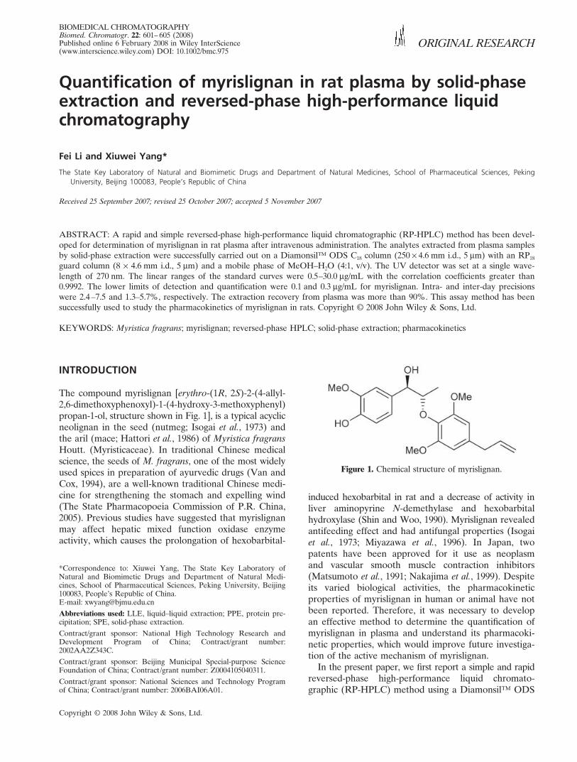
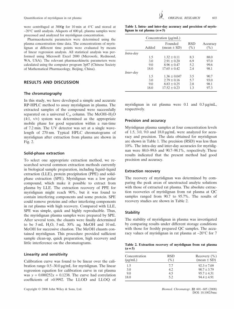
In this study, we have developed a simple and accurateRP-HPLC method to assay myrislignan in plasma. Theextracted samples of the compound were successfullyseparated on a universal C18 column. The MeOH–H2O(4:1, v/v) system was determined as the appropriatemobile phase for good separation within a run-timeof 7.2 min. The UV detector was set at a single wave-length of 270 nm. Typical HPLC chromatograms ofmyrislignan after extraction from plasma are shown inFig. 2.
Solid-phase extraction
To select one appropriate extraction method, we re-searched several common extraction methods currentlyin biological sample preparation, including liquid–liquidextraction (LLE), protein precipitation (PPE) and solid-phase extraction (SPE). Myrislignan was a low polarcompound, which make it possible to extract fromplasma by LLE. The extraction recovery of PPE formyrislignan might reach 90%, but it was found tocontain interfering components and some protein. SPEcould remove proteins and other interfering componentsin rat plasma with high recovery. Compared with LLE,SPE was simple, quick and highly reproducible. Thus,the myrislignan plasma samples were prepared by SPE.After several tests, the eluants were finally determinedto be 5 mL H2O, 5 mL 30% aq. MeOH and 10 mLMeOH for successive eluation. The MeOH eluants con-tained myrislignan. This procedure provided sufficientsample clean-up, quick preparation, high recovery andlittle interference on the chromatograms.
Linearity and sensitivity
Calibration curve was found to be linear over the cali-bration range 0.5–30.0 μg/mL for myrislignan. The linearregression equation for calibration curve in rat plasmawas y = 0.000252x + 0.1238. The curve had correlationcoefficients of >0.9992. The LLOD and LLOQ of
Table 1. Intra- and inter-day accuracy and precision of myris-lignan in rat plasma (n�=====�5)
Concentration (μg/mL)
Founded RSD AccuracyAdded (mean ± SD) (%) (%)
Intra-day1.5 1.32 ± 0.11 8.3 88.03.0 2.91 ± 0.20 6.9 97.09.0 8.96 ± 0.47 5.2 99.6
18.0 17.65 ± 0.42 2.4 98.1Inter-day
1.5 1.36 ± 0.047 3.5 90.73.0 2.79 ± 0.16 5.7 93.09.0 8.83 ± 0.25 2.8 98.1
18.0 17.52 ± 0.23 1.3 97.3
myrislignan in rat plasma were 0.1 and 0.3 μg/mL,respectively.
Precision and accuracy
Myrislignan plasma samples at four concentration levelsof 1.5, 3.0, 9.0 and 18.0 μg/mL were analyzed for accu-racy and precision. The data obtained for myrislignanare shown in Table 1. The precision (RSD) was less than10%. The intra-day and inter-day accuracies for myrislig-nan were 88.0–99.6 and 90.7–98.1%, respectively. Theseresults indicated that the present method had goodprecision and accuracy.
Extraction recovery
The recovery of myrislignan was determined by com-paring the peak areas of unextracted analyte solutionswith those of extracted rat plasma. The absolute extrac-tion recoveries of myrislignan from rat plasma at QCsamples ranged from 90.7 to 95.7%. The results ofrecovery studies are shown in Table 2.
Stability
The stability of myrislignan in plasma was investigatedby comparing results under different storage conditionswith those for freshly prepared QC samples. The accu-racy values of myrislignan in rat plasma at −20°C for 7
Table 2. Extraction recovery of myrislignan from rat plasma(n�=====�5)
Concentration RSD Recovery (%)(μg/mL) (%) (mean ± SD)
1.5 7.7 92.3�±�7.093.0 4.2 90.7�±�3.799.0 4.5 95.7�±�4.31
18.0 5.2 94.4�±�4.91
Copyright © 2008 John Wiley & Sons, Ltd. Biomed. Chromatogr. 22: 601–605 (2008)DOI: 10.1002/bmc
604 F. Li and X. YangORIGINAL RESEARCH
Figure 2. Typical chromatograms of the quantification of nyrislignanin rat plasma samples: (a) blank plama; (b) blank plasma sample spikedwith myrislignan; (c) plasma sample from a rat after 1 min of i.v. adminis-tration of myrislignan.
days and during freeze–thaw cycles were 88.8–96.0 and92.0–96.0%, respectively. All the results are shown inTable 3. The results of stability experiments indicatedthat myrislignan in rat plasma remained stable underthese conditions.
Application to pharmacokinetic study
The method presented here was successfully appliedto the pharmacokinetic studies of myrislignan in rats.
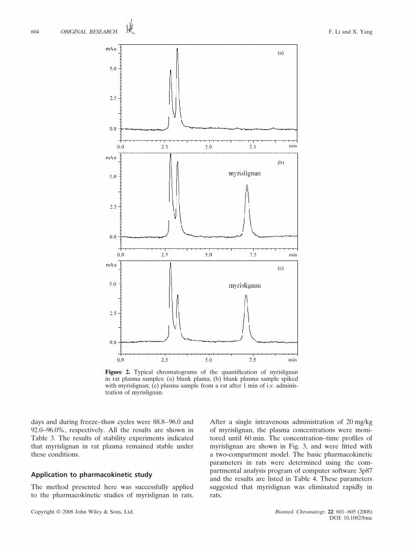
After a single intravenous administration of 20 mg/kgof myrislignan, the plasma concentrations were moni-tored until 60 min. The concentration–time profiles ofmyrislignan are shown in Fig. 3, and were fitted witha two-compartment model. The basic pharmacokineticparameters in rats were determined using the com-partmental analysis program of computer software 3p87and the results are listed in Table 4. These parameterssuggested that myrislignan was eliminated rapidly inrats.
Copyright © 2008 John Wiley & Sons, Ltd. Biomed. Chromatogr. 22: 601–605 (2008)DOI: 10.1002/bmc
Quantification of myrislignan in rat plasma 605ORIGINAL RESEARCH
Table 3. Stability of myrislignan in rat plasma (n ===== 5)
Concentration (μg/mL)
Found RSD AccuracyAdded (mean ± SD) (%) (%)
7 day stability1.5 1.44 ± 0.10 7.2 96.03.0 2.94 ± 0.13 4.4 98.09.0 8.28 ± 0.53 6.4 92.0
18.0 16.92 ± 0.69 4.1 94.0
Three freeze–thaw cycles1.5 1.44 ± 0.095 6.6 96.03.0 2.84 ± 0.19 6.7 94.59.0 7.99 ± 0.53 6.6 88.8
18.0 16.43 ± 0.58 3.5 91.3
CONCLUSION
An HPLC method has been developed to determinemyrislignan in rat plasma. The method consisted ofsample preparation by solid-phase extraction, followedby chromatographic separation with UV detector. Thechromatographic condition employed provided goodseparation of myrislignan without interfering peaks. Thismethod was simple, reproducible, specific and time-effective. It has been successfully used to determine thepharmacokinetic parameters of myrislignan in rat plasma.
Acknowledgments
This research was supported by National High Techno-logy Research and Development Program of China(2002AA2Z343C), Beijing Municipal Special-purposeScience Foundation of China (Z0004105040311) andthe National Sciences and Technology Program of China(2006BAI06A01).
REFERENCES
Hattori M, Hada S, Watahiki A, Ihara H, Shu YZ, Kakiuchi N,Mizuno T and Namba T. Studies on dental caries preventionby traditional medicines. X. Antibacterial action of phenolic com-ponents from mace against Streptococcus mutans. Chemical andPharmaceutical Bulletin 1986; 34: 3885 –3893.
Isogai A, Murakoshi S, Suzuki A and Tamura S. Isolation from nutmegof growth inhibitory substances to silkworm larvae. Agriculturaland Biological Chemistry 1973; 37: 889– 895.
Matsumoto A, Matsumoto T and Tokuda H. Lignans from maceas neoplasm inhibitors. Japanese Kokai Tokkyo Koho 1991; 4.
Miyazawa M, Kasahara H and Kameoka H. Antifungal activities of8-O-4′-neolignans from Myristica fragrans. Natural Product Letters1996; 8: 271–273.
Nakajima K, Yamazaki T, Kawashima K, Shinho Y, Kurashige T,Nohara T, Kinjo Y, Imakiire M and Nishimura M. Vascular smoothmuscle contraction inhibitors from Myristica. Japanese Kokai TokkyoKoho 1999; 8.
Shin KH and Woo WS. Inhibition and induction of hepatic mixedfunction oxidase by phenylpropanoids from the seeds of Myristicafragrans. Han’guk Saenghwa Hakhoechi 1990; 23: 122–127.
The State Pharmacopoeia Commission of P.R. China. Chinese Phar-macopoeia, Vol. 1, 2005; 102–103.
Van GC and Cox PA. Ethnobotany of nutmeg in the Spice Islands.Journal of Ethnopharmacology 1994; 42: 117–124.
Yang XW, Aihemaiti MMT, Li Q, Xu W, Yang Z and Xiao SY.Studies on chemical constituents from seeds of Myristica fragrans.Chinese Traditional and Herbal Drugs 2003; 34(suppl.): 93–96.
Table 4. Pharmacokinetic parameters of myrislignan after a singleintravenous administration of 20 mg/kg in rat plasma (n�=====�5)
Parameter Unit Value
t1/2 α min 4.74�±�1.27t1/2 β min 28.4�±�11.76Vc mg/(μg/mL) 1.03�±�0.076AUC μg min/mL 254.84�±�7.07Cl mg/min/(μg/mL) 0.078�±�0.0022
Figure 3. The concentration–time profiles of myrislignan afteri.v. administration at 20 mg/kg in rats (n = 5).