Embed Size (px)
Citation preview

Quantification of Feline calicivirus (FCV) Genome
Quantification of Feline calicivirus
Open Reading Frame 1
(orf1)
For general laboratory and research use only
150 tests
Quantification of Feline calicivirus (FCV) Genome
Introduction to Feline calcivirus Feline Calicivirus (FCV) is a single stranded RNA virus which typically causes a self-limiting oral and upper respiratory tract disease in cats. FCV belongs to the Caliciviridae family of viruses which also includes significant human pathogens such as norovirus and sapovirus. The genome is approximately 7.7 kb long, it is polyadenylated at the 3’ end and bound by a virally-encoded protein at the 5’-end. It encodes three open reading frames (ORFs). ORF 1 codes for the non-structural proteins including a viral protease and an RNA-dependent RNA polymerase. ORF 2 codes for the major capsid protein and shows sequence variability depending on strain, and ORF 3 encodes a minor structural protein. Cats can be infected with FCV via the nasal, oral or conjunctival routes. Indirect transmission is also possible, especially in an environment such as a cattery where secretions may contaminate cages, feed and personnel. The virus can persist for several weeks when on dried surfaces at room temperature and longer in colder wetter conditions. Most infected cats shed the virus in secretions within 30 days of being infected. Some remain carriers and shedders for life, although most will eliminate the disease. The virus predominantly replicates in the oral and respiratory tissues, although some strains vary in their tissue tropisms and pathogenicity and it may also affect the joints or skin. The most common clinical sign of the disease is oral ulcerations which are usually observed on the tip of the tongue and can rupture with necrosis of the overlying epithelium. Lesions can also be observed in the joints of infected cats which can lead to lameness as a result of acute synovitis. Other symptoms that are observed include conjunctivitis, sneezing, fever, anorexia, ocular and nasal discharge, excessive salivation, gingivitis and oedema. Diagnosis of FCV is difficult without specific tests because the symptoms are similar to other feline respiratory diseases, especially feline herpes virus.

Quantification of Feline calicivirus (FCV) Genome
Specificity The Techne Prime Pro kits for Feline calicivirus (FCV) genomes is designed for the in vitro quantification of FCV genomes. The kit is designed to have the broadest detection profile possible whilst remaining specific to the FCV genome. The primers and probe sequences in this kit have 100% homology with a broad range of clinically relevant reference sequences based on a comprehensive bioinformatics analysis. If you require further information, or have a specific question about the detection profile of this kit then please email [email protected] and our bioinformatics team will answer your question.
Quantification of Feline calicivirus (FCV) Genome
Kit Contents
• FCV specific primer/probe mix (150 reactions BROWN) FAM labeled, BHQ quenched
• FCV positive control template (for Standard curve RED)
• Internal extraction control RNA (150 reactions BLUE)
• Internal extraction control primer/probe mix (150 reactions BROWN)
VIC labelled
• Endogenous ACTB primer/probe mix (150 reactions BROWN)
FAM labeled, BHQ quenched
• Internal extraction control/FCV/ACTB RT primer mix (150 reactions GREEN) Required for two step protocol only
• RNAse/DNAse free water
Reagents and equipment to be supplied by the user Real-Time PCR Instrument
RNA extraction kit
This kit is designed to work well with all processes that yield high quality RNA with minimal PCR inhibitors.
Prime Pro
Lyophlised OneStep qRT-PCR MasterMix kit
Contains complete one step qRT-PCR MasterMix
Optional - Reverse Transcription kit and Mastermix
Although a one step RT-PCR protocol is recommended, this kit is designed to work well with a two step protocol. We recommend the use of Prime Pro Reverse Transcription kits and the use of Prime Pro 2x MasterMix for the two step protocol.
Pipettes and Tips
Vortex and centrifuge
Thin walled 0.2 ml PCR reaction tubes
Quantification of Feline calicivirus (FCV) Genome
Kit storage and stability This kit is stable at room temperature but should be stored at -20ºC on arrival. Techne does not recommend using the kit after the expiry date stated on the pack. Once the lyophilized components have been re-suspended, unnecessary repeated freeze/thawing should be avoided. The kit is stable for six months from the date of resuspension under these circumstances.
Suitable sample material All kinds of sample material suited for PCR amplification can be used. Please ensure the samples are suitable in terms of purity, concentration, and RNA/DNA integrity (An internal PCR control is supplied to test for non- specific PCR inhibitors). Always run at least one negative control with the samples. To prepare a negative-control, replace the template RNA sample with RNAse/DNAse free water.
Dynamic range of test Under optimal PCR conditions Techne detection kits have very high priming efficiencies of >95% and can detect less than 100 copies of target template.
Notices and disclaimers This product is developed, designed and sold for research purposes only. It is not intended for human diagnostic or drug purposes or to be administered to humans unless clearly expressed for that purpose by the Food and Drug Administration in the USA or the appropriate regulatory authorities in the country of use. During the warranty period Techne pathogen detection kits allow precise and reproducible data recovery combined with excellent sensitivity. For data obtained by violation to the general GLP guidelines and the manufacturer’s recommendations the right to claim under guarantee is expired. Black Hole Quencher”, “BHQ”, “CAL Fluor, “Quasar” and “Pulsar” are registered trademarks of Biosearch Technologies, Inc., Novato, CA. This technology is protected by U.S. and World-wide patents either issued or in application and is licensed and sold under agreement with Biosearch Technologies, Inc. These products are sold exclusively for R&D use by the purchaser. They may not be used for human or veterinary in vitro diagnostic (IVD) applications and they may not be re-sold, distributed or re-packaged without express written authorization from Biosearch Technologies Inc. PCR is a proprietary technology covered by several US and foreign patents. These patents are owned by Roche Molecular Systems Inc. and have been sub- licensed by PE Corporation in certain fields. Depending on your specific application you may need a license from Roche or PE to practice PCR. Additional information on purchasing licenses to practice the PCR process may be obtained by contacting the Director of Licensing at Roche Molecular Systems, 1145 Atlantic Avenue, Alameda, CA 94501 or Applied Biosystems business group of the Applera Corporation, 850 Lincoln Centre Drive, Foster City, CA 94404. In addition, the 5' nuclease assay and other homogeneous amplification methods used in connection with the PCR process may be covered by U.S. Patents 5,210,015 and 5,487,972, owned by Roche Molecular Systems, Inc, and by U.S. Patent 5,538,848, owned by The Perkin-Elmer Corporation. The purchase of Biosearch Technologies products does not, either expressly or by implication, provide a license to use this or other patented technology. Licensing information can be obtained by contacting the Director of Licensing, Applied Biosystems, 850 Lincoln Centre Drive, Foster City, CA 94404 or the Licensing Department at Roche Molecular Systems Inc., 1145 Atlantic Avenue, Alameda, CA 94501.”
Trademarks Techne ™ is a trademark of Bibby Scientific Ltd. The PCR process is covered by US Patents 4,683,195, and 4,683,202 and foreign equivalents owned by Hoffmann-La Roche AG. ABI, ABI PRISM® GeneAmp® and MicroAmp® are registered trademarks of the Applera Genomics (Applied Biosystems Corporation). BIOMEK® is a registered trademark of Beckman Instruments, Inc.; iCycler™ is a registered trademark of Bio-Rad Laboratories, Rotor-Gene is a trademark of Corbett Research. LightCycler™ is a registered trademark of the Idaho Technology Inc. GeneAmp®, TaqMan® and AmpliTaqGold® are registered trademarks of Roche Molecular Systems, Inc., The purchase of the Techne ™ reagents cannot be construed as an authorization or implicit license to practice PCR under any patents held by Hoffmann-LaRoche Inc.

Quantification of Feline calicivirus (FCV) Genome
Principles of the test Real-time PCR
A FCV specific primer and probe mix is provided and this can be detected through the FAM channel.
The primer and probe mix provided exploits the so-called TaqMan® principle. During PCR amplification, forward and reverse primers hybridize to the FCV DNA/cDNA. A fluorogenic probe is included in the same reaction mixture which consists of a DNA probe labeled with a 5`-dye and a 3`-quencher. During PCR amplification, the probe is cleaved and the reporter dye and quencher are separated. The resulting increase in fluorescence can be detected on a range of real-time PCR platforms.
One Step vs. Two step real-time PCR
When detecting/quantifying the presence of a target with an RNA genome Techne recommend the use of a one step qRT-PCR protocol. One step qRT-PCR combines the reverse transcription and real-time PCR reaction in a simple closed tube protocol. This saves significant bench time but also reduces errors. The sensitivity of a one step protocol is also greater than a two step because the entire biological sample is available to the PCR without dilution.
Positive control
For copy number determination and as a positive control for the PCR set up, the kit contains a positive control template. This can be used to generate a standard curve of FCV copy number / CT value. Alternatively the positive control can be used at a single dilution where full quantitative analysis of the samples is not required. Each time the kit is used, at least one positive control reaction must be included in the run. A positive result indicates that the primers and probes for detecting the target FCV gene worked properly in that particular experimental scenario. If a negative result is obtained the test results are invalid and must be repeated. Care should be taken to ensure that the positive control does not contaminate any other kit component which would lead to false-positive results. This can be achieved by handling this component in a Post PCR environment. Care should also be taken to avoid cross-contamination of other samples when adding the positive control to the run. This can be avoided by sealing all other samples and negative controls before pipetting the positive control into the positive control well.
Negative control
To confirm the absence of contamination, a negative control reaction should be included every time the kit is used. For this reaction, the RNAse/DNAse free water should be used instead of template. A negative result indicates that the reagents have not become contaminated while setting up the run. If a positive result is obtained the results should be ignored and the test samples repeated. Possible sources of contamination should first be explored and removed.
Quantification of Feline calicivirus (FCV) genomes
Internal RNA extraction control When performing RNA extraction, it is often advantageous to have an exogenous source of RNA template that is spiked into the lysis buffer. This control RNA is then co-purified with the sample RNA and can be detected as a positive control for the extraction process. Successful co-purification and real-time PCR for the control RNA also indicates that PCR inhibitors are not present at a high concentration.
A separate RT primer mix and a real-time PCR primer/probe mix are supplied with this kit to detect the exogenous RNA using real-time PCR. The PCR primers are present at PCR limiting concentrations which al lows multiplexing with the ta rge t sequence primers. Amplification of the control cDNA does not interfere with detection of the pathogen target cDNA even when present at low copy number. The Internal control is detected through the VIC channel and gives a CT value of 31+/-3 depending on the level of sample dilution.
Endogenous ACTB control To confirm extraction of a valid biological template, a primer and probe mix is included to detect the Actin Beta (ACTB) gene. Detection of ACTB is through the FAM channel and it is NOT therefore possible to perform a multiplex for ACTB and the pathogen primers. A poor ACTB signal may indicate that the sample did not contain sufficient biological material.
Carry-over prevention using UNG (optional)
Carry over contamination between PCR reactions can be prevented by including uracil-N- glycosylase (UNG) in the reaction mix. Some commercial mastermix preparations contain UNG or alternatively it can be added as a separate component. UNG can only prevent carry over from PCR reactions that include deoxyuridine triphosphate (dUTP) in the original PCR reaction. Alliance Bio Inc. recommend the application of 0.2U UNG per assay with a 15 minute incubation step at 37°C prior to amplification. The heat-labile UNG is then inactivated during the Taq polymerase activation step (95°C for 10 minutes).
Quantification of Feline calicivirus (FCV) genomes
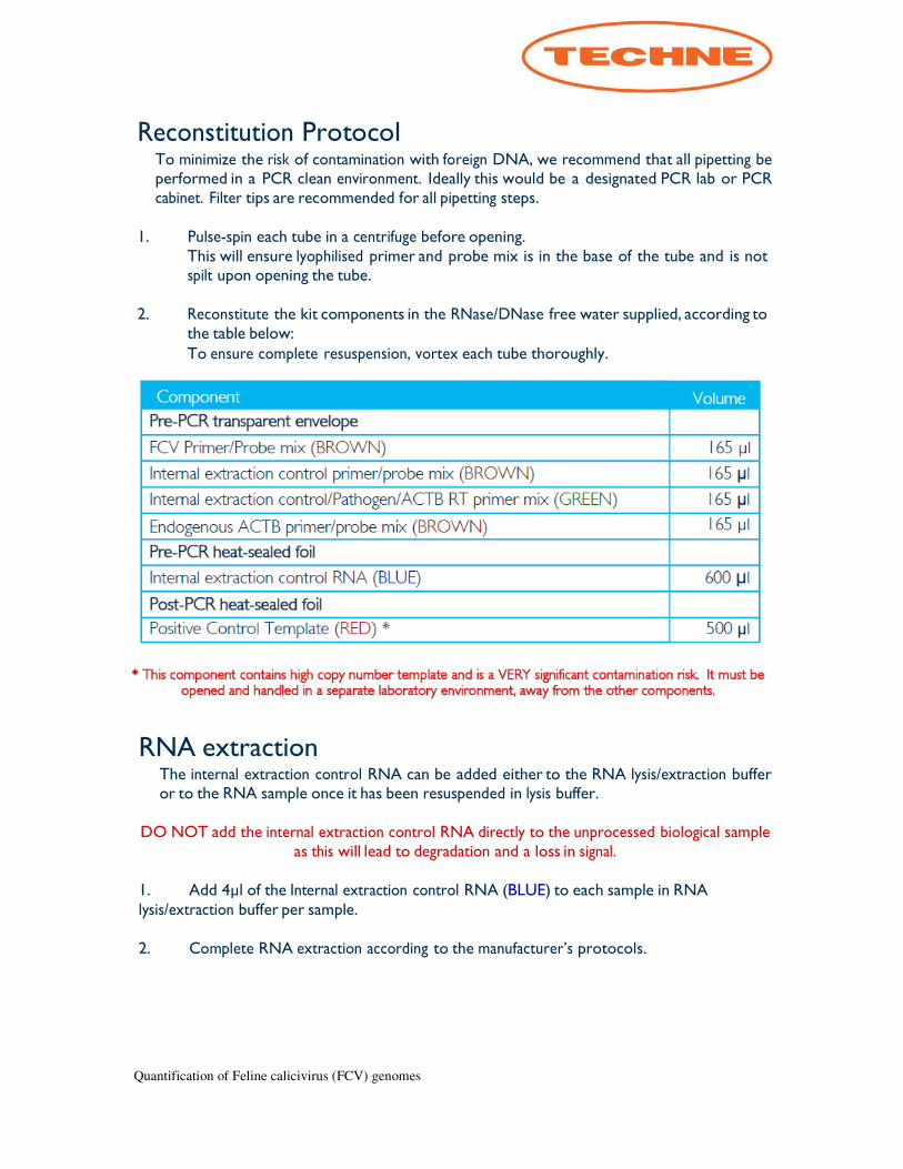
Reconstitution Protocol To minimize the risk of contamination with foreign DNA, we recommend that all pipetting be performed in a PCR clean environment. Ideally this would be a designated PCR lab or PCR cabinet. Filter tips are recommended for all pipetting steps.
1. Pulse-spin each tube in a centrifuge before opening.
This will ensure lyophilised primer and probe mix is in the base of the tube and is not spilt upon opening the tube.
2. Reconstitute the kit components in the RNase/DNase free water supplied, according to
the table below: To ensure complete resuspension, vortex each tube thoroughly.
RNA extraction The internal extraction control RNA can be added either to the RNA lysis/extraction buffer or to the RNA sample once it has been resuspended in lysis buffer.
DO NOT add the internal extraction control RNA directly to the unprocessed biological sample
as this will lead to degradation and a loss in signal.
1. Add 4µl of the Internal extraction control RNA (BLUE) to each sample in RNA lysis/extraction buffer per sample.
2. Complete RNA extraction according to the manufacturer’s protocols.
Quantification of Feline calicivirus (FCV) genomes
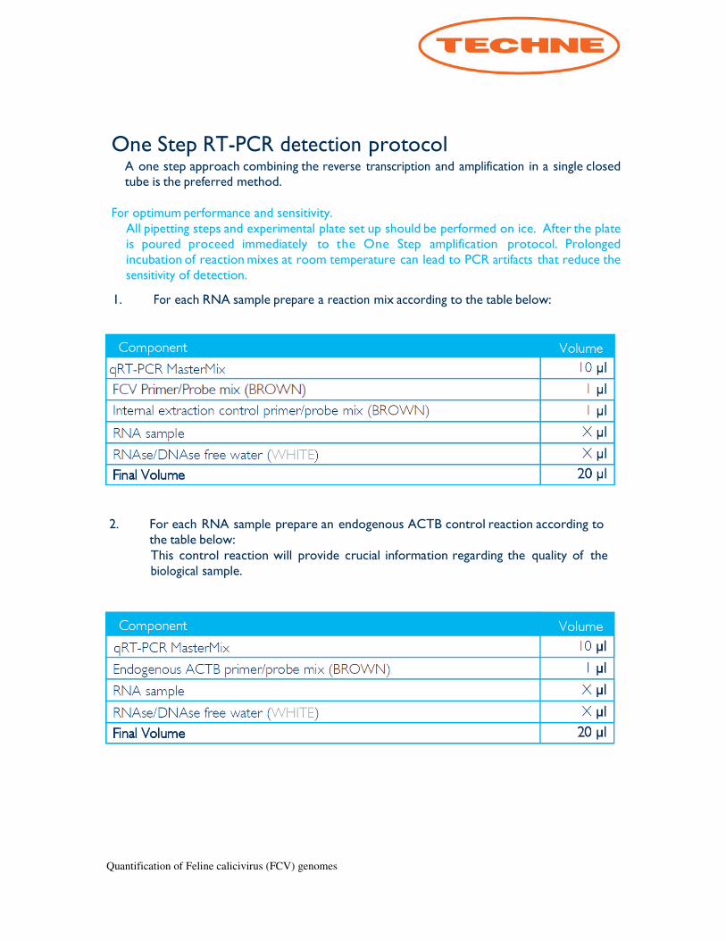
One Step RT-PCR detection protocol A one step approach combining the reverse transcription and amplification in a single closed tube is the preferred method.
For optimum performance and sensitivity.
All pipetting steps and experimental plate set up should be performed on ice. After the plate is poured proceed immediately to the One Step amplification protocol. Prolonged incubation of reaction mixes at room temperature can lead to PCR artifacts that reduce the sensitivity of detection.
1. For each RNA sample prepare a reaction mix according to the table below:
2. For each RNA sample prepare an endogenous ACTB control reaction according to the table below: This control reaction will provide crucial information regarding the quality of the biological sample.
Quantification of Feline calicivirus (FCV) genomes
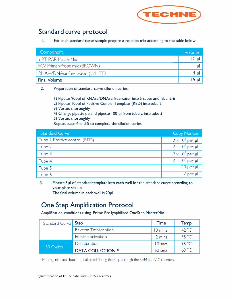
Standard curve protocol
1. For each standard curve sample prepare a reaction mix according to the table below
2. Preparation of standard curve dilution series.
1) Pipette 900µl of RNAse/DNAse free water into 5 tubes and label 2-6 2) Pipette 100µl of Positive Control Template (RED) into tube 2 3) Vortex thoroughly 4) Change pipette tip and pipette 100 µl from tube 2 into tube 3 5) Vortex thoroughly Repeat steps 4 and 5 to complete the dilution series
3. Pipette 5µl of standard template into each well for the standard curve according to
your plate set-up The final volume in each well is 20µl.
One Step Amplification Protocol Amplification conditions using Prime Pro
lyophilised OneStep MasterMix.

Quantification of Feline calicivirus (FCV) genomes
Alternative two step reverse transcription/real-time PCR protocol ReverseTranscription A reverse transcription primer (GREEN) is included and is designed for use with the a
reverse transcription kit.
1. After reverse transcription, prepare a reaction mix according to the table below for each
cDNA sample
2. Pipette 15µl of this mix into each well according to your real-time PCR experimental plate set up.
3. Prepare sample cDNA templates for each of your samples (suggested concentration
(5ng/µl) in RNAse/DNAse free water. If the concentration of RNA that was used to make the cDNA is not known, then dilute your RT reaction mix 1:5 (10µl of sample cDNA and 40µl of water).
4. Pipette 5µl of cDNA template into each well, according to your experimental plate setup.
The final volume in each well is 20µl. For negative control wells use 5µl of RNAse/DNAse free water.
Quantification of Feline calicivirus (FCV) genomes
Negative
control
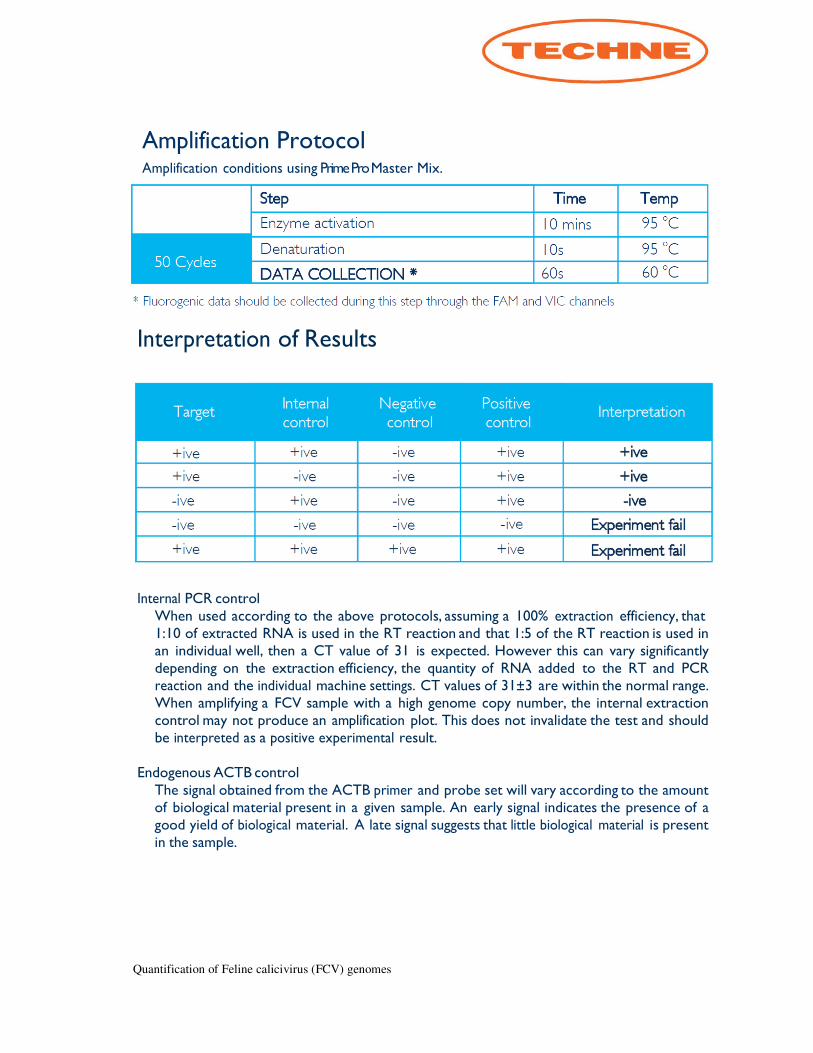
Amplification Protocol Amplification conditions using Prime Pro Master Mix.
Interpretation of Results
Internal PCR control
When used according to the above protocols, assuming a 100% extraction efficiency, that 1:10 of extracted RNA is used in the RT reaction and that 1:5 of the RT reaction is used in an individual well, then a CT value of 31 is expected. However this can vary significantly depending on the extraction efficiency, the quantity of RNA added to the RT and PCR reaction and the individual machine settings. CT values of 31±3 are within the normal range. When amplifying a FCV sample with a high genome copy number, the internal extraction control may not produce an amplification plot. This does not invalidate the test and should be interpreted as a positive experimental result.
Endogenous ACTB control
The signal obtained from the ACTB primer and probe set will vary according to the amount of biological material present in a given sample. An early signal indicates the presence of a good yield of biological material. A late signal suggests that little biological material is present in the sample.