Embed Size (px)
Citation preview
7 Westferry Circus ● Canary Wharf ● London E14 4HB ● United Kingdom
Telephone +44 (0)20 7418 8400 Facsimile +44 (0)20 7418 8416
E-mail [email protected] Website www.ema.europa.eu An agency of the European Union
© European Medicines Agency, 2010. Reproduction is authorised provided the source is acknowledged.
21 October 2010EMA/CHMP/SAWP/283298/2010Committee for Medicinal Products for Human Use (CHMP)
Qualification opinion ILSI/HESI submission of novel renal biomarkers for toxicity
Agreed by Scientific Advice Working Party February 2010
Adoption by CHMP for release for consultation 18 March 2010
End of consultation (deadline for comments) 31 July 2010
Adoption by CHMP 21 October 2010
Keywords Non-clinical, renal biomarkers, nephrotoxicity
Qualification Opinion ILSI/HESI Submission of Novel Renal Biomarkers for ToxicityEMA/CHMP/SAWP/283298/2010 2/18
Background
The European Medicines Agency qualification process is a new, voluntary, scientific pathway leading to
either a CHMP opinion or a Scientific Advice on innovative methods or drug development tools. It
includes qualification of biomarkers developed by consortia, networks, public/private partnerships,
learned societies or pharmaceutical industry for a specific intended use in pharmaceuticals R&D.
The HESI study evaluated four novel urinary biomarkers (BMs) of nephrotoxicity i.e. alpha-glutathione
S-transferase (α-GST), μ-glutathione S-transferase (μ-GST), renal papillary antigen-1 (RPA-1) and
clusterin, and compared their performance against the more traditional measurements for diagnosis of
nephrotoxicity.
The data presented in this report were all generated in single and repeated dose studies conducted in
male rats of two strains (Sprague-Dawley and Wistar) that are commonly used in preclinical toxicity
studies. The information obtained from these studies demonstrates the potential utility of these BMs
for use in rodent studies conducted to evaluate the potential target organ toxicity of compounds as
part of the preclinical safety assessment of candidate medicines.
Scope
HESI proposes that there is sufficient evidence to support the voluntary use of α-GST, RPA-1 and
clusterin along with currently used methods to gain further insight into renal injury when it is seen in
preclinical safety assessment studies in rats.
Specifically, HESI claims the following:
“The novel BMs evaluated have been shown not only to have utility for the detection of tubular injury
but some also provide useful information on the tubular site of injury
Clusterin is confirmed to have utility for detection of tubular injury without additional insight as to
location. The superiority of clusterin (compared with the reference BMs: BUN and sCr) was evident
when regeneration was present.
α-GST was shown to be superior to all of the reference markers for detection of injury to the
proximal tubule.
RPA-1 was shown to be superior to all of the reference markers for detection of injury to the
collecting duct.”
These claims are the subject of the CHMP qualification process described below. Measurements were
made for μ-GST (Yb1); however according to the Applicant, these data did not support a claim.
HESI Overall Strategy General Principles
Select compounds causing damage to specific portions of the nephron. HESI stated that correlation
of biomarker values with a defined histopathological phenotype is central to evaluation of the utility
of biomarkers to report nephrotoxic injury. The use of different compounds and different dosage
regimens (both unitary doses and duration of dosing) serves to produce varied pathology (different
sites and patterns of injury). Histopathology was the gold standard used to assess biomarker
performance. Immunohistochemistry was used to confirm location of specific lesions.
Use male (HW and SD) rats as the initial test species.
Select candidate biomarkers with promise based on literature and previous HESI programs.
Qualification Opinion ILSI/HESI Submission of Novel Renal Biomarkers for ToxicityEMA/CHMP/SAWP/283298/2010 3/18
Conduct pilot studies to define doses ranging from no observable effect dose to markedly toxic (i.e.
using dose-response information to obviate, in the first instance, the need for “negative” controls
other than vehicle).
Define procedures to be used for sample collection by each participating laboratory to ensure
optimal preservation of biomarkers and other analytes.
Develop biomarker assays where none exist and comprehensively validate the biomarker assays.
Design and execute single definitive studies for each selected nephrotoxin and rat strain and
exchange samples between six labs as part of a robust assay validation process.
Conduct according to GLP standards with the following exceptions:
- There would be no independent audit although each participating laboratory would be responsible
for conducting thorough quality control of their data.
- No formulation analysis or exposure assessment would be done since development of validated
assays would require disproportionate resource and time.
Where sample volumes permit, conduct auxiliary studies using ‘omics’ technologies to identify
additional biomarkers.
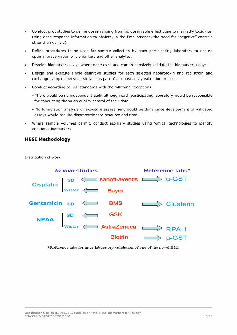
HESI Methodology
Distribution of work
Qualification Opinion ILSI/HESI Submission of Novel Renal Biomarkers for ToxicityEMA/CHMP/SAWP/283298/2010 4/18
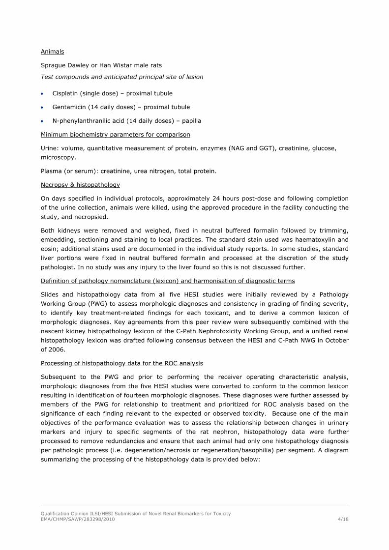
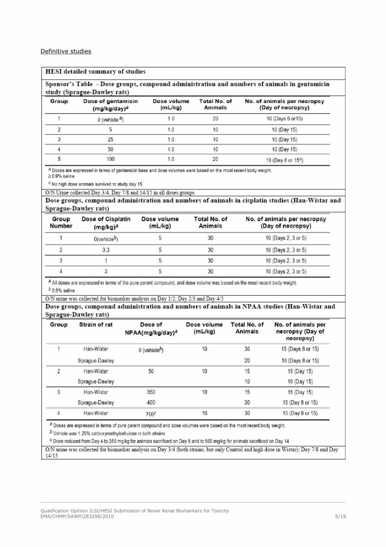
Animals
Sprague Dawley or Han Wistar male rats
Test compounds and anticipated principal site of lesion
Cisplatin (single dose) – proximal tubule
Gentamicin (14 daily doses) – proximal tubule
N-phenylanthranilic acid (14 daily doses) – papilla
Minimum biochemistry parameters for comparison
Urine: volume, quantitative measurement of protein, enzymes (NAG and GGT), creatinine, glucose,
microscopy.
Plasma (or serum): creatinine, urea nitrogen, total protein.
Necropsy & histopathology
On days specified in individual protocols, approximately 24 hours post-dose and following completion
of the urine collection, animals were killed, using the approved procedure in the facility conducting the
study, and necropsied.
Both kidneys were removed and weighed, fixed in neutral buffered formalin followed by trimming,
embedding, sectioning and staining to local practices. The standard stain used was haematoxylin and
eosin; additional stains used are documented in the individual study reports. In some studies, standard
liver portions were fixed in neutral buffered formalin and processed at the discretion of the study
pathologist. In no study was any injury to the liver found so this is not discussed further.
Definition of pathology nomenclature (lexicon) and harmonisation of diagnostic terms
Slides and histopathology data from all five HESI studies were initially reviewed by a Pathology
Working Group (PWG) to assess morphologic diagnoses and consistency in grading of finding severity,
to identify key treatment-related findings for each toxicant, and to derive a common lexicon of
morphologic diagnoses. Key agreements from this peer review were subsequently combined with the
nascent kidney histopathology lexicon of the C-Path Nephrotoxicity Working Group, and a unified renal
histopathology lexicon was drafted following consensus between the HESI and C-Path NWG in October
of 2006.
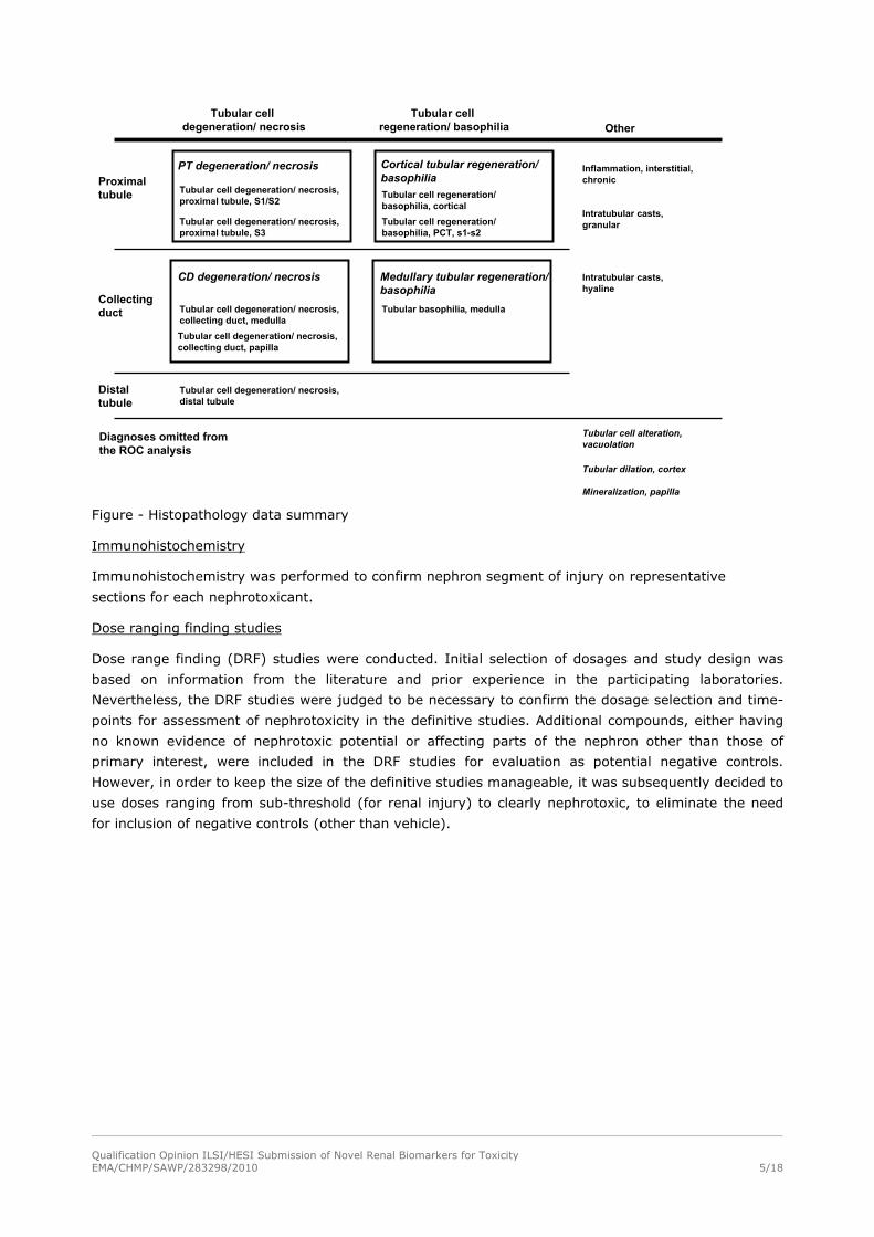
Processing of histopathology data for the ROC analysis
Subsequent to the PWG and prior to performing the receiver operating characteristic analysis,
morphologic diagnoses from the five HESI studies were converted to conform to the common lexicon
resulting in identification of fourteen morphologic diagnoses. These diagnoses were further assessed by
members of the PWG for relationship to treatment and prioritized for ROC analysis based on the
significance of each finding relevant to the expected or observed toxicity. Because one of the main
objectives of the performance evaluation was to assess the relationship between changes in urinary
markers and injury to specific segments of the rat nephron, histopathology data were further
processed to remove redundancies and ensure that each animal had only one histopathology diagnosis
per pathologic process (i.e. degeneration/necrosis or regeneration/basophilia) per segment. A diagram
summarizing the processing of the histopathology data is provided below:
Qualification Opinion ILSI/HESI Submission of Novel Renal Biomarkers for ToxicityEMA/CHMP/SAWP/283298/2010 5/18
Tubular cell degeneration/ necrosis, proximal tubule, S1/S2
Tubular cell degeneration/ necrosis, proximal tubule, S3
Tubular cell regeneration/ basophilia, cortical
Tubular cell degeneration/ necrosis, distal tubule
Tubular cell degeneration/ necrosis, collecting duct, medulla
Tubular cell degeneration/ necrosis, collecting duct, papilla
Tubular basophilia, medulla
Tubular cell regeneration/ basophilia, PCT, s1-s2
Tubular cell regeneration/ basophilia
Tubular cell degeneration/ necrosis
Proximal tubule
Collectingduct
Distal tubule
PT degeneration/ necrosis
CD degeneration/ necrosis
Cortical tubular regeneration/basophilia
Medullary tubular regeneration/basophilia
Intratubular casts, granular
Intratubular casts, hyaline
Inflammation, interstitial, chronic
Other
Tubular cell alteration, vacuolation
Tubular dilation, cortex
Mineralization, papilla
Diagnoses omitted from the ROC analysis
Figure - Histopathology data summary
Immunohistochemistry
Immunohistochemistry was performed to confirm nephron segment of injury on representative
sections for each nephrotoxicant.
Dose ranging finding studies
Dose range finding (DRF) studies were conducted. Initial selection of dosages and study design was
based on information from the literature and prior experience in the participating laboratories.
Nevertheless, the DRF studies were judged to be necessary to confirm the dosage selection and time-
points for assessment of nephrotoxicity in the definitive studies. Additional compounds, either having
no known evidence of nephrotoxic potential or affecting parts of the nephron other than those of
primary interest, were included in the DRF studies for evaluation as potential negative controls.
However, in order to keep the size of the definitive studies manageable, it was subsequently decided to
use doses ranging from sub-threshold (for renal injury) to clearly nephrotoxic, to eliminate the need
for inclusion of negative controls (other than vehicle).
Qualification Opinion ILSI/HESI Submission of Novel Renal Biomarkers for ToxicityEMA/CHMP/SAWP/283298/2010 6/18
Definitive studies
Qualification Opinion ILSI/HESI Submission of Novel Renal Biomarkers for ToxicityEMA/CHMP/SAWP/283298/2010 7/18
Statistical methods
Biomarker assay validation
The following aspects of the performance of the assays for each of the novel biomarkers under study
were assessed:
Repeatability (intra-run precision)
Intermediate precision (intra-lab precision)
Reproducibility (inter-lab precision)
Biomarker Qualification
Data Normalization
For all urinary markers, analyte concentrations for all animals were first normalised by dividing by the
corresponding urine creatinine concentration. All individual animal marker values (normalised to
creatinine in the case of urinary markers) were divided by the mean of the values in the concurrent
control (i.e. vehicle-dosed) animals. Thus, all marker values were expressed as a fold-change versus
the time-matched control group mean.
Assessment of variability of the novel BMs among control animals
Repeat urinary measurements for both Sprague-Dawley and Han-Wistar control animals were used to
estimate intra-animal and inter-animal variability for novel markers α-GST, μ-GST, RPA-1, and
clusterin. The statistical analysis was done, as described above, after normalization by division of the
analyte concentration by the corresponding urine creatinine concentration.
For each rat strain separately, creatinine-normalized analyte concentrations were analyzed by a one-
way random effects analysis of variance (ANOVA). Intra-animal and inter-animal variances were
calculated by equating observed and expected mean squares from the one-way random effects
ANOVA. Two-sided 95% confidence intervals were calculated using the standard Chi-squared method
for intra-animal variance and the modified large sample method for inter-animal variance. Negative
estimates and confidence bounds for the inter-animal variance were reported as zero.
Variance estimates and confidence intervals were converted to coefficient of variation (%CV) by
applying the square root transformation and dividing by the observed concentration mean.
Histopathology
Histopathology grades were assessed on a scale of 0 (no observed pathology) to 4 (severe pathology).
For each path, animals were defined as ‘Negative’ or ‘Positive’ as follows:
‘Negative’: Animals which were dosed with either vehicle or toxicant and with histopathology score =
0.
‘Positive’: Animals which were dosed with either vehicle or toxicant and with histopathology score > 0.
Data Exclusion
All animals from the high dose group (100 mg/kg) of the gentamicin study were excluded from all
statistical analyses. In this group, there were unscheduled sacrifices due to the poor clinical condition
of some animals (1 animal on day 6, 11 animals with controls on day 7 and the remaining 4 animals
with controls on days 8 and 10). Thus no high dose animals survived to day 14. It was therefore
Qualification Opinion ILSI/HESI Submission of Novel Renal Biomarkers for ToxicityEMA/CHMP/SAWP/283298/2010 8/18
considered that, despite the selection of the high dose based on a DRF study, this dosage exceeded a
maximum tolerated dose in this study.
Additionally, animals for which any individual biomarker result was missing were excluded from all
statistical analyses.
Descriptive Statistics
The number of animals classified as ‘Negative’ and ‘Positive’ was determined for each pathology, and
stratified by rat strain and toxicant.
For each marker and pathology, summary statistics (mean and standard deviation) were calculated for
‘Negative’ and ‘Positive’ animals, and stratified by rat strain and toxicant.
Receiver Operating Characteristic Curves
The discriminatory accuracy of each marker was assessed using receiver operating characteristic (ROC)
curve methods. The area under the ROC curve (AUCROC), a commonly used index of diagnostic
accuracy, was used to compare the performance of each marker.
For each marker and pathology, nonparametric point estimates and standard errors of the AUCROC
were calculated by rat strain and across both rat strains.
Using pooled data (across both rat strains), pairwise comparisons of AUCROC values for each novel
marker (α-GST, μ-GST, RPA-1, clusterin) versus each reference marker (BUN, SCr, NAG, protein) were
performed for each pathology and the corresponding two-sided p-value calculated.
For each novel marker separately, raw p-values were adjusted for multiple testing (i.e. pathologies and
reference markers) via the Hochberg procedure. No adjustment was made for the number of novel
markers considered.
Incremental Diagnostic Value
The incremental diagnostic value of each novel marker (α-GST, μ-GST, RPA-1, clusterin), when used in
conjunction with reference markers, was also assessed using ROC curves.
Two reference marker sets were considered:
1) BUN and serum creatinine
2) NAG and protein
For each pathology and reference marker set, a logistic regression model was fit:
logit (P) = α + β1*X1 + β 2*X2 where P denotes the probability that an animal is ‘Positive’ and {X1,
X2} denote the reference marker values. The AUCROC of the linear score (β 1*X1 + β 2*X2) was
calculated. Denote this by AUCROC(X1,X2). For each novel marker separately, the logistic regression
models above were then re-fit to include a term for the novel marker (denoted by X3). The AUCROC of
the linear score (β 1*X1 + β 2*X2 + β 3*X3) was calculated and denoted by AUCROC(X1, X2, X3).
For each novel marker and reference marker set, pairwise comparisons of AUCROC(X1,X2) versus
AUCROC(X1,X2,X3) were performed for each pathology and the corresponding two-sided p-value
calculated . Raw p-values were adjusted for multiple testing (i.e. pathologies and reference marker
sets) via the Hochberg procedure, for each novel marker separately.
Note that the linear scores derived above via logistic regression do not necessarily yield optimal
discriminatory accuracy. Other approaches are possible. However, logistic regression is commonly
utilized for deriving marker combinations and provides a practical and reasonable framework for
assessing the incremental diagnostic value of the novel markers under consideration.
Qualification Opinion ILSI/HESI Submission of Novel Renal Biomarkers for ToxicityEMA/CHMP/SAWP/283298/2010 9/18
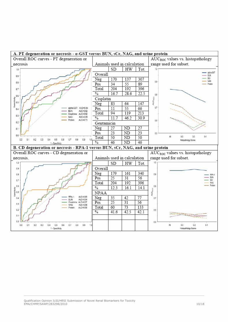
RESULTS – Diagnostic Performance
The submission includes data from a total of five studies using three compounds (cisplatin, gentamicin
and NPAA). Data submitted between April 2008 and April 2009. The HESI data on the analytical
validation, inter-animal and intra-animal variability and immunochemistry support the qualification
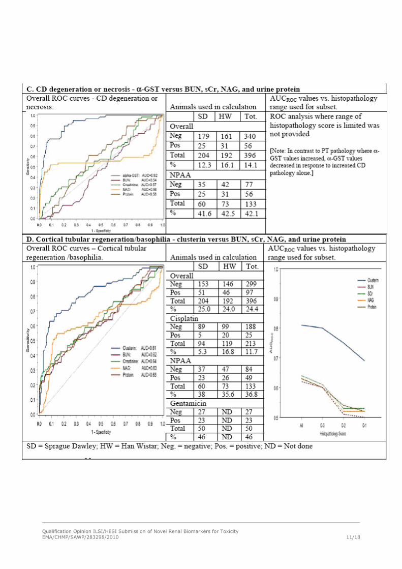
exercise with the caveats identified in the GAPS section below. The tables below (A,B,C,D) compiled by
the assessors focus on the overall ROC curves, the number of animals used for this calculation and the
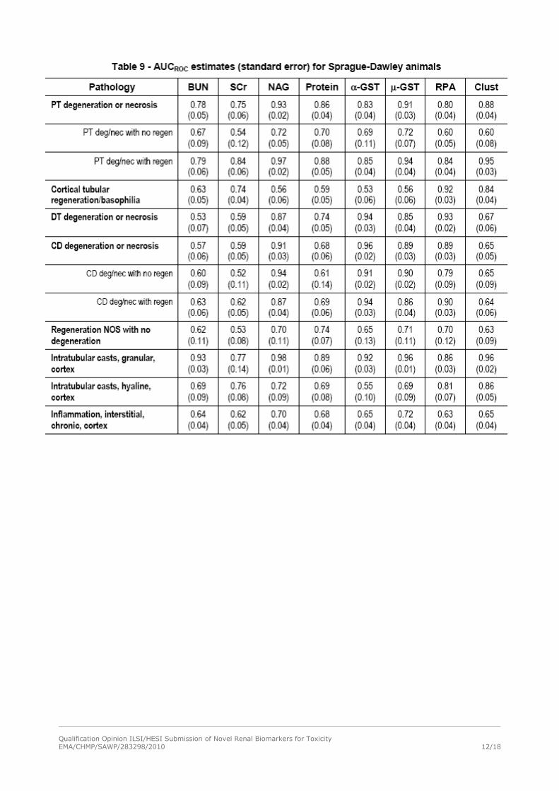
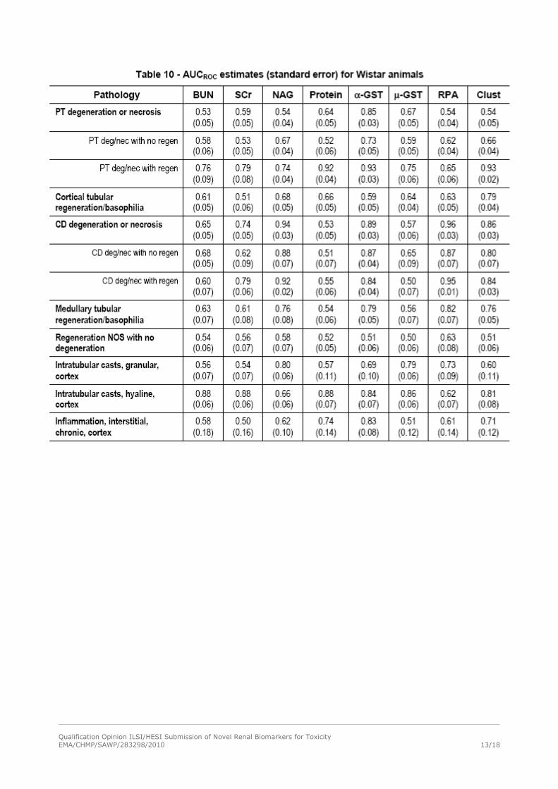
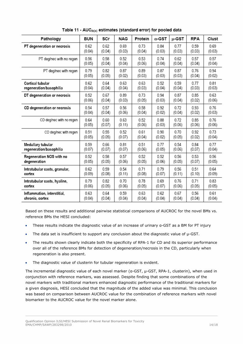
AUCROC values vs. histopathology range used for subset. Tables 9, 10 and 11 present the AUCROC
values for different pathologies. The full documentation on the statistical analysis, conducted according
to the methodology part, is not included in order to keep the document concise.
Qualification Opinion ILSI/HESI Submission of Novel Renal Biomarkers for ToxicityEMA/CHMP/SAWP/283298/2010 10/18
Qualification Opinion ILSI/HESI Submission of Novel Renal Biomarkers for ToxicityEMA/CHMP/SAWP/283298/2010 11/18
Qualification Opinion ILSI/HESI Submission of Novel Renal Biomarkers for ToxicityEMA/CHMP/SAWP/283298/2010 12/18
Qualification Opinion ILSI/HESI Submission of Novel Renal Biomarkers for ToxicityEMA/CHMP/SAWP/283298/2010 13/18
Qualification Opinion ILSI/HESI Submission of Novel Renal Biomarkers for ToxicityEMA/CHMP/SAWP/283298/2010 14/18
Based on these results and additional pairwise statistical comparisons of AUCROC for the novel BMs vs.
reference BMs the HESI concluded:
These results indicate the diagnostic value of an increase of urinary α-GST as a BM for PT injury
The data set is insufficient to support any conclusion about the diagnostic value of μ-GST.
The results shown clearly indicate both the specificity of RPA-1 for CD and its superior performance
over all of the reference BMs for detection of degeneration/necrosis in the CD, particularly when
regeneration is also present.
The diagnostic value of clusterin for tubular regeneration is evident.
The incremental diagnostic value of each novel marker (α-GST, μ-GST, RPA-1, clusterin), when used in
conjunction with reference markers, was assessed. Despite finding that some combinations of the
novel markers with traditional markers enhanced diagnostic performance of the traditional markers for
a given diagnosis, HESI concluded that the magnitude of the added value was minimal. This conclusion
was based on comparison between AUCROC value for the combination of reference markers with novel
biomarker to the AUCROC value for the novel marker alone.
Qualification Opinion ILSI/HESI Submission of Novel Renal Biomarkers for ToxicityEMA/CHMP/SAWP/283298/2010 15/18
Regulatory Data Assessment
The FDA and the European Medicines Agency contributed to the evaluation via the ad hoc appointed
Biomarkers Qualification Teams (QTs) providing (via written procedures and Joint Teleconferences/
meetings with the HESI representatives) elements for gap analysis, questions on the statistical
evaluations and drafting the conclusions.
Gaps identified by CHMP in the current qualification exercise
The QT assessed the data presented by the Applicant and identified some gaps in the qualification
exercise. The Applicant is encouraged to address the gaps in future investigations.
Analytical methods
- Some of the results of interference testing are missing [Hb, bilirubin and high salt for clusterin assay
and metals (mercury, cadmium, lead, lithium, gadolinium) for all assays].
- The impact of the criteria for repeatability, intermediate precision and reproducibility on the
diagnostic performance of the biomarkers was not evaluated.
Limitations of the studies to address specificity of the biomarkers for injury at a particular site
- The specificity of these BMs to kidney injury needs to be further investigated since other possible
target organs were not investigated (i.e. α-GST present in the liver also, clusterin in the cytoplasm of
interstitial macrophages within stomach, skeletal muscle, heart, tongue, as well as macrophages within
the medulla of thymus and lymph node of an untreated control rat). Liver toxicity could potentially
interfere in the evaluation of the diagnostic performance of these novel biomarkers. In the current
exercise the examination of liver was not standardised between studies and the statements that the
liver is not affected by the three compounds cannot be supported. In this context the assessment of
the liver should be standardised in future dose finding and definitive studies. Furthermore the testing
of an additional intermediate (between clearly toxic and non toxic) doses in the future dose finding
studies could help define a more appropriate control group and possibly increase the power of the
definitive studies to identify specific biomarkers of renal vs. liver toxicity.
- The number and type of nephrotoxic compounds in the studies was limited.
- There were no studies conducted with non-nephrotoxic compounds (e.g. hepatotoxins).
Reproducibility of experiments
The QT notices the inconsistency between dose-finding and definitive studies for gentamicin and NPAA
which makes the interpretation difficult.
Difference between strains and inference
The possibility of strain dependent sensitivity to nephrotoxicants and differential BMs response should
be further investigated. The QT notices differences in the histopathological finding between the two rat
strains. Based on the descriptive statistics strain differences in the BMs response are observed. For α-
GST the correlation between severity of histopathological findings and BM fold change is evident for
Wistar but not for Sprague Dawley, likewise (though perhaps not as clearly) for RPA-1. For clusterin
the correlation is more evident for Sprague Dawley. Inconsistency is also observed between strains in
the AUCROC values. Consequently pooling together the results from the two strains is not considered
optimal and complicates inference.
Extrapolation of findings to female rats is not possible
Only male animals were used which limits the scope of the qualification.
Qualification Opinion ILSI/HESI Submission of Novel Renal Biomarkers for ToxicityEMA/CHMP/SAWP/283298/2010 16/18
Unexpected findings
-Consistent with the immunohistochemistry localisation of α-GST to the proximal tubule, increases in
urinary α-GST were seen with PT injury in the absence of CD injury. However when isolated CD injury
was induced by NPAA, α-GST values were consistently decreased in urine in both strains and α-GST
was superior to all the reference BMs for the diagnosis of CD injury in the absence of PT injury. The
opposing effects of the proximal and collecting duct injury on α-GST levels are not adequately
understood and their impact on the diagnostic performance of the BM is not evaluated.
-It could be useful to revisit samples to understand elevation of biomarker levels in the absence of
histopathological changes.
Qualification Opinion ILSI/HESI Submission of Novel Renal Biomarkers for ToxicityEMA/CHMP/SAWP/283298/2010 17/18
CHMP Qualification Opinion
Clusterin was previously qualified by the FDA and the European Medicines agency after review of the
PSTC submission. (published report:
http://www.emea.europa.eu/pdfs/human/sciadvice/67971908en.pdf):
“The urinary kidney BMs (Kim-1, Albumin, Total Protein, β2-Microglobulin, Urinary Clusterin, Urinary
Trefoil Factor 3 and Urinary Cystatin C) are considered acceptable in the context of non-clinical drug
development for the detection of acute drug-induced nephrotoxicity, either tubular or glomerular with
associated tubular involvement.
They provide additional and complementary information to BUN and Serum Creatinine to correlate with
histo-pathological alterations considered to be the gold standard.
Additional data on the correlation between the BMs and the evolution and reversibility, of acute kidney
injury are needed. Also, further knowledge on species-specificity is required.”
The findings of the current HESI submission increase the level of evidence supporting the use of
Urinary Clusterin. Urinary Clusterin is a biomarker that may be used by Applicants to detect acute
drug-induced renal tubule alterations, particularly when regeneration is present, in male rats and can
be included along with traditional clinical chemistry markers and histopathology in GLP toxicology
studies which are used to support renal safety in clinical trials.
In addition the HESI data indicate that urinary RPA-1 is a biomarker that may be used to detect acute
drug-induced renal tubular alterations, particularly in the collecting duct, in male rats and can be
included along with traditional clinical chemistry markers and histopathology in GLP toxicology studies
which are used to support renal safety in clinical trials.
The QT acknowledges that the HESI data may support the use of urinary α-GST in detecting proximal
tubule injury in male rats. However the opposing effects of proximal and collecting duct injury on α-
GST levels raise uncertainty about the usefulness of this biomarker for detecting early mild renal
injury. Therefore before α-GST is qualified in this context further studies will be needed to evaluate the
mechanistic basis and usefulness of this BM.
CHMP Recommendations towards future qualification experiments
Methodological Considerations
- Replication of evidence: the conclusions drawn can be made more robust if replicated evidence is
available from another, similar series of experiments.
- Biomarkers Normalisation: For all urinary markers, analyte concentrations for all animals were first
normalised by dividing by the corresponding urine creatinine concentration. All individual animal
marker values (normalised to creatinine in the case of urinary markers) were divided by the mean of
the values in the concurrent control (i.e. vehicle-dosed) animals. Thus, all marker values were
expressed as a fold-change versus the time-matched control group mean. Urine creatinine
normalisation of BMs values is a standard practice and is considered acceptable. However
normalisation of the urinary BMs by the mean of the values in the concurrent control is not
recommended. It is acknowledged that this is done to minimise the impact of inter-study variability in
the BMs performance. However, the BMs should be normalised to the individual baseline BMs values.
Since urine baseline data was not collected in this experiment it is recommended to conduct this
normalisation in future studies. The Applicant argues that intra-animal variability is greater than inter-
animal, suggesting that baseline data may be of limited value with respect to variance reduction.
However both applicant and QT agree that collection of baseline data in future studies will be beneficial
Qualification Opinion ILSI/HESI Submission of Novel Renal Biomarkers for ToxicityEMA/CHMP/SAWP/283298/2010 18/18
to characterize the dynamic range for each marker and the effects of age, gender, diet and circadian
rhythm.
- Histopathology reading was not fully blinded. This is considered acceptable for the proposed
qualification context. Depending on the qualification claims a fully blinded histopathology reading might
be required. In any case methods to avoid bias in evaluating the standard of truth and the BM results
should be implemented and justified in all BMs’ submission.
- For the specific context of use a single section per kidney is adequate. However multiple sections
from each kidney could help elucidate the cases of BM elevation in absence of histopathology findings
or support a prodromal submission. The sponsor is encouraged to discuss the number of sections
needed in future experiments.
Recommendation for future claims
- BMs to report injury to the other parts of the nephron.
- Extension of work to non rodent species.
- Combinations of novel and/or conventional BMs to optimize diagnostic performance.
- Prodromal claims (BM to detect injury prior to histopathology changes).
- Claims on the reversibility.
- Claims following the chronic administration of nephrotoxicants.
- Investigation of site specific rather than aetiology specific BMs. These BMs could detect a lesion
regardless of the aetiology and manifestation.
HESI is encouraged to seek a qualification advice on these claims.
The extension of this exercise into the evaluation of use of novel BMs for renal injury in the
translational and clinical context is of great importance and could be also the topic of a future
qualification advice.