Embed Size (px)
Citation preview
Q fever Control Guideline Revised Jun 2019 Page 1 of 23
Q fever
NSW Control Guidelines for Public Health Units
Response summary Public health priority Sporadic cases: Routine. Action should be carried out as part of routine duties. Data entry should be
completed within 5 working days. Outbreak: High. Act as soon as possible, generally within one working day. Data entry should be commenced within 3 working days.
Case management Q fever cases can be treated with appropriate antibiotics. All notifications should be followed up to
ascertain the most likely source of infection, and to determine if there are any other linked cases. Management of co-exposed persons
Whilst person-to-person transmission is rare, there may be individuals who have been exposed to a common source (co-exposed). Identify co-exposed individuals (e.g. those at the same workplace), and advise them of the early signs and symptoms of Q fever to aid early diagnosis and treatment.
In responses to Q fever linked to workplace/occupational settings, [SafeWork NSW] should be involved, as [should] [NSW Department of Primary Industries] (if relevant); workers should be
assessed for immunity. Non-immune workers should not perform work that exposes them to Q fever risks until at least 15 days after vaccination against Q fever.
Contents 1. The disease ....................................................................................................................... 2 2. Routine prevention activities................................................................................................ 6 3. Surveillance objectives ........................................................................................................ 7 4. Data management .............................................................................................................. 8 5. Communications ................................................................................................................. 8 6. Case definition ................................................................................................................... 8 7. Laboratory testing .............................................................................................................. 9 8. Case management ............................................................................................................. 10 9. Environmental evaluation ................................................................................................... 11 10. Management of co-exposed persons ................................................................................. 12 11. Special situations ............................................................................................................. 12 12. References and additional sources of information ............................................................... 14 13. Appendices ..................................................................................................................... 16 14. Jurisdiction specific issues ................................................................................................ 16 Appendix 1: Q fever Factsheet ............................................................................................... 17
Revision history
Version Date Revised by Changes
1.0 1 Jul 2012
2.0 6 Jun 2019 Communicable Disease Branch
Update for consistency with the Q fever Series of National Guidelines (SoNG) v1.0 (endorsed April 2018, released 27 November 2018), localised for
NSW as indicated by [hard brackets].
Q fever Control Guideline Revised April 2019 Page 2 of 23
Appendix 2: PHU Q fever Checklist ......................................................................................... 19 Appendix 3: National Q fever Case Investigation Form ............................................................. 20 Appendix 4: Q fever Laboratory Diagnosis Flowchart ................................................................ 21 Appendix 5: Q fever Laboratory Result Interpretation ............................................................... 22
1. The disease
Infectious agents The infectious agent is Coxiella burnetii, an obligate intracellular Gram negative coccobacillus. It is a highly infective and efficient pathogen, with an extremely long biological half-life.1 The disease was first
described as Q (for query) fever in 1937 by Edward Derrick in Queensland,2 with the organism subsequently identified through culture almost simultaneously by Cox3 in the US and Burnet4 in Australia. The organism has since been found around the world (with the exception of Antarctica and possibly New
Zealand).5
Reservoir Cattle, sheep, and goats are the primary reservoirs for C. burnetii,6, 7 but a wide range of domestic 8, 9 and wild animals can be infected,10 including camels, llamas, alpacas, rodents, cats, dogs, horses, rabbits, pigs, buffalo, foxes, some birds, bandicoots, and kangaroos. Ticks are an important vector in
the transmission cycle in reservoir species.11 Clinical signs in animals include abortion, stillbirth, retention of fetal membranes, endometritis, infertility,
and pneumonia. Cattle are usually asymptomatic. Most wildlife species do not exhibit clinical signs of infection.12 Shedding of high numbers of organisms from infected animals occurs particularly with birth products (e.g. placental tissue and birth fluids).12 C. burnetii also can be shed in the urine, faeces, and milk of infected animals. Animals may eat the placenta after giving birth, and C. burnetii can survive
digestion and pass through an animal’s intestine, leading to the organism being discharged with the faeces. With transport of manure this can lead to the organism being spread widely in the environment.13, 14
C. burnetii not only exists in a variety of domestic and wild animal species, but also in the general environment (e.g. dust and soil).15 It is resistant to a variety of harsh environmental conditions, including
elevated temperatures, desiccation, osmotic shock, UV light, and chemical disinfectants.16 It can survive as an infectious agent on wool at 15–20oC for 9 months and on fresh meat in cold storage for more than a month.
Mode of transmission Respiratory route: the most common mode of transmission to humans is via the respiratory route
following inhalation of contaminated aerosols or dust ,17 arising from for example: o Parturient, slaughtered, or necropsied animals, particularly associated with birth products
(birth fluids, placental tissue, aborted/stillborn animals), and the evisceration component of butchering
o Dust residue contaminated by birth fluids, blood, faeces, or urine from infected animals
o C. burnetii can survive in dust for months to years. Windborne spread of contaminated dust can disperse the organism over several kilometres.18 Activities generating dust, such as herding, shearing, transport of animals, and mowing in or through areas where there are
livestock or wild animals, may precipitate human infections
Percutaneous route: infection can occur through subcutaneous and intramuscular inoculation,17, 19 for
example, following cuts with contaminated knives in the abattoir, or needle-stick injury when working with infected animals
Q fever Control Guideline Revised April 2019 Page 3 of 23
Foodborne: Consuming unpasteurised milk or unpasteurised milk products from infected animals has been suggested as a possible route for infection, although evidence is limited 7
Vector-borne: C. burnetii has been detected in numerous tick species in Australia,11, 20, 21 but human
infections from ticks have been infrequently documented,19, 21, 22 possibly through tick bites or inhalation of tick excreta.
Person-to-person transmission is very rare but can occur through: o Blood transfusion23 or bone marrow transplant24 o Vertical or perinatal transmission25
o Autopsy of infected cadavers5 o Sexual transmission26
Considering the major transmission routes of C. burnetii to humans, Q fever is not only thought to be a disease of occupational hazard (e.g. for farmers/abattoir workers), but also an environmental disease.27 C. burnetii has been listed as a Category B bioterrorism agent by the US Centers for Disease Control and Prevention,12, 28 due to its ease of production, survival in desiccation, and transmission through inhalation.
Incubation period Typically, the incubation period is 2–3 weeks, depending on the size of the infecting dose (range 4 days
to 6 weeks).7
Infectious period Person-to-person spread rarely occurs. Immunity following recovery from clinical illness may be life-long,12 with cell-mediated immunity lasting longer than humoral immunity. Antibodies are detectable for three to five years, but may persist for as long as 15 years.
Clinical presentation and outcome Following infection with C. burnetii, the majority of cases (60%) will be asymptomatic/subclinical
infections.19 Q fever may be present as an acute or chronic illness. Acute Q fever A person with acute Q fever can present with a variety of symptoms. The most common manifestation is an influenza-like illness which might occur in conjunction with hepatitis and/or pneumonia.5 Commonly reported signs and symptoms include fever, chills, sweats, severe headache (especially behind the eyes),
photophobia, weakness, anorexia, nausea, myalgia, cough, and weight loss. Patients can present with mild hepatitis associated with C. burnetii infection, which is more frequently acquired in sheep and goat-breeding areas.6
Pneumonia is an important manifestation of acute Q fever, ranging from mild to severe. Q fever pneumonia can, however, appear similar to other aetiologies of atypical pneumonia, such as those associated with Legionella or Mycoplasma, requiring consideration of differential diagnoses. Pneumonia is
less common in Australian than European cases, and upper respiratory tract involvement and tracheobronchitis seen with influenza are not typical features of Q fever.
A minority of infected cases (≤1%) may develop pericarditis, myocarditis,29 or neurologic complications (e.g. meningoencephalitis, encephalomyelitis).5 Infection in pregnant women (symptomatic or not) can lead to abortion, premature delivery, or low birth weight.30 The case fatality rate for untreated acute
cases is usually less than 1%.12
Q fever Control Guideline Revised April 2019 Page 4 of 23
Chronic Q fever Chronic Q fever can occur from one month to several years after acute illness, and sometimes without a
history of acute illness, as a result of persistence of C. burnetii infection in the host after a primary infection. Chronic Q fever may present as one of three major forms according to the focus of infection:
1. Endocarditis is the most serious manifestation of chronic Q fever, occurring in about 2% of acute Q fever patients.19 The most important factors associated with progression to endocarditis following primary Q fever infection are underlying valvular heart disease/valvular prosthesis.29, 31 Symptoms are
typically suggestive of cardiac involvement (heart failure or cardiac valve dysfunction), with histological features such as significant fibrosis and calcifications, slight inflammation and vascularisation, and small or non-visible vegetation. C. burnetii endocarditis is fatal if left untreated;
however, for cases with treatment, the ten year mortality rate is 19%.12
2. Osteoarticular infections. Bone and joint C. burnetii infections have been reported, occurring in less
than 1% of hospitalised Q fever cases.32 Osteomyelitis appears to present more frequently in children than in adults, with evidence of granulomatous bone lesions.29 Reported joint infections involve multiple locations, including wrist, tibia, ankle, shoulder, and prosthetic joints (the knee and hip).29
3. Vascular infections occur in patients with pre-existing aneurysms or vascular grafts after a primary
infection, and remain a severe disease with mortality rates between 18% and 26%.29 The abdominal
or thoracic aorta is the most frequent site for vascular infection.
Other related clinical syndromes Q fever fatigue syndrome (QFS) refers to systemic symptoms that fail to recover more than 12 months after the acute illness. Typical features of QFS include profound fatigue, arthralgia, myalgia, concentration and memory problems, sleeping problems, sweats, and headaches.33 QFS is the most
common sequela following acute infection in Australia, occurring in approximately 10–15% of patients with acute Q fever.19
Persons at increased risk of disease At-risk occupational groups (including contractors within the industries) are those with contact of
high-risk animals or animal products,19 including:
o Abattoir and meat workers (e.g. workers involved in slaughtering/skinning/meat processing/rendering, by-products workers, meat inspectors/packers, administration and maintenance workers) Agriculture, livestock and dairy farmers/workers
o Stockyard/feedlot workers and transporters of animals, animal products and waste o Shearers, wool classers/sorters, pelt and hide processors o Knackery workers
o Tannery workers o Laundry workers handling clothing from at-risk workplaces o Pet food manufacturing workers
o Veterinarians, veterinary nurses/students/researchers, and others who work with veterinary specimens
o Agriculture college staff and students (working with high-risk animals).
o Animal shooters/hunters o Laboratory personnel who work with materials containing viable C. burnetii (e.g. birth
products of infected animals/humans, tissue culture)
o Wildlife/zoo workers and animal trainers (working with high risk animals). o Dog/cat breeders, and anyone regularly exposed to parturient animals.
Other people at risk of Q fever through non-occupational, environmental exposures include: o Family members of the at-risk occupational groups described above, through exposures to
contaminated clothes, boots or equipment
Q fever Control Guideline Revised April 2019 Page 5 of 23
o People living on or in close proximity to a high risk industry (e.g. neighbouring livestock farms, stockyards housing cattle, sheep or goats,34, 35 meatworks,36 land being fertilised by
untreated animal manure) o Visitors to at risk environments (e.g. farms, abattoirs, animal saleyards, agricultural
shows) o People living or working near livestock transport routes with the potential to be exposed
to contaminated dust from the passing animals. o People involved in mowing which aerosolises dust potentially contaminated by animal
excreta, in areas where there are livestock or wild animals (e.g. kangaroos).
Persons at increased risk for chronic Q fever after experiencing an acute infection include:29, 31
o Immunosuppressed persons o Pregnant women o Persons with valvular heart disease/valvular prosthesis
o Persons with aneurysms/vascular grafts.
Disease occurrence and public health significance Q fever is a zoonotic disease that occurs around the world. The true incidence of disease is greater than that reported because of subclinical infection, as well as limited clinical suspicion and testing.
In Australia, there were around 500–800 notifications (2.5–5.0 per 100,000 population) annually in the 1990s.37 During 2001–2006, an Australian Government funded National Q Fever Management Program was implemented in Australia, which provided subsidised vaccination to at-risk groups, initially to abattoir
workers, contractors working in abattoirs, and sheep shearers; and subsequently to sheep, dairy, and beef cattle farmers, and their employees and family members working on farms.38 The program was concluded in late 2006. This program led to a substantial decrease in national Q fever notifications over
the period and beyond, from 792 cases (4.0 per 100,000 population) in 2002 to a nadir of 314 cases (1.4 per 100,000 population) in 2009. However, since cessation of the program there has been a gradual increase in annual Q fever notifications after 2010, reaching 551 cases (2.3 per 100,000 population) in 2016.37 Q fever notification rates are relatively high in Australia compared with European countries (0.18
per 100,000 population in 2014)39 and the US (0.04 per 100,000 population per year).40 The majority of Australian Q fever notifications have been reported from Queensland and New South
Wales, which accounted for 48% and 39% of total national notifications, respectively, during 2011–2015.37 The notification rate remains highest in south west/central west Queensland and northwest New South Wales,38 generally reflecting the intensity of local cattle, sheep, and goat husbandry, and
associated processing industries. Q fever outbreaks have been reported occasionally in Australia, generally related to occupational and/or
environmental exposures (Table 1).The largest reported Q fever outbreak in the world occurred in the Netherlands from 2007 to 2010, involving over 4,000 cases (including 28 deaths reported).41, 42 The outbreak was linked to dairy goat farms situated in and around densely populated areas. In the context
of this large outbreak, the Q fever notification rate peaked at 9.8 per 100,000 population per year in the Netherlands in 2009.43
Table 1: Summary of some Q fever outbreaks reported in Australia (up to 2015)
Outbreak setting Year Number of cases
Abattoir in Victoria44 1979 110 abattoir workers
Abattoir in NSW45, 46 1998 29 confirmed and 8 suspected cases
Goat farm in Queensland47 2003 5 cases
Animal saleyard in South Australia48 2004 25 cases exposed to infected sheep and dust
Q fever Control Guideline Revised April 2019 Page 6 of 23
Outbreak setting Year Number of cases
Cosmetics factory in49 Victoria 2006 4 cases linked to processing partially defrosted sheep placentas and fetal tissue
Abattoir in South Australia50 2007 5 confirmed cases and 1 possible fatal case
Veterinary hospital in NSW8 2010 9 veterinary personnel and 1 cat owner linked
to an infected cat undergoing a caesarean section
Farm in Victoria51 2011 5 cases involved in calving
Veterinary hospital in NSW52 2012 3 veterinary nurses attending to an infected dog undergoing a caesarean section
Goat dairy farm in Victoria13 2012–2014
18 confirmed cases (17 employees and 1 family member)
Remote rural town in NSW53 2014–2015
14 confirmed cases (3 in high risk occupations and 11 in non-animal related occupations)
Abattoir processing feral goats in Queensland (unpublished)
2015 9 abattoir workers
2. Routine prevention activities
Vaccination54 Q fever vaccine (Q-VAX®) has been available in Australia since 1989, with efficacy estimated at 83-100%.19 The vaccine is recommended for those at risk of infection with C. burnetii.
Immunisation of those in high risk occupational groups is the most effective preventive measure against Q fever. This includes everyone whose work exposes them to cattle, sheep, goats, kangaroos, camels, and other high risk animals and animal products (including products of conception). See Section 1
(Persons at increased risk of disease) for details of the at-risk occupations. In addition, people who are at risk of Q fever through non-occupational, environmental exposures (see Section 1) are also recommended for vaccination.
Work health and safety legislation places duties on employers to ensure the health and safety of their workers, so far as is reasonably practicable. Ideally, vaccination should occur at least 15 days before the
person starts working in an at-risk environment. People who visit high risk workplaces (even occasionally), such as tradespeople, labour hire workers, or occupational health staff, should also be vaccinated.
Pre-vaccination testing is imperative as a hypersensitivity reaction to the vaccine can result from previous (possibly unrecognised) exposure to the organism. A stringent pre-vaccination protocol must be
followed, which includes skin testing for cellular immunity, serological testing for humoral immunity, and a detailed history looking for previous laboratory-confirmed Q fever disease and previous vaccination. Persons who have worked for some time in the livestock or meat industries or another high risk
occupational group should be questioned particularly carefully. Pre-vaccination screening tests require expertise in both administration and interpretation. See the online version of the Australian Immunisation Handbook for current, detailed recommendations for pre-vaccination screening and vaccination.54
The lower acceptable age limit for Q fever vaccination is not known; however, it is not currently recommended for use in anyone aged less than 15 years. Q fever vaccination is not recommended
during pregnancy. In general, Q fever skin testing and vaccination should be avoided in individuals with impaired immunity – if exposure is unavoidable or highly likely, expert advice on vaccination should be sought for these cohorts.
Q fever Control Guideline Revised April 2019 Page 7 of 23
The Australian Q fever register is owned and funded by the Australian Meat Processor Corporation
(AMPC). It was established in 2001 to store information about Q fever vaccination status of people who have agreed to provide information (www.qfever.org). This website has a link to lists of Q fever vaccine service providers and a searchable database of the immune status of individuals who choose to submit their details.
Reducing exposures As well as vaccination as a preventive measure, individuals, companies and employers, and government
agencies can take steps to reduce the risk of exposure to Q fever through workplace design, safe work practices and town planning.
Workplace design55, 56 Engineering and design controls can be used in Q fever high risk areas (e.g. kill floors, offal rooms, slink rooms, yards, and pens) to minimise exposure, for example:
Installation of appropriate ventilation and dust suppression systems to reduce aerosols and dust from spreading
Structures, surfaces, machinery, and equipment should be designed to be easily cleaned Yard facilities for sheep, goats, and cattle should be situated well away from residential domestic
living areas.
Safe workplace practices55, 56 Require all workers, contractors, labour hire workers, and visitors to show proof of immunity to Q
fever.
Persons without evidence of immunity should preferably be refused entry to the workplace or higher risk areas; however, respiratory protective equipment (RPE) can be used as an interim or short-term control measure to protect non-immune workers, contractors, and visitors. The
minimum level of RPE is a properly fitted disposable P2 respirator. Handle animal products, waste, placentas, and aborted foetuses appropriately using personal
protective equipment (PPE), and dispose of birth products by deep burial. Wash animal body fluids from the work site and equipment. Where possible prevent animals from eating placental tissue and avoid using animal placental tissue in compost.
PPE and contaminated clothing/coveralls should be removed at the workplace, and appropriately bagged and washed on site, to reduce the risk of exposing non-vaccinated individuals and family members outside of the workplace. Equipment should not be removed from the workplace.
Maintain infection prevention and control principles – hands and arms should be washed thoroughly in soapy water after handling animals, animal products, and potentially contaminated
materials. Minimising dust and aerosols in slaughter and animal housing areas.
Town planning Town planning should consider the potential for windborne spread of Q fever and limit the encroachment of residential dwellings on existing likely sources of Q fever, including abattoirs, tanneries, stockyards,
and land that has historically been used for these purposes. Dust contaminated by the organisms can be carried downwind for several kilometres.7 In the Q fever outbreak settings in the Netherlands, the population risk of infection was substantially higher within five kilometres of infected dairy goat farms.57
3. Surveillance objectives 1. To monitor trends in Q fever with respect to time, population groups, geography, and risk factors. 2. To identify a likely source of infection so that the likelihood of further cases from the same source
can be minimised, such as in workplace settings.
3. To detect and guide immediate action and control measures for outbreaks to prevent further transmission.
Q fever Control Guideline Revised April 2019 Page 8 of 23
4. To guide the planning and implementation of policy, service provision, prevention strategies, and other public and animal health interventions.
4. Data management Within 5 working days of notification, enter cases onto the [NSW Notifiable Conditions Information Management System (NCIMS)]. In an outbreak setting, data should be entered within 3 working days following notification.
No evidence of reinfection has been documented, and full recovery from an acute Q fever infection usually confers life-long immunity. Chronic manifestations of Q fever following a primary infection are not
considered as reinfection. As such, there should be no second notification of Q fever from the same person.
5. Communications Public Health Units work collaboratively with healthcare providers and patients to ascertain Q fever
cases, complete the case investigation, identify further cases of similar exposures, and provide information and education on Q fever prevention and control (see 8 for details). In the context of responding to a Q fever outbreak or cases occurring in workplace settings, the
following jurisdictional government agencies should be included for information sharing and joint investigation (see Section 12 for details): [SafeWork NSW]
[NSW Department of Primary Industries] Local government authority Health authorities of neighbouring jurisdictions, when appropriate.
6. Case definition The case definition may have been updated since the publication of this guideline. Please check the case definitions webpage on the Australian Department of Health’s website (www.health.gov.au/internet/main/publishing.nsf/Content/cdna-casedefinitions.htm) for the latest
version.
Reporting Only confirmed cases should be notified. Confirmed case A confirmed case requires either:
1. Laboratory definitive evidence OR
2. Laboratory suggestive evidence AND clinical evidence. Laboratory definitive evidence
1. Detection of Coxiella burnetii by nucleic acid testing OR
2. Seroconversion or significant increase in antibody level to Phase II antigen in paired sera tested
in parallel in absence of recent Q fever vaccination OR
3. Detection of C. burnetii by culture (note this practice should be strongly discouraged except
where appropriate facilities and training exist: Section 8 - culture)
Q fever Control Guideline Revised April 2019 Page 9 of 23
Laboratory suggestive evidence Detection of specific IgM in the absence of recent Q fever vaccination.
Clinical evidence A clinically compatible disease.
The most recent Australian national notifiable diseases case definition for Q fever can be found at the Department of Health website (www.health.gov.au/casedefinitions).
Please note, the above Q fever case definition does not differentiate between acute and chronic Q fever, and potentially excludes some chronic Q fever cases due to exclusion of serology testing for antibodies to Phase I antigen in the definition.
7. Laboratory testing
Testing guidelines A series of blood specimens should be requested if acute Q fever infection is suspected and should
include: 1. Unclotted blood or serum for Q fever PCR (and possible culture), AND 2. Paired (acute and convalescent) serum/clotted blood specimens taken 2–3 weeks apart for
serology. The collection of convalescent sera from all cases is critical, even if the patient has since recovered.
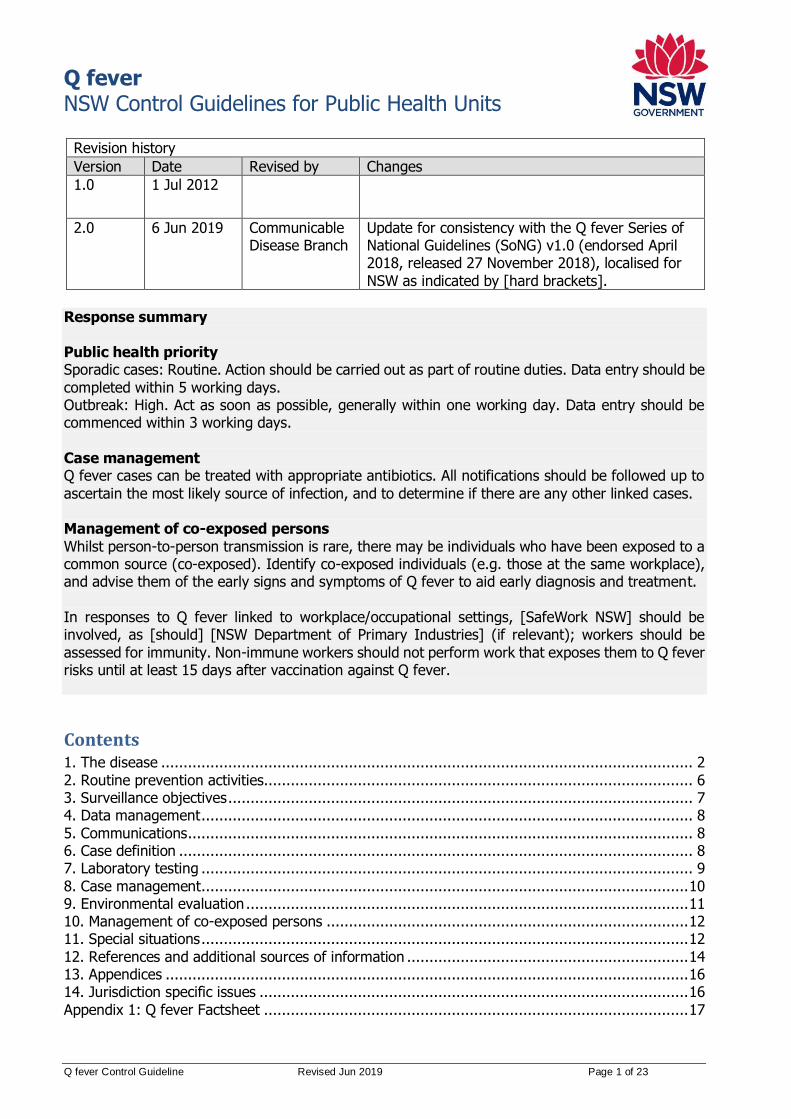
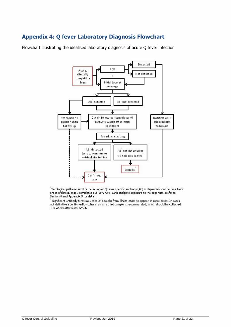
The steps for laboratory diagnosis are illustrated in Appendix 4. Further detail on tests and interpreting results is provided below, as well as on Public Health Laboratory Network (PHLN) laboratory case definitions website.
Consideration should also be given toward other zoonotic diseases based on risk exposures.
PCR testing In acute Q fever cases, the organism may be detected in blood up to 2 weeks after illness onset. If the patient presents within this period, unclotted blood or serum should be submitted for PCR (and possible
culture – see below). Whilst PCR offers a highly sensitive method for detecting both live and dead C. burnetii, a negative result alone does not exclude a Q fever diagnosis, and serological testing should be completed for all cases.
In chronic Q fever cases, C. burnetii DNA may be detected by PCR in peripheral blood mononuclear cells or in biopsy specimens from focally infected tissue (e.g. heart valves, bone, synovium). The sensitivity of
PCR in serum in patients with endocarditis or vascular infection is low to modest, in the order of 23–67%.58
PCR positive with negative serology results confirm acute Q fever, and theoretically convalescent serology is not indicated, however it is useful if serial testing is performed at intervals to screen for chronic infection.
Serology testing Indirect immunofluorescence assay (IFA) is the reference method, but the complement fixation test (CFT) and enzyme immunoassays (EIA) are also used to support diagnoses. For the diagnosis and follow
up of C. burnetii infection, IFA to both phase I and phase II antigens for subclasses IgM, IgG, and IgA is recommended.
It is important to note that single serology tests (EIA, CFT, or IFA) are unable to distinguish between acute, past and chronic infections, and antibody detection is highly dependent on the timing of specimen
Q fever Control Guideline Revised April 2019 Page 10 of 23
collection. Two serum/clotted blood samples should, therefore, always be collected if Q fever is suspected — one at presentation, and another 2–3 weeks later, even if the patient has since recovered.
Seroconversion usually occurs within 7–15 days after exposure, and ninety per cent (90%) of cases have seroconverted by the third week after exposure. Significant antibody titres may take 3–4 weeks from illness onset to appear in some cases. In cases not
definitively confirmed by other means, a third sample is recommended, which should be collected 3–4 weeks after fever onset.
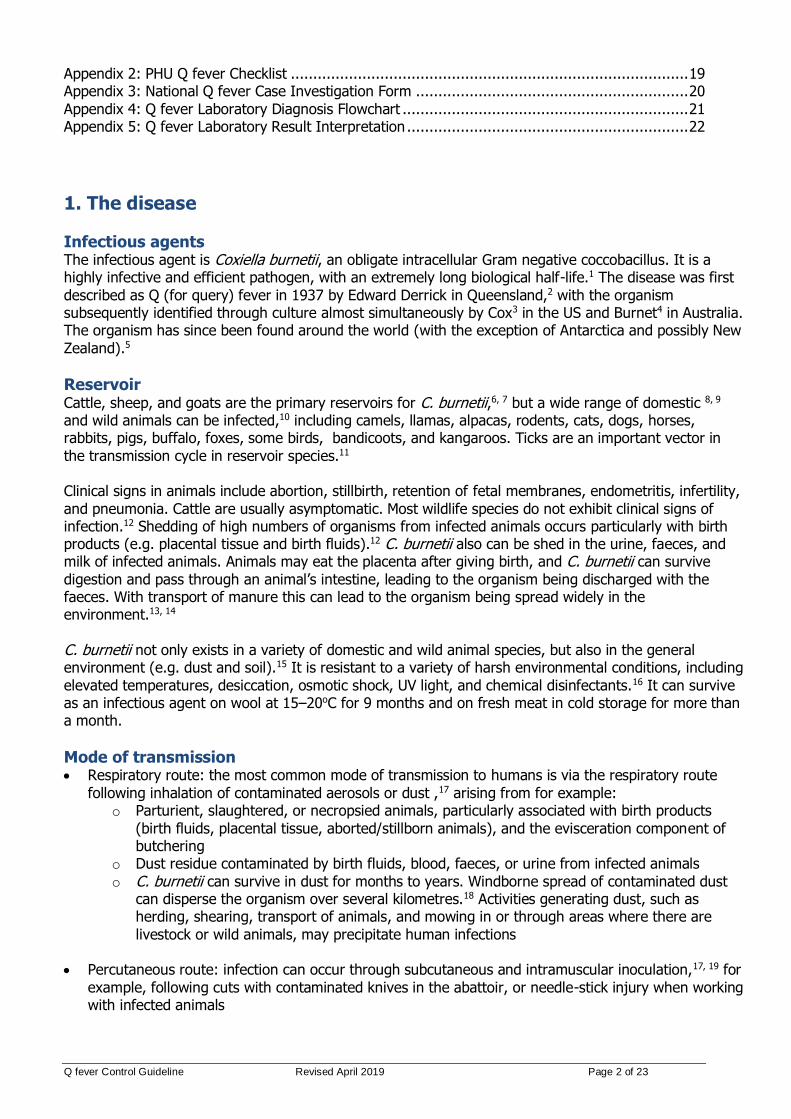
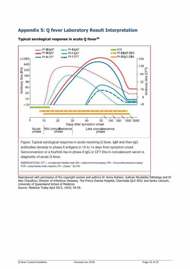
The interpretation of Q fever serology results can be challenging. There are different patterns of antibody response during the course of acute Q fever and chronic Q fever, specifically in terms of antibody subclasses to Phase I and Phase II antigens.19 For example, in acute Q fever cases, IgM to
Phase II antigen tends to rise first, followed by rise of IgG and IgA to Phase II antigen as detected by IFA at various times after the onset of disease.59 In contrast, for chronic Q fever (e.g. endocarditis), IgG, with or without IgA to Phase I antigen, is present at high titre.19, 60 A chart and reference tables are
provided at Appendix 5 to assist in interpreting serology results. Where required, Public Health Units should seek expert guidance in interpreting these results.
Management of acute Q fever includes the measurement of serial antibody titres over time. This can aid in identifying those at risk of developing chronic Q fever, and allow early interventions. It is preferable that confirmed cases have testing undertaken at a laboratory with such capacity.
Culture Isolation of C. burnetii by culture can only be performed by vaccinated staff in an appropriate physical
containment level 3 (PC3) facility.61 For this reason, culture is strongly discouraged except where appropriate facilities and training exist – currently culture is only performed at the Australian Rickettsial Reference Laboratory (ARRL), Geelong, Victoria.
8. Case management
Response times Sporadic cases: Routine response. Initial case investigation and data entry should be completed
within 5 working days. Outbreak: High priority response. The responsible Public Health Unit should act and notify the
jurisdictional Communicable Disease Branch as soon as possible, generally within 1 working day. Data entry should be commenced within 3 working days.
Response procedure Case investigation Public Health Unit staff carry out case investigation in collaboration with the case’s treating doctor and
the case. Information required from treating doctor:
Complete relevant sections of the Q fever Case Investigation Form (Appendix 3) with treating doctor to: Determine clinical details (including onset date, symptoms, acute or chronic presentation,
hospitalisation, and outcome) Determine previous vaccination status Request repeat serology to confirm the diagnosis, if required
Document case treatment and follow-up of those who are at higher risk of chronic Q fever (see Case treatment)
Obtain contact details of the case, and ensure that the case has been informed of their diagnosis and an understanding that a follow up interview will occur.
Q fever Control Guideline Revised April 2019 Page 11 of 23
Case interview and exposure investigation: Complete relevant sections of the Q fever Case Investigation Form (Appendix 3) with the case (or carer)
to: Explore risk factors for infection including occupational risk factors, contact with animals through
farming, hunting or other activities, and environmental exposures
Determine the possible source of infection Confirm vaccination history
Provide education (see below). If the case involves an occupational exposure, see Section 12 for additional actions.
Case treatment Commence empiric treatment if Q fever is clinically suspected. Do not wait for laboratory results. Refer to latest edition of the Therapeutic Guidelines: Antibiotic.
A two week course of oral doxycycline is generally used to treat acute Q fever. Trimethoprim+sulfamethoxazole is recommended for pregnant women until 32 weeks of gestation, even if recovered, to prevent fetal and maternal complications.34
After treatment of C. burnetii primary infection, it is recommended to screen for risk factors of chronic Q fever infection, including pre-existing valvular heart disease/valvular prosthesis, vascular
aneurysms/vascular grafts, and immunosuppression.29 A cardiac assessment, which may include echocardiography, is recommended to assess whether there are underlying abnormalities of the heart valves. Those who are at higher risk of chronic Q fever should be monitored serologically and clinically at
3, 6, 9, 12, 18, and 24 months after acute infection. There is a body of evidence to suggest antibiotic prophylaxis for 12 months in case of cardiac valve problems/valvular prosthesis may be of benefit.29
In chronic disease (e.g. endocarditis), prolonged combination therapy (with addition of hydroxychloroquine) and cardiac surgery may be required. Expert advice from an infectious diseases physician and other specialist physicians should be sought as appropriate.
Education The case should be advised of the nature of the infection and its mode of transmission, and of
appropriate precautions necessary to prevent others from becoming infected from exposure to the same source. The case should also be advised about seeking medical care if symptoms do not resolve following completion of treatment, or new symptoms develop that may indicate a complication or a
chronic Q fever infection (Appendix 1: Q fever Factsheet). Isolation and restriction Exclusion of infected persons is not required. Q fever is rarely transmissible from person to person. Active case finding See Section 12 for active case finding in special situations.
9. Environmental evaluation C. burnetii is highly resistant to desiccation and may remain viable in dust for more than a year. It is killed by heat (>63 degrees Celsius for 30 minutes), and some disinfectants including hydrogen peroxide,
sodium hypochlorite (at concentrations of greater than 5 per cent), and 2 per cent formaldehyde. A 1:100 dilution of household bleach is also an effective solution.29
Appropriate animal and environmental management plays an important role in reducing transmission of C. burnetii to humans. Lessons and experience drawn from Q fever outbreaks in Victoria13 and the
Q fever Control Guideline Revised April 2019 Page 12 of 23
Netherlands,14 which were linked to goat dairy farms, point to effective measures in reducing Q fever risk, including:
Immediate removal of animal abortive and birth materials and safe disposal by deep burial Appropriate treatment of animal manure: no removal of manure from the deep litter sheds or
yards for at least one month after the kidding season; composting manure or alternatively storing manure for three months prior to spreading on farm land for fertiliser
Manure should be covered during storage and transport and must be under-ploughed
immediately when spreading on farm land There is no vaccination available for use in animals in Australia. Where available, vaccination has
been used in female animals prior to their first pregnancy62 to prevent them becoming a source of C. burnetti transmission, and human infection.
10. Management of co-exposed persons
Identification of co-exposed persons In occupational settings and outbreaks, active case finding among identified co-exposed persons should be considered (Section 12). The aim of identifying co-exposed persons is to alert them to the possibility
that they could develop disease due to a common source exposure.
Co-exposure definition A co-exposed person is defined as anyone who may have experienced the same occupational, animal, or environmental exposures as the case, or who may have been exposed to contaminated items associated with the case (e.g. clothing/boots). Person-to-person transmission is extremely unlikely. Co-exposed
persons may include people at the workplace (including those without direct contact with animals or animal products) and home.
Prophylaxis Vaccination during the incubation period does not prevent the disease. Post-exposure antibiotic prophylaxis is not recommended.
Education Q fever information (Appendix 1) should be provided to co-exposed persons with advice to seek medical
attention should they develop symptoms. Q fever vaccination should be recommended to all non-immune workers in high-risk occupations.
Isolation and restriction Those workers/co-exposed persons with the same exposure that do not have immunity to Q fever through natural infection or vaccination should not visit the setting, enter high risk workplaces or
perform work that exposes them to Q fever risks without wearing a properly fitted particulate respirator (e.g. disposable P2 respirator).
11. Special situations In addition to the generic case and co-exposed person follow-up requirements described above, further
actions are required in the following instances:
Cases and outbreaks linked to workplace/occupational settings Q fever case investigation may identify a plausible link with a workplace (such as an abattoir or dairy farm). Two cases or more within a three-month period in an at-risk workplace is considered a workplace outbreak.
Q fever Control Guideline Revised April 2019 Page 13 of 23
Responses to cases occurring in workplace settings (including outbreaks) need to be carried out in collaboration between the Public Health Unit, [SafeWork NSW], and the [NSW Department of Primary
Industries] if relevant. Unvaccinated Public Health Unit staff are not to be exposed to Q fever risks as part of the disease investigation and response. Immediate responses include working with the employer and management to:
Conduct active case finding in the at-risk setting, including urgent testing of workers with a current or recent clinically compatible illness. Laboratory-definitive evidence should be
actively pursued for all suspected cases, including obtaining convalescent sera from ill workers with a single negative serology result (even if they have since recovered)
Assess vaccination status of all workers (if not already known), institute a Q fever
vaccination program urgently if one is not in place, and maintain written records of employee Q fever status and vaccination
Restrict non-vaccinated workers (including not working in a high risk area for Q fever until at least 15 days after vaccination against Q fever); or (if restricting is not possible) provide other interim means of personal protective equipment such as use of a properly fitted
particulate respirator (e.g. disposable P2 respirator). The role of [SafeWork NSW] is to investigate and identify unsafe working conditions, and to monitor and
enforce compliance. This may involve a site visit and discussions with the employer. [SafeWork NSW] may, in consultation with the health department and [NSW Department of Primary Industries], provide information and advice to the employer.
Community clusters/Family clusters The term ‘cluster’ is taken to mean the occurrence of more cases than expected in the community,
where sources of infection are not apparent. For example, the year to date number of Q fever notifications from a region is over 2 standard deviations more than the previous five year mean over the same period.
The goal of community cluster detection is to further explore potential sources of infection and risk factors for Q fever in a broader community context, thereby informing public health action to interrupt
transmission and prevent further cases. Elements of the cluster response include:
In-depth analysis of epidemiological information of cases, e.g. age-specific rates, region-specific rates, timeline and mapping of cases and possible exposure sites and sources. In small populations it may be more useful to focus on the number of cases rather than the rate
Consider environmental and meteorological conditions, such as use of animal manure as fertiliser on farm land, wind direction, and rain patterns in recent weeks, to determine possible high risk
zones. Consider defining 1 km/5 km zones around the potential or identified source Work collaboratively with GPs and hospital emergency departments in the local area, to be alert
for Q fever cases and initiate active case finding
If potential sources of infection are suspected, work collaboratively with relevant organisations (e.g. environmental health, animal health authority, workplace health and safety authority) to
conduct risk assessment and take actions to minimise/eliminate risks Depending on the extent of the cluster, consider liaising with the Australian Red Cross Blood
Service and other appropriate national institutes as blood donation services in cluster locations may need to be restricted
Consider targeted vaccination programs to reduce the risk of disease in groups identified at
higher risk. If a significant outbreak, additional funds may be available to support the vaccination strategy.
Q fever Control Guideline Revised April 2019 Page 14 of 23
12. References and additional sources of information 1. Williams JC. Infectivity, virulence, and pathogenicity of Coxiella burnetii for various hosts. In:
Williams JC, Thompson HA, eds. Q fever: the biology of Coxiella burnetii. Boca Raton, Florida: CRC Press; 1991:21-71.
2. Derrick EH. "Q" fever, a new fever entity: clinical features, diagnosis and laboratory investigation.
Med J Aust. 1937;2:281-99. 3. Davis GE, Cox HR. A filter-passing infectious agent isolated from ticks. Public Health Rep.
1938;53:2259-67.
4. Burnet FM, Freeman M. Experimental studies on the virus of "Q" fever. Med J Aust. 1937;2:299-305.
5. Maurin M, Raoult D. Q fever. Clin Microbiol Rev. 1999;12(4):518-53.
6. Marrie TJR, D. Coxiella burnetii (Q Fever). In: Bennett JED, R.; Blaser, M.J., ed. Mandell, Douglas, and Bennett's Principles and Practice of Infectious Diseases. Philadelphia, PA: Elsevier; 2015:2208-16.
7. Parker NR, Barralet JH, Bell AM. Q fever. Lancet. 2006;367(9511):679-88.
8. Kopecny L, Bosward KL, Shapiro A, Norris JM. Investigating Coxiella burnetii infection in a breeding cattery at the centre of a Q fever outbreak. J Feline Med Surg. 2013;15(12):1037-45.
9. Shapiro AJ, Norris JM, Heller J, Brown G, Malik R, Bosward KL. Seroprevalence of Coxiella burnetii
in Australian dogs. Zoonoses Public Health. 2016;63(6):458-66. 10. Wildlife Health Australia. Q Fever in Australian Wildlife Fact Sheet. 2013. 11. Cooper A, Stephens J, Ketheesan N, Govan B. Detection of Coxiella burnetii DNA in wildlife and
ticks in northern Queensland, Australia. Vector Borne Zoonotic Dis. 2013;13(1):12-6. 12. Heymann DL, ed. Control of Communicable Diseases Manual. 20th ed. Washington: American
Public Health Association; 2015.
13. Bond KA, Vincent G, Wilks CR, Franklin L, Sutton B, Stenos J, et al. One Health approach to controlling a Q fever outbreak on an Australian goat farm. Epidemiol Infect. 2015:1-13.
14. Hermans T, Jeurissen L, Hackert V, Hoebe C. Land-applied goat manure as a source of human Q-
fever in the Netherlands, 2006-2010. PLoS One. 2014;9(5):e96607. 15. Tozer SJ, Lambert SB, Strong CL, Field HE, Sloots TP, Nissen MD. Potential animal and
environmental sources of Q fever infection for humans in Queensland. Zoonoses Public Health. 2014;61(2):105-12.
16. McCaul TF. The developmental cycle of Coxiella burnetii. In: Williams JCT, H.A., ed. Q fever: The Biology of Coxiella burnetii. Boca Raton, FL: CRC Press; 1991:224-58.
17. Raoult D, Marrie T, Mege J. Natural history and pathophysiology of Q fever. Lancet Infect Dis. 2005;5(4):219-26.
18. Tissot-Dupont H, Amadei MA, Nezri M, Raoult D. Wind in November, Q fever in December. Emerg Infect Dis. 2004;10(7):1264-9.
19. Marmion B. A guide to Q fever and Q fever vaccination. Victoria: CSL Biotherapies; 2009. 20. Graves SR, Islam A. Endemic Q Fever in New South Wales, Australia: A Case Series (2005-2013).
Am J Trop Med Hyg. 2016;95(1):55-9. 21. Duron O, Sidi-Boumedine K, Rousset E, Moutailler S, Jourdain E. The Importance of Ticks in Q
Fever Transmission: What Has (and Has Not) Been Demonstrated? Trends Parasitol. 2015;31(11):536-52.
22. Graves S, Jackson C, Hussain-Yusuf H, Vincent G, Nguyen C, Stenos J, et al. Ixodes holocyclus Tick-Transmitted Human Pathogens in North-Eastern New South Wales, Australia. Trop Med Infect Dis. 2016;1(1):4.
23. Pantanowitz L, Telford SR, Cannon ME. Tick-borne diseases in transfusion medicine. Transfus Med. 2002;12(2):85-106.
24. Kanfer E, Farrag N, Price C, MacDonald D, Coleman J, Barrett AJ. Q fever following bone marrow
transplantation. Bone Marrow Transplant. 1988;3(2):165-6. 25. Racult D, Stein A. Q fever during pregnancy--a risk for women, fetuses, and obstetricians. N Engl J
Med. 1994;330(5):371.
26. Milazzo A, Hall R, Storm PA, Harris RJ, Winslow W, Marmion BP. Sexually transmitted Q fever. Clin Infect Dis. 2001;33(3):399-402.
Q fever Control Guideline Revised April 2019 Page 15 of 23
27. Hartzell JD, Wood-Morris RN, Martinez LJ, Trotta RF. Q fever: epidemiology, diagnosis, and treatment. Mayo Clin Proc. 2008;83(5):574-9.
28. Biological and chemical terrorism: strategic plan for preparedness and response. Recommendations of the CDC Strategic Planning Workgroup. MMWR Recomm Rep. 2000;49(RR-4):1-14.
29. Eldin C, Melenotte C, Mediannikov O, Ghigo E, Million M, Edouard S, et al. From Q Fever to Coxiella burnetii Infection: a Paradigm Change. Clin Microbiol Rev. 2017;30(1):115-90.
30. Carcopino X, Raoult D, Bretelle F, Boubli L, Stein A. Managing Q fever during pregnancy: the benefits of long-term cotrimoxazole therapy. Clin Infect Dis. 2007;45(5):548-55.
31. Fenollar F, Fournier PE, Carrieri MP, Habib G, Messana T, Raoult D. Risks factors and prevention of
Q fever endocarditis. Clin Infect Dis. 2001;33(3):312-6. 32. Raoult D, Tissot-Dupont H, Foucault C, Gouvernet J, Fournier PE, Bernit E, et al. Q fever 1985-
1998. Clinical and epidemiologic features of 1,383 infections. Medicine (Baltimore). 2000;79(2):109-23.
33. Morroy G, Keijmel SP, Delsing CE, Bleijenberg G, Langendam M, Timen A, et al. Fatigue following Acute Q-Fever: A Systematic Literature Review. PLoS One. 2016;11(5):e0155884.
34. Anderson A, Bijlmer H, Fournier PE, Graves S, Hartzell J, Kersh GJ, et al. Diagnosis and management of Q fever--United States, 2013: recommendations from CDC and the Q Fever Working Group. MMWR Recomm Rep. 2013;62(RR-03):1-30.
35. Karki S, Gidding HF, Newall AT, McIntyre PB, Liu BC. Risk factors and burden of acute Q fever in older adults in New South Wales: a prospective cohort study. Med J Aust. 2015;203(11):438.
36. Palmer C, McCall B, Jarvinen K, Krause M, Heel K. "The dust hasn't settled yet": the National Q
fever Management Program, missed opportunities for vaccination and community exposures. Aust N Z J Public Health. 2007;31(4):330-2.
37. National Notifiable Diseases Surveillance System 2016;Pages
www9.health.gov.au/cda/source/rpt_4_sel.cfm on 14 January 2016. 38. Gidding HF, Wallace C, Lawrence GL, McIntyre PB. Australia's national Q fever vaccination
program. Vaccine. 2009;27(14):2037-41.
39. EFSA (European Food Safety Authority) and ECDC (European Centre for Disease Prevention and Control). The European Union summary report on trends and sources of zoonoses, zoonotic agents and food-borne outbreaks in 2014. EFSA Journal. 2015;13(12):191.
40. Centers for Disease Control and Prevention (CDC);Pages www.cdc.gov/qfever/stats/index.html on 13 March 2016.
41. Delsing CE, Kullberg BJ, Bleeker-Rovers CP. Q fever in the Netherlands from 2007 to 2010. Neth J Med. 2010;68(12):382-7.
42. Kampschreur LM, Delsing CE, Groenwold RH, Wegdam-Blans MC, Bleeker-Rovers CP, de Jager-Leclercq MG, et al. Chronic Q fever in the Netherlands 5 years after the start of the Q fever epidemic: results from the Dutch chronic Q fever database. J Clin Microbiol. 2014;52(5):1637-43.
43. EFSA (European Food Safety Authority) and ECDC (European Centre for Disease Prevention and Control). The European Union Summary Report on Trends and Sources of Zoonoses, Zoonotic Agents and Food-borne Outbreaks in 2009. EFSA Journal. 2011;9(3):378.
44. Buckley B. Q fever epidemic in Victorian general practice. Med J Aust. 1980;1(12):593-5. 45. Q fever outbreak in an abattoir in Cooma, NSW. Commun Dis Intell. 1998;22(10):222. 46. Gilroy N, Formica N, Beers M, Egan A, Conaty S, Marmion B. Abattoir-associated Q fever: a Q fever
outbreak during a Q fever vaccination program. Aust N Z J Public Health. 2001;25(4):362-7. 47. Miller M, Roche P, Yohannes K, Spencer J, Bartlett M, Brotherton J, et al. Australia's notifiable
diseases status, 2003 annual report of the National Notifiable Diseases Surveillance System.
Commun Dis Intell Q Rep. 2005;29(1):1-61. 48. O'Connor BA, Tribe IG, Givney R. A windy day in a sheep saleyard: an outbreak of Q fever in rural
South Australia. Epidemiol Infect. 2015;143(2):391-8.
49. Wade AJ, Cheng AC, Athan E, Molloy JL, Harris OC, Stenos J, et al. Q fever outbreak at a cosmetics supply factory. Clin Infect Dis. 2006;42(7):e50-2.
50. Q FEVER - AUSTRALIA (SOUTH AUSTRALIA) (03): FATALITY. 2007.
51. Gullan LN, Cowie BC, Franklin LJ. Outbreak of Q fever related to changed farming practices, Victoria, Australia. Victorian Infectious Diseases Bulletin. 2013;16:15-9.
Q fever Control Guideline Revised April 2019 Page 16 of 23
52. Gibbons GC, White PJ. 2012;Pages www.asid.net.au/documents/item/216. 53. Archer BN, Hallahan C, Stanley P, Seward K, Lesjak M, Hope K, et al. Atypical outbreak of Q fever
affecting low-risk residents of a remote rural town in New South Wales. Commun Dis Intell Q Rep. 2017;41(2):E125-E33.
54. Australian Government Department of Health. The Australian Immunisation Handbook. Canberra: Australian Government Department of Health; 2013.
55. Workplace Health and Safety Queensland 2015;Pages www.worksafe.qld.gov.au/agriculture/workplace-hazards/diseases-from-animals/q-fever.
56. SafeWork NSW 2016;Pages www.safework.nsw.gov.au/health-and-safety/safety-topics-a-
z/diseases/q-fever. 57. Schimmer B, Ter Schegget R, Wegdam M, Zuchner L, de Bruin A, Schneeberger PM, et al. The use
of a geographic information system to identify a dairy goat farm as the most likely source of an
urban Q-fever outbreak. BMC Infect Dis. 2010;10:69. 58. Wegdam-Blans MC, Kampschreur LM, Delsing CE, Bleeker-Rovers CP, Sprong T, van Kasteren ME,
et al. Chronic Q fever: review of the literature and a proposal of new diagnostic criteria. J Infect.
2012;64(3):247-59. 59. Chaudhuri A, Robson J. Q fever queries and answers. Medicine Today. 2013;14(4):54-9. 60. Fournier PE, Marrie TJ, Raoult D. Diagnosis of Q fever. J Clin Microbiol. 1998;36(7):1823-34.
61. Standards Australia Limited/Standards New Zealand. Australian/New Zealand Standard. Safety in laboratories Part 3: Microbiological safety and containment AS/NZS 2243.3:2010. 2010.
62. Hogerwerf L, van den Brom R, Roest HI, Bouma A, Vellema P, Pieterse M, et al. Reduction of
Coxiella burnetii prevalence by vaccination of goats and sheep, The Netherlands. Emerg Infect Dis. 2011;17(3):379-86.
13. Appendices Appendix 1: Q fever Factsheet Appendix 2: PHU Q fever Checklist
Appendix 3: National Q fever Case Investigation Form Appendix 4: Q fever Laboratory Diagnosis Flowchart Appendix 5: Q fever Laboratory Result Interpretation
14. Jurisdiction specific issues Links to State and Territory Public Health Legislation, the Biosecurity Act 2015 and the National Health Security Act 2007. www.health.gov.au/internet/main/publishing.nsf/Content/cda-state-legislation-links.htm
Q fever Control Guideline Revised April 2019 Page 17 of 23
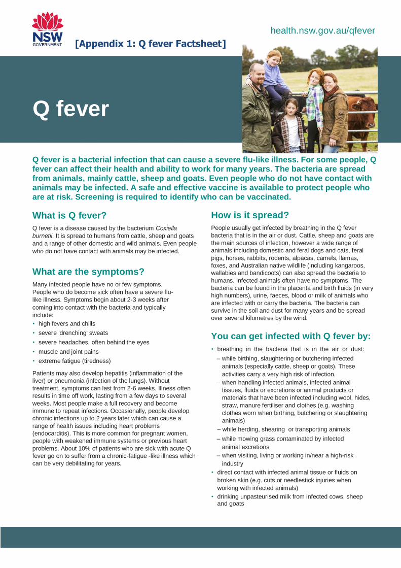
Q fever Q fever is a bacterial infection that can cause a severe flu-like illness. For some people, Q fever can affect their health and ability to work for many years. The bacteria are spread from animals, mainly cattle, sheep and goats. Even people who do not have contact with animals may be infected. A safe and effective vaccine is available to protect people who are at risk. Screening is required to identify who can be vaccinated.
What is Q fever? Q fever is a disease caused by the bacterium Coxiella
burnetii. It is spread to humans from cattle, sheep and goats
and a range of other domestic and wild animals. Even people
who do not have contact with animals may be infected.
What are the symptoms? Many infected people have no or few symptoms.
People who do become sick often have a severe flu-
like illness. Symptoms begin about 2-3 weeks after
coming into contact with the bacteria and typically
include: • high fevers and chills • severe 'drenching' sweats • severe headaches, often behind the eyes • muscle and joint pains • extreme fatigue (tiredness) Patients may also develop hepatitis (inflammation of the
liver) or pneumonia (infection of the lungs). Without
treatment, symptoms can last from 2-6 weeks. Illness often
results in time off work, lasting from a few days to several
weeks. Most people make a full recovery and become
immune to repeat infections. Occasionally, people develop
chronic infections up to 2 years later which can cause a
range of health issues including heart problems
(endocarditis). This is more common for pregnant women,
people with weakened immune systems or previous heart
problems. About 10% of patients who are sick with acute Q
fever go on to suffer from a chronic-fatigue -like illness which
can be very debilitating for years.
How is it spread? People usually get infected by breathing in the Q fever
bacteria that is in the air or dust. Cattle, sheep and goats are
the main sources of infection, however a wide range of
animals including domestic and feral dogs and cats, feral
pigs, horses, rabbits, rodents, alpacas, camels, llamas,
foxes, and Australian native wildlife (including kangaroos,
wallabies and bandicoots) can also spread the bacteria to
humans. Infected animals often have no symptoms. The
bacteria can be found in the placenta and birth fluids (in very
high numbers), urine, faeces, blood or milk of animals who
are infected with or carry the bacteria. The bacteria can
survive in the soil and dust for many years and be spread
over several kilometres by the wind.
You can get infected with Q fever by: • breathing in the bacteria that is in the air or dust: – while birthing, slaughtering or butchering infected
animals (especially cattle, sheep or goats). These
activities carry a very high risk of infection. – when handling infected animals, infected animal
tissues, fluids or excretions or animal products or
materials that have been infected including wool, hides,
straw, manure fertiliser and clothes (e.g. washing
clothes worn when birthing, butchering or slaughtering
animals) – while herding, shearing or transporting animals
– while mowing grass contaminated by infected
animal excretions – when visiting, living or working in/near a high-risk
industry • direct contact with infected animal tissue or fluids on
broken skin (e.g. cuts or needlestick injuries when
working with infected animals) • drinking unpasteurised milk from infected cows, sheep
and goats2018
[Appendix 1: Q fever Factsheet]
health.nsw.gov.au/qfever
Q fever Control Guideline Revised April 2019 Page 18 of 23
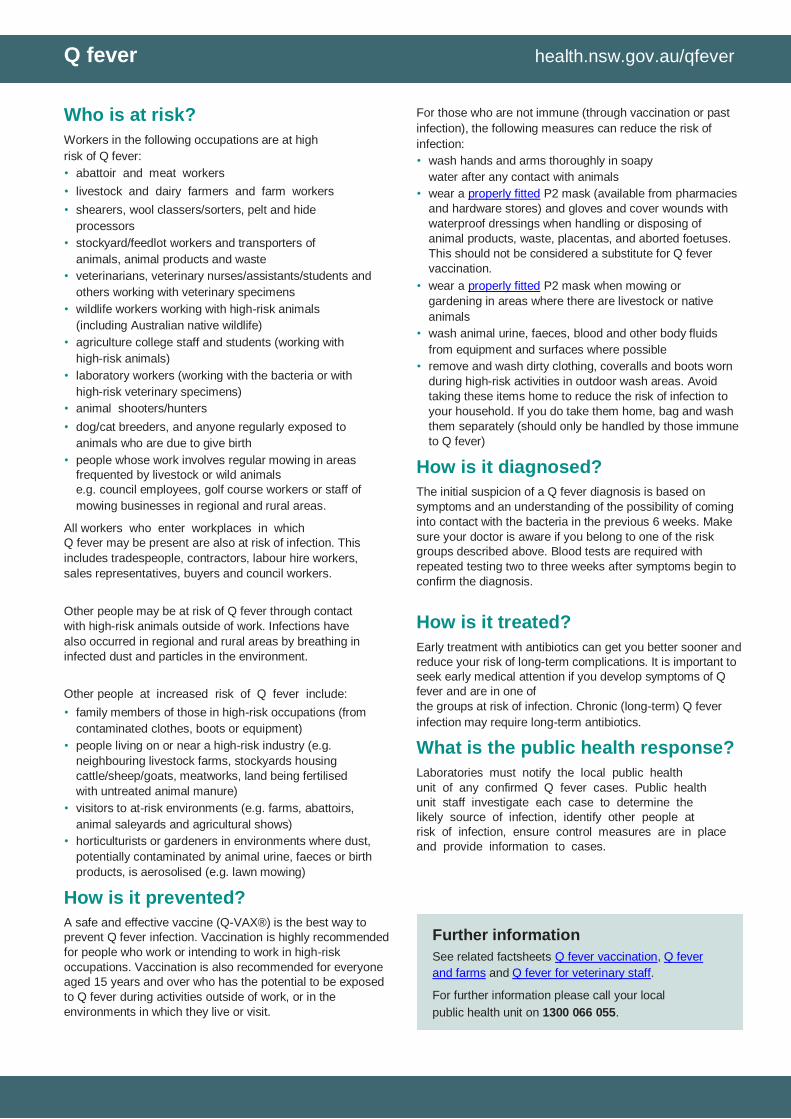
Q fever health.nsw.gov.au/qfever
Who is at risk? Workers in the following occupations are at high
risk of Q fever: • abattoir and meat workers • livestock and dairy farmers and farm workers • shearers, wool classers/sorters, pelt and hide
processors • stockyard/feedlot workers and transporters of
animals, animal products and waste • veterinarians, veterinary nurses/assistants/students and
others working with veterinary specimens • wildlife workers working with high-risk animals
(including Australian native wildlife) • agriculture college staff and students (working with
high-risk animals) • laboratory workers (working with the bacteria or with
high-risk veterinary specimens) • animal shooters/hunters • dog/cat breeders, and anyone regularly exposed to
animals who are due to give birth • people whose work involves regular mowing in areas
frequented by livestock or wild animals
e.g. council employees, golf course workers or staff of
mowing businesses in regional and rural areas. All workers who enter workplaces in which Q fever may be present are also at risk of infection. This
includes tradespeople, contractors, labour hire workers,
sales representatives, buyers and council workers.
Other people may be at risk of Q fever through contact
with high-risk animals outside of work. Infections have
also occurred in regional and rural areas by breathing in
infected dust and particles in the environment.
Other people at increased risk of Q fever include: • family members of those in high-risk occupations (from
contaminated clothes, boots or equipment) • people living on or near a high-risk industry (e.g.
neighbouring livestock farms, stockyards housing
cattle/sheep/goats, meatworks, land being fertilised
with untreated animal manure) • visitors to at-risk environments (e.g. farms, abattoirs,
animal saleyards and agricultural shows) • horticulturists or gardeners in environments where dust,
potentially contaminated by animal urine, faeces or birth
products, is aerosolised (e.g. lawn mowing)
How is it prevented? A safe and effective vaccine (Q-VAX®) is the best way to
prevent Q fever infection. Vaccination is highly recommended
for people who work or intending to work in high-risk
occupations. Vaccination is also recommended for everyone
aged 15 years and over who has the potential to be exposed
to Q fever during activities outside of work, or in the
environments in which they live or visit.
For those who are not immune (through vaccination or past
infection), the following measures can reduce the risk of
infection: • wash hands and arms thoroughly in soapy
water after any contact with animals • wear a properly fitted P2 mask (available from pharmacies
and hardware stores) and gloves and cover wounds with
waterproof dressings when handling or disposing of
animal products, waste, placentas, and aborted foetuses.
This should not be considered a substitute for Q fever
vaccination. • wear a properly fitted P2 mask when mowing or
gardening in areas where there are livestock or native
animals • wash animal urine, faeces, blood and other body fluids
from equipment and surfaces where possible • remove and wash dirty clothing, coveralls and boots worn
during high-risk activities in outdoor wash areas. Avoid
taking these items home to reduce the risk of infection to
your household. If you do take them home, bag and wash
them separately (should only be handled by those immune
to Q fever)
How is it diagnosed? The initial suspicion of a Q fever diagnosis is based on
symptoms and an understanding of the possibility of coming
into contact with the bacteria in the previous 6 weeks. Make
sure your doctor is aware if you belong to one of the risk
groups described above. Blood tests are required with
repeated testing two to three weeks after symptoms begin to
confirm the diagnosis.
How is it treated? Early treatment with antibiotics can get you better sooner and
reduce your risk of long-term complications. It is important to
seek early medical attention if you develop symptoms of Q
fever and are in one of the groups at risk of infection. Chronic (long-term) Q fever
infection may require long-term antibiotics.
What is the public health response? Laboratories must notify the local public health unit of any confirmed Q fever cases. Public health unit staff investigate each case to determine the likely source of infection, identify other people at risk of infection, ensure control measures are in place and provide information to cases.
Further information
See related factsheets Q fever vaccination, Q fever
and farms and Q fever for veterinary staff.
For further information please call your local
public health unit on 1300 066 055.
Q fever Control Guideline Revised Jun 2019 Page 19 of 23
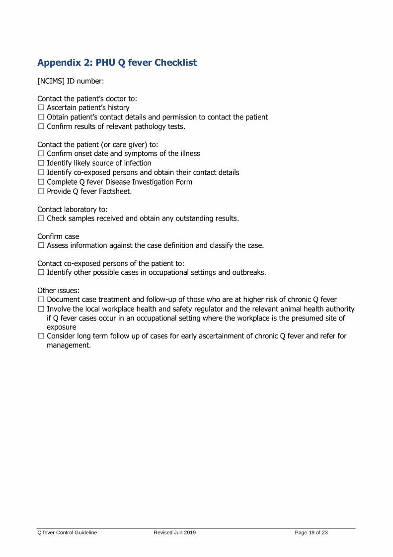
Appendix 2: PHU Q fever Checklist [NCIMS] ID number:
Contact the patient’s doctor to:
☐ Ascertain patient’s history
☐ Obtain patient’s contact details and permission to contact the patient
☐ Confirm results of relevant pathology tests.
Contact the patient (or care giver) to:
☐ Confirm onset date and symptoms of the illness
☐ Identify likely source of infection
☐ Identify co-exposed persons and obtain their contact details
☐ Complete Q fever Disease Investigation Form
☐ Provide Q fever Factsheet.
Contact laboratory to:
☐ Check samples received and obtain any outstanding results.
Confirm case
☐ Assess information against the case definition and classify the case.
Contact co-exposed persons of the patient to:
☐ Identify other possible cases in occupational settings and outbreaks.
Other issues:
☐ Document case treatment and follow-up of those who are at higher risk of chronic Q fever
☐ Involve the local workplace health and safety regulator and the relevant animal health authority
if Q fever cases occur in an occupational setting where the workplace is the presumed site of
exposure
☐ Consider long term follow up of cases for early ascertainment of chronic Q fever and refer for
management.
Q fever Control Guideline Revised Jun 2019 Page 20 of 23
Appendix 3: Q fever Case Investigation Form
[TBA - pending finalisation]
Q fever Control Guideline Revised Jun 2019 Page 21 of 23
Appendix 4: Q fever Laboratory Diagnosis Flowchart
Flowchart illustrating the idealised laboratory diagnosis of acute Q fever infection
Q fever Control Guideline Revised Jun 2019 Page 22 of 23
Appendix 5: Q fever Laboratory Result Interpretation Typical serological response in acute Q fever59
Reproduced with permission of the copyright owners and authors Dr Jenny Robson, Sullivan Nicolaides Pathology and Dr Alex Chaudhuri, Director of Infectious Diseases, The Prince Charles Hospital, Chermside QLD 4032 and Senior Lecturer, University of Queensland School of Medicine. Source: Medicine Today April 2013; 14(4): 54-59.
Q fever Control Guideline Revised Jun 2019 Page 23 of 23
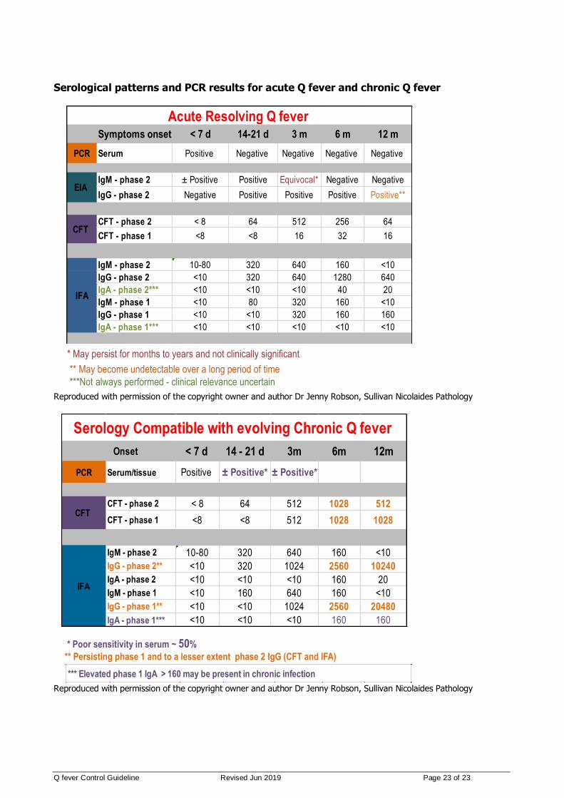
Serological patterns and PCR results for acute Q fever and chronic Q fever
Reproduced with permission of the copyright owner and author Dr Jenny Robson, Sullivan Nicolaides Pathology
Reproduced with permission of the copyright owner and author Dr Jenny Robson, Sullivan Nicolaides Pathology
Symptoms onset < 7 d 14-21 d 3 m 6 m 12 m
PCR Serum Positive Negative Negative Negative Negative
IgM - phase 2 ± Positive Positive Equivocal* Negative Negative
IgG - phase 2 Negative Positive Positive Positive Positive**
CFT - phase 2 < 8 64 512 256 64
CFT - phase 1 <8 <8 16 32 16
IgM - phase 2 10-80 320 640 160 <10
IgG - phase 2 <10 320 640 1280 640
IgA - phase 2*** <10 <10 <10 40 20
IgM - phase 1 <10 80 320 160 <10
IgG - phase 1 <10 <10 320 160 160
IgA - phase 1*** <10 <10 <10 <10 <10
** May become undetectable over a long period of time
***Not always performed - clinical relevance uncertain
Acute Resolving Q fever
EIA
CFT
IFA
* May persist for months to years and not clinically significant * May persist for months to years and not clinically significant
** May become undetectable over a long period of time
***Not always performed - clinical relevance uncertain
* Poor sensitivity in serum ~ 50%
** Persisting phase 1 and to a lesser extent phase 2 IgG (CFT and IFA)
*** Elevated phase 1 IgA > 160 may be present in chronic infection
Onset < 7 d 14 - 21 d 3m 6m 12m
PCR Serum/tissue Positive ± Positive* ± Positive*
CFT - phase 2 < 8 64 512 1028 512
CFT - phase 1 <8 <8 512 1028 1028
IgM - phase 2 10-80 320 640 160 <10
IgG - phase 2** <10 320 1024 2560 10240
IgA - phase 2 <10 <10 <10 160 20
IgM - phase 1 <10 160 640 160 <10
IgG - phase 1** <10 <10 1024 2560 20480
IgA - phase 1*** <10 <10 <10 160 160
Serology Compatible with evolving Chronic Q fever
CFT
IFA