Embed Size (px)
Citation preview
PyMOL Tutorial Notes
What is a PDB file?
Go through the Ubiquitin pdb file: 1UBQ.pdb https://bit.ly/2o5uqqa (link to slides showing 1UBQ.pdb) “The primary information stored in the PDB archive consists of coordinate files for biological molecules. These files list the atoms in each protein, and their 3D location in space. These files are available in several formats (PDB, mmCIF, XML). A typical PDB formatted file includes a large "header" section of text that summarizes the protein, citation information, and the details of the structure solution, followed by the sequence and a long list of the atoms and their coordinates. The archive also contains the experimental observations that are used to determine these atomic coordinates.” (https://pdb101.rcsb.org/learn/guide-to-understanding-pdb-data/introduction )
Basic PyMOL Controls
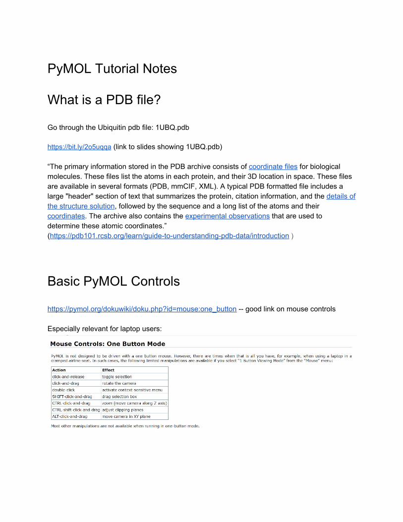
https://pymol.org/dokuwiki/doku.php?id=mouse:one_button -- good link on mouse controls Especially relevant for laptop users:
Examining basic PyMOL controls with 3BTA.pdb
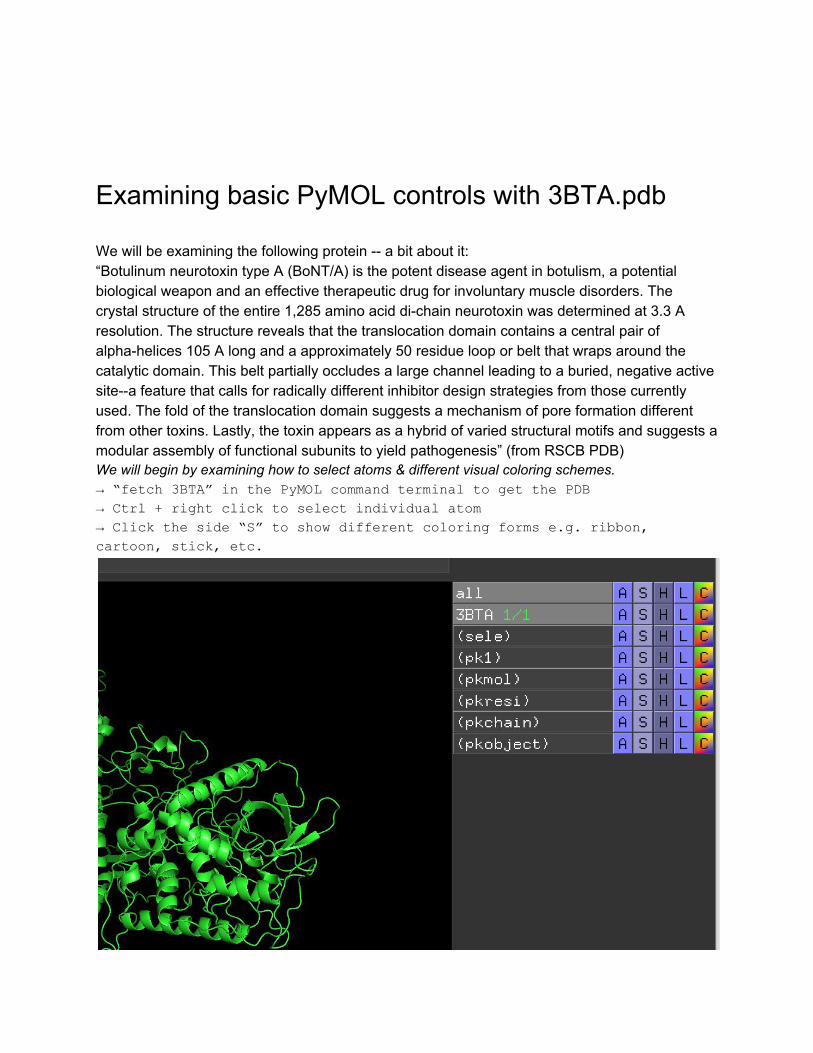
We will be examining the following protein -- a bit about it: “Botulinum neurotoxin type A (BoNT/A) is the potent disease agent in botulism, a potential biological weapon and an effective therapeutic drug for involuntary muscle disorders. The crystal structure of the entire 1,285 amino acid di-chain neurotoxin was determined at 3.3 A resolution. The structure reveals that the translocation domain contains a central pair of alpha-helices 105 A long and a approximately 50 residue loop or belt that wraps around the catalytic domain. This belt partially occludes a large channel leading to a buried, negative active site--a feature that calls for radically different inhibitor design strategies from those currently used. The fold of the translocation domain suggests a mechanism of pore formation different from other toxins. Lastly, the toxin appears as a hybrid of varied structural motifs and suggests a modular assembly of functional subunits to yield pathogenesis” (from RSCB PDB) We will begin by examining how to select atoms & different visual coloring schemes. → “fetch 3BTA” in the PyMOL command terminal to get the PDB
→ Ctrl + right click to select individual atom
→ Click the side “S” to show different coloring forms e.g. ribbon,
cartoon, stick, etc.
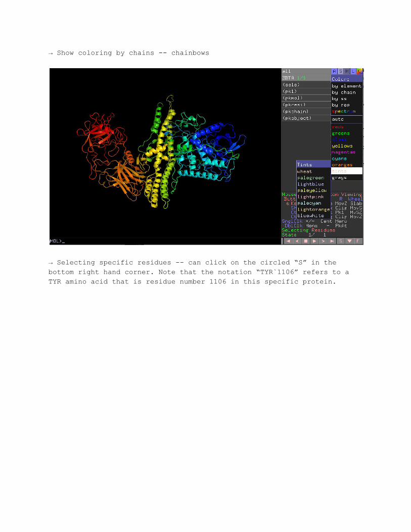
→ Show coloring by chains -- chainbows
→ Selecting specific residues -- can click on the circled “S” in the
bottom right hand corner. Note that the notation “TYR`1106” refers to a
TYR amino acid that is residue number 1106 in this specific protein.
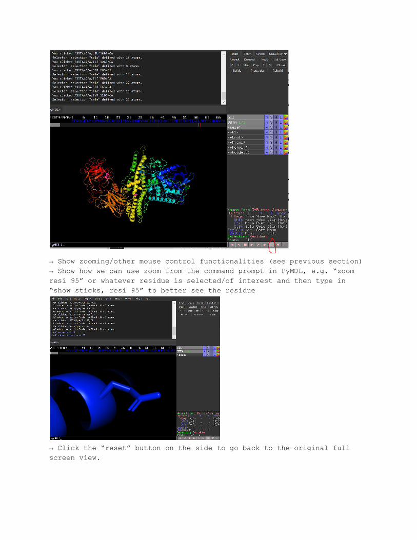
→ Show zooming/other mouse control functionalities (see previous section)
→ Show how we can use zoom from the command prompt in PyMOL, e.g. “zoom
resi 95” or whatever residue is selected/of interest and then type in
“show sticks, resi 95” to better see the residue
→ Click the “reset” button on the side to go back to the original full
screen view.
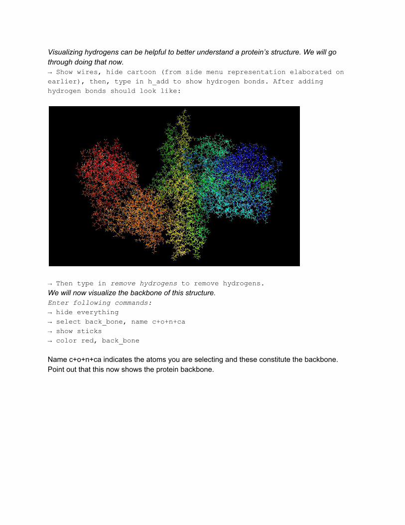
Visualizing hydrogens can be helpful to better understand a protein’s structure. We will go through doing that now. → Show wires, hide cartoon (from side menu representation elaborated on
earlier), then, type in h_add to show hydrogen bonds. After adding
hydrogen bonds should look like:
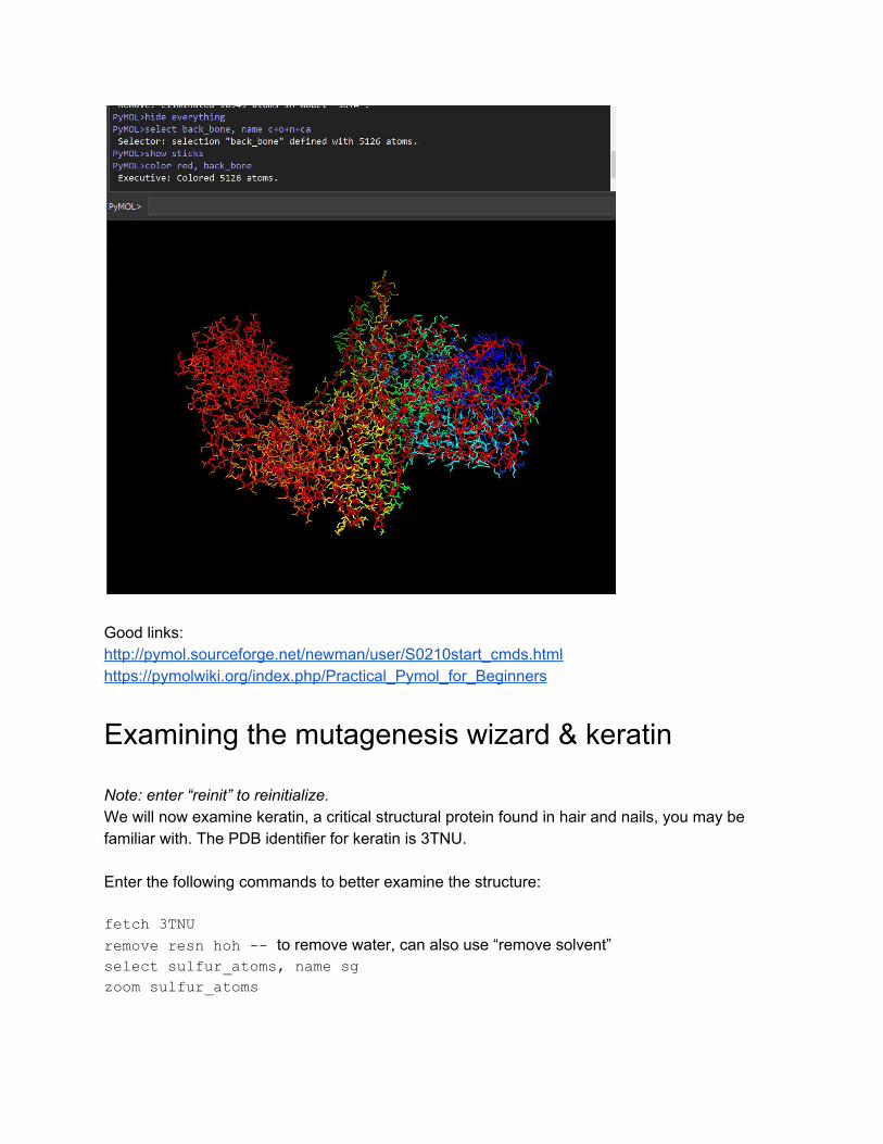
→ Then type in remove hydrogens to remove hydrogens. We will now visualize the backbone of this structure. Enter following commands:
→ hide everything
→ select back_bone, name c+o+n+ca
→ show sticks
→ color red, back_bone
Name c+o+n+ca indicates the atoms you are selecting and these constitute the backbone. Point out that this now shows the protein backbone.
Good links: http://pymol.sourceforge.net/newman/user/S0210start_cmds.html https://pymolwiki.org/index.php/Practical_Pymol_for_Beginners
Examining the mutagenesis wizard & keratin
Note: enter “reinit” to reinitialize. We will now examine keratin, a critical structural protein found in hair and nails, you may be familiar with. The PDB identifier for keratin is 3TNU. Enter the following commands to better examine the structure:
fetch 3TNU
remove resn hoh -- to remove water, can also use “remove solvent” select sulfur_atoms, name sg
zoom sulfur_atoms
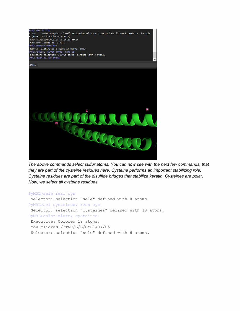
The above commands select sulfur atoms. You can now see with the next few commands, that they are part of the cysteine residues here. Cysteine performs an important stabilizing role; Cysteine residues are part of the disulfide bridges that stabilize keratin. Cysteines are polar. Now, we select all cysteine residues.
PyMOL>sele resi cys
Selector: selection "sele" defined with 0 atoms.
PyMOL>sel cysteines, resn cys
Selector: selection "cysteines" defined with 18 atoms.
PyMOL>color slate, cysteines
Executive: Colored 18 atoms.
You clicked /3TNU/B/B/CYS`407/CA
Selector: selection "sele" defined with 6 atoms.
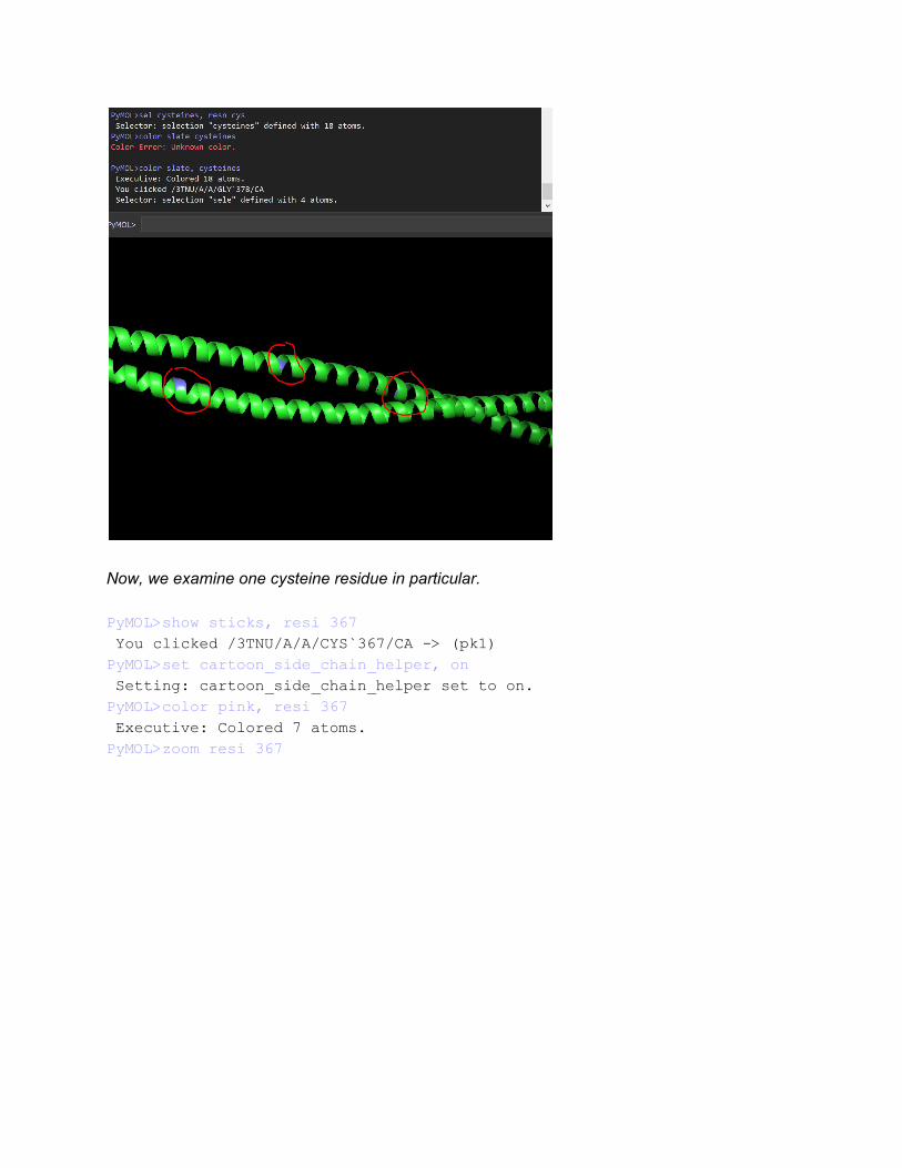
Now, we examine one cysteine residue in particular. PyMOL>show sticks, resi 367
You clicked /3TNU/A/A/CYS`367/CA -> (pk1)
PyMOL>set cartoon_side_chain_helper, on
Setting: cartoon_side_chain_helper set to on.
PyMOL>color pink, resi 367
Executive: Colored 7 atoms.
PyMOL>zoom resi 367
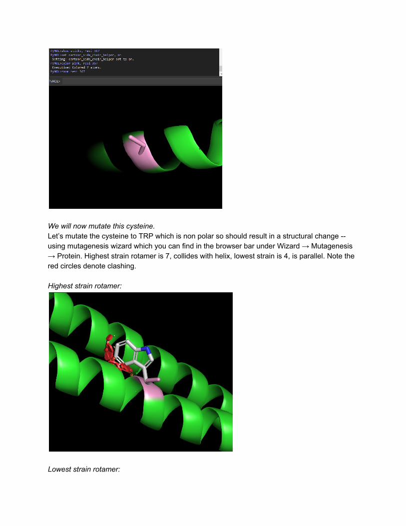
We will now mutate this cysteine. Let’s mutate the cysteine to TRP which is non polar so should result in a structural change -- using mutagenesis wizard which you can find in the browser bar under Wizard → Mutagenesis → Protein. Highest strain rotamer is 7, collides with helix, lowest strain is 4, is parallel. Note the red circles denote clashing. Highest strain rotamer:
Lowest strain rotamer:
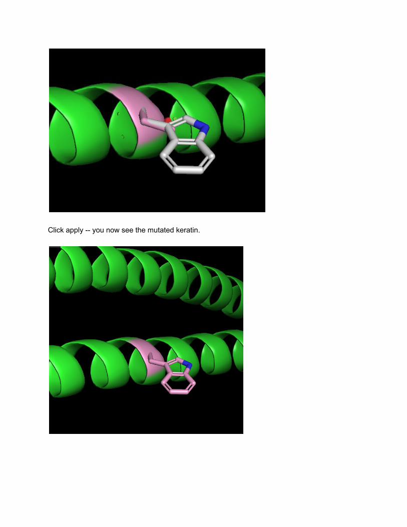
Click apply -- you now see the mutated keratin.
Examining ricin, a toxin found in castor beans http://pdb101.rcsb.org/motm/161 -- we will run through this. You can: → fetch 2aai → remove solvent → note parts “A” and “B” as identified in the article -- how would you use PyMOL to visualize them differently using what we have learned? < can use the shift + click + drag to select A and the color command to color> → visualize the disulfides as spheres with the GUI