Embed Size (px)
Citation preview
Purification of hepatitis B surface antigen virus-like particles from recombinant Pichia pastoris and
in vivo analysis of their immunogenic properties.
Item Type Article
Authors Gurramkonda, Chandrasekhar; Zahid, Maria; Nemani, SatishKumar; Adnan, Ahmad; Gudi, Satheesh Kumar; Khanna, Navin;Ebensen, Thomas; Lünsdorf, Heinrich; Guzmán, Carlos A; Rinas,Ursula
Citation Purification of hepatitis B surface antigen virus-like particlesfrom recombinant Pichia pastoris and in vivo analysis of theirimmunogenic properties. 2013, 940:104-11 J. Chromatogr. BAnalyt. Technol. Biomed. Life Sci.
DOI 10.1016/j.jchromb.2013.09.030
Journal Journal of chromatography. B, Analytical technologies in thebiomedical and life sciences
Rights Archived with thanks to Journal of chromatography. B, Analyticaltechnologies in the biomedical and life sciences
Download date 15/02/2022 04:32:32
Link to Item http://hdl.handle.net/10033/305192
This is a pre- or post-print of an article published inGurramkonda, C., Zahid, M., Nemani, S.K., Adnan, A., Gudi, S.K., Khanna, N., Ebensen, T., Lünsdorf, H.,
Guzman, C.A., Rinas, U.Purification of hepatitis B surface antigen virus-like particles from recombinant Pichia pastoris and in vivo
analysis of their immunogenic properties.Journal of Chromatography B, Volume 940, 1 December 2013,
Pages 104–111.
1
Purification of hepatitis B surface antigen virus-like 1
particles from recombinant Pichia pastoris and in 2
vivo analysis of their immunogenic properties 3
4
Chandrasekhar Gurramkonda 1,2,3*
, Maria Zahid 4, Satish Kumar Nemani
4, Ahmad 5
Adnan 1,5
, Satheesh Kumar Gudi 2, Navin Khanna
2, Thomas Ebensen
6, Heinrich 6
Lünsdorf 6, Carlos A. Guzmán
6, and Ursula Rinas
1,4* 7
8 1
Department of Structural Biology, Helmholtz Centre for Infection Research, 9
Braunschweig, Germany 10 2 International Centre for Genetic Engineering and Biotechnology, New Delhi, India 11
3 Current address: Technology Research Centre, Department of Chemical, 12
Biochemical and Environmental Engineering, University of Maryland Baltimore 13
County, Baltimore, USA 14 4
Institute of Technical Chemistry – Life Sciences, University of Hannover, 15
Hannover, Germany 16 5
Current address: Department of Chemistry, GC University Lahore, Pakistan 17 6
Department of Vaccinology and Applied Microbiology, Helmholtz Centre for 18
Infection Research, Braunschweig, Germany 19
20
21
22
23
24
25
26
27
*Corresponding authors 28 [email protected]; [email protected] (CG) 29
[email protected] (UR) 30
31
32
2
Abstract 33
Following earlier studies on high-level intracellular production of hepatitis B surface 34
antigen (HBsAg) using recombinant Pichia pastoris, we present here in detail an 35
enhanced method for the purification of recombinant HBsAg virus-like particles 36
(VLPs). We have screened various detergents for their ability to promote the 37
solubilization of recombinant intracellular HBsAg. In addition, we have analyzed the 38
effect of cell disruption and extraction regarding their impact on the release of 39
HBsAg. Our results show that introduction of the mild nonionic detergent Tween 20 40
in the initial process of cell lysis at ~ 600 bars by high pressure homogenization lead 41
to the best results. The subsequent purification steps involved polyethylene glycol 42
precipitation of host cell contaminants, hydrophobic adsorption of HBsAg to 43
colloidal silica followed by ion-exchange chromatography and either isopycnic 44
density ultracentrifugation or size exclusion chromatography for the recovery of the 45
VLPs. After final KSCN treatment and dialysis, a total yield of ~3% with a purity of 46
>99% was reached. The pure protein was characterized by electron microscopy, 47
showing the presence of uniform VLPs which are the pre-requisite for 48
immunogenicity. The intramuscular co-administration of HBsAg VLPs, with either 49
alum or a PEGylated-derivative of the toll-like receptor 2/6 agonist MALP-2, to mice 50
resulted in the elicitation of significantly higher HBsAg-specific IgG titers as well as 51
a stronger cellular immune response compared to mice vaccinated with a gold 52
standard vaccine (EngerixTM
). These results show that P. pastoris derived HBsAg 53
VLPs exhibit a high potential as a superior biosimilar vaccine against hepatitis B. 54
Keywords 55
Hepatitis B surface antigen virus-like particles; Aerosil-380; Ion-exchange 56
chromatography; Ultracentrifugation; Size-exclusion chromatography; Electron 57
microscopy; Adjuvant; Vaccine 58
3
1. Introduction 59
The development of a safe recombinant hepatitis B vaccine has led to the inclusion 60
of hepatitis B vaccination in the national infant immunization schedules of 61
approximately 160 countries [1]. Recombinant DNA technology was used to produce 62
hepatitis B surface antigen (HBsAg) in form of virus-like particles (VLPs) using the 63
yeast Saccharomyces cerevisiae leading to the development of a so-called “second” 64
generation hepatitis B vaccine and the first recombinant subunit vaccine available 65
[2]. This formulation of the hepatitis B vaccine has been on the market since 1986. 66
Initially, HBsAg VLPs of ~22 nm were purified from the plasma of asymptomatic 67
HBV carriers, but due to safety issues and restricted supply, the “first” generation 68
plasma-derived vaccines are no longer in use [2]. Nowadays, as patents have expired, 69
“third” generation “biosimilar” recombinant HBsAg VLP-based vaccines are being 70
introduced into the market by a variety of new manufacturers which try to make the 71
vaccine also more affordable to developing countries [2]. 72
As HBsAg is a very hydrophobic protein, secretion is inefficient in yeast and 73
high-level production has been only achieved as intracellular product. The 74
purification of recombinant HBsAg from yeast cultures is well documented [3-25], 75
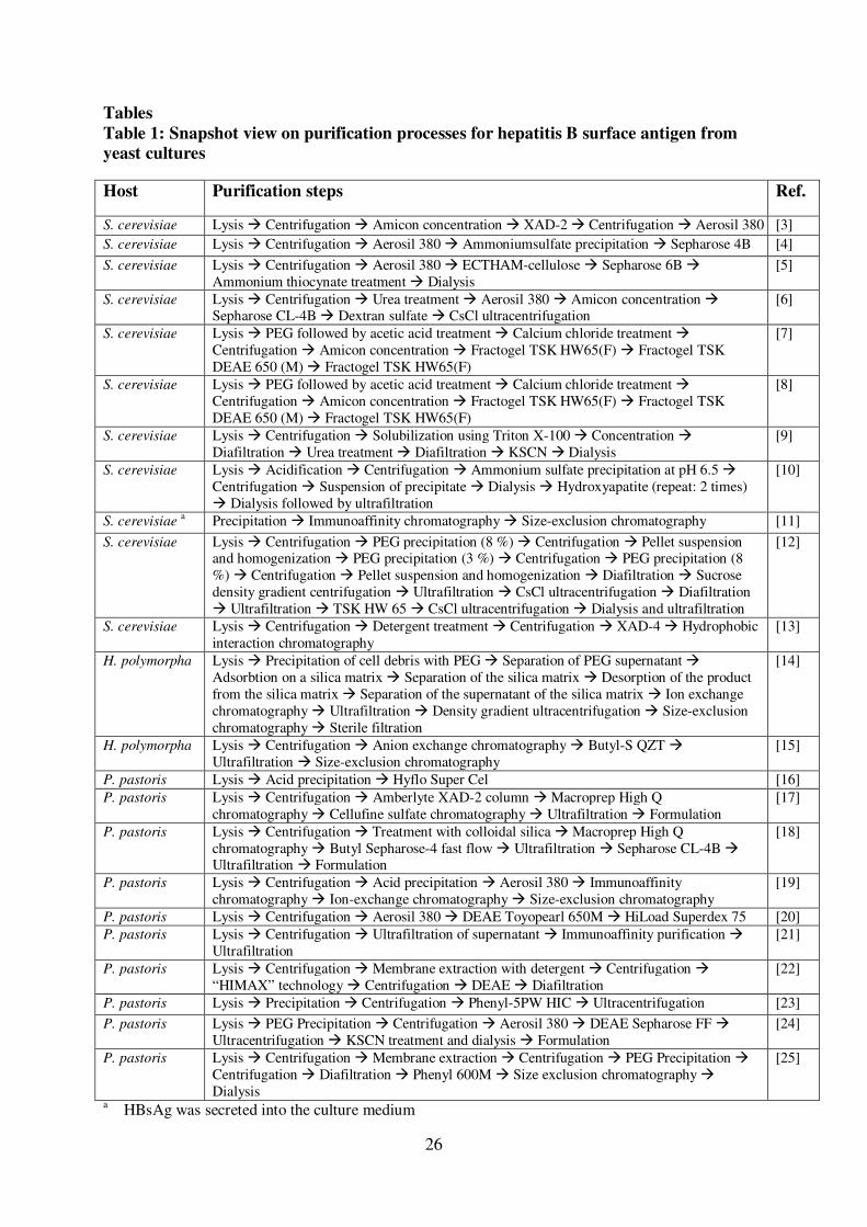
[see Tables 1 and 2] and several studies have shown that purified yeast-derived 76
HBsAg can assemble into characteristic ~22 nm VLPs [26-29]. These particles are 77
highly immunogenic and capable of eliciting potent neutralizing antibodies as they 78
mimic the conformation of native viruses but lack the viral genome and can be used 79
as safe and cheap vaccine [26, 30-32]. 80
Previously, we have reported a simple fed-batch technique which leads to the 81
production of ~6-7 g/l HBsAg, with 30% in a “soluble” form competent for assembly 82
into VLPs [29]. Although, the purification of HBsAg VLPs was reported before in 83
4
the “Materials and Methods section” [24], optimization studies of the extraction 84
conditions, details of the purification of HBsAg VLPs and the final characterization 85
of their immunogenic properties were not reported. Here, a simple strategy is 86
outlined for the purification of HBsAg leading to VLPs with satisfactory yields, high 87
purity and excellent quality. Finally, we provide evidence in mice about the superior 88
immunogenic properties of these HBsAg VLPs as a parenteral subunit vaccine in 89
combination with either alum or a novel adjuvant, the TLR2/6 agonist MALP-2. 90
91

5
2. Materials and methods 92
2.1 Strain and culture conditions 93
The P. pastoris strain GS115 carrying 8 copies of the HBsAg gene under the control 94
of the AOX1 promoter has been described previously [33]. The cells were grown on 95
defined medium in a fed-batch procedure as described before [29]. Briefly, the cells 96
were first grown in a batch procedure on glycerol (initial concentration 95 g/l). After 97
depletion of glycerol, production of HBsAg was initiated through the addition of 98
methanol to a final concentration of 6 g/l and the methanol concentration was kept 99
constant at 6 g/l by continuous methanol feeding throughout the entire production 100
phase. 101
102
2.2 Purification of recombinant HBsAg 103
After harvesting, cells were pelleted by centrifugation at 4,225 g for 15 min at room 104
temperature (1 liter culture broth OD600 ~240 corresponding to ~80 g dry cell mass 105
and ~200 g wet cell mass). The cell pellet was resuspended in the same volume of ice 106
cold buffer [20 mM sodium phosphate buffer, pH 8.0, 5 mM EDTA] for removal of 107
media components and other contaminants and recentrifuged. 108
109
2.2.1 Step 1: Cell lysis and detergent mediated solubilization of HBsAg 110
For the initial detergent optimization studies, a cell pellet (corresponding to 1 ml OD 111
100 culture broth) was resuspended with glass beads [0.5 g of ~ 0.5 mm size] in 1 ml 112
of a basic lysis buffer [10 mM sodium phosphate buffer, pH 8.0, 5 mM EDTA, 113
500 mM NaCl, 8% glycerol]. This basic lysis buffer was additionally supplemented 114
with 0-2% detergents [Tween 20 or Triton X-100 or CHAPS or NP-40 or sodium 115
deoxycholate] for detergent testing and the whole mixture incubated at 4oC using a 116
6
thermomixer. In pilot scale studies, cell lysis was essentially carried out as described 117
previously [24]. The washed cell pellet from 1 liter culture broth was resuspended in 118
1 liter ice-cold lysis buffer [25 mM phosphate buffer, pH 8.0, 5 mM EDTA, 0.6% 119
(v/v) Tween-20] and the pre-cooled cell suspension disrupted by high pressure 120
homogenization (Gaulin Lab 60, APV Gaulin, Germany) using four cycles at 600 bar 121
and ~4°C. Cell lysis was confirmed by microscopy. 122
123
2.2.2 Step 2: Polyethylene glycol (PEG) precipitation 124
To the lysate collected after high pressure homogenization, a 5 M NaCl solution was 125
slowly added within 30 min to a final concentration of 500 mM followed by the 126
addition of polyethylene glycol 6000 (S. D. Fine-Chem, India, 50% w/v) to a final 127
concentration of 5% (w/v). This suspension was stirred for 2 h at 4°C and 128
precipitation was then allowed to occur for 12-16 h at 4°C without stirring. The 129
suspension was then clarified by centrifugation at 4°C and 4,225 g for 15 min. 130
131
2.2.3 Step 3: Aerosil-380 adsorption 132
Prior to use, Aerosil-380 (Evonik, Hanau, Germany) was pre-equilibrated, e.g. 133
washed twice, with 25 mM sodium phosphate buffer, pH 7.2, 500 mM NaCl 134
(centrifuged at 4,225 g for 15 min and 4°C). The clarified supernatant obtained after 135
PEG precipitation (removal of host cell proteins and other host contaminants) was 136
mixed with Aerosil-380 (0.13 g of dry Aerosil-380 pre-equilibrated per g initial wet 137
cell mass). This suspension was stirred for 4 h at 4°C and centrifuged at 4°C and 138
4,225 g for 15 min. The pellet (corresponding to 1 liter of initial culture broth) was 139
washed twice with 25 mM phosphate buffer (pH 7.2), centrifuged as above, finally 140
resuspended in 800 ml of 50 mM sodium carbonate-bi-carbonate buffer, pH 10.8, 1.2 141

7
M urea and kept at 37°C for 12 h with stirring. This suspension was then centrifuged 142
at 25°C and 15,180 g for 60 min and the supernatant pH adjusted to pH 8.5 for better 143
removal of silica particles (Aerosil-380) and the solution clarified by vacuum-144
filtration (0.45 µm) before proceeding to the next step. 145
146
2.2.4 Step 4: Ion-exchange chromatography 147
The clarified Aerosil-380 eluate was further processed by anion exchange 148
chromatography. An XK column (Amersham Pharmacia Biotech, Sweden) packed 149
with 200 ml of DEAE Sepharose FF (GE Healthcare) and pre-equilibrated with 50 150
mM Tris-HCl, pH 8.5 (conductivity ~3.2 mS/cm) was employed and the column 151
loaded with the Aerosil-380 eluate (~800 ml) using a flow rate of 4 ml/min. After 152
loading, the column was washed with washing buffer [50 mM Tris-HCl, pH 8.5, 153
conductivity ~ 3.2 mS/cm] until the absorbance at 280 nm in the eluate returned to 154
baseline. The bound HBsAg was eluted using a salt step [50 mM Tris-HCl, pH 8.5, 155
500 mM NaCl, conductivity ~50 mS/cm]. The protein containing fractions 156
(absorbance at 280 nm) were analyzed by SDS-PAGE. 157
158
2.2.5 Step 5: Isopycnic density ultracentrifugation and size-exclusion 159
chromatography 160
To the pooled HBsAg-containing fractions obtained after ion-exchange 161
chromatography, CsCl was added to a final density of 1.2 d/ml. This solution was 162
ultra-centrifuged (Sorval rotor: TV865B) at 236,525 g for 12 h at ~ 23°C without 163
break. Alternatively, size-exclusion chromatography was used for further 164
purification. The DEAE Sepharose FF eluate was concentrated by ultrafiltration 165
(Vivaspin membrane 10,000 MWCO, Sartorius Stedium Biotech GmbH, Germany) 166
8
and loaded onto a pre-equilibrated Sephacryl S-300 (Hiprep 26/60) pre-packed 167
column. Elution was carried out with PBS (pH 7.2) and monitored at 280 nm. 168
169
2.2.6 Step 6: Potassium thiocyanate (KSCN) treatment and dialysis of the final bulk 170
The HBsAg positive fractions were pooled and treated with KSCN to a final 171
concentration of 1.2 M. The mixture was stirred at 37°C for ~ 4 h. The KSCN treated 172
HBsAg was extensively dialyzed against PBS (pH 7.2) and the final pure protein (the 173
so called bulk protein) filter sterilized and used for immunization studies. 174
175
2.3 Analytical methods for HBsAg determination 176
2.3.1 Quantitative analysis of HBsAg by ELISA 177
The concentration of HBsAg in cell extracts and other samples was determined using 178
a quantitative Sandwich ELISA (Hepanostika HBsAg Ultra, Biomerieux, The 179
Netherlands) following the manufacturer’s instructions. This ELISA was originally 180
developed for analyzing HBsAg in human sera and most likely detects preferentially 181
the immunogenic (“bioactive”) versions of HBsAg (e.g. VLPs and rod-shaped 182
structures). The clarified samples were diluted appropriately with a buffer containing 183
0.1% BSA in PBS (pH 7.2) and analyzed in triplicates. For calibration, a dilution 184
series containing 0 to 1 ng/ml of HBsAg standard (NIBSC code number – 00/588) 185
and 0 to 100 ng/ml of in-house prepared pure HBsAg was employed. All samples 186
were analyzed in triplicates. 187
188
2.3.2 Quantitative analysis of HBsAg by RP-HPLC 189
The amount of HBsAg was also analyzed by reversed phase-high performance liquid 190
chromatography (RP-HPLC), essentially as reported previously [34]. This assay 191
9
detects all conformational versions of HBsAg. Using the described conditions for 192
sample preparation and chromatography [29], the standard HBsAg (NIBSC code 193
number – 00/588) as well as the purified HBsAg eluted at a retention time of 10.9 194
min. The standard HBsAg was used for calibration. The proteins eluting at 10.9 min 195
(standard and purified HBsAg) were collected, dried to remove traces of organic 196
solvents, and subjected to SDS-PAGE analysis and immunoblotting using a linear 197
epitope-specific anti-HBsAg in-house monoclonal antibody to confirm the presence 198
of HBsAg. 199
200
2.4 Other protein analytical methods 201
The protein concentration in total cell extracts and other samples was determined 202
using the bicinchoninic acid (BCA) method [35]. SDS-PAGE analysis was 203
performed as reported [29]. Electron microscopy of HBsAg VLPs was carried out as 204
described previously [24, 29]. 205
206
2.5 In vivo immunization studies 207
2.5.1 Mice 208
Female BALB/c (H-2d) mice 6-8 weeks old were purchased from Harlan (Germany). 209
All animal experiments in this study were performed in agreement with the local 210
government of Lower Saxony (Germany) with the permission No. 33.11.42502-04-211
017/08. 212
213
2.5.2 Immunization protocols 214
The mice were immunized by the i.m. route on days 0, 14 and 28 with Engerix™ (2 215
µg; GSK, England) or 2 µg of HBsAg VLPs alone or co-administered with alum 216
10
(1:1) or PEGylated MALP-2 (10 µg/dose) in a total volume of 50 µl of PBS [36]. 217
Mixing of antigen and adjuvant in PBS was performed 30 min before the i.m. 218
injection into the right hind leg. The optimal dose of the adjuvant was determined in 219
preliminary studies (data not shown). Animals in the negative control group received 220
only PBS. 221
222
2.5.3 Detection of antigen-specific IgG in the sera 223
The HBsAg VLP-specific antibodies were determined in the serum samples by 224
ELISA using microtitre plates coated with 100 µl/well of the respective antigen (2 225
µg/ml in 0.05 M carbonate buffer, pH 9.6, as previously described [37]. 226
227
2.5.4 Measurement of cellular proliferation 228
The spleens of vaccinated mice were aseptically removed, single-cell suspensions 229
were prepared and the erythrocytes lysed by 2 min incubation in ACK buffer. The 230
cells were washed twice and adjusted to 2 x 10⁶ cells/ml in complete RPMI medium 231
containing 10% fetal bovine serum, 100 U/ml penicillin and 100 µg/ml streptomycin. 232
The splenocytes were seeded at 100 µl/well (1 x 10⁵) in a U-bottomed 96-well 233
microtitre plate (Sarstedt, Germany) and cultured in quadruplicate for 4 days in the 234
presence of different concentrations of HBsAg VLPs, 5 µg/ml concanavalin A or 235
medium alone [38, 39]. 236
237
2.5.5 ELISPOT assay 238
For the determination of the amount of cytokine secreting T helper cells in the 239
spleen, the murine IFN-γ, IL-2, IL-4 and IL-17 ELISpot kits (BD Pharmingen, USA) 240
were used according to the manufacturer’s instructions. Spleen cells (1 x 10⁶ or 5 x 241
11
105 per well) were incubated for 24 h (IFNγ) up to 48 h (IL-2, IL-4 and IL-17) in the 242
absence or in the presence of the HBsAg VLPs with a concentration of 2 µg/ml. 243
Then, cells were removed and the plates were processed. Colored spots were counted 244
with an ELISpot reader (C.T.L.) and analyzed using the ImmunoSpot image analyzer 245
software v3.2 [40]. 246
247
2.5.6 Statistical analysis 248
The statistical significance of the differences observed between the different 249
experimental groups was analyzed using the Student’s unpaired t-test and the non-250
parametric Mann-Whitney test of SigmaStat 3.10 (Build 3.10.0) or alternatively with 251
Graph Pad Prism 5 for Windows (Version 5.04) using the two-way ANOVA test. 252
Differences were considered significant at p < 0.05. 253
254
12
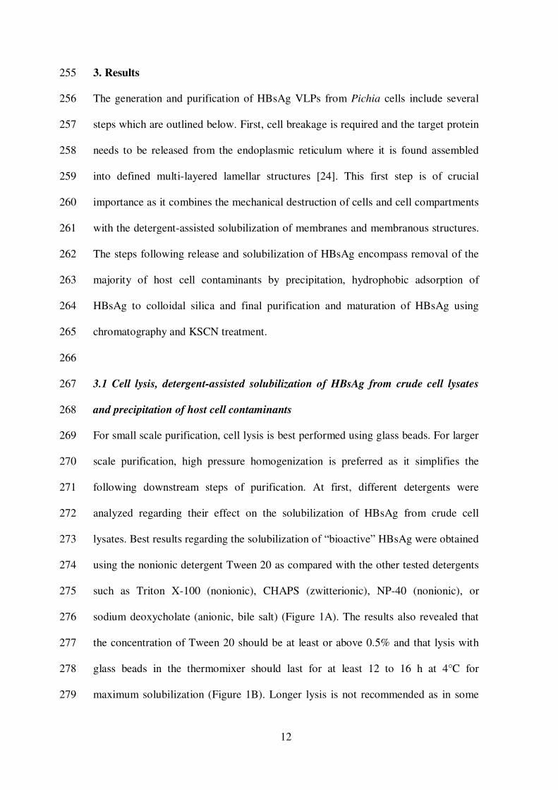
3. Results 255
The generation and purification of HBsAg VLPs from Pichia cells include several 256
steps which are outlined below. First, cell breakage is required and the target protein 257
needs to be released from the endoplasmic reticulum where it is found assembled 258
into defined multi-layered lamellar structures [24]. This first step is of crucial 259
importance as it combines the mechanical destruction of cells and cell compartments 260
with the detergent-assisted solubilization of membranes and membranous structures. 261
The steps following release and solubilization of HBsAg encompass removal of the 262
majority of host cell contaminants by precipitation, hydrophobic adsorption of 263
HBsAg to colloidal silica and final purification and maturation of HBsAg using 264
chromatography and KSCN treatment. 265
266
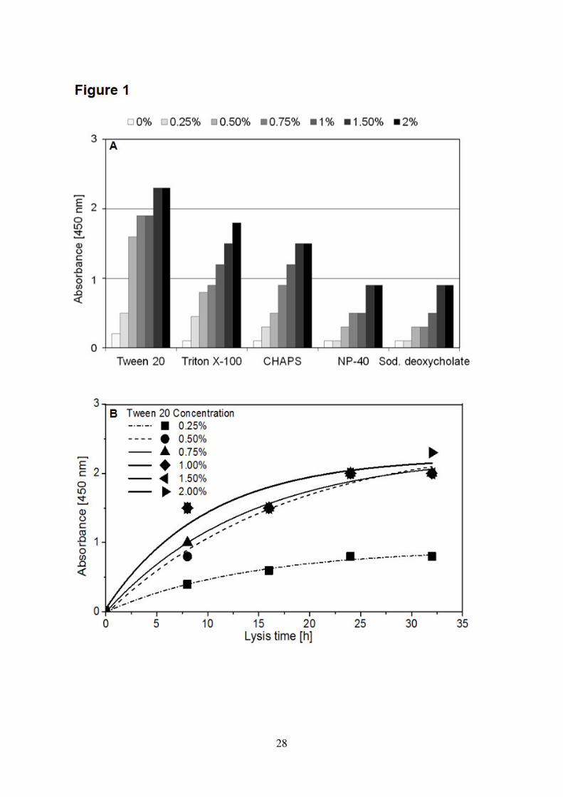
3.1 Cell lysis, detergent-assisted solubilization of HBsAg from crude cell lysates 267
and precipitation of host cell contaminants 268
For small scale purification, cell lysis is best performed using glass beads. For larger 269
scale purification, high pressure homogenization is preferred as it simplifies the 270
following downstream steps of purification. At first, different detergents were 271
analyzed regarding their effect on the solubilization of HBsAg from crude cell 272
lysates. Best results regarding the solubilization of “bioactive” HBsAg were obtained 273
using the nonionic detergent Tween 20 as compared with the other tested detergents 274
such as Triton X-100 (nonionic), CHAPS (zwitterionic), NP-40 (nonionic), or 275
sodium deoxycholate (anionic, bile salt) (Figure 1A). The results also revealed that 276
the concentration of Tween 20 should be at least or above 0.5% and that lysis with 277
glass beads in the thermomixer should last for at least 12 to 16 h at 4°C for 278
maximum solubilization (Figure 1B). Longer lysis is not recommended as in some 279
13
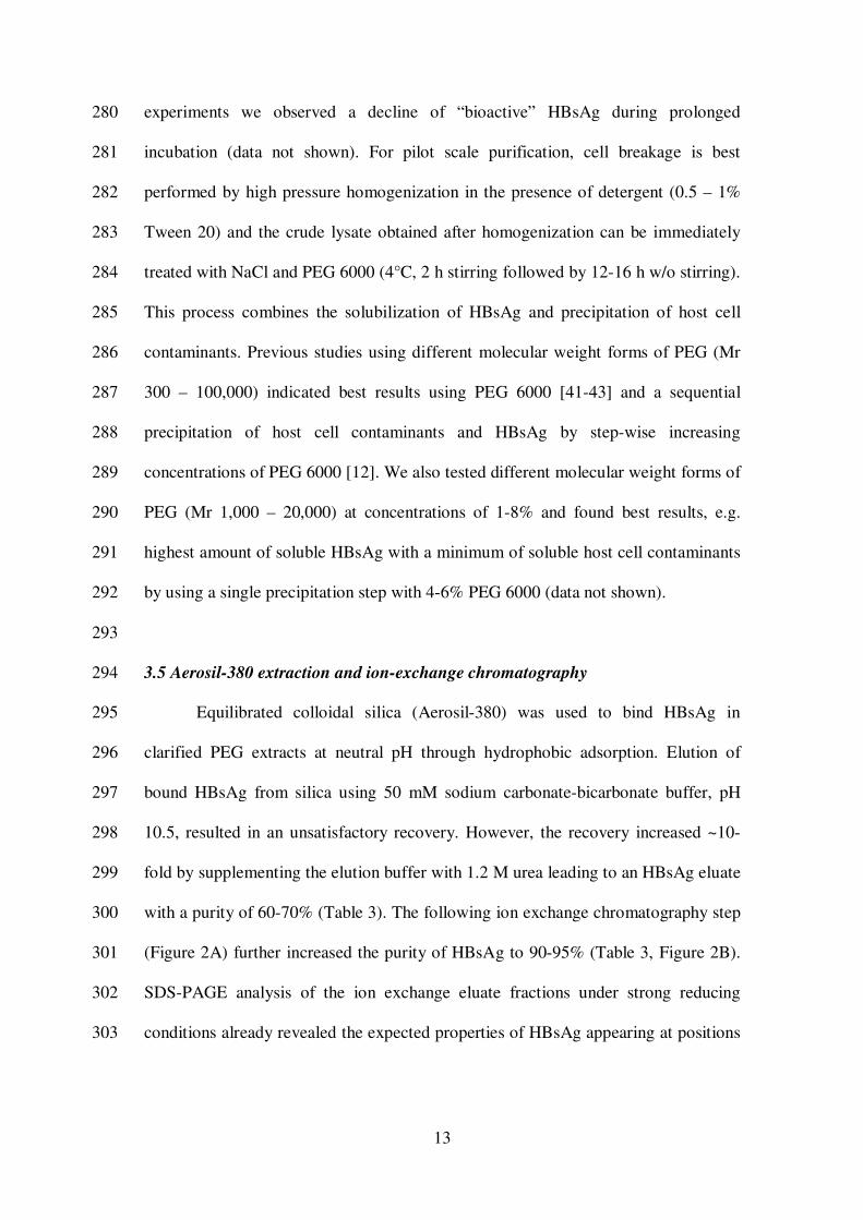
experiments we observed a decline of “bioactive” HBsAg during prolonged 280
incubation (data not shown). For pilot scale purification, cell breakage is best 281
performed by high pressure homogenization in the presence of detergent (0.5 – 1% 282
Tween 20) and the crude lysate obtained after homogenization can be immediately 283
treated with NaCl and PEG 6000 (4°C, 2 h stirring followed by 12-16 h w/o stirring). 284
This process combines the solubilization of HBsAg and precipitation of host cell 285
contaminants. Previous studies using different molecular weight forms of PEG (Mr 286
300 – 100,000) indicated best results using PEG 6000 [41-43] and a sequential 287
precipitation of host cell contaminants and HBsAg by step-wise increasing 288
concentrations of PEG 6000 [12]. We also tested different molecular weight forms of 289
PEG (Mr 1,000 – 20,000) at concentrations of 1-8% and found best results, e.g. 290
highest amount of soluble HBsAg with a minimum of soluble host cell contaminants 291
by using a single precipitation step with 4-6% PEG 6000 (data not shown). 292
293
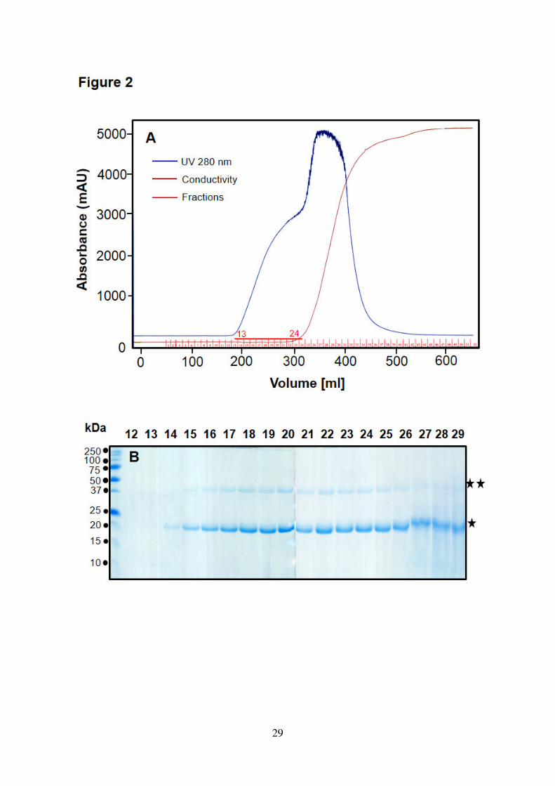
3.5 Aerosil-380 extraction and ion-exchange chromatography 294
Equilibrated colloidal silica (Aerosil-380) was used to bind HBsAg in 295
clarified PEG extracts at neutral pH through hydrophobic adsorption. Elution of 296
bound HBsAg from silica using 50 mM sodium carbonate-bicarbonate buffer, pH 297
10.5, resulted in an unsatisfactory recovery. However, the recovery increased ~10-298
fold by supplementing the elution buffer with 1.2 M urea leading to an HBsAg eluate 299
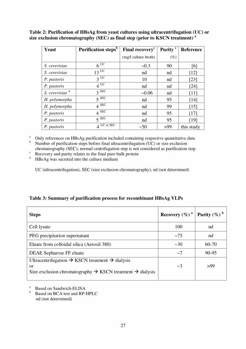
with a purity of 60-70% (Table 3). The following ion exchange chromatography step 300
(Figure 2A) further increased the purity of HBsAg to 90-95% (Table 3, Figure 2B). 301
SDS-PAGE analysis of the ion exchange eluate fractions under strong reducing 302
conditions already revealed the expected properties of HBsAg appearing at positions 303
14
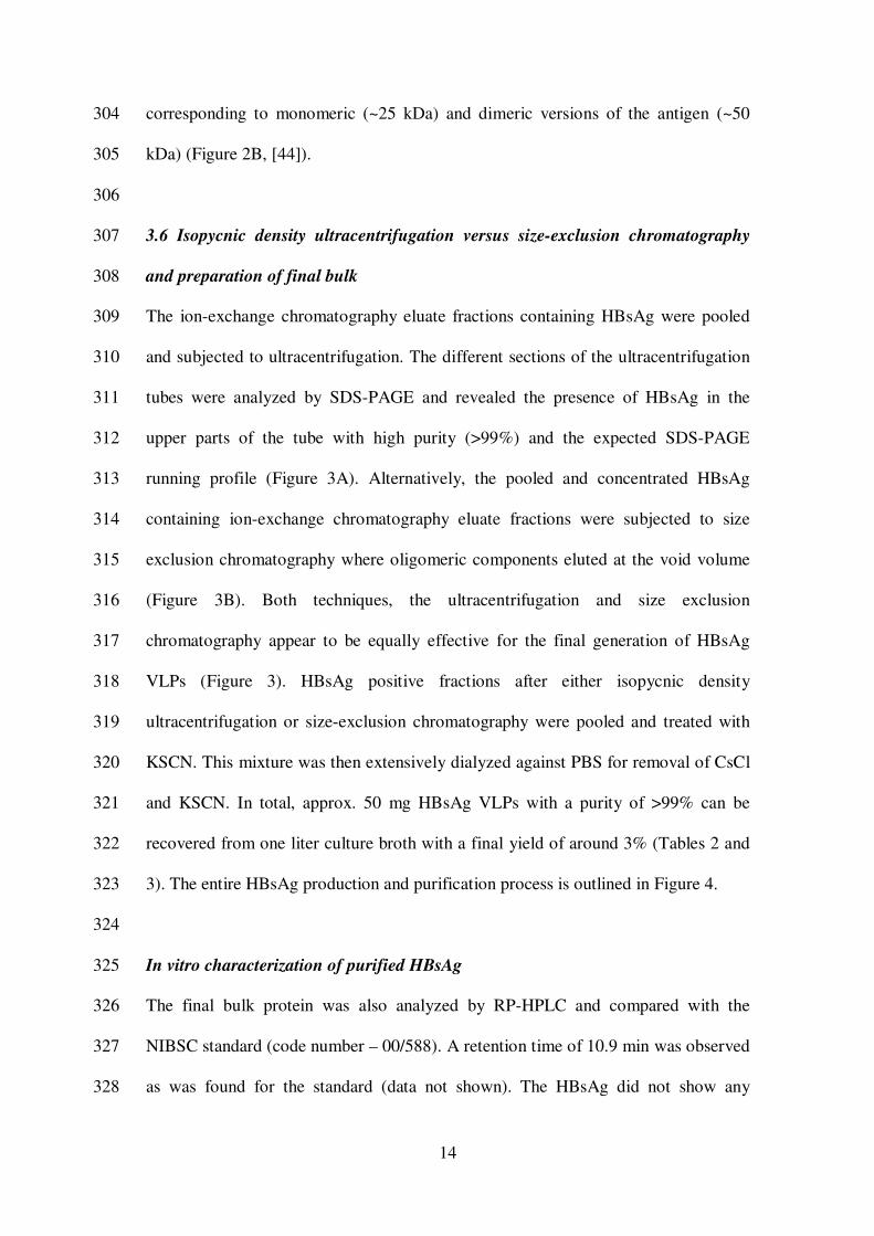
corresponding to monomeric (~25 kDa) and dimeric versions of the antigen (~50 304
kDa) (Figure 2B, [44]). 305
306
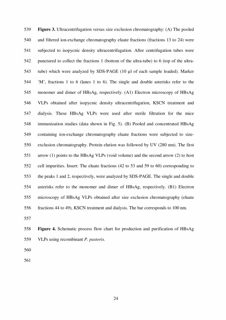
3.6 Isopycnic density ultracentrifugation versus size-exclusion chromatography 307
and preparation of final bulk 308
The ion-exchange chromatography eluate fractions containing HBsAg were pooled 309
and subjected to ultracentrifugation. The different sections of the ultracentrifugation 310
tubes were analyzed by SDS-PAGE and revealed the presence of HBsAg in the 311
upper parts of the tube with high purity (>99%) and the expected SDS-PAGE 312
running profile (Figure 3A). Alternatively, the pooled and concentrated HBsAg 313
containing ion-exchange chromatography eluate fractions were subjected to size 314
exclusion chromatography where oligomeric components eluted at the void volume 315
(Figure 3B). Both techniques, the ultracentrifugation and size exclusion 316
chromatography appear to be equally effective for the final generation of HBsAg 317
VLPs (Figure 3). HBsAg positive fractions after either isopycnic density 318
ultracentrifugation or size-exclusion chromatography were pooled and treated with 319
KSCN. This mixture was then extensively dialyzed against PBS for removal of CsCl 320
and KSCN. In total, approx. 50 mg HBsAg VLPs with a purity of >99% can be 321
recovered from one liter culture broth with a final yield of around 3% (Tables 2 and 322
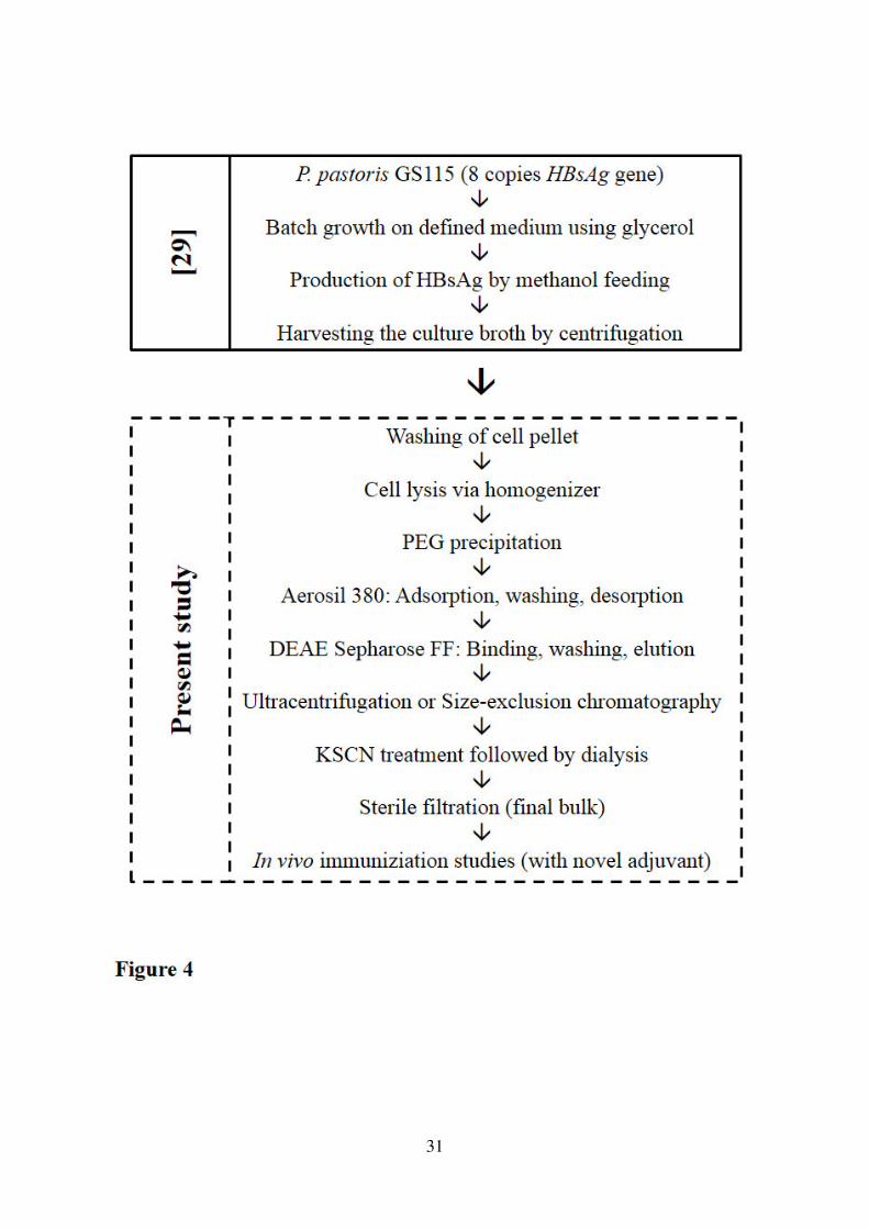
3). The entire HBsAg production and purification process is outlined in Figure 4. 323
324
In vitro characterization of purified HBsAg 325
The final bulk protein was also analyzed by RP-HPLC and compared with the 326
NIBSC standard (code number – 00/588). A retention time of 10.9 min was observed 327
as was found for the standard (data not shown). The HBsAg did not show any 328
15
binding to lectins, thus proving absence of glycosylation (data not shown). Finally, 329
electron microscopy of pure HBsAg, obtained using either ultracentrifugation or 330
size-exclusion chromatography, revealed in both cases the presence of the 331
characteristic icosahedral symmetrical structures with a diameter of ~22 nm, the so-332
called HBsAg “VLPs” (included in Figure 3). 333
334
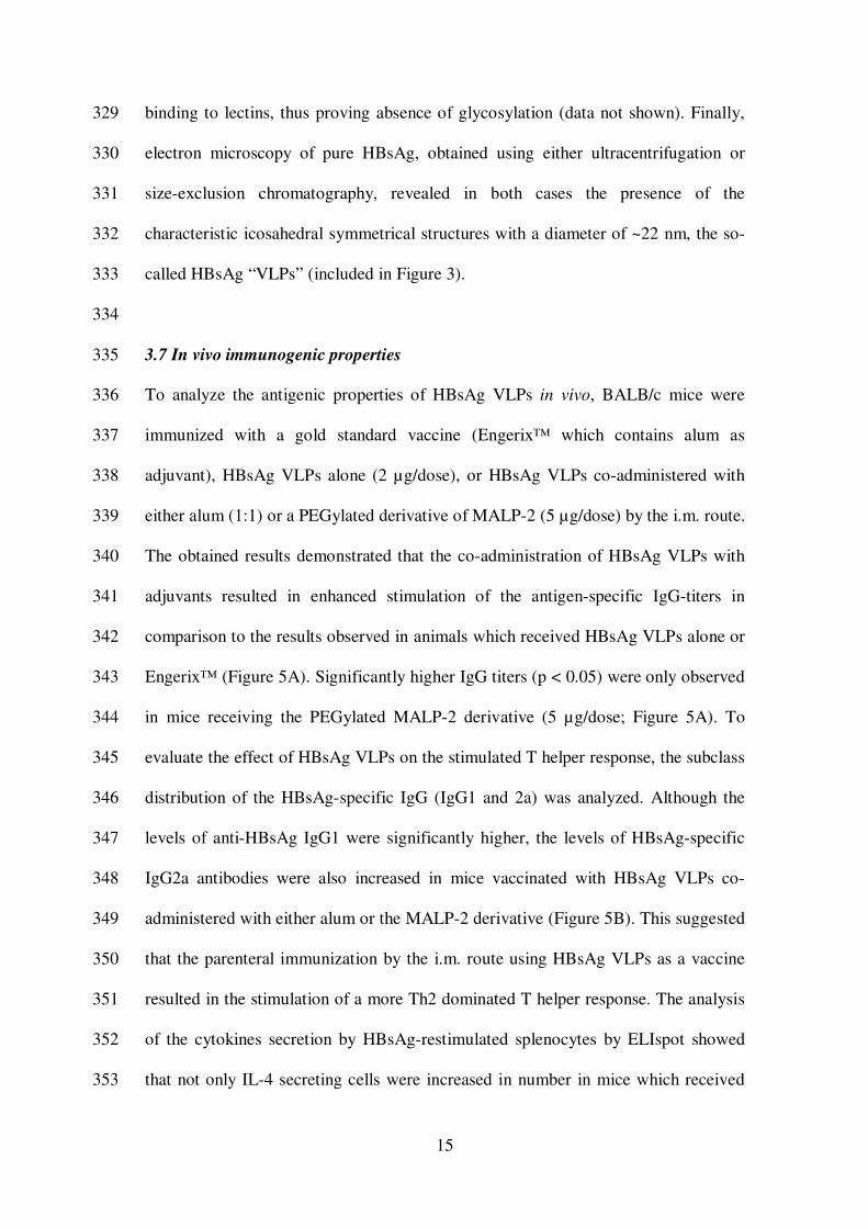
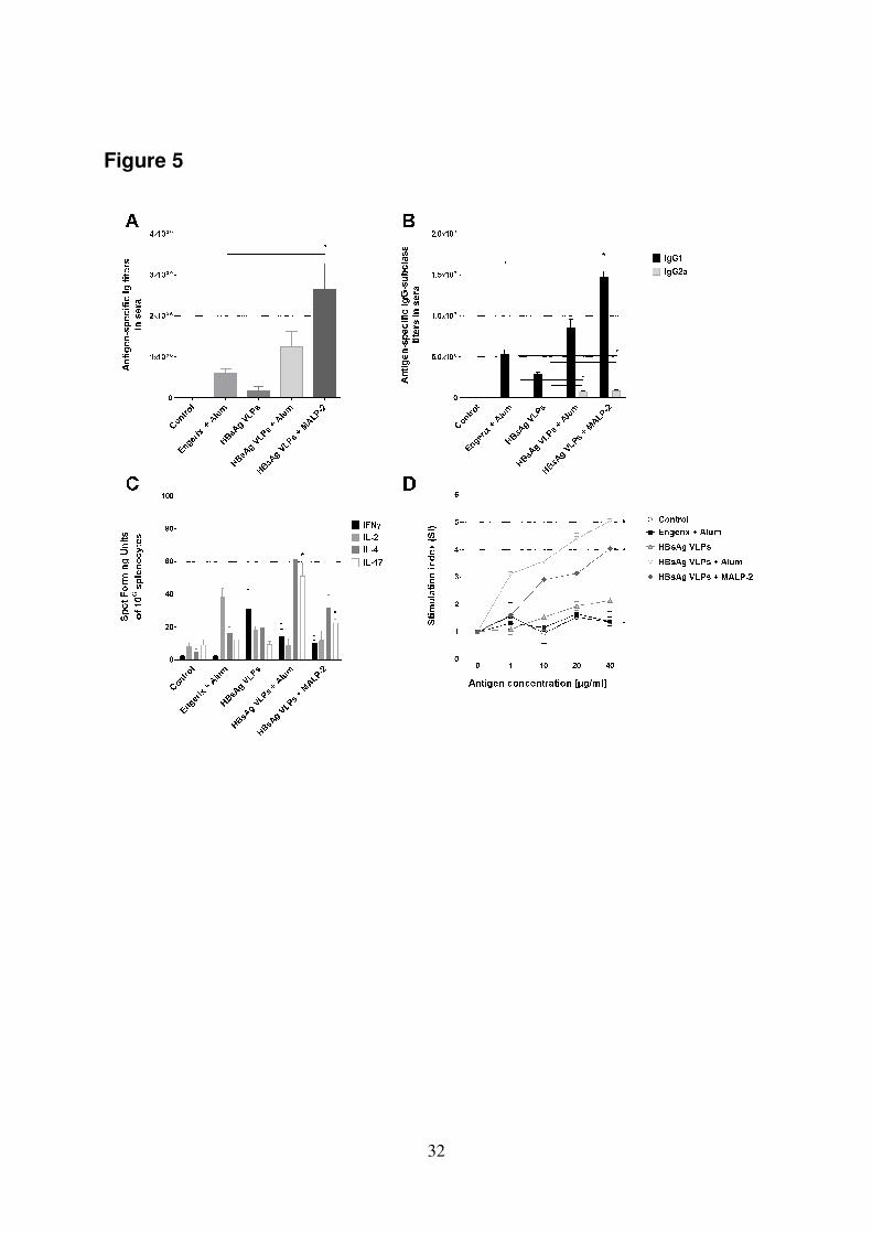
3.7 In vivo immunogenic properties 335
To analyze the antigenic properties of HBsAg VLPs in vivo, BALB/c mice were 336
immunized with a gold standard vaccine (Engerix™ which contains alum as 337
adjuvant), HBsAg VLPs alone (2 µg/dose), or HBsAg VLPs co-administered with 338
either alum (1:1) or a PEGylated derivative of MALP-2 (5 µg/dose) by the i.m. route. 339
The obtained results demonstrated that the co-administration of HBsAg VLPs with 340
adjuvants resulted in enhanced stimulation of the antigen-specific IgG-titers in 341
comparison to the results observed in animals which received HBsAg VLPs alone or 342
Engerix™ (Figure 5A). Significantly higher IgG titers (p < 0.05) were only observed 343
in mice receiving the PEGylated MALP-2 derivative (5 µg/dose; Figure 5A). To 344
evaluate the effect of HBsAg VLPs on the stimulated T helper response, the subclass 345
distribution of the HBsAg-specific IgG (IgG1 and 2a) was analyzed. Although the 346
levels of anti-HBsAg IgG1 were significantly higher, the levels of HBsAg-specific 347
IgG2a antibodies were also increased in mice vaccinated with HBsAg VLPs co-348
administered with either alum or the MALP-2 derivative (Figure 5B). This suggested 349
that the parenteral immunization by the i.m. route using HBsAg VLPs as a vaccine 350
resulted in the stimulation of a more Th2 dominated T helper response. The analysis 351
of the cytokines secretion by HBsAg-restimulated splenocytes by ELIspot showed 352
that not only IL-4 secreting cells were increased in number in mice which received 353
16
the HBsAg VLPs co-administered with alum or the MALP-2 derivative as compared 354
to the control groups, but the HBsAg-specific IL-17, IFNγ and IL-2 secreting cells 355
were also increased (Figure 5C). To further characterize the capacity of HBsAg 356
VLPs to induce the cellular immune responses, spleen cells isolated from vaccinated 357
mice on day 42 were re-stimulated in vitro with HBsAg VLPs and their proliferation 358
capacity was then assessed. A strong dose-dependent proliferative response was only 359
observed in mice vaccinated with HBsAg VLPs co-administered with alum (SI >4) 360
or the PEGylated MALP-2 derivative (SI >4), as shown in Figure 5D. In contrast, no 361
or only marginal responses were observed with cells derived from mice vaccinated 362
with either HBsAg VLPs alone (SI >2), Engerix™ or PBS (SI <2). The differences 363
observed between the results obtained with adjuvanted HBsAg VLPs compared with 364
those obtained from either the control, non-adjuvanted HBsAg VLPs or Engerix™ 365
vaccinated mice were significantly higher (p < 0.05). 366
367
17
4. Discussion 368
HBsAg is a very hydrophobic protein with long stretches of connected 369
hydrophobic amino acids. Only recently it was shown that HBsAg – when produced 370
in yeast, e.g. P. pastoris – does not assemble into VLPs within the cell as was 371
assumed previously nor does it insert in significant amounts into ER membranes as 372
was also proposed. Evidence has been presented that the HBsAg remains in the 373
endoplasmic reticulum (ER) where it does not form VLPs but where a major fraction 374
assembles into well-ordered multi-layered lamellar structures [24]. The layering 375
order of HBsAg in these lamellar structures strongly suggests the presence of well-376
ordered HBsAg subunits [24], which should be solubilizable without getting 377
structurally disordered to reassemble into VLPs under appropriate conditions. The 378
remainder of HBsAg forms non-structured aggregates in the ER, which are only 379
solubilizable using extremely harsh protein-structure breaking conditions [29]. 380
Thus, cell breakage and release of HBsAg form the ER in a “bioactive” form 381
competent for VLP assembly is the major objective for the first step of the 382
purification procedure. Usually, the nonionic detergent Triton X-100 is employed for 383
solubilization of HBsAg form yeast homogenates, e.g. [9, 13, 45, 46]. However, 384
there have been early indications that the nonionic detergent Tween 20 might be less 385
harsh to “intact” HBsAg compared to Triton X-100 [6]. In our hands Tween 20 386
released more “bioactive” HBsAg from the yeast homogenate compared to Triton X-387
100 as measured by an HBsAg Sandwich ELISA developed for the determination of 388
HBsAg particles in human plasma. We do not have a straight forward explanation 389
why Tween 20 performs better compared to Triton X-100 in releasing “bioactive” 390
HBsAg. Tween 20 is considered a milder detergent being less effective in membrane 391
solubilization compared to Triton X-100 [47-49]. Moreover, addition of Tween 20 to 392
18
protein formulations has proven to be effective in preventing shear induced 393
aggregation of antibodies [50] and also aggregation of murine polyomavirus VLPs 394
during storage [51]. Thus, replacement of Triton X-100 by Tween 20 presumably 395
helps to reduce shear stress induced denaturation and “irreversible” aggregation of 396
HBsAg during mechanical cell breakage. The other important objective for the first 397
downstream purification steps relates to the removal of host cell contaminants. As we 398
aimed for releasing “bioactive” HBsAg into the soluble fraction of the cell 399
homogenate host cell contaminants should be transferred preferably to the insoluble 400
fraction of the lysate. Substitution of Triton X-100 by Tween 20 presumably also 401
helps to achieve this objective as it is less effective in (host cell) membrane 402
solubilization. The intended transfer of host cell contaminants into the insoluble 403
fraction is further accomplished by addition of 5% PEG 6000, a hydrophilic nonionic 404
polymer, which is known to precipitate the majority of host cell proteins, 405
polysaccharides, and nucleic acids but not the HBsAg when employed at this 406
concentration [8]. After precipitate removal, the following purification steps e.g. 407
hydrophobic adsorption to colloidal silica, desorption from silica and subsequent 408
chromatography and final maturation are with minor modifications in accordance 409
with previously published procedures. The final yields are certainly in need of 410
improvement but we would expect high robust yields under standardized industrial 411
GMP production and purification conditions. The quality of the final product, 412
however, is outstanding as it outperforms, in particular when adjuvanted with the 413
novel adjuvant MALP-2, the gold standard HBsAg vaccine Engerix™ in stimulating 414
humoral and cellular immune responses. 415
416
19
Acknowledgements 417
This work was supported by institutional core funds of the Helmholtz Centre for 418
Infection Research and ICGEB and an Indo-German collaborative grant 419
(International Bureau of the BMBF, DLR, IND 03/009). Maria Zahid and Ahmad 420
Adnan wish to express their gratitude to the Deutscher Akademischer 421
Austauschdienst (DAAD) of Germany and the Higher Education Commission (HEC) 422
of Pakistan for their fellowships. 423
424
20
425
Reference List 426
427
[1] World Health Organization - Hepatitis B Fact sheet N° 204 (2008). 428
[2] L.J.Frost, M.R.Reich, Access: How do good health technologies get to 429
poor people in poor countries. Creative Commons Attribution-430
Noncommercial-Share Alike 3.0 United States License, 2008, p. 67. 431
[3] D.E. Wampler, E.D. Lehman, J. Boger, W.J. McAleer, E.M. Scolnick, 432
Proc. Natl. Acad. Sci. USA 82 (1985) 6830. 433
[4] Y. Uemura, T. Ohmura, A. Ohmizu, A. Sumi, W. Ohtani, Y. Sakanishi, 434
H. Morise, H. Arimura, T. Suyama, US Patent 4,694,074 (1987). 435
[5] A. Friedman, E.D. Lehman, W.J. McAleer, T.F. Schaefer, E.M. Scolnick, 436
D.E. Wampler, US Patent 4,707,542 (1987). 437
[6] F. van Wijnendaele, G. Simonet, US Patent 4,649,192 (1987). 438
[7] F. van Wijnendaele, D. Gilles, G. Simonet, US Patent 4,683,294 (1987). 439
[8] F. van Wijnendaele, D. Gilles, G. Simonet, US Patent 4,857,317 (1989). 440
[9] S. Yamazaki, US Patent 5,001,915 (1991). 441
[10] O. Nobuya, M. Kyosuke, J. Fukusaburo, M. Hiroshi, EP 0 156 242 B1 442
(1992). 443
[11] S. Kuroda, T. Miyazaki, S. Otaka, Y. Fujisawa, Appl. Microbiol. 444
Biotechnol. 40 (1993) 333. 445
[12] J.H. Hsieh, S.C. Shih, W.K. Chi, Y.D. Chu, A.N. Lin, US Patent 446
5,462,863 (1995). 447
[13] G.S. Kee, J. Jin, B. Balasundaram, D.G. Bracewell, N.S. Pujar, N.J. 448
Titchener-Hooker, Biotechnol. Prog. 26 (2010) 26. 449
[14] M. Pointek, M. Weniger, US Patent 6,428,984 B1 (2002). 450
[15] Y. Huang, J. Bi, Y. Zhang, W. Zhou, Y. Li, L. Zhao, Z. Su, Protein Expr. 451
Purif. 56 (2007) 301. 452
[16] A. Agraz, Y. Quinones, N. Exposito, F. Brena, J. Madruga, E. Penton, L. 453
Herrera, Biotechnol. Bioeng. 42 (1993) 1238. 454
[17] R.K. Venkata, P.K. Satyanarayana, S.K. Venkat, Indian Patent 455
IN180249 (1998). 456
[18] R.K. Venkata, P.K. Satyanarayana, S.K. Venkat, S.A. Venkata, Indian 457
Patent IN180250 (1998). 458
21
[19] E. Hardy, E. Martinez, D. Diago, R. Diaz, D. Gonzalez, L. Herrera, J. 459
Biotechnol. 77 (2000) 157. 460
[20] N. Bardiya, Anaerobe 12 (2006) 194. 461
[21] S. Ottone, X. Nguyen, J. Bazin, C. Berard, S. Jimenez, O. Letourneur, 462
Protein Expr. Purif. 56 (2007) 177. 463
[22] V.K. Srinivas, E. Krishnamurthy, Indian Patent IN202961 (2007). 464
[23] R. Liu, Q. Lin, Y. Sun, X. Lu, Y. Qiu, Y. Li, X. Guo, Appl. Biochem. 465
Biotechnol. 158 (2009) 432. 466
[24] H. Lünsdorf, C. Gurramkonda, A. Adnan, N. Khanna, U. Rinas, Microb. 467
Cell Fact. 10 (2011) 48. 468
[25] A. Patil, N. Khanna, J. Chromatogr. B 898 (2012) 7. 469
[26] P. Valenzuela, A. Medina, W.J. Rutter, G. Ammerer, B.D. Hall, Nature 470
298 (1982) 347. 471
[27] W.J. McAleer, E.B. Buynak, R.Z. Maigetter, D.E. Wampler, W.J. Miller, 472
M.R. Hilleman, Nature 307 (1984) 178. 473
[28] J.M. Cregg, J.F. Tschopp, C. Stillman, R. Siegel, M. Akong, W.S. Craig, 474
R.G. Buckholz, K.R. Madden, P.A. Kellaris, G.R. Davis, B.L. Smiley, J. 475
Cruze, G. Torregrossa, G. Velicelebi, G.P. Thill, Bio/Technology 5 (1987) 476
479. 477
[29] C. Gurramkonda, A. Adnan, T. Gäbel, H. Lünsdorf, A. Ross, S.K. 478
Nemani, S. Swaminathan, N. Khanna, U. Rinas, Microb. Cell Fact. 8 479
(2009) 13. 480
[30] E.V. Grgacic, D.A. Anderson, Methods 40 (2006) 60. 481
[31] M.F. Bachmann, G.T. Jennings, Nat. Rev. Immunol. 10 (2010) 787. 482
[32] A. Roldao, M.C. Mellado, L.R. Castilho, M.J. Carrondo, P.M. Alves, 483
Expert. Rev. Vaccines. 9 (2010) 1149. 484
[33] A. Vassileva, D.A. Chugh, S. Swaminathan, N. Khanna, Protein Expr. 485
Purif. 21 (2001) 71. 486
[34] D.O. O'Keefe, A.M. Paiva, Anal. Biochem. 230 (1995) 48. 487
[35] P.K. Smith, R.I. Krohn, G.T. Hermanson, A.K. Mallia, F.H. Gartner, 488
M.D. Provenzano, E.K. Fujimoto, N.M. Goeke, B.J. Olson, D.C. Klenk, 489
Anal. Biochem. 150 (1985) 76. 490
[36] H. Weigt, P.F. Muhlradt, A. Emmendorffer, N. Krug, A. Braun, 491
Immunobiology 207 (2003) 223. 492
22
[37] S. Borsutzky, V. Fiorelli, T. Ebensen, A. Tripiciano, F. Rharbaoui, A. 493
Scoglio, C. Link, F. Nappi, M. Morr, S. Butto, A. Cafaro, P.F. Muhlradt, 494
B. Ensoli, C.A. Guzman, Eur. J. Immunol. 33 (2003) 1548. 495
[38] C. Link, R. Gavioli, T. Ebensen, A. Canella, E. Reinhard, C.A. Guzman, 496
Eur. J. Immunol. 34 (2004) 899. 497
[39] F. Rharbaoui, B. Drabner, S. Borsutzky, U. Winckler, M. Morr, B. 498
Ensoli, P.F. Muhlradt, C.A. Guzman, Eur. J. Immunol. 32 (2002) 2857. 499
[40] P.D. Becker, S. Fiorentini, C. Link, G. Tosti, T. Ebensen, A. Caruso, C.A. 500
Guzman, Vaccine 24 (2006) 5269. 501
[41] J. Vnek, A.M. Prince, US Patent 3,951,937 (1976). 502
[42] A.R. Neurath, A.M. Prince, A. Lippin, US Patent 3,994,870 (1976). 503
[43] T. Ohmura, T. Fujiwara, A. Ohmizu, S. Funakoshi, US Patent 4,565,697 504
(1986). 505
[44] Q. Zhao, Y. Wang, D. Freed, T.M. Fu, J.A. Gimenez, R.D. Sitrin, M.W. 506
Washabaugh, Hum. Vaccin. 2 (2006) 174. 507
[45] P.J. Kniskern, A. Hagopian, US Patent 5,614,384 (1997). 508
[46] G.S. Kee, N.S. Pujar, N.J. Titchener-Hooker, Biotechnol. Prog. 24 (2008) 509
623. 510
[47] S. Schuck, M. Honsho, K. Ekroos, A. Shevchenko, K. Simons, Proc. Natl. 511
Acad. Sci. USA 100 (2003) 5795. 512
[48] R.W. Egan, J. Biol. Chem. 251 (1976) 4442. 513
[49] M. Alfalah, G. Wetzel, I. Fischer, R. Busche, E.E. Sterchi, K.P. Zimmer, 514
H.P. Sallmann, H.Y. Naim, J. Biol. Chem. 280 (2005) 42636. 515
[50] T.W. Patapoff, O. Esue, Pharm. Dev. Technol. 14 (2009) 659. 516
[51] J. Mohr, Y.P. Chuan, Y. Wu, L.H. Lua, A.P. Middelberg, Methods 60 517
(2013) 248. 518
519
520 521
23
Figure legends 522
523
Figure 1. Detergent solubilization of HBsAg from cell lysates: (A) Cells were lysed 524
with glass beads in basic lysis buffer additionally containing detergents at the 525
indicated concentrations and incubated at 4°C in a thermomixer for 48 h. The final 526
amount of “bioactive” HBsAg released into the soluble lysate fraction is given in 527
relative units of the Sandwich ELISA readout. (B) The time-dependent release of 528
“bioactive” HBsAg into the soluble fraction of the lysate as followed by the 529
Sandwich-ELISA. 530
531
Figure 2. Anion exchange chromatography (DEAE Sepharose FF): (A) Elution of 532
bound proteins during ion exchange chromatography. The eluate fractions 13 to 24 533
were pooled (each fraction 12 ml), filtered and subjected to the next purification step. 534
(B) Analysis of eluate fractions 12 to 29 by SDS-PAGE (10 µl of each sample 535
loaded). The single and double asterisks refer to the monomer and dimer of HBsAg, 536
respectively. 537
538
24
Figure 3. Ultracentrifugation versus size exclusion chromatography: (A) The pooled 539
and filtered ion-exchange chromatography eluate fractions (fractions 13 to 24) were 540
subjected to isopycnic density ultracentrifugation. After centrifugation tubes were 541
punctured to collect the fractions 1 (bottom of the ultra-tube) to 6 (top of the ultra-542
tube) which were analyzed by SDS-PAGE (10 µl of each sample loaded). Marker 543
‘M’, fractions 1 to 6 (lanes 1 to 6). The single and double asterisks refer to the 544
monomer and dimer of HBsAg, respectively. (A1) Electron microscopy of HBsAg 545
VLPs obtained after isopycnic density ultracentrifugation, KSCN treatment and 546
dialysis. These HBsAg VLPs were used after sterile filtration for the mice 547
immunization studies (data shown in Fig. 5). (B) Pooled and concentrated HBsAg 548
containing ion-exchange chromatography eluate fractions were subjected to size-549
exclusion chromatography. Protein elution was followed by UV (280 nm). The first 550
arrow (1) points to the HBsAg VLPs (void volume) and the second arrow (2) to host 551
cell impurities. Insert: The eluate fractions (42 to 53 and 59 to 60) corresponding to 552
the peaks 1 and 2, respectively, were analyzed by SDS-PAGE. The single and double 553
asterisks refer to the monomer and dimer of HBsAg, respectively. (B1) Electron 554
microscopy of HBsAg VLPs obtained after size exclusion chromatography (eluate 555
fractions 44 to 49), KSCN treatment and dialysis. The bar corresponds to 100 nm. 556
557
Figure 4. Schematic process flow chart for production and purification of HBsAg 558
VLPs using recombinant P. pastoris. 559
560
561
25
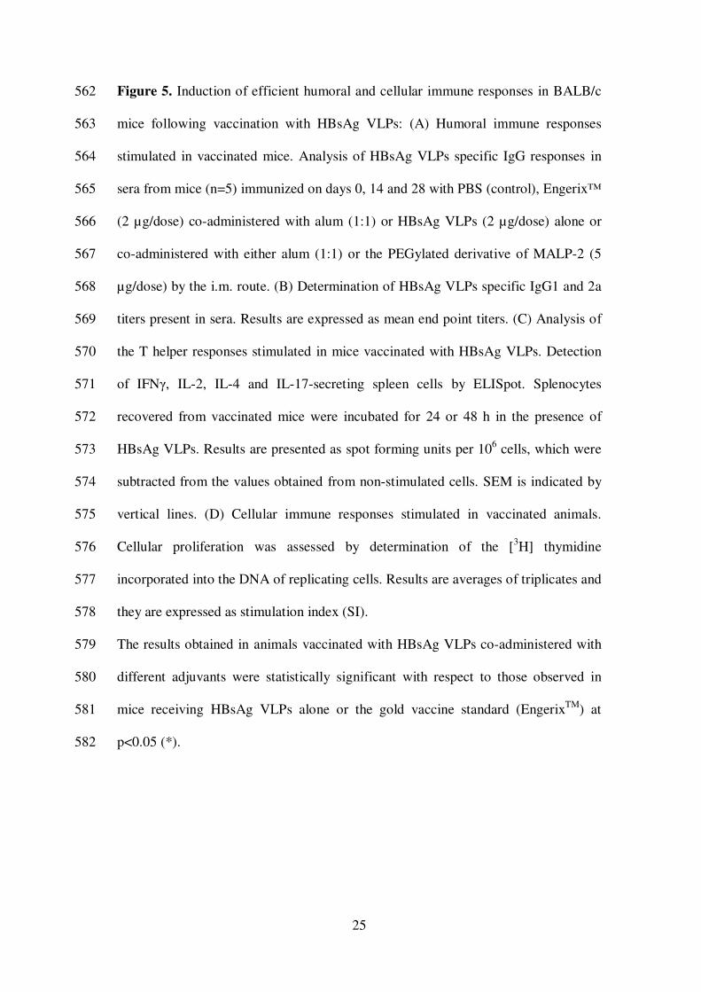
Figure 5. Induction of efficient humoral and cellular immune responses in BALB/c 562
mice following vaccination with HBsAg VLPs: (A) Humoral immune responses 563
stimulated in vaccinated mice. Analysis of HBsAg VLPs specific IgG responses in 564
sera from mice (n=5) immunized on days 0, 14 and 28 with PBS (control), Engerix™ 565
(2 µg/dose) co-administered with alum (1:1) or HBsAg VLPs (2 µg/dose) alone or 566
co-administered with either alum (1:1) or the PEGylated derivative of MALP-2 (5 567
µg/dose) by the i.m. route. (B) Determination of HBsAg VLPs specific IgG1 and 2a 568
titers present in sera. Results are expressed as mean end point titers. (C) Analysis of 569
the T helper responses stimulated in mice vaccinated with HBsAg VLPs. Detection 570
of IFNγ, IL-2, IL-4 and IL-17-secreting spleen cells by ELISpot. Splenocytes 571
recovered from vaccinated mice were incubated for 24 or 48 h in the presence of 572
HBsAg VLPs. Results are presented as spot forming units per 106 cells, which were 573
subtracted from the values obtained from non-stimulated cells. SEM is indicated by 574
vertical lines. (D) Cellular immune responses stimulated in vaccinated animals. 575
Cellular proliferation was assessed by determination of the [3H] thymidine 576
incorporated into the DNA of replicating cells. Results are averages of triplicates and 577
they are expressed as stimulation index (SI). 578
The results obtained in animals vaccinated with HBsAg VLPs co-administered with 579
different adjuvants were statistically significant with respect to those observed in 580
mice receiving HBsAg VLPs alone or the gold vaccine standard (EngerixTM
) at 581
p<0.05 (*). 582
26
Tables
Table 1: Snapshot view on purification processes for hepatitis B surface antigen from
yeast cultures
Host Purification steps Ref.
S. cerevisiae Lysis � Centrifugation � Amicon concentration � XAD-2 � Centrifugation � Aerosil 380 [3]
S. cerevisiae Lysis � Centrifugation � Aerosil 380 � Ammoniumsulfate precipitation � Sepharose 4B [4]
S. cerevisiae Lysis � Centrifugation � Aerosil 380 � ECTHAM-cellulose � Sepharose 6B �
Ammonium thiocynate treatment � Dialysis
[5]
S. cerevisiae Lysis � Centrifugation � Urea treatment � Aerosil 380 � Amicon concentration � Sepharose CL-4B � Dextran sulfate � CsCl ultracentrifugation
[6]
S. cerevisiae Lysis � PEG followed by acetic acid treatment � Calcium chloride treatment �
Centrifugation � Amicon concentration � Fractogel TSK HW65(F) � Fractogel TSK
DEAE 650 (M) � Fractogel TSK HW65(F)
[7]
S. cerevisiae Lysis � PEG followed by acetic acid treatment � Calcium chloride treatment �
Centrifugation � Amicon concentration � Fractogel TSK HW65(F) � Fractogel TSK
DEAE 650 (M) � Fractogel TSK HW65(F)
[8]
S. cerevisiae Lysis � Centrifugation � Solubilization using Triton X-100 � Concentration �
Diafiltration � Urea treatment � Diafiltration � KSCN � Dialysis
[9]
S. cerevisiae Lysis � Acidification � Centrifugation � Ammonium sulfate precipitation at pH 6.5 �
Centrifugation � Suspension of precipitate � Dialysis � Hydroxyapatite (repeat: 2 times)
� Dialysis followed by ultrafiltration
[10]
S. cerevisiae a Precipitation � Immunoaffinity chromatography � Size-exclusion chromatography [11]
S. cerevisiae Lysis � Centrifugation � PEG precipitation (8 %) � Centrifugation � Pellet suspension and homogenization � PEG precipitation (3 %) � Centrifugation � PEG precipitation (8
%) � Centrifugation � Pellet suspension and homogenization � Diafiltration � Sucrose
density gradient centrifugation � Ultrafiltration � CsCl ultracentrifugation � Diafiltration
� Ultrafiltration � TSK HW 65 � CsCl ultracentrifugation � Dialysis and ultrafiltration
[12]
S. cerevisiae Lysis � Centrifugation � Detergent treatment � Centrifugation � XAD-4 � Hydrophobic
interaction chromatography
[13]
H. polymorpha Lysis � Precipitation of cell debris with PEG � Separation of PEG supernatant �
Adsorbtion on a silica matrix � Separation of the silica matrix � Desorption of the product
from the silica matrix � Separation of the supernatant of the silica matrix � Ion exchange
chromatography � Ultrafiltration � Density gradient ultracentrifugation � Size-exclusion
chromatography � Sterile filtration
[14]
H. polymorpha Lysis � Centrifugation � Anion exchange chromatography � Butyl-S QZT �
Ultrafiltration � Size-exclusion chromatography
[15]
P. pastoris Lysis � Acid precipitation � Hyflo Super Cel [16]
P. pastoris Lysis � Centrifugation � Amberlyte XAD-2 column � Macroprep High Q
chromatography � Cellufine sulfate chromatography � Ultrafiltration � Formulation
[17]
P. pastoris Lysis � Centrifugation � Treatment with colloidal silica � Macroprep High Q
chromatography � Butyl Sepharose-4 fast flow � Ultrafiltration � Sepharose CL-4B � Ultrafiltration � Formulation
[18]
P. pastoris Lysis � Centrifugation � Acid precipitation � Aerosil 380 � Immunoaffinity
chromatography � Ion-exchange chromatography � Size-exclusion chromatography
[19]
P. pastoris Lysis � Centrifugation � Aerosil 380 � DEAE Toyopearl 650M � HiLoad Superdex 75 [20]
P. pastoris Lysis � Centrifugation � Ultrafiltration of supernatant � Immunoaffinity purification �
Ultrafiltration
[21]
P. pastoris Lysis � Centrifugation � Membrane extraction with detergent � Centrifugation �
“HIMAX” technology � Centrifugation � DEAE � Diafiltration
[22]
P. pastoris Lysis � Precipitation � Centrifugation � Phenyl-5PW HIC � Ultracentrifugation [23]
P. pastoris Lysis � PEG Precipitation � Centrifugation � Aerosil 380 � DEAE Sepharose FF �
Ultracentrifugation � KSCN treatment and dialysis � Formulation
[24]
P. pastoris Lysis � Centrifugation � Membrane extraction � Centrifugation � PEG Precipitation �
Centrifugation � Diafiltration � Phenyl 600M � Size exclusion chromatography �
Dialysis
[25]
a HBsAg was secreted into the culture medium
27
Table 2: Purification of HBsAg from yeast cultures using ultracentrifugation (UC) or
size exclusion chromatography (SEC) as final step (prior to KSCN treatment) a
Yeast Purification stepsb Final recovery
c
(mg/l culture broth)
Purity c
(%)
Reference
S. cerevisiae 6 UC
~0.3 90 [6]
S. cerevisiae 13 UC
nd nd [12]
P. pastoris 3 UC
10 nd [23]
P. pastoris 4 UC
nd nd [24]
S. cerevisiae d 3
SEC ~0.06 nd [11]
H. polymorpha 5 SEC
nd 95 [14]
H. polymorpha 4 SEC
nd 99 [15]
P. pastoris 4 SEC
nd 95 [17]
P. pastoris 5 SEC
nd 95 [19]
P. pastoris 4 UC or SEC
~50 >99 this study
a Only references on HBsAg purification included containing respective quantitative data
b Number of purification steps before final ultracentrifugation (UC) or size exclusion
chromatography (SEC); normal centrifugation step is not considered as purification step c
Recovery and purity relates to the final pure bulk protein d
HBsAg was secreted into the culture medium
UC (ultracentrifugation), SEC (size exclusion chromatography), nd (not determined)
Table 3: Summary of purification process for recombinant HBsAg VLPs
Steps Recovery (%) a Purity (%)
b
Cell lysate 100 nd
PEG precipitation supernatant ~75 nd
Eluate from colloidal silica (Aerosil 380) ~30 60-70
DEAE Sepharose FF eluate ~7 90-95
Ultracentrifugation � KSCN treatment � dialysis
or
Size-exclusion chromatography � KSCN treatment � dialysis
~3
>99
a Based on Sandwich-ELISA
b Based on BCA test and RP-HPLC
nd (not determined)
28
29

30
31
32
Figure 5