Embed Size (px)
Citation preview
THE Joumnr. cm BIOLOGICAL CHEMISTRY Vol. 246, No. 16, Issue of August 25, PP. 5031-5039, 1971
Printed in U.S.A.
Purification and Specificity of Porcine Enterokinase
(Received for publication, January 11, 1971)
S. MAROUX, J. BARATTI, AND P. DESNUELLE*
From the Institut de Biochimie et de Biologie MolBculaire du Centre National de la Recherche i%ienti$que, 13 Marseille, France
SUMMARY
Enterokinase (enteropeptidase, EC 3.4.4.8) has been purified from porcine duodenal extracts by chromatography on DEAE-cellulose, carboxymethyl cellulose, Sephadex G-100, and Sephadex G-200. The final product was found to be homogeneous by a number of tests. Its specific activity against trypsinogen (nanomoles of trypsinogen activated per 30 min per mg) was 6550, which represented a 650-fold purilication over the first clear extract. The prepa- ration was still contaminated, however, by traces of amino- peptidase activity.
Porcine enterokinase activates bovine trypsinogen much better than does bovine trypsin (K, 6 times lower and koat 2000 times higher). This property arises from the fact that the specificity site of enterokinase recognizes in trypsinogen not merely the basic residue of the -Lys-Ile- bond (residues 6 and 7) which is split during activation of the zymogen but also recognizes the sequence -Asp,-Lys (residues 2 to 6) which is present in all of the trypsinogens so far studied. By contrast, aspartyl residues are known to slow down the action of trypsin on the zymogen (autoactivation). The sequence -Asp,,-Lys appears to be essential for the inter- action of enterokinase with peptide or protein substrates and inhibitors. For example, the enzyme does not cleave the peptide Val-Alaz-Lys-Ile-Val-Gly and it does not activate chymotrypsinogen A. It does not hydrolyze the fully denatured S-carboxymethyl derivative of chymotryp- sinogen and it cleaves exclusively the -Lys-Ile- bond (residues 6 and 7) in S-carboxymethylated bovine tryp- sinogen. Moreover, enterokinase is not inhibited by relatively high concentrations of soybean trypsin inhibitor or the two pancreatic inhibitors described by Kunitz and Kazal.
On the other hand, enterokinase is similar to trypsin in several respects. For example, it is inhibited by oc-tosylly- sine chloromethyl ketone and by diisopropylphosphoro- fluoridate, which suggests that an active serine and a histidine residue participate in the catalytic action of enterokinase. Moreover, enterokinase cleaves the synthetic trypsin sub- strates benzoylarginine ethyl ester and tosylarginine methyl ester. This probably means that the specificity site of enterokinase is composed of several subsites. One, possibly similar to that of trypsin, is responsible for the binding of low molecular weight basic substrates and inhibitors. The
* To whom requests for reprints should be addressed. Present address, C.N.R.S.-C.B.M. 31, Chemin Joseph Aiguier 13-Mar- seille, France.
others, responsible for the interactions with aspartyl side chains, participate in the selective union of the enzyme with peptides or protein substrates and inhibitors.
The properties of enterokinase suggest that it initiates the activation of trypsinogen which occurs in the duodenum. Therefore, it appears to be a key enzyme of digestion since trypsin is responsible for the activation of all of the other pancreatic zymogens.
As early as 1899, Schepowalnickow in Pavlov’s laboratory dis- covered that a proteolytic activity was generated in pancreatic juice upon addition of duodenal juice. The agent responsible for this effect, enterokinase (enteropeptidase, EC 3.4.4. S), was first considered by Waldschmidt-Leitz to be a sort of cofactor adding to a component of pancreatic origin to form active tryp- sin. The isolation by Kunitz and Northrop (1) of active trypsin in a crystalline state and the demonstration by the same authors that this enzyme arose directly from pure trypsinogen by auto- activation proved later that this assumption was not correct.
A first attempt to purify enterokinase from porcine duodenal extracts was made in 1939 by Kunitz (2, 3), who showed that it was a distinct enzyme readily activating pure crystalline tryp- sinogen at pH 6 to 9 by a first order reaction. The procedure was later improved by Yamashina (4, 5) who used several pre- cipitations of the enzyme by ethanol and acetone in the presence of metal ions. Although still impure, the preparations thus ob- tained permitted the Japanese author to find (6) that entero- kinase was a glycoprotein activating bovine trypsinogen by the cleavage of the same Lys-He bond (between residues 6 and 7) as does trypsin (7, 8) or an acidic proteinase from Aspergillus sa’itoi (9).
All of the trypsinogens so far identified contain 4 adjacent aspartyl residues just before the Lys-Ile bond whose cleavage induces activation. By using a number of synthetic model pep- tides (lo), this quite unusual sequence was recently shown to slow down the hydrolysis of the Lys-He bond by trypsin (11,12) and it was consequently assumed to provide trypsinogen with an additional protection against autoactivation in pancreas and pancreatic juice. The protective effect was not observed when activation was performed with the Aspergillus proteinase (12).
Enterokinase acts in the intestine in which no protection against trypsin is required or even desirable. A stimulating hypothesis, therefore, is that, in sharp contrast to trypsin, entero-
5031
by guest on February 3, 2019http://w
ww
.jbc.org/D
ownloaded from
5032 XpeciJicity of Enterokinase Vol. 246, No. 16
0 10 20 30 TiME (MiN)
J
FIG. 1. Quantitative determination of enterokinase with bovine trypsinogen as substrate. The tryptic activity resulting from incubation at DH 5.6 and 25” of trgpsinogen with entero- kinase-containing preparations is measure~titri~etrically against BzArg-OEt. For experimental details, see text.
kinase is built in such a way as to recognize specifically the aspartyl sequence of trypsinogen. In other words, the aspartyl sequence would be a permanent feature of all trypsinogens irre- spective of the species in which they are synthesized, because this sequence provides the signal for fast activation.
This hypothesis has been shown to be correct by using highly purified enterokinase preparations as well as suitable chemical and physicochemical techniques.
MATERIALS AND METHODS
Enzymes, Inhibitors, and Jclodel Peptides-The same batch of bovine trypsinogen (Worthington, once crystallized, Lot 7 HQ) was used throughout this work. Its potential specific activity against BzArg-OEt’ was 45 pmoles min-’ mg-I. The concentra- tions of the zymogen and of trypsin were estimated spect,rophoto- met,rically at 280 nm by using ext,inction coefficient values (E&) of 13.9 and 14.4, respectively.
The pancreatic trypsin inhibitor (Kunitz) was a gift of Labora- toires Choay (Paris) to Professor M. Lazdunski. The Kasal’s inhibitor sample was kindly provided by Dr. I,. Greene (Brook- haven Laboratories, Long Island, New York).
The model peptides, Lys-Ile-Val-Gly, Asp-Lys-Ile-Val- Gly, Asps-Lys-Ile-Val-Gly, Val-Asp2-Lys-Ile-Val-Gly, and Val-A&Lys-Ile-Val-Gly, were synthesized by Svarda and Bricas according to methods already described (10). The pep- tide Val-Asp*-Lys was isolated by filtration through Sephadex G-25 (fine) and chromatography on Domes 50-X2 of the mixture resulting from the activation of bovine trypsinogen. The pep- tide Val-Asp4 was prepared from Peptide Val-Asp-Lys by cleavage of the COOH-terminnl lysine residue by carboxypepti- dase B and it was purified by chromatography on Dowex 50-X2.
Estimation of Trypsin Activity-The activity of trypsin was estimated titrimetrically at pH 7.9 and 25” with t.he aid of a recording Radiometer PI-I-stat model TTTl. Substrates were 2 or 25 nlM solutions of BzArg-OEt or TosArg-OMe, respectively. The specific activity of pure trypsin against BzArg-OEt was 50 pmoles min-’ mg-I. The trypsin unit (BzArg-OEt or TosArg- 01’Ie) was defined as the amount of enzyme hydrolyzing 1 pmole of these substrates per min under the conditions of the test.
1 The abbreviations used are: BzArg-OEt , benzoyl-L-arginine ethyl ester; TosArg-OMe, tosyl-L-arginine methyl ester; DFP, diisopropylphosphorofluoridate; CM-cellulose, carboxymethyl cellulose; TLCK, I-chloro-3-tosylamido-7-amino-2-heptanone.
Estimation of Enterokinase Activity-Enterokinase was often estimated through its activating effect on trypsinogen. The enterokinase solution (J: ml), a 50 mM sodium citrate-citric acid buffer ((2 Z) ml, pH 5.6), and 0.5 ml of a 1 mg per ml trypsinogen solution in 1 mM HCl were mixed and incubated for 30 min at 25”. The reaction was stopped by addition of 50 ~1 of 1.5 M HCl. A l- or 2-ml sample of the resulting solution was used for each trypsin assay with BzArg-OEt as substrate. The enterokinase unit (trypsinogen) was defined as the amount of enzyme which, under the conditions of the test, activated 1 nmole of trypsinogen in 30 min.
The enterokinase-catalyzed activation of trypsinogen per- formed under these conditions is shown by Fig. 1 to be a zero order reaction for more than 30 min. A strict linearity during this period was observed when the amount of enterokinase was between 1 and 4 units. At higher enzyme concentrations, tryp- sin activity was no longer proportional to added enterokinase.
Enterokinase was also found to hydrolyze the trypsin subst,rate BzArg-OEt. In some experiments described below, BzArg-OEt had certain advantages over trypsinogen so that a second entero- kinase unit (BzArg-OEt) was defined as the amount of enzyme hydrolyzing 1 nmole of this substrat,e per min.
Estimation of Aminopeptidase Activity-As shown below, our purest enterokinase preparations were still contaminated by traces of an aminopeptidase. This latter activity was measured by a 5-hour incubation at room temperature with a 2 mg per ml solution of the peptide L-Leu-Gly in 0.5a/, ammonium bicar- bonate containing 0.05% merseptyl,2 followed by a determination of the liberated glycine in an automatic analyzer. The amino- peptidase unit was the amount of enzyme hydrolyzing 1 pmole of the above peptide in 5 hours.
Estimation of Protein Confent-Results given by the method of Lowry et al. (13) are certainly wrong for a glycoprotein such as enterokinase. Nevertheless, this method was used rather than the dry weight method which required too great an amount of enzyme. Pure bovine trypsinogen was arbitrarily chosen as the reference protein. The extinction coefficient (Ep&) at 280 nm of pure enterokinase was calculated in this way to be 17.8.
Radioactivity Determinations-The samples of chymotrypsin-a! and enterokinase labeled with [32P]DFP were counted in water through the Tcherenkov effect and with the aid of a Packard Tri-Carb liquid scintillation spectrometer model 574.
Peptide dlaps-Electrophoresis-chromatography assays were performed on Whatman No. 1 paper. Electrophoresis was run for 90 min at 36 volts per cm at pH 3.5 in a pyridine-acetic acid- water mixture (1: lo:289 by volume). The butanol-l-acetic acid-pyridine-water solvent system (15 : 3 : 10 : 12 by volume) was used for chromatography.
RESULTS
Puri$cation of Porcine Enteroliinase-The procedure described below has been designed to obtain the relatively small amount of highly purified enterokinase necessary for the completion of this work. Further experiments are presently in progress to improve the method and to prepare the large enzyme quantities required for future structural investigat,ions. Fractional precipitations in ammonium sulfate and by organic solvents according to Kunitz (2, 3) and Yamashina (5, 6) were not used because they caused
2 Added as a preservative during incubations of long duration.
by guest on February 3, 2019http://w
ww
.jbc.org/D
ownloaded from
hue of August 25, 1971 S. Maroux, J. Bamtti, and P. Desnuelle 5033
NUMBER OF FRACTIONS
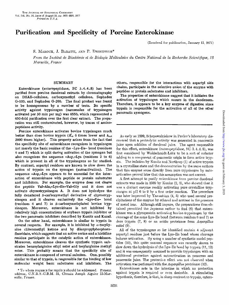
Fro. 2. Second chromatography on DEAE-cellulose. The DEAE-cellulose (Whatman No. 11) column, 4 X 10 cm, equili- brated with a 10 mM Tris-acetat,e buffer (pH 6.0), 50 mM in NaCI, was eluted by a 50 + 400 mM linear NaCl concentration gradient (volume of the two chambers, 400 ml). The specific activities of enterokinase marked along t,he corresponding peak were measured with trypsinogen as substrate (cf. Fig. 1). Elution rate, 55 ml per hour. Volume of fractions, 7 ml.
severe losses in enzyme activity for a relatively low purification effect.
About 80 porcine duodena were collected in the slaughterhouse immediately after the death of the animals. They were kept at 0”; ul)on arrival in the laboratory each was rinsed twice with 25 ml of cold wat,er. The extracts were pooled and filtered through gnuzl?. The filtrate was acidified to pH 5.0 by addition of glacial acetic acid. 111 further esperiments were performed at 4”. Aft,er standing for 1 hour, the precipitate was removed by a 30- min centrifugation at 27,000 X ‘g in a Sorvall centrifuge model RC 2 13 equipped with a GSA rotor. The pH 5 supernatant (about 4 liters) was pumped into a DEAE-cellulose (Whatman No. 11) column, 6 X 10 cm, equilibrated with a 10 rnnf Tris- acetat,e buffer, pl-I 6.0, 50 mlcI in NaCl. After a preliminary washing of the column with 300 ml of buffer, elution was per- formed at the same pH by a 50 to 400 mM linear NaCl concen- tration gradient. Under these conditions, the enterokinase ac- tivity emerged from the column at a NaC1 concentrat.ion of about 180 ni>I. lifter dialysis of the pooled fractions and lyophiliza- tion, about 1 g of a brown powder was obtained.
The powder was dissolved in 50 ml of an ammoniutn acetate- acetic acid buffer (pH 5.0), 50 rnM in acetic acid, the insoluble fraction was spun down at. 30,000 X g for 15 min, and the clear superiiatant was charged onto a CM-cellulose column, 4 X 60 cm, equilibrated with the acetate buffer. The unretarded frac- tions containing enterokinase were lyophilized to give 200 mg of a white powder.
This powder was dissolved in 15 ml of a 10 mM Tris-acetate buffer, pH 6.0, 500 m&l in NaCl, and the solution was filtered through a Sephades G-100 column, 3 X 200 cm, equilibrated wit,11 t,he satne buffer. Enterokinase migrated unretarded, and during the filtration it, was separated from the last traces of
TABLE I Flow sheet of purijication procedure
The last Sephadex G-200 filtration of the procedure (Fig. 3) is not included in the table because of the relatively heavy and variable losses observed during this step.
Aqueous extract. . . . . . . . . . pH 5.0 supernatant . . . . . . First DEAE-cellulose chromatog-
raphy........................... Chromatography on CM-cellulose.. Filtration through Sephadex G-100. Second DEAE-cellulose chroma-
tography . . , . . . . . . . . . . . . . . .
No. of enterokinase
units
130,000 97,500 10
58,500 60 26,990 300 23,400 557
19,500 1,350
Sp+fic a,‘t:;“,‘:d’
rypsinogen
Yield
% 100
75
45 20 18
15
R6150
: :, 2 \
6770 $ Q"600 w'
1.2 IL 1.6 BREAKTHROUGH VOLUMES
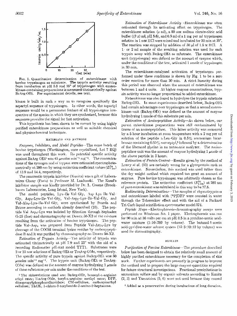
FIG. 3. First filtration through Sephadex G-200. The Sepha- dex G-200 column, 3 X 400 cm, was equilibrated and eluted by a 10 mM Tris-acetate buffer (pH 6.0), 500 mM in NaCl. The specific activities of enterokinase marked along t,he peak were measured with trypsinogen as substrate (cf. Fig. 1). Elution rate, 12 ml per hour. Breakthrough volume of the column, 900 ml.
trypsin and from nucleic acids. It was still contaminated, how- ever, by a major component detectable by disc electrophoresis (7.5% gel; pH 8.6). This latter was removed by a second chro- matography on a DEAE-cellulose column, 4 X 10 cm, performed under the same conditions as the first. Enterokinase is shown in Fig. 2 to emerge from this column as a single peak. All of the fractions under the peak gave a single band by disc electrophore- sis and displayed a constant specific activity (trypsinogen) of 1350 units per mg. Table I indicates that at this stage the over-all yield of activity was about 15% of the activity in the initial extract. The purification was about 135-fold over the pH 5 supernatant.
In spite of the evidence of homogeneity reported above, the preparations thus obtained (29 mg) were not yet pure. They did not hydrolyze p-nitrophenylacetate, a substrate common to many proteases and esterases, but they were found to split the dipep-
by guest on February 3, 2019http://w
ww
.jbc.org/D
ownloaded from
5034 Specificity of Enterokinase Vol. 246,-No. 16
BREAKTHROUGH VOLUMES
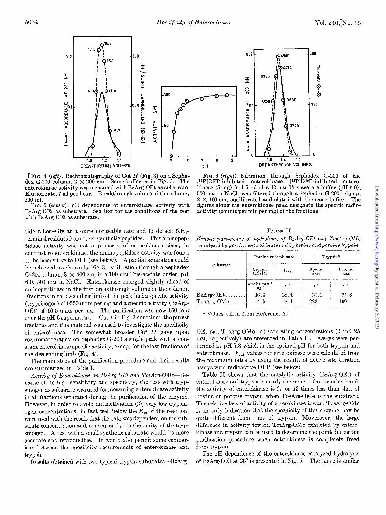
t FIG. 4 (left). Rechromatography of Cut ZZ (Fig. 3) on a Sepha- dex G-200 column, 2 X 200 cm. Same buffer as in Fig. 3. The enterokinase activity was measured with BzArg-OEt as substrate. Elution rate, 7 ml per hour. Breakthrough volume of the column, 200 ml.
FIG. 5 (cenfer). pH dependence of enterokinase activity with BzArg-OEt as substrate. See text for the conditions of the test with BzArg-OEt as substrate.
tide n-Leu-Gly at a quite noticeable rate and to detach NHT terminal residues from other synthetic peptides. This aminopep- tidase activity was not a property of enterokinase since, in contrast to enterokinase, the aminopeptidase activity was found to be insensitive to DFP (see below). A partial separation could be achieved, as shown by Fig. 3, by filtration through a Sephadex G-200 column, 3 x 400 cm, in a 100 rnrvr Tris-acetate buffer, pH 6.0, 500 nuz in NaCI. Enterokinase emerged slightly ahead of aminopeptidase in the first breakthrough volume of the column. Fractions in the ascending limb of the peak had a specific activity (trypsinogen) of 6550 units per mg and a specific activity (BzArg- OEt) of 16.0 units per mg. The purification was now 650-fold over the pH 5 supernatant. Cut I in Fig. 3 contained the purest fractions and this mat,erial was used to investigate the specificity of enterokinase. The somewhat broader Cut II gave upon rechromatography on Sephadex G-200 a single peak with a con- stant enterokinase specific activity, except for the last fractions of the descending limb (Fig. 4).
The main steps of the purification procedure and their results are summarized in Table I.
Activity of Enterokinase on BzArg-OEt and TosArg-OMe-Be- cause of its high sensitivity and specificity, the test with tryp- sinogen as substrate was used for measuring enterokinase activity in all fractions separated during the purification of the enzyme. However, in order to avoid autoactivation (2), very low trypsin- ogen concentrations, in fact well below the K, of the reaction, were used with the result that the rate was dependent on the sub- strate concentration and, consequently, on the purity of the tryp- sinogen. A test with a small synthetic substrate would be more accurate and reproducible. It would also permit some compar- ison between the specificity requirements of enterokinase and trypsin.
Results obtained with two typical trypsin substrates-BzArg-
PH
1.0 1.2 1.1 BREAKTHROUGH VOLUMES
Fro. 6 (right). Filtration through Sephadex G-200 of the [32P]DFP-inhibited enterokinase. [32P]DFP-inhibited entero- kinase (5 mg) in 1.5 ml of a 10 mM Tris-acetate buffer (pH 6.0), 500 mM in NaCl, was filtered through a Sephadex G-200 column, 2 X 100 cm, equilibrated and eluted with the same buffer. The figures along the enterokinase peak designate the specific radio- activity (counts per min per mg) of the fractions.
TABLE II
Kinetic parameters of hydrolysis of BzArg-OEt and TosArg-OMe catalyzed by porcine enterokinase and by bovine and porcine trypsin
Substrate
Porcine enterokinase
Specific &at activity
Trypsin”
Bovine POKiIR &at &at
BzArg-OEt . . TosArg-OMe.. :::I ‘:I: 1 “:I: 1 2::‘” 1 1::‘”
0 Values taken from Reference 14.
OEt and TosArg-OMe-at saturating concentrations (2 and 25 mM, respectively) are presented in Table II. Assays were per- formed at pH 7.8 which is the optimal pH for both trypsin and enterokinase. /c,,~ values for enterokinase were calculated from the maximum rates by using the results of active site titration assays with radioactive DFP (see below).
Table II shows that the catalytic activity (BzArg-OEt) of enterokinase and trypsin is nearly the same. On the other hand, the activity of enterokinase is 27 or 13 times less than that of bovine or porcine trypsin when TosArg-OMe is the substrate. The relative lack of activity of enterokinase toward TosArg-OMe is an early indication that the specificity of this enzyme may be quite different from that of trypsin. Moverover, the large difference in activity toward TosArg-OMe exhibited by entero- kinase and trypsin can be used to determine the point during the purification procedure when enterokinase is completely freed from trypsin.
The pH dependence of the enterokinase-catalyzed hydrolysis of BzArg-OEt at 25” is presented in Fig. 5. The curve is similar
by guest on February 3, 2019http://w
ww
.jbc.org/D
ownloaded from
Issue of August 25, 1971 S. Maroux, J. Baratti, and P. Demuelle 5035
eu ou TiME (MiN) 11 S (rnM-’ 1
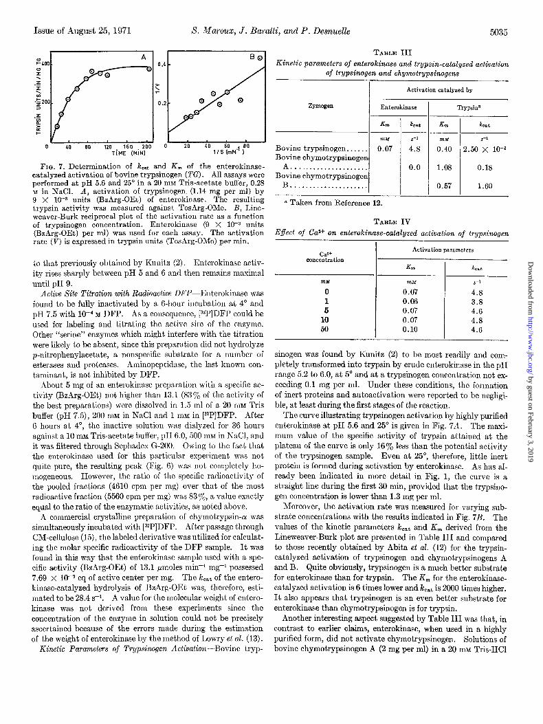
FIG. 7. Determination of k,,t and K, of the enterokinase- catalyzed activation of bovine trypsinogen (TG). All assays were performed at pH 5.6 and 25” in a 20 mM Tris-acetate buffer, 0.28 M in NaCl. A, activation of trypsinogen. (1.14 mg per ml) by 9 X 10eJ units (BzArg-OEt) of enterokinase. The resulting trypsin activity was measured against TosArg-OMe. B, Line- weaver-Burk reciprocal plot of the activation rate as a function of trypsinogen concentration. Enterokinase (9 X 10-s units (BzArg-OEt) per ml) was used for each assay. The activation rate (V) is expressed in trypsin units (TosArg-OMe) per min.
to that previously obtained by Kunitz (2). Enterokinase activ- ity rises sharply between pH 5 and 6 and then remains maximal until pH 9.
Active Site Titration with Radioactive DFP-Enterokinase was found to be fully inactivated by a B-hour incubation at 4” and pH 7.5 with low4 M DFP. As a consequence, [32P]DFP could be used for labeling and titrating the active site of the enzyme. Other “serine” enzymes which might interfere with the titration were likely to be absent, since this preparation did not hydrolyze p-nitrophenylacetate, a nonspecific substrate for a number of esterases and proteases. Aminopeptidase, the last known con- taminant, is not inhibited by DFP.
About 5 mg of an enterokinase preparation with a specific ac- tivity (BzArg-OEt) not higher than 13.1 (83yc of the activity of the best preparations) were dissolved in 1.5 ml of a 20 mM Tris buffer (pH 7.5), 200 mM in NaCl and 1 mM in [32P]DFP. After 6 hours at 4”, the inactive solution was dialyzed for 36 hours against a 10 mM Tris-acetate buffer, pH 6.0, 500 mM in NaCl, and it was filtered through Sephadex G-200. Owing to the fact that the enterokinase used for this particular experiment was not quite pure, the resulting peak (Fig. 6) was not completely ho- mogeneous. However, the ratio of the specific radioactivity of the pooled fractions (4610 cpm per mg) over that of the most radioactive fraction (5560 cpm per mg) was 83%,, a value exactly equal to the ratio of the enzymatic activit,ies. as noted above.
A commercial crystalline preparation of chymotrypsin-a! was simultaneously incubated with [32P]DFP. After passage through CM-cellulose (15), the labeled derivative was utilized for calculat- ing the molar specific radioactivity of the DFP sample. It was found in this way that the enterokinase sample used with a spe- cific activity (BzArg-OEt) of 13.1 pmoles min-r mg+ possessed 7.69 x lop9 eq of active center per mg. The &at of the entero- kinase-catalyzed hydrolysis of BzArp-OEt was, therefore, esti- mated to be 28.4 s-l. A value for the molecular weight of entero- kinase was not derived from these experiments since the concentration of the enzyme in solution could not be precisely ascertained because of the errors made during the estimation of the weight of enterokinase by the method of Lowry et al. (13).
Kinetic Parameters of Trypsirwgen Activation-Bovine tryp-
TABLE III Kinetic parameters of enterokinase and trupsin-catalyzed activation
of trypsinogen and chymotrypsinogens
Activation catalyzed by
zymogen Enterokinase
&?I &at
?nM s-1
Bovine trypsinogen.. . . _ . 0.07 4.8 Bovine chymotrypsinogen
A . . . . . . . . . . . . . . . . . . . . 0.0 Bovine chymotrypsinogen
B . . . . . . . . . . . . . . . . . . _ .
I Trypsin”
K?fl
?ndl
0.40
1.08
0.57
hat
s-1
2.50 X 1O-3
0.18
1.60
a Taken from Reference 12.
TABLE IV Effect of Ca2+ on enterokinase-catalyzed activation of trypsinogen
ca2+ concentration
?md
0 1 6
10 50
- I Activation parameters
. - Klf2 &at
?nM s-1
0.07 4.8 0.06 3.8 0.07 4.6 0.07 4.8 0.10 4.6
sinogen was found by Kunitz (2) to be most readily and com- pletely transformed into trypsin by crude enterokinase in the pH range 5.2 to 6.0, at 5” and at a trypsinogen concentration not ex- ceeding 0.1 mg per ml. Under these conditions, the formation of inert proteins and autoactivation were reported to be negligi- ble, at least during the first stages of the reaction.
The curve illustrating trypsinogen activation by highly purified enterokinase at pH 5.6 and 25’ is given in Fig. 711. The maxi- mum value of the specific activity of trypsin attained at the plateau of the curve is only 16% less than the potential activity of the trypsinogen sample. Even at 25”, therefore, little inert protein is formed during activation by enterokinase. As has al- ready been indicated in more detail in Fig. 1, the curve is a straight line during the first 30 min, provided that the trypsino- gen concentration is lower than 1.3 mg per ml.
Moreover, the activation rate was measured for varying sub- strate concentrations with the results indicated in Fig. 7B. The values of the kinetic parameters kcst and K, derived from the Lineweaver-Burk plot are presented in Table III and compared to those recently obtained by Abita et al. (12) for the trypsin- catalyzed activation of trypsinogen and chymotrypsinogens A and B. Quite obviously, trypsinogen is a much better substrate for enterokinase than for trypsin. The K, for the enterokinase- catalyzed activation is 6 times lower and keat is 2000 times higher. It also appears that trypsinogen is an even better substrate for enterokinase than chymotrypsinogen is for trypsin.
Another interesting aspect suggested by Table III was that, in contrast to earlier claims, enterokinase, when used in a highly purified form, did not activate chymotrypsinogen. Solutions of bovine chymotrypsinogen A (2 mg per ml) in a 20 m&t Tris-HCl
by guest on February 3, 2019http://w
ww
.jbc.org/D
ownloaded from
5036
0.i
SpeciJicity of Enterokinase Vol. 246, No. 16
40 l/S rnt”l-’
80
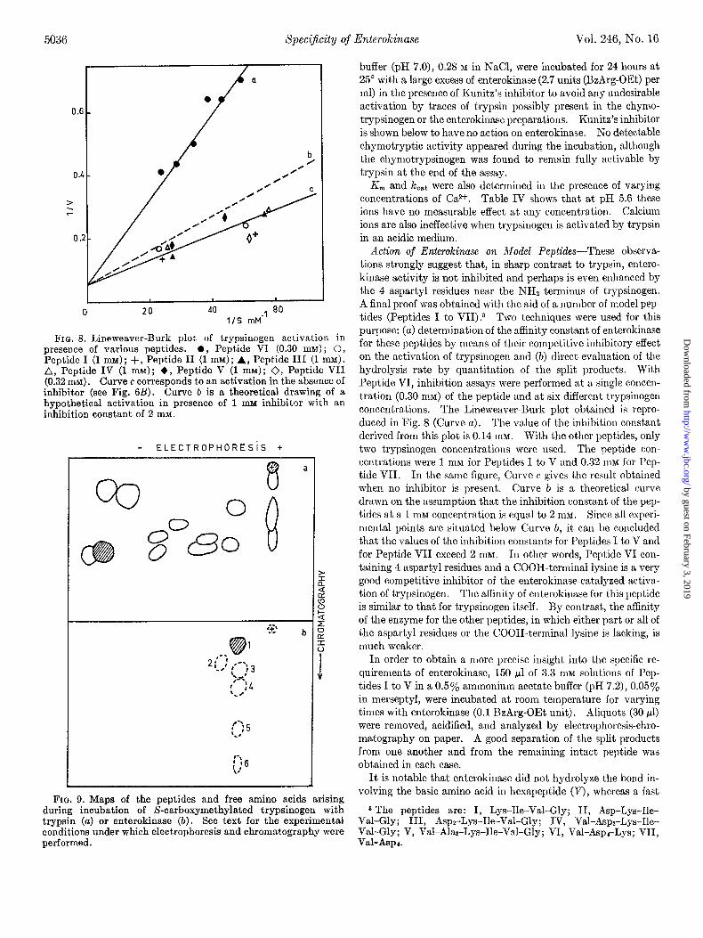
FIQ. 8. Lineweaver-Burk plot of trypsinogen activation in presence of various peptides. 0, Peptide VI (0.30 mM) ; 0, Peptide I (1 mM); +, Peptide II (1 mM); A, Peptide III (1 mM). A, Peptide IV (1 mM); +, Peptide V (1 mM); 0, Peptide VII (0.32 mM). Curve c corresponds to an activation in the absence of inhibitor (see Fig. 6B). Curve b is a theoretical drawing of a hypothetical activation in presence of 1 mu inhibitor with an inhibition constant of 2 mM.
- ELECTROPHORESiS +
+
8
a
0
I
b
FIG. 9. Maps of the peptides and free amino acids arising during incubation of S-carboxymethylated trypsinogen with trypsin (a) or enterokinase (b). See text for the experimental conditions under which electrophoresis and chromatography were performed.
buffer (pH 7.0), 0.28 M in NaCl, were incubated for 24 hours at 25” with a large excess of enterokinase (2.7 units (BzArg-OEt) per ml) in the presence of Kunitz’s inhibitor to avoid any undesirable activation by traces of trypsin possibly present in the chymo- trypsinogen or the enterokinase preparations. Kunitz’s inhibitor is shown below to have no action on enterokinase. No detectable chymotryptic activity appeared during the incubation, although the chymotrypsinogen was found to remain fully activable by trypsin at the end of the assay.
Km and koat were also determined iu the presence of varying concentrations of Ca2+. Table IV shows that at pH 5.6 these ions have no measurable effect at any concentration. Calcium ions are also ineffective when trypsinogen is activated by trypsin in an acidic medium.
Action of Enterokinase on Model Peptides-These observa- tions strongly suggest that, in sharp contrast to trypsin, entero- kinase activity is not inhibited and perhaps is even enhanced by the 4 aspartyl residues near the NH2 terminus of trypsinogen. A final proof was obtained with the aid of a number of model pep- tides (Peptides I to VII).3 Two techniques were used for this purpose: (a) determination of the affinity constant of enterokinase for these peptides by means of their competitive inhibitory effect on the activation of trypsinogen and (b) direct evaluation of the hydrolysis rate by quantitation of the split products. With Peptide VI, inhibition assays were performed at a single concen- tration (0.30 mM) of the peptide and at six different trypsinogen concentrations. The Lineweaver-Burk plot obtained is repro- duced in Fig. 8 (Curve a). The value of the inhibition constant derived from this plot is 0.14 mM. With the other peptides, only two trypsinogen concentrations were used. The peptide con- centrations were 1 mM for Pept,ides I to V and 0.32 mM for Pep- tide VII. In the same figure, Curve c gives the result obtained when no inhibitor is present. Curve Z, is a theoretical curve drawn on the assumption that the inhibition constant of the pep- tides at a 1 mM concentration is equal to 2 RIM. Since all experi- mental points are situated below Curve b, it can be concluded that the values of the inhibition constants for Peptides I to V and for Peptide VII exceed 2 ITIM. In other words, Peptide VI con- taining 4 aspartyl residues and a COOH-terminal lysine is a very good competitive inhibitor of the enterokinase-catalyzed activa- tion of trypsinogen. The affinity of enterokinase for this peptide is similar to that for trypsinogen itself. By contrast, the affinity of the enzyme for the other peptides, in which either part or all of the aspartyl residues or the COOH-terminal lysine is lacking, is much weaker.
In order to obtain a more precise insight into the specific re- quirements of enterokinase, 150 ~1 of 3.3 mM solutions of Pep- tides I to V in a 0.5% ammonium acetate buffer (pH 7.2), 0.05% in merseptyl, were incubated at room temperature for varying times with enterokinase (0.1 BzArg-OEt unit). Aliquots (30 ~1) were removed, acidified, and analyzed by electrophoresis-chro- matography on paper. A good separation of the split products from one another and from the remaining intact pept.ide was obtained in each case.
It is notable that enterokinase did not hydrolyze the bond in- volving the basic amino acid in hexapeptide (V), whereas a fast
3 The peptides are: I, Lys-Be-Val-Gly; II, Asp-Lys-Ile- Val-Gly; III, Aspz-Lys-Ile-Val-Gly; IV, Val-Asp,-Lys-Ile- Val-Gly; V, Val-Ala,-Lys-Ile-Val-Gly; VI, Val-Asp,-Lys; VII, Val-Asp,.
by guest on February 3, 2019http://w
ww
.jbc.org/D
ownloaded from
Issue of August 25, 1971 S. Maroux, J. Baratti, and P. Desnuelle 5037
cleavage of the same bond was observed (total hydrolysis in 2 hours) in Peptide IV. The only difference between these pep- tides is that, in the first, 2 alanines replace 2 aspartyl residues. Enterokinase also hydrolyzed at a quite noticeable rate aspartyl Peptide III (total hydrolysis in 4 hours) and more slowly Pep- tides II (total hydrolysis in 24 hours) and I (50% hydrolysis in 24 hours).
Hydrolysis of S-Carboxymethylated Chymotrypsirwgen A and Trypsinogen-The specificities of enterokinase and trypsin have also been compared with the aid of two very large peptide chains of known sequence, the S-carboxymethylated derivatives of bovine chymotrypsinogen A and trypsinogen. A suspension of 1.3 mg of each in 100 ~1 of a 0.5% ammonium acetate-acetic acid buffer (pH 7.0), 0.0531, in merseptyl, was incubated at room tem- perature for 24 hours with enterokinase or trypsin (0.5 BzArg- OEt unit). After acidification, the soluble peptides were sepa- rated by electrophoresis-chromatography on paper as described previously.
The peptide map obtained after incubation of S-carboxy- methylated trypsinogen with trypsin is reproduced in Fig. Qa. Fourteen well separated spots are revealed. As already pointed out by Pech&re et al. (16), trypsin is unable to split the bond linking lysine-6 and isoleucine-7 in S-carboxymethylated tryp- sinogen, with the consequence that no spot corresponding to the “activation” hexapeptide, Val-AsprLys, is visible on Map Qa.
Map Qb shows the result obtained from S-carboxymethylated trypsinogen after digestion with enterokinase. A single strong spot (Spot 1) is visible which corresponds to the “activation” hexapeptide. The much weaker spots, 6,4,6, and 6, correspond to free glycine, threonine, valine, and leucine, respectively, which are located near the NH2 terminus of trypsin and consequently are assumed to be liberated by the aminopeptidase present as a contaminant. Spot S is free serine which appears as a result of the cleavage by aminopeptidase of a NH&erminal serine residue in the “inert” proteins (17). The Val-Asp,-Lys spot is clearly visible after only a lo-min incubation of S-carboxymethylated trypsinogen with enterokinase.
Digestion of S-carboxymethylated chymotrypsinogen A by trypsin gave rise to 14 soluble peptides which were well separated from each other by electrophoresis-chromatography. In sharp contrast, however, neither peptides nor free amino-acids could be detected on the maps derived from the soluble portion of the enterokinase digests of this compound. The absence of peptides suggested that none of the 18 peptide bonds involving the basic amino acids in fully denatured chymotrypsinogen A was attacked by enterokinase. The absence of free amino acids arose from the fact that the NHs-terminal S-carboxymethyl cysteine residue of the chain was a poor substrate for aminopeptidase. This result provided an independent confirmation of the fact that chymo- trypsinogen A is not cleaved by enterokinase. Had cleavage occurred, aminopeptidase would have acted upon the newly created NHa-terminal residues.
Interaction with Natural Trypsin Inhibitors-Inhibition of trypsin by some “natural” inhibitors represents a good example of highly selective protein-protein interaction. It was, therefore, interesting to know whether these inhibitors also affect the ac- tivity of pure enterokinase.
Enterokinase (1.7 BzArg-OEt units) in 4.5 ml of a 5 mM Tris buffer (pH 7.9), 0.04 M in NaCl, was incubated at 25” for 30 min with a lo-fold molar excess of soybean trypsin inhibitor or a
c 0 20 40 $0 80
TIME (MIN)
J
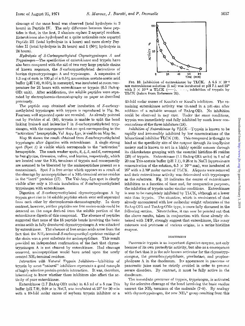
FIG. 10. Inhibition of enterokinase by TLCK. A 6.5 X 10⁻⁴ rnM enterokinase solution (1 ml) was incubated at pH 7.1 and 25’ with 2 X lo+ M TLCK (---). - - -, inhibition of trypsin by TLCK (taken from Reference 20).
40-fold molar excess of Kunitz’s or Kasal’s inhibitors. The re- maining enterokinase activity was titrated in a pa-stat after addition of a suitable amount of BzArg-OEt. No inhibition could be observed in any case. Under the same conditions, trypsin was immediately and fully inhibited by much lower con- centrations of the three inhibitors (18).
Inhibition of Enterokinase by TLCK-Trypsin is known to be rapidly and irreversibly inhibited by low concentrations of the bifunctional inhibitor TLCK (19). This compound is thought to bind at the specificity site of the enzyme through its tosyllysine moiety and is known to act in a highly specific manner through its chloroketone grouping with the essential histidine-46 residue (20) of trypsin. Enterokinase (1.1 BzArg-OEt units) in 1 ml of 20 lllM Tris-acetate buffer (pH 7.1), 0.28 XI in NaCl (approximate concentration of the solution, 6.5 x lo+ mM), was incubated at 25” with a 3.103 molar excess of TLCK. Aliquots were removed and their enterokinase activity was determined with trypsinogen as the substrate. Fig. 10 indicates the course of enterokinase inhibition as a function of time and, for comparative purposes, the inhibition of trypsin under similar conditions. Enterokinase is seen to be completely inhibited by TLCK, although at a lower rate than trypsin. The situation, which is reminiscent of that already encountered with low molecular weight substrates of the Bzhrg-OEt and TosArg-OMe type, is more fully discussed in the following section. Nevertheless, it can now be pointed out that the above results, taken in conjunction with those already ob- tained with DFP, strongly suggest that enterokinase, like many esterases and proteases of various origins, is a serine-histidine enzyme.
DISCUSSION
Pancreatic trypsin is an important digestive enzyme, not only because of its own proteolytic activity, but also as a consequence of the fact that it is the sole known activator for the chymotryp- sinogens, the procarboxypeptidases, proelastase, and prophos- pholipase A in the duodenum. Its appearance in pancreas or pancreatic juice must be strictly avoided in order to prevent severe disorders. By contrast, it must be fully active in the duodenum.
The immediate precursor of trypsin, trypsinogen, is activated by the selective cleavage of the bond involving the basic residue nearest the NH2 terminus of the molecule (7-Q). By analogy with chymotrypsinogen, the new NH3+ group resulting from this
by guest on February 3, 2019http://w
ww
.jbc.org/D
ownloaded from
5038 Specificity of Enterokinase Vol. 246, No. 16
cleavage can be assumed to favor the appearance of a functional active site through the formation of an ionic bridge with a car- boxylate of the molecule (21, 22).
As a result of Kunitz’s extensive and now classical investiga- tions (23), the best known mode of activation of trypsinogen is autoactivation catalyzed by trypsin. However, the 4 aspartyl residues situated just before the strategic lysyl bond exert a very pronounced inhibitory effect upon the action of trypsin (11, 12). As a consequence, it may be assumed that, at the beginning of the process when the ratio of trypsin to trypsinogen is low, autoacti- vation has a limited significance in vivo.
On the other hand, duodenum has been shown to synthesize an enzyme, enterokinase, which can activate trypsinogen by a first order reaction (l-3). Enterokinase, the sugar content of which is about 30% (3, 6), has been reported to be a brush border en- zyme in rat intestine. Consequently, its main site of action is likely to be the surface membrane of the villous epithelial cells rather than the duodenal lumen (24). In spite of this strict localization and of the relatively small amount of enterokinase in the intestine, this enzyme may initiate the activation of tryp- sinogen in &JO, provided that its action is much more efficient than that of trypsin.
This has been found to be the case. The determination of the kinetic parameters of the enterokinase-catalyzed activation of trypsinogen at pH 5.6 showed that trypsinogen is a much better substrate for enterokinase than for trypsin. K,,, is 6 times lower and kcst is 2000 times higher. Moreover, a more precise picture of the specificity requirements of enterokinase was obtained by the use of several model peptides similar in structure to the NH,- terminal sequence of bovine trypsinogen or differing from it, either by the number of aspartyl residues, or by the replacement of these residues by alanine, or finally by the lack of lysine. The results showed that the affinity of enterokinase for its substrates depends upon the presence not only of a lysine residue but also upon the presence of the adjacent aspartyl residues.
Therefore, enterokinase appears to recognize not merely, as does t.rypsin, the aliphatic carbon chain and the basic amino group of lysine, but the entire polyaspartyl-lysine structure. The aspartyl residues, which slow down the action of trypsin, constitute for enterokinase the signal for a fast and highly specific activation. The unusually narrow specificity of enterokinase is shown by the fact that this enzyme hydrolyzes none of the bonds involving the 18 basic residues in fully denatured S-carboxy- methylated chymotrypsinogen A. In S-carboxymethylated trypsinogen, it hydrolyzes exclusively the lysyl bond nearest the NH2 terminus which has 4 aspartyl residues adjacent to it. For trypsin to cleave this bond, trypsinogen must be in the native form (25). Moreover, trypsin splits indiscriminately at all of the basic residues in fully denatured trypsinogen and other pro- teins.
The absolute requirement of enterokinase for a polyaspartyl- lysine structure is not observed with low molecular weight sub- strates such as BzArg-OEt and TosArg-OMe. Similarly, entero- kinase is inhibited by TLCK nearly as rapidly as trypsin. This latter property and the strong inhibitory effect exerted by low concentrations of DFP suggest that a histidine and a serine resi- due are involved in the catalytic site of enterokinase.
All of these results are consistent with the view that the cata- lytic site of enterokinase is very similar to, if not identical with, that of trypsin and of a number of other proteases and esterases.
However, the specificity site of this enzyme is apparently com- posed of several subsites. One, specific for lysine, is probably similar to the specificity site of trypsin. It is responsible for the binding of small substrates (BzArg-OEt, TosArg-OMe) and for the binding of the inhibitor TLCK by the enzyme. The others are necessary for enterokinase to interact with peptides or protein substrates and inhibitors. These latter compounds cannot even bind to the lysine-specific subsite, unless they contain a poly- aspartyl-lysine sequence. This suggests that the subsites act in concert to bring about a correct positioning of the interacting components.
The importance of each of the 4 aspartyl residues (Asp-2 to Asp-5) in the binding of trypsinogen to enterokinase is at present difficult to assess. At first site, it might be postulated that Asp-4 and Asp-5, which are located just before Lys-6, are particularly important since their replacement by alanine (as in Peptide V) completely inhibits the action of enterokinase. The residue Asp-3 would be less essential since its replacement by valine (as in Peptide IV) merely reduces the affinity of the enzyme for the substrate.
In conclusion, the activation of the pancreatic zymogens in the duodenum in tivo appears to be a “cascade” process. It is very likely that trypsinogen is first activated by enterokinase. The trypsin then formed continues the activation of trypsinogen and induces the activation of the other zymogens. In this respect, the conversion of pancreatic zymogens into active enzymes for purposes of digestion is comparable to the successive activations of the 10 factors involved in blood coagulation (26). The com- parison may also be extended to the activation of plasma pro- thrombin, fibrinogen (26), plasminogen (27), prokallikrein (28), and kininogen (29). In most of these cases, trypsin can bring about the transformation, although at a relatively low rate and with a relatively poor specificity. However, the enzymes re- sponsible for these conversions in viva are much more efficient. They are also much more specific since each reaction requires a separate enzyme. Like enterokinase, the blood enzymes can therefore be expected to recognize not merely a basic residue but a broader structural unit.
It will be interesting in the future to know whether lysine may be replaced by arginine and aspartic acid by glutamic acid in the structural unit recognized by enterokinasc.
The essential role played by enterokinase in the activation of pancreatic precursors in viva has been recently emphasized by the finding that a young girl with an enterokinase deficiency was suffering from quite severe gastrointestinal disorders (30)
Acknowledgments-We wish to thank Dr. M. Rovery and Professor M. Lazdunski for helpful discussions.
1. KUNITZ, M., AND NORTHROP, J. H., J. Gen. Physid., 19, 991 (1936) .’
3: 4. 5. 6. 7.
KUNITZ, M., J. Gen. Physiol., 22,429 (1939). KUNITZ. M, J. Gen. Phvsiol., 22,447 (1939). YAMAS&N~, I., Arch. kerni; ‘7, 539 (1954). YAMASHINA, I., Arch. Kemi, 9, 225 (1956). YAMASHINA, I., Acta Chem. &and., 10,739 (1956). ROVERY, M., FABRE, C., AND DESNUELLE, P., Biochim. Bio-
8. phys. Ada, 12, 547 (1953).
DAVIE, E. W., AND NEURATH, H., J. Biol. Chem., 2l2, 515 (1955).
REFERENCES
by guest on February 3, 2019http://w
ww
.jbc.org/D
ownloaded from
Issue of August 25, 1971 X. Maroux, J. Baratti, ancl P. Desnuelle 5039
9. GABELOTEAU, C., AND DESNUELLE, P., Biochim. Biophys. Acta, 42, 230 (1960).
10. SVARDA, J., AND BRICAS, E., Bull. Sot. Chim. France, 2423 (1968).
11. D~&AA&, M., DESNUELLE, P., LAZDUNSKI, M., BRICAS, E AND SVARDA, J., Biochem. Biophys. Res. Commun., 29, 235 (1967).
12. ABITA, J. P., DELAAGE, M., L.UDUNSKI, M., AND SVARDA, J., Eur. J. Biochem., 8, 314 (1969).
13. LOWRY, 0. H., ROSEBROUGH, N. J., LEWIS FARR, A. L., AND RANDALL, R. J., J. Biol. Chem., 193, 265 (1951).
14. LAZDUNSKI, M., Bull. Sot. Chim. Biol., 47, 301 (1965). 15. ROVERY, M., BIANCHETTA, J., AND DESNUELLE, P., Biochim.
Biophys. Acta, in press. 16. PECHERE, J.-F., DIXON, G. H., MAYBURY, R. H., AND NEU-
RATH, H., J. Biol. Chem., 233, 1364 (1958). 17. KUNITZ, M., J. Gen. Physiol., 22, 293 (1939). 13. VINCENT, J. P., LAZDUNSKI, M., AND DILAAGE, M., Eur. J.
Biochem., 12, 250 (1970). 19. SHAW, E., MARES-GUIA, M., AND COHEN, W., Biochemistry 4,
2219 (1965). 20. PETRA, P. H., COHEN, W., AND SHAW, E. N., Biochem. Biophys.
Res. Commun., 21, 612 (1965).
21. SIGLER, P. B., BLOW, D. M., MATTHEWS, B. W. AND HENDER- SON, R., J. Mol. Biol., 36, 143 (1968).
22. LAZDUNSKI, M., DELAAQE, M., ABITA, J. P., AND VINCENT, J. P., in P. DESNUELLE, H. NEURATH, AND M. OTTESON (Editors), Structure-function rezationships in proteolytic enzymes. Munskgaard, Copenhagen 1969, p. 42.
23. NORTHROP, J. H., KUNITZ, M., AND HERRIOTT, R., Crystalline enzymes, Ed. 2, Columbia University Press, New York, 1948.
24. NORDSTROM, C., AND DAHLQVIST, A., Biochim. Biophys. Acta, 198, 621 (1970).
25. GABELOTEAU, C., AND DESNUELLE, P., Arch. Biochem. Bio- phys., 89, 475 (1957).
26. ESNOUF, M. P., AND MACFARLANE, R. C., Advan. Enzymol., 20, 255 (1968).
27. SUMMARIA, L., HSIEH, B., AND ROBBINS, K. C., J. BioZ. Chem., 242, 4279 (1967).
28. NA&SAWA,‘ S., .TAKAHASHI, H., KOIDA, M., SUZUKI, T., AND SCHOENMAKERS, J. G. G., Biochem. Biophys. Res. Commun., 32,644 (1968).
29. KATO, H., AND Suzug~, T., Biochem. Biophys. Res. Commun., 32, 800 (1968).
30. HADORN, B., TARLOW, M. J., LLOYD, J. K. AND WOLFF, 0. H., Lancet, 812 (1969).
by guest on February 3, 2019http://w
ww
.jbc.org/D
ownloaded from
S. Maroux, J. Baratti and P. DesnuellePurification and Specificity of Porcine Enterokinase
1971, 246:5031-5039.J. Biol. Chem.
http://www.jbc.org/content/246/16/5031Access the most updated version of this article at
Alerts:
When a correction for this article is posted•
When this article is cited•
to choose from all of JBC's e-mail alertsClick here
http://www.jbc.org/content/246/16/5031.full.html#ref-list-1
This article cites 0 references, 0 of which can be accessed free at
by guest on February 3, 2019http://w
ww
.jbc.org/D
ownloaded from