Embed Size (px)
Citation preview
JOURAUL OF BACTERIOLOGY, Dec. 1992, p. 7689-76960021-9193/92/237689-08$02.00/0Copyright © 1992, American Society for Microbiology
Vol. 174, No. 23
Purification and Characterization of a Mutant DnaB ProteinSpecifically Defective in ATP Hydrolysis
PARESH SHRIMANKAR,t LEIF STORDAL, AND RUSSELL MAURER*
Department ofMolecular Biology and Microbiology, Case Western ReserveUniversity School of Medicine, Cleveland, Ohio 44106-4960
Received 5 August 1992/Accepted 2 October 1992
The dnaB gene of Escherichia coli encodes an essential DNA replication enzyme. Fueled by the energyderived from the hydrolysis ofATP to ADP + P1, this enzyme unwinds double-stranded DNA in advance of theDNA polymerase. While doing so, it intermittently stimulates primase to synthesize an RNA primer for anOkazaki frmMent. To better understand the structural basis of these and other aspects of DnaB function, wehave initiated a study of mutant DnaB proteins. Here, we report the purification and characterization of amutant DnaB protein (RC231) containing cysteine in place of arginine at residue 231. The mutant proteinattains a stable, properly folded structure that allows association of six promoters to form a hexamer, as is alsotrue for wild-type DnaB. Further, the mutant protein interacts with ATP, the nonhydrolyzable ATP analogadenosine-5'-O-(3-thiotriphosphate) (ATP'yS), ADP, and poly(dT), and it stimulates primase action. It is,however, profoundly deficient in ATP hydrolysis, helicase activity, and replication activity at the chromosomalorigin of replication. In addition, while general prnming reactions with wild-type DnaB and ATP elicited thesynthesis of short primers, reactions with DnaB and ATPyS or with RC231 and either ATP or ATP'ySstimulated the synthesis of significantly longer primers. On the basis of these observations, we suggest thatprimase interacts directly with DnaB throughout primer synthesis during general priming, until dissociation ofDnaB from DNA or ATP hydrolysis by DnaB disrupts the interaction and leads to primer termination.
The DnaB protein of Escherichia coli is required forreplication of the bacterial chromosome and many plasmidsand bacteriophage DNAs (14). Native DnaB protein is ahexamer of identical subunits encoded by the dnaB gene (6,25). Its participation in DNA replication begins when a DnaBhexamer associates with up to six molecules of a specifictransferase, the DnaC protein (13). This complex is compe-tent to transfer DnaB to a region of single-stranded DNA(ssDNA) at the chromosomal origin of replication. Whenbound to ssDNA, DnaB exhibits a potent ATPase activity(yielding ADP + Pi) (2, 26) and is thought to migrate in a5'-to-3' direction along the single strand to which it is bound,thereby entering the replication fork (7, 17). As this actioncontinues, the double-stranded DNA at the fork becomesunwound, thus moving the fork. DnaB is the only helicaserequired to reconstitute DNA replication in vitro dependenton the chromosomal origin of replication (oriC), but it ispossible that other helicases (Rep, UvrD, PriA) play sup-porting or backup roles in vivo (19).
In addition to its role as a helicase, DnaB criticallyinfluences the action of another essential replication en-zyme, the DnaG primase. Primase synthesizes short RNAsthat are extended by DNA polymerase (which cannot startchains de novo) to produce classical Okazaki fragments, atleast on the lagging strand (4). Primase does not associatespontaneously with either single-stranded or double-stranded naked DNA; rather, other proteins, including DnaB(1, 22), must first be present on the DNA. It is thought thatthe association of primase with the replication fork is dy-namic, occurring while DnaB continues to unwind double-
* Corresponding author.t Present address: Department of Genetics, Washington Univer-
sity School of Medicine, St. Louis, MO 63110.
stranded DNA (31). However, there is no clear evidence thatprimase interacts with DnaB directly.Although these functions of DnaB have been investigated
in detail, little is known about the structural determinants ofDnaB function. Partial trypsin digestion of DnaB showedthat it could be digested into two relatively stable domains of12,000 Da (N terminal) and 33,000 Da (C terminal), with lossof a 45-amino-acid linker region (23). The amino-terminaldomain was missing 14 amino acids from its N-terminal end.The carboxyl-terminal domain was shown to retain nucleo-tide binding, ssDNA-dependent ATPase activity and theability to form hexamers. None of the other activities ofnative DnaB was found in either of the two domains. Thehelicase activity of DnaB was unknown at the time.With the goal of better understanding the structural basis
for DnaB function, our laboratory has isolated dominant-lethal'mutations in the dnaB gene (21). The dominant-lethalphenotype predicts that the encoded proteins, although notfully functional, are structurally intact since they interferewith host DNA replication. Such proteins are excellentcandidates for analysis in vitro because they are likely toexhibit a specific alteration of function. Here, we report theoverexpression, purification, and characterization of onesuch mutant DnaB protein, which contains cysteine in placeof arginine at position 231. This protein was found to have aprimary defect in ATP hydrolysis.
MATERIALS AND METHODS
Materials. Except where indicated otherwise, chemicalsand enzymes were obtained from commercial suppliers andwere reagent grade or equivalent. Polyclonal antibodyagainst DnaB and a sample of purified DnaB were generousgifts from Linda Reha-Krantz, and primase was generouslyprovided by Charles McHenry. Poly(dT) used in theseexperiments was characterized as having an average chain
7689
on July 17, 2018 by guesthttp://jb.asm
.org/D
ownloaded from
7690 SHRIMANKAR ET AL.
length of 167 on the basis of velocity sedimentation, accord-ing to the manufacturer (Pharmacia-LKB). However, thispoly(dT) migrated more slowly than a 693-base polymerasechain reaction product during electrophoresis over a se-quencing gel. In fact, its migration was indistinguishablefrom that of a separate lot of poly(dT) characterized by themanufacturer as having average length 824. That thepoly(dT) chain length was indeed much greater than 167 wasconfirmed by an independent technique in which (dA)15,annealed at random to the poly(dT), was extended to the endof the template with Sequenase. This reaction produced aladder of products, the longest of which was at least severalhundred bases longer than 167.
Buffers. Buffer A was 50 mM Tris-HCl (pH 7.5). Buffer Bcontained 50 mM Tris-HCl (pH 7.5), 10% (wt/vol) sucrose,and 2 mM EDTA. Buffer C contained 50 mM Tris-HCl (pH7.5), 5 mM MgCl2, 30mM KCl, 1 mM ATP, and 25% (wt/vol)glycerol. Buffer D (10x) contained 400 mM 4-(2-hydroxy-ethyl)-1-piperazineethanesulfonic acid (HEPES)-KOH (pH7.5), 500 p,g of bovine serum albumin (BSA) per ml, 110 mMmagnesium acetate, and 34 mM ATP. Buffer E contained 20mM potassium phosphate (pH 6.5), 1 mM dithiothreitol, and0.5 mM EDTA. Buffer F contained 89 mM Tris-HCl (pH8.0), 89 mM boric acid, and 2 mM EDTA.
Bacterial strains. MG1655 is an F- prototroph of E. coliK-12 obtained from G. Weinstock (11). RM990 (dnaB+) andits dnaB22(Ts) derivative RM1031 have been describedpreviously (21). RM2528 is a recA56 srl-300::TnlO deriva-tive of RM990. BL21 is an F- ompT rB- mB- derivative ofE. coli B. BL21(DE3) carries a X prophage bearing a copy ofthe T7 RNA polymerase gene under heterologous (lac)control. BL21 and BL21(DE3) were obtained from A.Rosenberg and F. W. Studier (29).
Cloning, subcloning, and mutagenesis of the dnaB gene.From a clone of the E. coli dnaB gene in XSE6 (9), a 3.6-kbBglII fragment containing the entire dnaB gene was sub-cloned into M13mpl8 (32). By using oligonucleotide-directedmutagenesis (16), codon 231 of the dnaB gene in M13mpl8was changed to encode cysteine instead of the wild-typearginine, a change identical to the one in dnaB129, a strongdominant-lethal mutation in the Salmonella typhimuriumdnaB gene (21). The mutant E. coli gene will be designateddnaB720, and its encoded protein will be referred to asRC231. The XSE6 and M13mpl8 clones of dnaB eachcomplemented the temperature-sensitive phenotype of E.coli RM1031, whereas dnaB720 failed to show complemen-tation.
Overexpression and purification. Using oligonucleotide-directed mutagenesis, we generated an NcoI site (5'-CCA&lG-3') overlapping the initiator methionine codon.DNA fragments (from either the wild-type dnaB gene ordnaB720) starting from this NcoI site up to a BamHI site 3'of the gene were subcloned into overexpression plasmidpET-3d (dnaB) or pET-lld (dnaB720) (29). The dnaB anddnaB720 genes were thus placed under T7 transcriptionaland translational signals. The wild-type DnaB protein wasoverexpressed from plasmid pET-3d in a BL21(DE3) hostthat also harbored plasmid pLysS (29). No overexpression ofDnaB was seen in the absence of plasmid pLysS. The T7lysozyme encoded by plasmid pLysS enhanced the stabilityof plasmid pET-3d with the dnaB insert, presumably byinhibiting background T7 RNA polymerase activity and theassociated DnaB expression. Cell growth was even moresensitive to background expression of RC231. Even in thetightly regulated pET-lld vector, the dnaB720 gene could beexpressed in the cell only in the presence of plasmid pLysS.
Wild-type DnaB and RC231 were purified by the sameprocedure, throughout which the two proteins behavedidentically. Purification was carried out by modifying theprocedure of Khatri et al. (12). Briefly, cells were lysed bythe action of the pLysS-encoded lysozyme, aided by afreeze-thaw step. The lysate supernatant was fractionated byprecipitation with ammonium sulfate (ultrapure; Schwartz/Mann). The fraction that was soluble at 20% and insoluble at40% ammonium sulfate saturation contained DnaB. Thisfraction was desalted and further fractionated over a DEAE-Sephacel (Pharmacia-LKB) ion-exchange column, whichwas washed with 150 mM KCl in buffer C and eluted with alinear gradient of 190 to 400 mM KCl in buffer C. In somepreparations, alternatively, the column was washed at 190mM KCl in buffer C, then washed at 240 mM KCl in bufferC, and eluted with a gradient of 240 to 400 mM KCl in bufferC. The peak fractions were pooled and were essentiallypure. In some preparations, the DEAE-Sephacel chromatog-raphy was repeated or the pooled fractions were similarlychromatographed over a Mono-Q column (Pharmacia-LKB)to obtain sufficient purity. Although these preparations werefree of detectable contaminating proteins, we recently foundsubstantial amounts of RNA in some wild-type DnaB andRC231 preparations, including some of the preparations usedfor the experiments described here. Polyribonucleotides areknown not to affect the ATPase activity of DnaB protein (2).We have found that this RNA can be removed from thepurified protein by either hydroxyapatite chromatography,glycerol gradient centrifugation, or chromatography onMono-Q.ATPase assay. Assay of hydrolyzed ATP was carried out
by a modification of the procedure described earlier (8, 10).A final reaction volume (180 ,ul) contained 50 mM Tris-HCl(pH 7.5), 10 mM MgCl2, 75 nmol (as nucleotide; =25 p,g2 ofM13 ssDNA or poly(dT) (when included), 2.1 mM [y- 2P]ATP at 700 cpm/nmol (obtained by mixing labeled ATP at 10Ci/mmol [New England Nuclear] with unlabeled ATP [Sig-ma]), and 1.35 ,ug of either DnaB or RC231. The reactionmixtures, minus protein, were assembled on ice and warmedto 37°C for 5 min, and the reactions were initiated by theaddition of protein and maintained at 37°C thereafter. Atvarious times, a 25-pl aliquot was withdrawn from thereaction mixture and processed as follows. ATP hydrolysisin the aliquot was stopped by the addition of 100 pl of 20 mMsilicotungstic acid in 20 mM sulfuric acid and 240 ,ul of 1 mMpotassium phosphate (pH 7.0) at 0°C. To this were addedsequentially 100 ,ul of 5% (wt/vol) ammonium molybdate in 4M sulfuric acid, 60 pl of 1:1 acetone-5% (wt/vol) trichloro-acetic acid, and 400 p,l of 1:1 isobutanol-benzene. The tubeswere vortexed at high speed for 30 s, phases were allowed toseparate for 15 min, and the amount of Pi in the upperorganic phase was estimated by counting a 100-p,l aliquot ofthe upper phase. The total Pi released was adjusted for atotal volume of 350 p,l in the upper organic phase.
Helicase assay. The helicase substrate was prepared byhybridization of an end-labeled 24-mer oligonucleotide to asingle-stranded M13 template and isolating the hybrid asdescribed previously (12). The helicase reaction was carriedout as follows. In a total volume of 15 ,ul were included 1.5p,l of lOx buffer D, 30 to 50 fmol of helicase substrate, and-24 pmol of DnaB or -14 to 56 pmol of RC231. After beingassembled on ice, the mixture was incubated at 37°C for 10min and then immediately loaded on a 20% polyacrylamidegel in buffer F and run at 4°C for 90 to 120 min at 150 V. Theresults were evaluated by autoradiography.
Partial trypsin digestion. Partial trypsin digestion was
J. BACTERIOL.
on July 17, 2018 by guesthttp://jb.asm
.org/D
ownloaded from
DnaB PROTEIN DEFECTIVE IN ATP HYDROLYSIS 7691
carried out by a modification of a previously describedprocedure (24). A 12.5-,ug sample of DnaB or RC231 wasdigested with 13.5 pg of trypsin (Sigma) in a total volume of11 pl. Incubation was at 0°C for 30 min. The reaction wasstopped by adding 10 ,ul of 3% sodium dodecyl sulfate(SDS)-40 mM dithiothreitol, and the digestion products wereresolved by electrophoresis through an SDS-polyacrylamidegel (10% acrylamide) and stained with Coomassie stain.Identification of cleavage products was confirmed by amino-terminal microsequencing (20).
Glycerol gradient centrifugation. DnaB or RC231 wasanalyzed by centrifugation of 100 p,g of protein over a 20 to45% (wt/vol) glycerol gradient in buffer E-200 mM KCl-5mM ATP at 50,000 rpm for 20 h at 4°C in a Beckman SW50.1rotor. Fractions (180 pul) were collected from the bottom ofthe tube, and the protein concentration was assayed by usingthe Bradford reagent. Markers used were horse heart cy-tochrome c, BSA, and bovine liver catalase.
General priming assay. General priming was done essen-tially as described previously (1). Each reaction mixturecontained 19 mM Tris (pH 7.5), 3.8% (wt/vol) sucrose, 7.7mM dithiothreitol, 38 p,g of BSA per ml, 7.7 mM MgCl2,3.8 pug of rifampin per ml, 3.8 p,g of primase per ml, 770p,M (unlabeled) ATP or adenosine-5'-O-(3-thiotriphosphate(ATP-yS), 10 p,Ci of [a-32P]ATP (>3,000 Ci/mmol; Amer-sham), 69 p,g of poly(dT) per ml, and 65 p.g of DnaB orRC231 per ml in a total volume of 26 pl. Incubation was for30 min at 37°C. From the original reaction tube, an aliquot of8 p.l was denatured by heat and then fractionated over a 16%polyacrylamide-7 M urea gel and evaluated by autoradiog-raphy; duplicate 5-p,l aliquots were quantitated by binding toDE-81 filters (Whatman) (27).
RESULTS
Cloning and overexpression of the dominant-lethal mutant(dnaB720) of the E. coli dnaB gene. Dominant-lethal muta-tions in the S. typhimurium dnaB gene have been described(21). These mutant proteins have been proposed to interferewith DNA replication through their residual functionalityand thus are promising candidates for structure-functionanalysis. For this study, we re-created one of the mutationsin the E. coli dnaB gene to take advantage of a long historyof biochemical work on the E. coli protein. The S. typhimu-rium and E. coli proteins are 93% identical, and the genes arefunctionally interchangeable in vivo, giving reason to thinkthat any one mutation would exhibit a similar phenotype ineither genetic background. The mutation that we chose hadthree distinct properties (as an S. typhimurium mutation)that dictated its selection for this study. First, the mutationhad a strong phenotype in vivo, suggesting that its biochem-ical phenotype would be obvious. Second, its dominance invivo was observed from 30 to 42°C, suggesting that theprotein would be stable upon overexpression and purifica-tion. Finally, the mutation was located in a consensusnucleotide binding fold, suggesting explicit possibilities forthe biochemical defect. All three of these suggestions werein fact borne out with the purified E. coli mutant protein.To overexpress a strong dominant-lethal mutant gene such
as dnaB720, it was essential to clone it downstream from astrong but very tightly repressed promoter because evenminimal expression of RC231 was inhibitory to cell growth(see Materials and Methods). Figure 1 (lanes a and b) showsthe level of overexpression both of the wild-type DnaB andof RC231, the mutant protein encoded by dnaB720 havingcysteine in place of arginine at residue 231. The wild-type
iSl_~.9 52 kD)a
a e
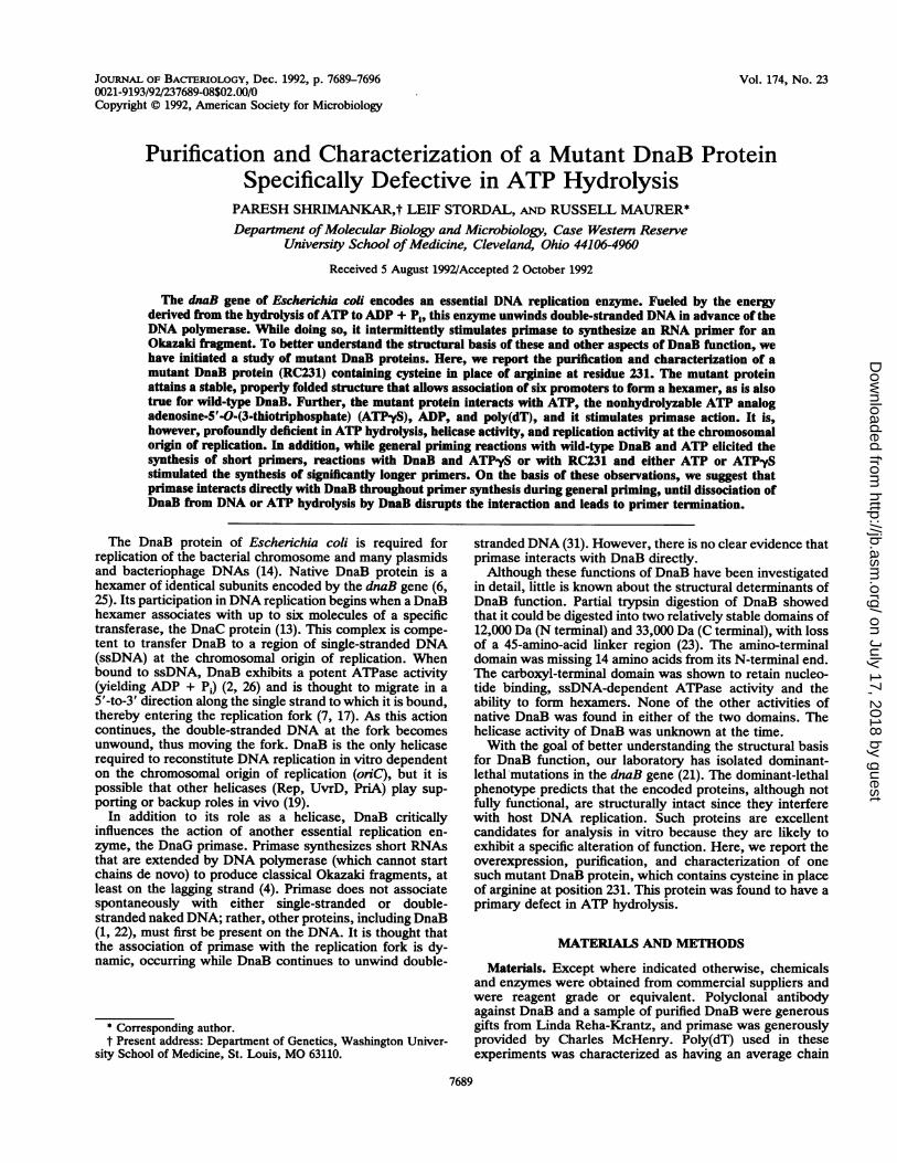
FIG. 1. Expression and purification of DnaB and RC231. Sam-ples were fractionated over an SDS-polyacrylamide gel. Lanes: a,total cell lysate showing overproduction of DnaB protein 4 h afterinduction with isopropyl-1-thio-o-D-galactopyranoside; b, total celllysate showing overproduction of RC231 7 h after induction; c, totalcell lysate showing absence of overproduction of RC231 in theabsence of plasmid pLysS 7 h after induction; d, purified wild-typeDnaB (-30 p.g); e, purified RC231 (-30 p.g).
protein accumulated to be the most abundant cellular pro-tein, whereas RC231 accumulated to a lesser but still signif-icant extent, as judged from Coomassie-stained gels.
Purification. The wild-type DnaB and RC231 proteinswere purified as described in Materials and Methods. Thepurification scheme did not rely on any functional propertiesof DnaB such as ATP binding or DNA binding. This allowedthe purification of RC231 without regard to its functionality.Purification of both proteins was followed by gel electropho-resis of fractionated samples. The two proteins were purifiedto a high degree (>95% purity; Fig. 1, lanes d and e). Thewild-type protein thus obtained was highly active in anoriC-dependent replication reaction reconstituted in vitro(data not shown) and in various partial assays as shownbelow.
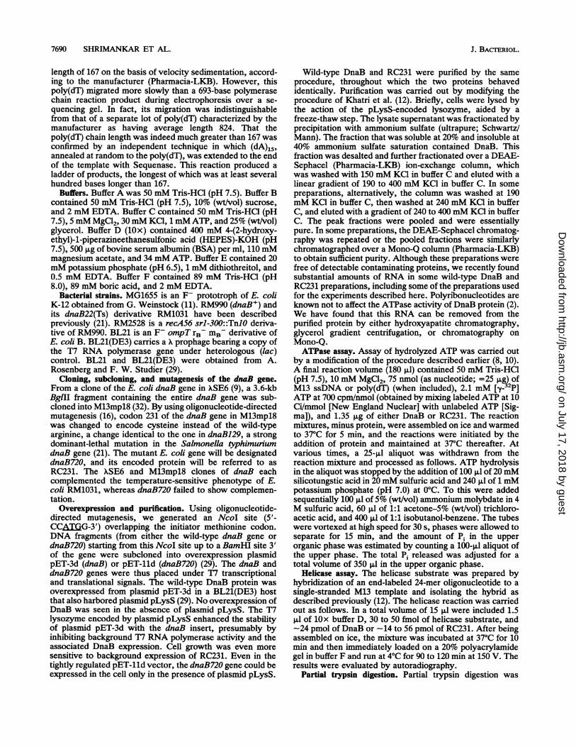
Structural studies. The wild-type DnaB protein of E. coliexists as a stable hexamer in solution (6, 25). When RC231protein was analyzed by sedimentation over a glycerolgradient, its rate of sedimentation was identical to that ofwild-type DnaB, with a sedimentation coefficient of 11.3S ineach case (Fig. 2). Using a Stokes radius value of 60 A (1 A= 0.1 nm) (25), we calculated the native molecular weight ofeach protein from the Svedberg equation to be 290,000(±412,000). This molecular weight estimate, which might below for reasons discussed elsewhere (25), is most consistentwith a hexameric structure. The calculated molecular massof a DnaB hexamer is 313,590 Da.
Partial trypsin digestion of wild-type DnaB protein yieldstwo stable domains: an ~--12,000-Da amino-terminal domainand an -33,000-Da carboxyl-terminal domain (24). Thecarboxyl-terminal domain accumulates only in the presenceof a nucleotide. Wild-type DnaB and RC231 from which ATP(included during purification) had been stripped by gelfiltration were subjected to trypsin digestion with or withoutadded nucleotides (Fig. 3). The major digestion productswere similar for the two proteins, as would be expected ifthey were similarly folded. In the absence of added nucleo-tide, the carboxyl-terminal domains of both proteins wererapidly degraded. However, addition of either ATP or ADP
VOL. 174,0 1992
on July 17, 2018 by guesthttp://jb.asm
.org/D
ownloaded from
7692 SHRIMANKAR ET AL.
Catalase
20
18
16
-a 1 4
-5
C 10a.
8 -
0
1- 6 -
4-
2
0
BSA Cytochrome c
D(a
L..
EC
-*- DraB
--- RC231
Fraction #
FIG. 2. Glycerol gradient centrifugation of DnaB and RC231 (seeMaterials and Methods). Each marker protein (beef liver catalase[232,000 Da], BSA [66,000 Da], and cytochrome c [12,400 Da]) wasrun in an independent tube at the same time, and its peak position inits gradient is indicated by an arrow.
dramatically stabilized both proteins against trypsin diges-tion, and the carboxyl-terminal domain transiently accumu-lated. Since residue 231 is in the carboxyl-terminal domain ofthe protein, these results show that the mutant protein iscapable of binding ATP and ADP.Thus, the mutant and wild-type proteins exhibited identi-
cal sedimentation behavior and were similar in their sensi-tivities to trypsin and in their interactions with ATP andADP. All of these observations point to native folding ofRC231.
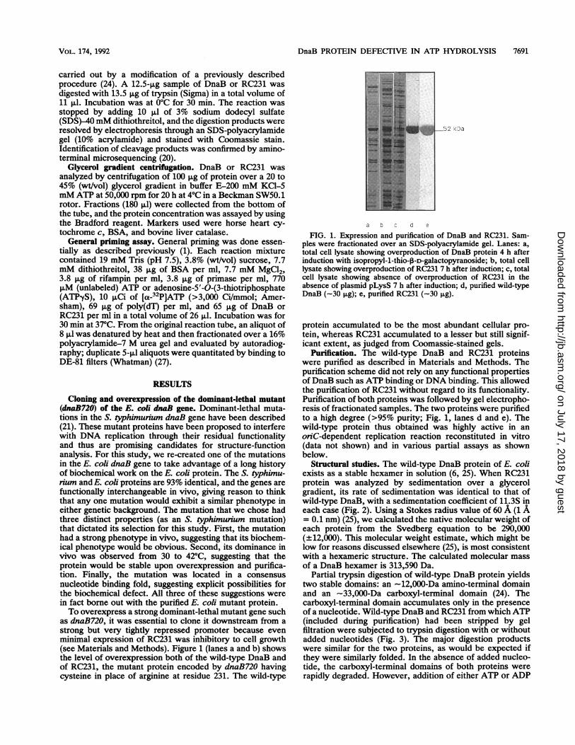
Functional studies. (i) ATP hydrolysis. Wild-type DnaB hasa potent ssDNA-dependent ATPase activity and a muchweaker DNA-independent ATPase activity (2, 26). EitherM13 viral DNA or poly(dT) will serve as effector (Fig. 4A).RC231 protein exhibited no ATP hydrolysis above the assaybackground even with M13 DNA or poly(dT) (Fig. 4B; note
Time (minutes)
40
'n.U
E75
30
20
0 5 10 15 20 25 30 35
Time (minutes)
FIG. 4. ATP hydrolysis. All sample data have been corrected forPi detected at time zero. This assay background was generallybetween 100 and 200 pmol except in the DnaB-M13 assay, in whichit was 515 pmol. (A) Activity of DnaB with M13 ssDNA cofactor(i), with poly(dT) cofactor (0), and without DNA (i); (B) activityof RC231 with M13 ssDNA cofactor (A), with poly(dT) cofactor (E),and without DNA (A). For reference, the data for DnaB in theabsence of DNA are repeated in panel B (-).
+ + + + DnaB
+ + + + RC231
- + + + - + + + Trypsin
ATP ATP ADP NONE ATP AlP AOP NONE Nucleotide
* ,Full-length.-W~ N-terminal
14 amino acids
C-terminal
fragment
.. . Es 2_ _-Trypsin
FIG. 3. Partial trypsin digestion of DnaB and RC231. The prod-ucts of each reaction were fractionated over SDS-polyacrylamide(10%) gels. The figure is a composite of two experiments, with thealignment between the two gels indicated. The DnaB proteins, priorto digestion, were stripped of ATP (present during purification) bygel filtration chromatography. The reaction mixtures were com-posed as indicated above the lanes. ATP or ADP, if indicated, wasadded at 5 mM. The N-terminal -12-kDa fragment was not resolvedin these particular gels but was observed in other gels.
picomole scale). RC231 protein added to an M13-dependentATPase reaction with wild-type DnaB had little effect onhydrolysis (data not shown). Therefore, the absence ofhydrolytic activity in RC231 cannot be explained by thepresence of an ATPase inhibitor; instead, RC231 must beinherently affected in ATP hydrolysis. Taken together withevidence indicating that RC231 can bind ATP (i.e., thetrypsin digestion experiments in which the ATP concentra-tion was only twice that used in the hydrolysis assay, andalso the general priming assay described below, in which theATP concentration was one-third of that in the hydrolysisassay), these results indicate that RC231 is unable to com-plete the hydrolysis of a bound ATP. This failure could be atthe level of catalysis or at the level of binding to the allostericeffector, ssDNA. Marszalek and Kaguni (18) report thatRC231 does not bind M13 DNA under conditions similar tothose used in this study, but the strong activity of RC231 insupporting general priming on poly(dT) indicates that themutant protein binds poly(dT) significantly. The concentra-tion of poly(dT) in the priming assay is half of that in theATPase assay. Thus, even when bound to effector ssDNA,RC231 is unable to hydrolyze ATP. We conclude that thedefect in RC231 is at the level of catalysis, since there is no
B.
)o-..
P0
'0
J. BACTERIOL.
on July 17, 2018 by guesthttp://jb.asm
.org/D
ownloaded from
DnaB PROTEIN DEFECTIVE IN ATP HYDROLYSIS 7693
.++- +
rid Substrate
II:_.~ Unwound Oligonucleotide
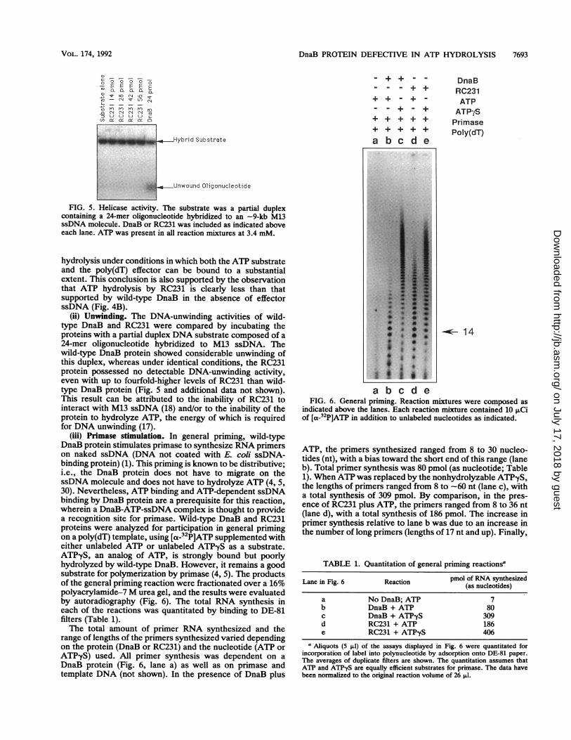
FIG. 5. Helicase activity. The substrate was a partial duplexcontaining a 24-mer oligonucleotide hybridized to an -9-kb M13ssDNA molecule. DnaB or RC231 was included as indicated aboveeach lane. ATP was present in all reaction mixtures at 3.4 mM.
hydrolysis under conditions in which both the ATP substrateand the poly(dT) effector can be bound to a substantialextent. This conclusion is also supported by the observationthat ATP hydrolysis by RC231 is clearly less than thatsupported by wild-type DnaB in the absence of effectorssDNA (Fig. 4B).
(ii) Unwinding. The DNA-unwinding activities of wild-type DnaB and RC231 were compared by incubating theproteins with a partial duplex DNA substrate composed of a24-mer oligonucleotide hybridized to M13 ssDNA. Thewild-type DnaB protein showed considerable unwinding ofthis duplex, whereas under identical conditions, the RC231protein possessed no detectable DNA-unwinding activity,even with up to fourfold-higher levels of RC231 than wild-type DnaB protein (Fig. 5 and additional data not shown).This result can be attributed to the inability of RC231 tointeract with M13 ssDNA (18) and/or to the inability of theprotein to hydrolyze ATP, the energy of which is requiredfor DNA unwinding (17).
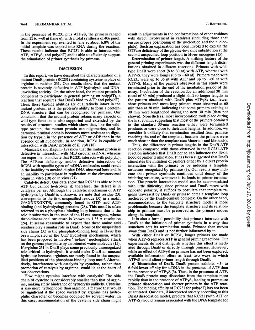
(iii) Primase stimulation. In general priming, wild-typeDnaB protein stimulates primase to synthesize RNA primerson naked ssDNA (DNA not coated with E. coli ssDNA-binding protein) (1). This priming is known to be distributive;i.e., the DnaB protein does not have to migrate on thessDNA molecule and does not have to hydrolyze ATP (4, 5,30). Nevertheless, ATP binding and ATP-dependent ssDNAbinding by DnaB protein are a prerequisite for this reaction,wherein a DnaB-ATP-ssDNA complex is thought to providea recognition site for primase. Wild-type DnaB and RC231proteins were analyzed for participation in general primingon a poly(dT) template, using [ot-32P]ATP supplemented witheither unlabeled ATP or unlabeled ATP-yS as a substrate.ATP-yS, an analog of ATP, is strongly bound but poorlyhydrolyzed by wild-type DnaB. However, it remains a goodsubstrate for polymerization by primase (4, 5). The productsof the general priming reaction were fractionated over a 16%polyacrylamide-7 M urea gel, and the results were evaluatedby autoradiography (Fig. 6). The total RNA synthesis ineach of the reactions was quantitated by binding to DE-81filters (Table 1).The total amount of primer RNA synthesized and the
range of lengths of the primers synthesized varied dependingon the protein (DnaB or RC231) and the nucleotide (ATP orATPyS) used. All primer synthesis was dependent on aDnaB protein (Fig. 6, lane a) as well as on primase andtemplate DNA (not shown). In the presence of DnaB plus
+ + + +++ ++.+
a b c d e.I.
DnaBRC231ATPATP ySPrimasePoly(dT)
i4
a b c d eFIG. 6. General priming. Reaction mixtures were composed as
indicated above the lanes. Each reaction mixture contained 10 uCiof [a-32P]ATP in addition to unlabeled nucleotides as indicated.
ATP, the primers synthesized ranged from 8 to 30 nucleo-tides (nt), with a bias toward the short end of this range (laneb). Total primer synthesis was 80 pmol (as nucleotide; Table1). When ATP was replaced by the nonhydrolyzable ATPyS,the lengths of primers ranged from 8 to -60 nt (lane c), witha total synthesis of 309 pmol. By comparison, in the pres-ence of RC231 plus ATP, the primers ranged from 8 to 36 nt(lane d), with a total synthesis of 186 pmol. The increase inprimer synthesis relative to lane b was due to an increase inthe number of long primers (lengths of 17 nt and up). Finally,
TABLE 1. Quantitation of general priming reactionsa
Lane in Fig. 6 Reaction pmol of RNA synthesized(as nucleotides)
a No DnaB; ATP 7b DnaB + ATP 80c DnaB + ATP-yS 309d RC231 + ATP 186e RC231 + ATP-yS 406
a Aliquots (5 pl) of the assays displayed in Fig. 6 were quantitated forincorporation of label into polynucleotide by adsorption onto DE-81 paper.The averages of duplicate filters are shown. The quantitation assumes thatATP and ATPyS are equally efficient substrates for primase. The data havebeen normalized to the original reaction volume of 26 1.
_Z E P E EE-D: CL a c) c] aLo.rco N o 0
0 - C\ sl- Ut)C
0 n pn tn n a:-0 N N N aa L) (I U UL
1. ) ....... - Hybr
VOL. 174, 1992
.. .1
on July 17, 2018 by guesthttp://jb.asm
.org/D
ownloaded from
7694 SHRIMANKAR ET AL.
in the presence of RC231 plus ATPyS, the primers rangedfrom 11 to -60 nt (lane e), with a total synthesis of 406 pmol.In the experiment represented in lane e, about 6.8% of theinitial template was copied into RNA during the reaction.These results indicate that RC231 is able to interact withATP, ATP-yS, and poly(dT) and is able to efficiently supportthe stimulation of primer synthesis by primase.
DISCUSSION
In this report, we have described the characterization of amutant DnaB protein (RC231) containing cysteine in place ofarginine at residue 231. Our results show that the mutantprotein is severely defective in ATP hydrolysis and DNA-unwinding activity. On the other hand, the mutant protein iscompetent to participate in general priming on poly(dT), areaction that requires that DnaB bind to ATP and poly(dT).Thus, these binding abilities are qualitatively intact in themutant protein, as is the protein's ability to form a protein-DNA structure that can be recognized by primase. Ourconclusion that the mutant protein retains many aspects ofwild-type function is also supported and extended by theresults of structural studies which show that, like the wild-type protein, the mutant protein can oligomerize, and itscarboxyl-terminal domain becomes more resistant to diges-tion by trypsin in the presence of ATP or ADP than in theabsence of all nucleotide. In addition, RC231 is capable ofinteraction with DnaC protein of E. coli (18).Marszalek and Kaguni (18) show that the mutant protein is
defective in interaction with M13 circular ssDNA, whereasour experiments indicate that RC231 interacts with poly(dT).The ATPase deficiency and/or defective interaction ofRC231 with specific types of ssDNA molecules could resultin the inability to unwind duplex DNA observed here and inan inability to participate in replication at the chromosomalorigin in vitro (18) or in vivo (21).
Catalytic defect. Our data show that RC231 is able to bindATP but cannot hydrolyze it; therefore, the defect is incatalysis per se. Although the catalytic mechanism of ATPhydrolysis by DnaB is unknown, we note that residue 231corresponds to the first unspecified residue (X) in a motif,G/AXXXXGKT/S, commonly found in GTP- and ATP-binding (and hydrolyzing) proteins (28). This motif is oftenreferred to as a phosphate-binding loop because that is therole it subserves in the case of the H-ras oncogene, whosethree-dimensional structure is known to 1.35-A resolution(24). It seems reasonable to expect that these amino acidresidues play a similar role in DnaB. None of the unspecifiedside chains (X) in the phosphate-binding loop in H-ras hasbeen implicated in the GTP hydrolysis mechanism, whichhas been proposed to involve "in-line" nucleophilic attackon the gamma phosphate by an oriented water molecule (15).If arginine 231 in DnaB plays some previously unrecognizedrole critical to hydrolysis, it would make DnaB an unusualhydrolase because arginines are rarely found in the unspec-ified positions of the phosphate-binding loop motif. Alterna-tively, interference with catalysis by cysteine, rather thanpromotion of catalysis by arginine, could lie at the heart ofour observations.How might cysteine interfere with catalysis? The side
chain of cysteine is considerably smaller than that of argin-ine, making steric hindrance of hydrolysis unlikely. Cysteineis also more hydrophobic than arginine, a feature that wouldbe significant if the space vacated by arginine has hydro-philic character or becomes occupied by solvent water. Inthis case, accommodation of the cysteine side chain might
result in adjustments in the conformations of other residueswith direct involvement in catalysis (including those thatensure proper positioning of the nucleotide and the nucleo-phile). Such an explanation has been invoked to explain theGTPase deficiency of the glycine-to-valine substitution at thesecond unspecified loop position in H-ras oncogene (15).
Determination of primer length. A striking feature of thegeneral priming experiments was the different length distri-butions obtained in different reactions. Primers with wild-type DnaB were short (8 to 30 nt) with ATP, whereas withATP,yS, they were longer (up to -60 nt). Primers made withRC231 were up to 36 nt with ATP and up to -60 nt withATPyS. Many of the primers observed in this study wereterminated prior to the end of the incubation period of theassay. Incubation of the reaction for an additional 30 min(total of 60 min) produced a slight shift to longer lengths inthe pattern obtained with DnaB plus ATP-yS; i.e., fewershort primers and more long primers were observed at 60min than at 30 min, indicating that some primers existing at30 min were lengthened during the next 30 min (data notshown). Nonetheless, most incorporation took place duringthe first 20 min, suggesting that most of the primers obtainedin the standard 30-min reaction either were terminatedproducts or were close to their final lengths. In addition, weconsider it unlikely that termination resulted from primasereaching the end of the template, because the primers weremuch shorter than the template (see Materials and Methods).Thus, the difference in primer lengths in the DnaB-ATP
reaction compared with those observed in the RC231-ATPreaction indicates that DnaB per se can influence the likeli-hood of primer termination. It has been suggested that DnaBstimulates the initiation of primers either by a direct proteininteraction with the primase or by inducing a templatestructure recognized by primase (3). Our results may indi-cate that primer synthesis continues until decay of theinitiating structure, whatever it is, leads to primer termina-tion. The protein interaction model can be accommodatedwith little difficulty; since primase and DnaB move withopposite polarity, it suffices to postulate that template re-gions traversed by DnaB or primase enter a template loopanchored by the DnaB-primase complex. On the other hand,accommodation to the template structure model is moreproblematic because the template structure and its relation-ship to primase must be preserved as the primase movesalong the template.
It is also a formal possibility that primase interacts withDnaB at the initiation of primer synthesis in a way thatsomehow sets its termination mode. Primase then movesaway from DnaB and is not further influenced by it.With either DnaB or RC231, longer primers are made
when ATPyS replaces ATP in general priming reactions. Ourexperiments do not distinguish whether this effect is medi-ated through DnaB or directly through primase. However,while an effect of ATP-yS on primase has not been explored,available information offers at least two ways in whichATP-yS could affect primer length through DnaB.
(i) Dissociation of DnaB. DnaB protein exhibits -3- to6-fold-lower affinity for ssDNA in the presence of ATP thanin the presence of ATP-yS (3). Thus, in the presence of ATP,the DnaB protein may dissociate from the template morerapidly than in the presence of ATPyS, leading to prematureprimase dissociation and shorter primers in the ATP reac-tion. The binding affinity of RC231 for poly(dT) has not beenquantitated. Our data, if interpreted strictly according to thisDnaB dissociation model, predicts that RC231 (with ATP orATP-yS) would remain associated with the DNA template for
J. BACTERIOL.
on July 17, 2018 by guesthttp://jb.asm
.org/D
ownloaded from
DnaB PROTEIN DEFECTIVE IN ATP HYDROLYSIS 7695
a longer time than does DnaB plus ATP, thus allowing thesynthesis of longer primers.
(ii) Effect of DnaB ATPase activity on primer lengths. Thecardinal feature of the DnaB dissociation model, namely, theobligatory dependence of primer termination on DnaB dis-sociation from the template, has not been proven. Therefore,we consider an alternative model in which DnaB, through itsATPase, actively ejects primase without leaving the DNA.This is an economical model because it explains both theprotein effect (DnaB versus RC231) and the nucleotide effect(ATP versus ATP-yS). There are two variations on this idea.With respect to the first variation, a conformation effect, it isknown that the conformation of DnaB changes with eachcycle of ATP hydrolysis (24), and in one case it has beenshown that primase binds more tightly to a DnaB-ssDNA-nucleotide complex that is not hydrolytically active than toan active complex (3). Thus, primase may interact stronglywith DnaB-ATP-poly(dT) but weakly with DnaB-ADP-poly(dT). Each cycle of ATP hydrolysis would then beassociated with a high probability of primase release andhence primer termination. The second variation postulates a
mobility effect. In addition to cycling through conforma-tional changes, a DnaB molecule undergoing rounds of ATPhydrolysis when bound to ssDNA is likely to migrate on theDNA (17). This mobility of the protein on DNA coulddestabilize a bound primase molecule that is in the process ofRNA synthesis. Either of these effects would account for thesynthesis of primers that are short in reactions with DnaBplus ATP. In contrast, the other three combinations, whichare deficient in nucleotide hydrolysis, apparently do notcycle through conformational changes or migrate on ssDNA,thus providing a stable recognition structure for primase andallowing synthesis of longer primers.Primer termination in the simplified general priming reac-
tions may reflect a combination of mechanisms. ATP hydro-lysis is unlikely to be the sole mechanism because blockinghydrolysis by using ATPyS produces a different primerlength distribution than does blocking hydrolysis by substi-tuting RC231 for DnaB. However, at authentic replicationforks, DnaB is unlikely to dissociate from the DNA becauseof its presumed processive action (7, 17). Therefore, we
suggest that ATP hydrolysis by DnaB and its associatedmobility on DNA could influence primer termination atauthentic replication forks. This suggestion does not pre-
clude the possibility that other replication proteins such as
DNA polymerase III holoenzyme (33) could also affectprimer length and could be the predominant effectors invivo.
Genetic dominance of RC231. The inability of RC231 tohydrolyze ATP and to unwind DNA provide a sufficientexplanation for the failure to support DNA replication invitro, as reported previously (18). However, the observationin that study that RC231 competes with wild-type DnaB forDnaC protein, yet is delivered inefficiently to the oriCregion, may be incomplete or misleading in explaining thecharacteristic genetic dominance of the mutant. In vivo, instrains that are heterodiploid for dnaB, monomers of bothRC231 and the functional DnaB are synthesized simulta-neously and, one could imagine, assemble into hetero-oligomers. These hetero-oligomers could be competent to betransferred to the oniC region through the action of DnaCand would be expected to stimulate primase, as judged fromthe results presented here. However, they may fail in a
subsequent step of replication (primer termination, migra-tion on ssDNA, or unwinding), leading to stalled replicationforks. In this report, we have not described the properties of
such hetero-oligomers; even in experiments in which RC231and wild-type DnaB were both present in a given reaction(18), they were added as separately purified proteins. Thereis no clear evidence that subunit exchange takes place underthese conditions. While a detailed explanation of geneticdominance will have to await further experimentation, theresults presented here and elsewhere (18) illuminate severalways in which ATP hydrolysis is central to the replicationactivity of DnaB.
ACKNOWLEDGMENTS
We thank Jarek Marszalek and Jon Kaguni for their interest in thisproject and for communicating results prior to publication. Weespecially thank our colleague Miriam Lifsics for extensive guidanceand many helpful discussions.
This work was supported by Public Health Service grants AI19942and GM47111 from the National Institutes of Health.
REFERENCES1. Arai, K., and A. Kornberg. 1979. A general priming system
employing only DnaB protein and primase for DNA replication.Proc. Natl. Acad. Sci. USA 76:4308-4312.
2. Arai, K., and A. Kornberg. 1981. Mechanism of DnaB protein.II. ATP hydrolysis by DnaB protein dependent on single- ordouble-stranded DNA. J. Biol. Chem. 256:5253-5259.
3. Arai, K., and A. Kornberg. 1981. Mechanism of dnaB proteinaction. III. Allosteric role of ATP in the alteration of DNAstructure by dnaB protein in priming replication. J. Biol. Chem.256:5260-5266.
4. Arai, K., and A. Kornberg. 1981. Mechanism of dnaB proteinaction. IV. General priming ofDNA replication by dnaB proteinand primase compared with RNA polymerase. J. Biol. Chem.256:5267-5272.
5. Arai, K., R. Low, and A. Kornberg. 1981. Movement and siteselection for priming by the primosome in phage +X174 DNAreplication. Proc. Natl. Acad. Sci. USA 78:707-711.
6. Arai, K., S. Yasuda, and A. Kornberg. 1981. Mechanism ofdnaB protein action. I. Crystallization of properties of DnaBprotein, an essential replication protein in Escherichia coli. J.Biol. Chem. 256:5247-5252.
7. Baker, T., B. Funnell, and A. Kornberg. 1987. Helicase action ofdnaB protein during replication from the Escherichia coli chro-mosomal origin in vitro. J. Biol. Chem. 262:6877-6885.
8. Conway, T., and F. Lipmann. 1964. Characterization of aribosome-linked guanosine triphosphate in Escherichia coli ex-tracts. Proc. Natl. Acad. Sci. USA 52:1462-1466.
9. Elledge, S., and G. Walker. 1985. Plasmid vectors for identifi-cation of genes by complementation of Escherichia coli mu-tants. J. Bacteriol. 162:777-783.
10. Grifo, J. A., R. D. Abramson, C. A. Sailer, and W. C. Merrick.1984. RNA-stimulated ATPase activity of eukaryotic initiationfactors. J. Biol. Chem. 259:8648-8654.
11. Guyer, M. S., R. R. Reed, J. A. Steitz, and K. B. Low. 1980.Identification of a sex-factor affinity site in E. coli as y8. ColdSpring Harbor Symp. Quant. Biol. 45:135-140.
12. Khatri, G. S., T. MacAllister, P. R. Sista, and D. Bastia. 1989.The replication terminator protein of E. coli is a DNA sequence-specific contra-helicase. Cell 59:667-674.
13. Kobori, J., and A. Kornberg. 1982. The Eschenichia coli dnaCgene product. III. Properties of the DnaB-DnaC protein com-plex. J. Biol. Chem. 257:13770-13775.
14. Kornberg, A., and T. Baker. 1990. DNA replication. W. H.Freeman Co., New York.
15. Krengel, U., I. Schlichting, A. Scherer, R. Schumann, M. Frech,J. John, W. Kabsch, E. F. Pai, and A. Wittinghofer. 1990.Three-dimensional structures of H-ras p21 mutants: molecularbasis for their inability to function as signal switch molecules.Cell 62:539-548.
16. Kunkel, T. 1985. Rapid and efficient site-specific mutagenesiswithout phenotypic selection. Proc. Natl. Acad. Sci. USA82:488-492.
VOL. 174, 1992
on July 17, 2018 by guesthttp://jb.asm
.org/D
ownloaded from
7696 SHRIMANKAR ET AL.
17. LeBowitz, J., and R. McMacken. 1986. The Eschenichia coliDnaB replication protein is a DNA helicase. J. Biol. Chem.261:4738-4748.
18. Marszalek, J., and J. M. Kaguni. 1992. Defective replicationactivity of a dominant-lethal dnaB gene product from Esche-richia coli. J. Biol. Chem. 267:19334-19340.
19. Matson, S., and K. Kaiser-Rogers. 1990. DNA helicases. Annu.Rev. Biochem. 59:289-329.
20. Matsudaira, P. 1987. Sequence from picomole quantities ofproteins electroblotted onto polyvinylidene difluoride mem-branes. J. Biol. Chem. 262:10035-10038.
21. Maurer, R., and A. Wong. 1988. Dominant-lethal mutations inthe dnaB helicase gene of Salmonella typhimunum. J. Bacteriol.170:3682-3688.
22. McMacken, R., and A. Kornberg. 1978. A multi-enzyme systemfor priming the replication of 4X174 viral DNA. J. Biol. Chem.253:3313-3319.
23. Nakayama, N., N. Arai, Y. Kaziro, and K. Arai. 1984. Structuraland functional studies of the DnaB protein using limited prote-olysis. Characterization of domains for DNA-dependent ATPhydrolysis and for protein association in the primosome. J. Biol.Chem. 259:88-96.
24. Pai, E. F., U. Krengel, G. A. Petsko, R. S. Goody, W. Kabsch,and A. Wittinghofer. 1990. Refined crystal structure of thetriphosphate conformation of H-ras p21 at 1.35A resolution:implications for the mechanism of GTP hydrolysis. EMBO J.9:2351-2359.
25. Reha-Krantz, L., and J. Hurwitz. 1978. The dnaB gene productof Escherichia coli. I. Purification, homogeneity, and physical
properties. J. Biol. Chem. 253:4043-4050.26. Reha-Krantz, L., and J. Hurwitz. 1978. The dnaB gene product
of Escherichia coli. II. Single-stranded DNA-dependent ribonu-cleoside triphosphatase activity. J. Biol. Chem. 253:4051-4057.
27. Rowen, L., and A. Kornberg. 1978. Primase, the dnaG protein ofEscherichia coli. An enzyme which starts DNA chains. J. Biol.Chem. 253:758-764.
28. Saraste, M., P. Sibbald, and A. Wittinghofer. 1990. TheP-loop-a common motif in ATP- and GTP-binding proteins.Trends Biochem. Sci. 15:430-434.
29. Studier, F. W., A. H. Rosenberg, J. J. Dunn, and J. W.Dubendorf. 1990. Use of T7 RNA polymerase to direct theexpression of cloned genes. Methods Enzymol. 185:60-88.
30. Wahle, E., R. S. Lasken, and A. Kornberg. 1989. The dnaB-dnaC replication protein complex ofEscherichia coli. II. Role ofthe complex in mobilizing dnaB functions. J. Biol. Chem.264:2469-2475.
31. Wu, C. A., E. L. Zechner, and K. J. Marians. 1992. Coordinatedleading- and lagging-strand synthesis at the Escherichia coliDNA replication fork. I. Multiple effectors act to modulateOkazaki fragment size. J. Biol. Chem. 267:4030-4044.
32. Zagursky, R, and M. Berman. 1984. Cloning vectors that yieldhigh levels of single-stranded DNA for rapid DNA sequencing.Gene 27:183-191.
33. Zechner, E. L., C. A. Wu, and K. J. Marians. 1992. Coordinatedleading- and lagging strand synthesis at the Escherichia coliDNA replication fork. III. A polymerase-primase interactiongoverns primer size. J. Biol. Chem. 267:4054-4063.
J. BACTERIOL.
on July 17, 2018 by guesthttp://jb.asm
.org/D
ownloaded from


![OSA Archivum - [300-1-2]-1954-7689 · 2012. 12. 20. · AGRTCÜLTVRE Forestry TTEM wo.7689/54 septeñber 9 1/13321 POLAND /0200/ /0204/ sAWMTL1, CONT)TTTOWS SOURCE 49-year—old ethnic-German](https://img.pdfslide.us/doc/110x75/6117a206d821da043f2971e2/osa-archivum-300-1-2-1954-7689-2012-12-20-agrtcoeltvre-forestry-ttem-wo768954.jpg)