Embed Size (px)
Citation preview

International Journal of Science and Research (IJSR) ISSN (Online): 2319-7064
Index Copernicus Value (2013): 6.14 | Impact Factor (2013): 4.438
Volume 4 Issue 2, February 2015
www.ijsr.net Licensed Under Creative Commons Attribution CC BY
Purification and Biochemical Characterization of L-
Asparaginase from Aspergillus niger and Evaluation
of Its Antineoplastic Activity
Vaishali Dange1, Swati Peshwe
2
1Department of Biotechnology, Shivchhatrapati College, Aurangabad, India
2Department of Microbiology, Government Institute of Science, Aurangabad, India
Abstract: L-asparaginase is a chemotherapeutic drug used in the treatment of lymphoblastic leukemia. In the present study, the
extracellular L-asparaginase produced by strain Aspergillus niger was purified, characterized. Moreover, its antiproliferative activity
was evaluated. The apparent molecular weight of the enzyme was found to be 136 kDa. The optimal pH and temperature for the enzyme
were 9.0°C and 40°C, respectively. The enzyme retained 100% of the activity at 40°C for 120 min. L-asparaginase against human
normal cells did not show cytotoxicity. However, in the human leukemia cell line A431 the antiproliferative effects of L-asparaginase
was observed after 96 h of incubation. For the first time, an L-asparaginase from fungus was evaluated as an antitumor agent in human
cells lines and further investigations should be conducted to improve the knowledge about this enzyme.
Keywords: Aspergillus niger; L-Asparaginase; Antineoplasic Activity; Leukemia cell line viz. A431
1. Introduction
The enzyme L-asparaginase (L-asparagine amino hy-
drolase, E.C. 3.5.1.1) is an important component in the
treatment of pediatric acute lymphoblastic leukemia (ALL)
and catalyzes the hydrolysis of asparagine into aspartic acid
and ammonia. This conversion provokes the asparagine
starvation in the blood plasma and induces the death of
malignant cells, since they are unable to synthesize
asparagine and reduced levels of asparagine inhibits protein
synthesis in leukemic cells [1]. Current studies of L-
asparaginase therapy have also started in adults [2]. The
effect and half-life of L-asparaginase depends on some
factors such as antibody formation, plasmatic proteases,
formation of asparagine via asparagine synthetase and
microbial source [3]. Over the years, several bacterial L-
asparaginases have been reported and only a few reports
about L-asparaginase produced by filamentous fungi have
been made. Among these reports are the L-asparaginase
production by Aspergillus tamari, A. terreus [4], A. niger
[5], A. nidulans [6], and in some yeast, but their
antiproliferative activities were not analyzed. Currently,
there are three asparaginases preparations available for
therapy, two of them are native and produced by the bacteria
Escherichia coli and Erwinia chrysanthemi. The other one,
also from E. coli is conjugated and its elimination half life is
approximately six days, five times longer than the native E.
coli and nine times longer than the Erwinia preparations [2,
7]. The bacterial L-asparaginases are targets of antibodies
and proteases, moreover side effects are observed during the
treatment using this enzyme. Great efforts have been made
to modify and immobilize these L-asparaginases in order to
decrease their immunogenicity effects and increase their
half-life. L-asparaginase from other sources, like eukaryotic
microorganisms, should lead to an enzyme with less adverse
effects. In this study, the extracellular L-asparaginase
produced by Aspergillus niger was purified, biochemically
characterized, and the antiproliferative activity of enzyme
was evaluated against two leukemic cells lines.
2. Material and Methods
2.1 Microorganism
Aspergillus niger was isolated from soil. The fungus has
been maintained by weekly transfers on slants of PDA
medium.
2.2. L-Asparaginase Production
Enzyme production was optimized in Czapek Dox’s
modified liquid medium in two steps: the pre fermentation
medium containing 0.2% (w/v) glucose, 2% (w/v) L-proline,
0.2% (w/v) NH4NO3, 0.15% (w/v) KH2PO4, 0.05% (w/v)
KCl, 0.05% (w/v) MgSO4·7H2O and 0.001% (w/v)
ZnSO4·7H2O, FeSO4·7H2O and CuSO4·5H2O, the pH was
adjusted to 8.5 with KOH, this medium was inoculated with
1 × 10⁷ spores·mL
−1 and incubated at 120 rpm for 17 h at
30°C. The culture was filtrated; the mycelium was collected
and inoculated in the fermentative medium, which was
similar to the medium used in previous step except for the
absence of NH4NO3. The culture was reincubated for 96
hours at the same previous conditions.
2.3. L-Asparaginase Assay
L-asparaginase activity was determined according Drainas
and Pateman [6] and modified as follows: 0.6 mL 20
mmol·L−1
Tris-HCl buffer, pH 8.0; 0.2 mL 100 mmol·L−1
stock L-asparagine solution; 0.2 mL 1 mol·L−1
stock
hydroxylamine solution; and 1mL culture broth were mixed
and incubated at 37°C and 120 rpm. After 30 min 0.5 mL
ferric chloride reagent [10% (w/v) FeCl3 plus 5% (w/v)
trichloroacetic acid in 0.66 mol·L−1 HCl] was added. One
unit of L-asparaginase is the amount of enzyme that re-
Paper ID: OCT141573 564

International Journal of Science and Research (IJSR) ISSN (Online): 2319-7064
Index Copernicus Value (2013): 6.14 | Impact Factor (2013): 4.438
Volume 4 Issue 2, February 2015
www.ijsr.net Licensed Under Creative Commons Attribution CC BY
leases 1 mmol NH3 or aspartic acid per minute at 37°C at
the specific conditions just mentioned. The reaction mixture
contained 0.5 mL of 0.04 M of L-glutamine; 0.5 mL of 0.5
M Tris-HCl buffer; pH 7.2; 0.5 -1.0 mL culture broth
(concentrated or not) and distilled water a final volume of
2.0 mL. It was incubated at 37°C for 30 - 60 min. and the
reaction was stopped with 0.5 mL of 1.5 M of trichloroacetic
acid. In fact 0.1 mL of mixture just mentioned and 0.2 mL of
Nessler’s reagent were added to 3.7 mL of distilled water.
After 20 min, the absorption was measured at 450 nm.
2.4. Determination of Protein Concentration
The concentration of protein was determined by the
Bradford [9] method with bovine serum albumin as a
standard.
2.5. Separation and Purification of L-Asparaginase
The extracellular L-asparaginase was purified in three
chromatographic steps, and after each step, the fractions
were analyzed for activity and absorbance at 280 nm.
Step 1: The culture fluid was separated from mycelium by
filtration. Then 900 mL was dialyzed against 5 mmol·L−1
Tris-HCl buffer, pH 8.0 (buffer A) overnight at 4°C, and 20
mL were applied to a DEAE—Sepharose Fast Flow column
(2.5 × 22.5 cm) pre-equilibrated with 20 mmol·L−1 Tris-
HCl buffer, pH 8.0 (buffer B). The bound proteins were
eluted by step-wise increases in NaCl (100 and 150
mmol·L−1) at flow rate of 120 mL·h−1. Fractions (5.0 mL)
were collected.
Step 2: The fractions with L-asparaginase collected from
DEAE—Sepharose Fast Flow were pooled, dialyzed against
buffer A, and loaded on a Sephacryl S-200 HR column (1.0
× 58.0 cm). The protein elution was performed with the
buffer B containing 150 mmol·L−1 NaCl at a flow rate of
9.6 mL·h−1. Fractions (2.0 mL) were collected.
Step 3: The fractions from step 2 that contained L-
asparaginase were pooled, dialyzed against buffer A, and
applied again on a Sephacryl S-200 HR column (1.0 × 5.8
cm) however the flow rate was reduced at 6.0 mL·h−1. The
fractions (0.8 mL) were pooled and dialyzed against buffer
A over- night at 4°C. The samples were assayed and used for
further characterization.
2.6. Electrophoresis Analysis
Polyacrilamide gel electrophoresis (SDS-PAGE) as
described by Laemmli [11] was performed using 4% (w/v)
stacking gel and 7.5% (w/v) acrylamide slab gel at a
constant current of 20 mA. Protein bands were stained with
silver nitrate according to Blum et al. [12].
2.7. Characterization of Purified L-Asparaginase
The optimum values of pH and temperature of purified
enzyme were determined over a pH range of 2.2 - 10.6
(citrate-phosphate buffer [pH 2.2 - 7.8], Tris-HCl buffer [pH
8.2 - 9.0], and carbonate-bicarbonate buffer [pH 9.5 - 10.6])
and temperatures from 25°C to 60°C. Km was determined
from double reciprocal plots (Linewear-Burk) incubating the
pure enzyme with different concentrations of substrate at
temperature and pH optima. The molecular mass was
estimated by chromatography on Sephacryl S-200 HR using
different protein molecular weight markers: cytochrome C
(12.4 kDa), α-lactalbumin (14.2 kDa), bovine serum albumin
(66 kDa), alcohol dehydrogenase (150 kDa), and β-amylase
(200 kDa). The column was pre-equilibrated with buffer B,
the proteins were eluted with the same buffer containing 150
mmol·L−1
NaCl at a flow rate of 6.0 mL·h−1
. The thermo
stability of purified L-asparaginase was determined by pre-
incubating the enzymes in 100 mmol·L−1
Tris-HCl buffer
(pH 8.0) at different temperatures (40°C, 50°C, and 60°C)
for 120 min. Samples were collected at 15, 30, 60, 90, and
120 min, and the residual activity was assayed. Proteolytic
resistance was evaluated after digestion of 180 μg of pure
asparaginase and modified with 60 μg of bovine trypsin in a
total volume of 1.0 ml at 37°C. Samples were collected from
each solution at 5, 10, 15, 30, and 60 min and assayed for
the residual activity. All the stability studies were repeated
in triplicate and values are shown as mean ± SD.
2.8. Cell Culture and Cell Preparation
The human leukemia cell line A431, Stong et al. [13] were
purchased from the American Type Culture Collection and
were maintained in RPMI (GIBCO, USA) supplemented
with 10% fetal calf serum, 100 U/mL penicillin and 100
μg/mL streptomycin. Human peripheral blood mononuclear
cells (PBMC) were purified from heparinized venous blood
drawn from healthy donors. PBMC were isolated by
centrifugation on Ficoll-Paque (Pharmacia-LKB, Uppsala,
Sweden) density gradients (1.077 g/mL) at 1000 rpm for 15
min at room temperature and subsequently resuspended in
RPMI. All cell cultures were incubated at 37°C in a 5% CO2
humidified atmosphere. The counting and cell viability tests
were determined using the test of Trypan blue exclusion.
2.9. Proliferation Assay
Cells were seeded in 96-well plates at 1 × 10⁴ cells per well.
After 24 h, L-asparaginase was added at concentrations of
12.5 μg/mL, 25 μg/mL, 50 μg/mL, 100 μg/mL and 200
μg/mL. At different time points (48, 72, and 96 h) of
continuous drug exposure, 10 μl of MTT dye (3 mg/mL was
added in each well. The plates were incubated for 2 h at
37°C and the formazan product was measured at 450 nm by
using a microplate reader (Bio-Rad Laboratories). The
experients were performed in triplicate in three independent
sets. Values are shown as mean ± SD. Cell survival was
calculated by subtracting the background absorbance of
media alone and then dividing the absorbance of test wells
by the absorbance of the control (untreated) wells.
3. Results
3.1. Biochemical Characterization of L-Asparaginase
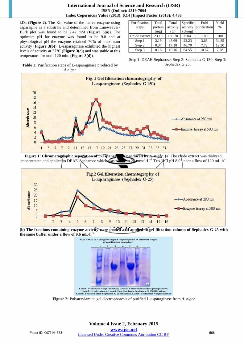
The purification of L-asparaginase was carried out by three
steps with a final yield of 7.28% and a purification fold of
10.67 (Table 1, Figure 1). The molecular weight of the
native enzyme determined by its mobility on the gel
filtration column and SDS-PAGE was estimated to be 136
Paper ID: OCT141573 565
International Journal of Science and Research (IJSR) ISSN (Online): 2319-7064
Index Copernicus Value (2013): 6.14 | Impact Factor (2013): 4.438
Volume 4 Issue 2, February 2015
www.ijsr.net Licensed Under Creative Commons Attribution CC BY
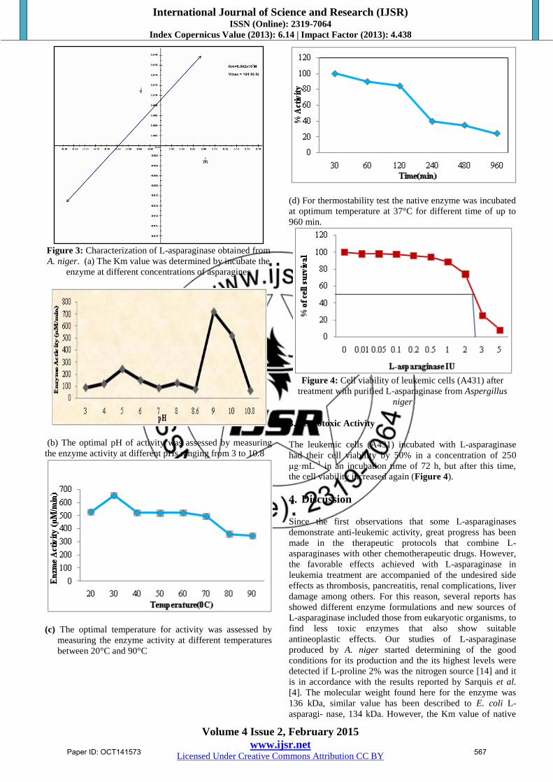
kDa (Figure 2). The Km value of the native enzyme using
asparagine as a substrate and determined from Lineweaver-
Burk plot was found to be 2.42 mM (Figure 3(a)). The
optimum pH for enzyme was found to be 9.0 and at
physiological pH the enzyme retained 70% of maximum
activity (Figure 3(b)). L-asparaginase exhibited the highest
levels of activity at 37°C (Figure 3(c)) and was stable at this
temperature for until 120 min. (Figure 3(d)).
Table 1: Purification steps of L-asparaginase produced by
A.niger
Purification
steps
Total
protein
(mg)
Total
activity
(U)
Specific
activity
(U/mg)
Fold
purification
Yield
%
Crude extract 23.10 139.70 6.04 1.00 100
Step 1 2.19 48.69 22.23 3.68 34.85
Step 2 0.37 17.18 46.70 7.72 12.30
Step 3 0.16 10.16 64.55 10.67 7.28
Step 1: DEAE-Sepharose; Step 2: Sephadex G 150; Step 3:
Sephadex G 25.
Figure 1: Chromatographic separation of L-asparaginase produced by A. niger. (a) The crude extract was dialyzed,
concentrated and applied to DEAE-Sepharose which was eluted in 20 mmol·L−1
Tris-HCl pH 8.0 under a flow of 120 mL·h−1
(b) The fractions containing enzyme activity were pooled and applied to gel filtration column of Sephadex G-25 with
the same buffer under a flow of 9.6 mL·h−1
Figure 2: Polyacrylamide gel electrophoresis of purified L-asparaginase from A. niger
Paper ID: OCT141573 566
International Journal of Science and Research (IJSR) ISSN (Online): 2319-7064
Index Copernicus Value (2013): 6.14 | Impact Factor (2013): 4.438
Volume 4 Issue 2, February 2015
www.ijsr.net Licensed Under Creative Commons Attribution CC BY
Figure 3: Characterization of L-asparaginase obtained from
A. niger. (a) The Km value was determined by incubate the
enzyme at different concentrations of asparagines
(b) The optimal pH of activity was assessed by measuring
the enzyme activity at different pHs ranging from 3 to 10.8
(c) The optimal temperature for activity was assessed by
measuring the enzyme activity at different temperatures
between 20°C and 90°C
(d) For thermostability test the native enzyme was incubated
at optimum temperature at 37°C for different time of up to
960 min.
Figure 4: Cell viability of leukemic cells (A431) after
treatment with purified L-asparaginase from Aspergillus
niger
3.2 Cytotoxic Activity
The leukemic cells (A431) incubated with L-asparaginase
had their cell viability by 50% in a concentration of 250
μg·mL−1
in an incubation time of 72 h, but after this time,
the cell viability increased again (Figure 4).
4. Discussion
Since the first observations that some L-asparaginases
demonstrate anti-leukemic activity, great progress has been
made in the therapeutic protocols that combine L-
asparaginases with other chemotherapeutic drugs. However,
the favorable effects achieved with L-asparaginase in
leukemia treatment are accompanied of the undesired side
effects as thrombosis, pancreatitis, renal complications, liver
damage among others. For this reason, several reports has
showed different enzyme formulations and new sources of
L-asparaginase included those from eukaryotic organisms, to
find less toxic enzymes that also show suitable
antineoplastic effects. Our studies of L-asparaginase
produced by A. niger started determining of the good
conditions for its production and the its highest levels were
detected if L-proline 2% was the nitrogen source [14] and it
is in accordance with the results reported by Sarquis et al.
[4]. The molecular weight found here for the enzyme was
136 kDa, similar value has been described to E. coli L-
asparagi- nase, 134 kDa. However, the Km value of native
Paper ID: OCT141573 567

International Journal of Science and Research (IJSR) ISSN (Online): 2319-7064
Index Copernicus Value (2013): 6.14 | Impact Factor (2013): 4.438
Volume 4 Issue 2, February 2015
www.ijsr.net Licensed Under Creative Commons Attribution CC BY
L-asparaginase was found to be 2.42 mM, while that from E.
coli is 0.0125 mM [15]. It shows that the L-asparaginase
from A. niger has less affinity for asparagine than L-
asparaginase from E. coli but the L-asparaginase from A.
niger was capable to inhibit the prolifiration of leukemia
cells. According to Panosyan et al. [16] the effective
deamination of glutamine by L-asparaginase appears to
contribute to the decrease of asparagine depletion by
depriving the asparagine synthetase of glutamine, the
precursor asparagine biosynthesis. Bacterial L-asparaginase,
used in therapeutic protocols, has low glutaminase activity
but toxicity reactions are attributed to this activity [17, 18].
Herein L-glutaminase activity was not detected in crude
enzyme (concentrated or not) produced by A. niger even
after 60 min of the reaction. This should contribute
significantly to diminution of side effects and it may be
helpful in clinical practice.
The antiproliferative effects of L-asparaginase produced by
A. niger was evaluated after 24, 48, 72 and 96 h of
incubation of A431 leukemia cell line. This L-asparaginase
caused 50% reduction in cell viability after 72 h on the cell
line HL-60 and after 96 h on the cell line RS4;11 (Figure 4).
Interestingly, cell proliferation of HL-60 cell increased after
72 h and this can be associated with multiple adaptive
cellular mechanisms. Studies have demonstrated increased
asparagine synthetase (AS) expression in cells treated with
L-asparaginase. It has been hypothesized that this elevated
activity allows these leukemia cells to become resistant to
the treatment. Moreover, other adaptive processes may
provide a substrate to asparagine synthetase such as
aspartate or glutamine, which derive from intracellular and
extracellular sources [19,20].
The results of the present study clearly indicate that the L-
asparaginase produced by A. niger has a molecular weight
similar to the E. coli, does not present glutaminase activity.
Moreover, this L-asparaginase caused antiproliferative
effects on A431 leukemia cell line. Altogether, these data
prompted further investigations into the L-asparaginase
produced by A. niger.
5. Conclusion
Phe characterization of the enzyme revealed an optimum at
pH 9.0. This property of enzyme makes clear that enzyme
produced by Aspergillus niger under the present study has
effective carcinostatic property, because the physiological
pH is one of the perquisites for anti tumor activity. The
optimum temperature for L-asparaginase activity was found
to be 370C which is the physiological temperature. This
property of enzyme is most suitable for complete elimination
of asparagines from the body when tumor patient is treated
with L-asparaginase in-vivo. Even though the enzyme
showed maximum activity at body temperature and
physiological pH and its considerable stability over a wide
range of pH and temperature makes it highly favorable to be
exploited as a potent anticancer agent.
References
[1] Narta, U. K., Kanwar, S. S., and Azmi, W. (2007),
“Pharmacological and Clinical Evaluation of L-
Asparaginase in the Treatment of Leukemia,” Critical
Reviews in Oncology/Hematology, 61(3), 208-221.
[2] Patil, S., Coutsouvelis, J. and Spencer, A. (2011),
“Asparaginase in the Management of Adult Acute
Lymphoblastic Leukemia: Is It Used Appropriately?”
Cancer Treatment Reviews, 37(3), 202-207.
[3] Panetta, J. C., Gajjar, N., HijiyaHak, , L. J., Cheng, C.,
Liu, W., Pui, C. H., and Relling, M., V., (2009),
“Comparison of Native E. coli and PEG-Asparaginase
Pharmacokinetics and Pharmacodynamics in Pediatric
Acute Lymphoblastic Leukemia,” Clinical
Pharmacology and Therapeutics, 86(6) , 651-658.
[4] Sarquis, M. I., Oliveira, E. M., Santos, A. S. and Costa,
G. L., (2004), “Production of L-Asparaginase by
Filamentous Fungi,” Memorias do Instituto Oswaldo
Cruz, 99(5), 489-492.
[5] Mishra, A., (2006), “Production of L-Asparaginase, an
Anticancer Agent, from Aspergillus niger Using
Agricultural Waste in Solid State Fermentation,”
Applied Biochemistry and Biotechnology, 135(1), 33-
42.
[6] Drainas, C. and Pateman, J., A., (1977), “L-
Asparaginase Activity in the Fungus Aspergillus
nidulans,” Biochemical Society Transactions, 41, 1365-
1371.
[7] Pieters, R., S., Hunger, P., Boos, J., Rizzari, C.,
Silverman, L., Baruchel, A., Goekbuget, N., Schrappe
M. and Ching-Hon. Pui, (2011), “L-Asparaginase
Treatment in Acute Lymphoblastic Leukemia,” Cancer,
117(2), 239-249.
[8] Imada, A., Igarasi S., Nakahama, K. and Isono, M.
(1973), “Asparaginase and Glutaminase Activities of
Microorganisms,” Journal Genetics Microbiology,
76(1), 85-99.
[9] Bradford, M., (1976), “A Rapid and Sensitive Method
for the Quantification of Microgram Quanties of Protein
Utilizing the Principle of Protein Dye Binding,”
Analitical Biochemistry, 72 (1-2), 248-254.
[10] Soares, A., L., Guimarães, G., M., Polakiewicz, B.,
Pitombo, R., N., M., and Abrahão-Neto, J., (2002),
“Effects of Polyethylene Glycol Attachment on
Physicochemical and Biological Stability of E. coli L-
Asparaginase,” International Journal of Pharmaceutics.
237(1-2), 163- 170.
[11] Laemmli, U., K., (1970), “Cleavage of Structural
Proteins during the Assembly of the Head of
Bacteriophage T4,” Nature, 227 (5259) 680-685.
[12] Blum, H., Beier, H., and Gross, H., J., (1987),
“Improved Silver Staining of Plant Proteins, RNA and
DNA in Polyacrylamide Gels,” Electrophoresis, 8(2),
93- 99.
[13] Stong, R., C., Korsmeyer, S., J., Parkin, J., L., Arthur
D., C. and Kersey, J., H., (1985), “Human Acute
Leukemia Cell Line with the t(4:11) Chromosomal
Rearrangement Exhibits B Lineage and Monocytic
Characteristics,” Blood, 65(1), 21-31.
[14] Foster, V., C., and Said, S., (2006), “The Influence of
Nitrogen Source on L-Asparaginase Production by
Strain Isolated from Soil,” VII Seminário Brasileiro de
Tecnologia Enzimática, Caxias do Sul, 21-24 May, 124.
[15] Avramis, V., I., and Panosyan, E., H., (2005),
“Pharmacokinetic/Pharmacodynamic Relationships of
Asparaginase Formulations: The Past, the Present and
Paper ID: OCT141573 568
International Journal of Science and Research (IJSR) ISSN (Online): 2319-7064
Index Copernicus Value (2013): 6.14 | Impact Factor (2013): 4.438
Volume 4 Issue 2, February 2015
www.ijsr.net Licensed Under Creative Commons Attribution CC BY
Recommendations for the Future,” Clinical
Pharmacokinetics, 44(4), 367-393.
[16] Panosyan, E., H., Grigoryan, R., S., Ayramis, I., A.,
Seibel, N., L., Gaynon, P., S, Siegel, S. E., H., Finger, J.
and Ayramis, V., I., (2004), “Deamination of Glutamine
Is a Prerequisite for Optimal Asparagine Deamination
by Asparaginases in Vivo (CCG-1961),” Anticancer
Research, 24(2), 1121-1125.
[17] Ollenschlager, G., Roth, E., Linkescht, W., Jansen, S.,
Simmel A., and Modder, B. (1988), “Asparaginase-
Induced Derange- ments of Glutamine Metabolism: The
Pathogenetic Basis for Some Drug-Related Side-
Effects,” European Journal of Clinical Investigation,
18(5), 512- 516.
[18] Offman, M., N., Krol, M., Patel, N., Krishnan, S., Liu,
J., Z., Saha, V. and Bates, P., A., (2011) “Rational
Engineering of L- Asparaginase Reveals Importance of
Dualactivity for Cancer Cell Toxicity,” Blood, 117(5),
1614-1621.
[19] Aslanian, A., M., Fletcher, B., S. and Kilberg, M., S.,
(2001) “Asparagine Synthetase Expression Alone Is
Sufficient to Induce L-Asparaginase Resistance in
MOLT-4 Human Leukaemia Cells,” Biochemical
Journal, 357(1), 321-328.
[20] Aslanian, A., M., and Kilberg, M., S., (2001) “Multiple
Adaptive Mechanisms Affect Asparagine Synthetase
Substrate Availability in Asparaginase-Resistant
MOLT-4 Leukaemia Cells,” Biochemical Journal,
358(1), 59-67.
Paper ID: OCT141573 569