Embed Size (px)
Citation preview
Pulse Oximetry for the Diagnosis and Prediction for Surgical Exploration in the Pulseless Perfused Hand as a Result of Supracondylar Fractures of
the Distal HumerusReuben Chee Cheong Soh, MBBS, D. Khawn Tawng, MBBS, Arjandas Mahadev, MBBS
Department of Orthopaedic Surgery, KK Women’s and Children’s Hospital, Singapore
Original Article Clinics in Orthopedic Surgery 2013;5:74-81 • http://dx.doi.org/10.4055/cios.2013.5.1.74
Received February 6, 2012; Accepted May 9, 2012Correspondence to: Reuben Chee Cheong Soh, MBBSDepartment of Orthopaedic Surgery, KK Women’s and Children’s Hospital, Level 4 Children’s Tower, 100 Bukit Timah Road, Singapore 229899Tel: +65-6394-2171, Fax: +65-6291-9232E-mail: [email protected]
The pulseless pediatric supracondylar fracture of the humerus is often treated with emergent operative stabi-lization with closed reduction and percutaneous pinning
Background: The management of the pulseless perfused hand in association with a supracondylar humerus fracture following operative stabilisation remains controversial. Previous authors have suggested the use of color-flow duplex monitoring, magnetic resonance angiography and segmental pressure monitoring as objective steps to ascertain blood flow following adequate internal fixation. We examine the use of the waveform of the pulse oximeter in objectively determining a perfused limb and in predicting the need for surgical exploration in patients who present with a pulseless perfused hand after operative stabilisation for supra-condylar fracture of the humerus.Methods: A retrospective review of all supracondylar fractures over a 60 month duration (2005-2009) in our instituition was per-formed. Each electronic record was reviewed and limbs which had absent radial pulse following admission were identified. X-ray films of each of the patients were reviewed. A search using the Pubmed database was performed with the following keywords, supracondylar humerus fracture, pediatric, pulseless, vascular injury, arterial repair.Results: In this series of pulseless perfused hands following operative fixation of supracondylar fracture, a total of 26 patients were reviewed. All were Gartland grade III extension type fractures. Postoperative pulse oximeter waveforms were present in all but 4 patients. These patients subsequently had exploration of the brachial artery with significant findings. In the remaining 22 pa-tients, waveforms were present and the child had return of the radial pulse soon after operative fixation without any further need for surgical exploration. At 24 months follow-up, all children were well with no neurovascular compromise.Conclusions: The presence of a waveform on a pulse oximeter is a sensitive and easily available modality in determining vascu-lar perfusion as compared to other more complex investigations. The high sensitivity of this test will allow surgeons to objectively determine the requirement for surgical exploration of the brachial artery. Keywords: Supracondylar humeral fracture, Pulseless hand, Pediatric, Pulse oximetry, Vascular repair
(CRPP).1) In younger children, the use of clinical assess-ment and ultrasonographic diagnosis may be difficult due to the fretful nature of the child. This forms the basis of investigating the clinical usefulness of a pulse oximeter in pulseless perfused hands both in diagnosis and in deciding for surgical exploration postfixation.
At presentation to the emergency department, three scenarios manifest: 1) a pulseless hand, 2) a hand with intact but diminished pulse volume in comparison with the contralateral side, 3) a hand with an intact radial pulse.
Copyright © 2013 by The Korean Orthopaedic AssociationThis is an Open Access article distributed under the terms of the Creative Commons Attribution Non-Commercial License (http://creativecommons.org/licenses/by-nc/3.0)
which permits unrestricted non-commercial use, distribution, and reproduction in any medium, provided the original work is properly cited.Clinics in Orthopedic Surgery • pISSN 2005-291X eISSN 2005-4408
75
Soh et al. Pulse Oximetry in Pulseless Hand after Supracondylar Humeral FractureClinics in Orthopedic Surgery • Vol. 5, No. 1, 2013 • www.ecios.org
Postfixation, three scenarios manifest: 1) a poorly perfused hand with an absent radial pulse. This group will most likely require brachial artery exploration, 2) a perfused hand with a good radial pulse. This group does not need any further intervention, 3) a perfused hand with no radial pulse. This is the group of patients which we are interested in and will be referred to as the “pulseless perfused hand.”
Our study looked specifically at the presence or ab-sence of a waveform of the pulse oximeter in a child who presents with a pulseless hand and the waveform following adequate stabilization via CRPP.
There is no consensus on the treatment of the pulse-less perfused hand following operative fixation. Various treatments such as observation,2) arteriography,3,4) mag-netic resonance angiography,4) emergent exploration5-8) and delayed exploration9) have been suggested following fracture stabilization.
The purpose of our study is first to determine the usefulness of pulse oximetry in diagnosis of the pulseless hand and second, its usefulness as a predictor for the need of brachial artery exploration in children who were found to have pulseless perfused hands.
METHODS
This is an Institutional Review Board (IRB) approved ret-rospective sequential review of case notes and X-rays of all children admitted to our institution between 2005 and 2009 with a displaced supracondylar humerus fracture. All children were followed at least 24 months.
Upon admission to the emergency department, all children were examined clinically and had a pulse oxim-eter probe placed over the ipsilateral hand. The inclusion criteria was a pulseless perfused hand following a supra-
condylar fracture of the distal humerus as defined by an objective finding of no waveform on the Nellcor N395 pulse oximeter (Coviden, Boulder, CO, USA).
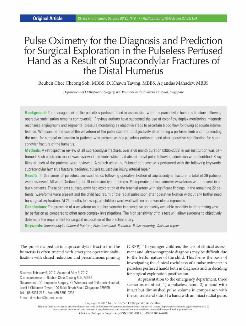
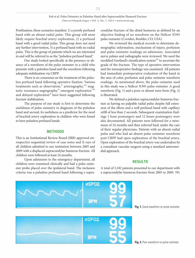
We reviewed the medical records to determine de-mographic information, mechanism of injury, perfusion and pulse oximeter readings on admission. Associated nerve palsies and radiographs were reviewed. We used the modified Gartland’s classification system10) to ascertain the grade of the fracture. The type of operative intervention and the intraoperative findings was examined. All patients had immediate postoperative evaluation of the hand in the area of color, perfusion and pulse oximeter waveform readings. As mentioned above, the pulse oximeter used in this study was a Nellcor N395 pulse oximeter. A good waveform (Fig. 1) and a poor or absent wave form (Fig. 2) is illustrated.
We defined a pulseless supracondylar humerus frac-ture as having no palpable radial pulse despite full exten-sion of the elbow and a well perfused hand with capillary refill of less than 2 seconds. Subsequent examination find-ings 1 hour postsurgery and 12 hours postsurgery were also documented. All patients were followed for a mini-mum of 24 months and then referred back under the care of their regular physicians. Patients with an absent radial pulse and who had an absent pulse oximeter waveform post CRPP had open exploration of the brachial artery. Open exploration of the brachial artery was undertaken by a consultant vascular surgeon using a standard anterome-dial approach.
RESULTS
A total of 3,182 patients presented to our department with a supracondylar humerus fracture from 2005 to 2009. 781
Fig. 1. Good waveform on pulse oximeter.
Fig. 2. Poor waveform on pulse oximeter.
76
Soh et al. Pulse Oximetry in Pulseless Hand after Supracondylar Humeral FractureClinics in Orthopedic Surgery • Vol. 5, No. 1, 2013 • www.ecios.org
Tabl
e 1.
Sum
mar
izing
26
Patie
nts w
ith P
ulse
less
and
Per
fuse
d Ha
nds P
ost R
educ
tion
and
Fixat
ion
Subj
ect
Age
(y
r + m
o)Ge
nder
Nat
ure
of in
jury
Gartl
and
clas
sific
atio
n an
d fra
ctur
e ty
pe a
nd
disp
lace
men
tN
erve
in
jury
Frac
ture
st
abili
zatio
n, n
o. o
f K-
wire
s (m
ed/la
t)
Wav
efor
m
follo
win
g
oper
ativ
e re
duct
ion
Brac
hial
arte
ry
expl
orat
ion
fin
dngs
Post
oper
ativ
e RO
MFo
llow
-up
dura
tion
(mo)
15
+ 0
MFe
ll fro
m th
e sli
deIII
, ext
ensio
n, p
oste
rola
tera
lNo
2 (1
/1)
Pres
ent
Full
30
210
+ 1
1M
Fell
from
the
slide
III, e
xten
sion,
pos
terio
rNo
2 (1
/1)
Pres
ent
Full
26
311
+ 7
MFe
ll of
f whi
le p
layin
g pi
ggyb
ack
III, e
xten
sion,
pos
tero
late
ral
No3
(1/2
)Pr
esen
tFu
ll27
44
+ 1
FSl
ippe
d at
hom
eIII
, ext
ensio
n, p
oste
rom
edia
lNo
2 (1
/1)
Pres
ent
Full
25
53
+ 5
FFe
ll ou
t of t
he b
edIII
, ext
ensio
n, p
oste
rior
No2
(1/1
)Pr
esen
tFu
ll24
69
+ 6
MFe
ll fro
m th
e m
onke
y bar
III, e
xten
sion,
pos
tero
late
ral
No3
(1/2
)Pr
esen
tFu
ll25
74
+ 4
FFe
ll fro
m th
e ch
air
III, e
xten
sion,
pos
tero
med
ial
No2
(1/1
)Pr
esen
tFu
ll30
83
+ 9
FFe
ll of
f the
raili
ng
III, e
xten
sion,
pos
tero
med
ial
No3
(1/2
)Pr
esen
tNM
26
95
+ 4
MFe
ll fro
m th
e m
onke
y bar
III, e
xten
sion,
pos
tero
med
ial
Yes,
AIN
an
d ra
dial
2 (1
/1)
Abse
ntHa
emat
oma
Redu
ced
30
106
+ 5
MFe
ll fro
m th
e m
onke
y bar
III, e
xten
sion,
pos
tero
med
ial
No2
(1/1
)Pr
esen
tFu
ll25
119
+ 2
FFe
ll do
wn
whi
le ru
nnin
gIII
, ext
ensio
n, p
oste
rola
tera
lNo
2 (1
/1)
Pres
ent
NM36
123
+ 8
FFe
ll in
scho
olIII
, ext
ensio
n, p
oste
rior
No2
(1/1
)Ab
sent
Kink
ing
of th
e br
achi
al a
rtery
Full
34
137
+ 4
MFe
ll w
hile
jum
ping
on
the
sofa
III, e
xten
sion,
pos
terio
rYe
s, AI
N2
(1/1
)Pr
esen
tNM
35
142
+ 5
MSl
ippe
d at
hom
eIII
, ext
ensio
n, p
oste
rom
edia
lYe
s, AI
N2
(1/1
)Pr
esen
tNM
27
155
+ 7
MFe
ll fro
m th
e m
onke
y bar
III, e
xten
sion,
pos
terio
rYe
s, AI
N3
(1/2
)Pr
esen
tFu
ll25
167
+ 4
FFe
ll of
f the
chai
rIII
, ext
ensio
n, p
oste
rior
Yes,
AIN
3 (0
/3)
Pres
ent
Full
25
1710
+ 2
MFe
ll fro
m m
etal
raili
ngIII
, ext
ensio
n, p
oste
rom
edia
lYe
s, AI
N2
(1/1
)Pr
esen
tRe
duce
d25
1811
+ 1
0M
Fell
off t
he ch
air
III, e
xten
sion,
pos
tero
late
ral
No2
(1/1
)Pr
esen
tFu
ll26
197
+ 11
MFe
ll in
pla
ygro
und
III, e
xten
sion,
pos
tero
late
ral
No2
(1/1
)Pr
esen
tFu
ll28
203
+ 1
MFe
ll of
f the
chai
rIII
, ext
ensio
n, p
oste
rola
tera
lNo
2 (1
/1)
Pres
ent
Full
25
215
+ 1
FFe
ll of
f the
chai
rIII
, ext
ensio
n, p
oste
rior
No2
(1/1
)Ab
sent
Kink
ing
of th
e br
achi
al a
rtery
Redu
ced
38
2212
+ 1
0M
Fell
from
the
mon
key b
arIII
, ext
ensio
n, p
oste
rom
edia
lYe
s, AI
N2
(1/1
)Pr
esen
tFu
ll24
235
+ 0
MFe
ll fro
m th
e m
onke
y bar
III, e
xten
sion,
pos
tero
med
ial
No2
(1/1
)Pr
esen
tNM
30
244
+ 9
FFe
ll in
pla
ygro
und
III, e
xten
sion,
pos
tero
med
ial
No3
(0/3
)Pr
esen
tNM
26
256
+ 9
MFe
ll fro
m th
e m
onke
y bar
III, e
xten
sion,
pos
tero
late
ral
No2
(1/1
)Pr
esen
tLim
ited
25
265
+ 5
MTr
ippe
d an
d fe
llIII
, ext
ensio
n, p
oste
rmed
ial
No2
(1/1
)Ab
sent
Arte
ry e
ntra
pped
in
# si
teFu
ll36
ROM
: ran
ge o
f mot
ion,
AIN
: ant
erio
r int
eros
seus
ner
ve, N
M: n
ot m
entio
ned
in fo
llow
-up
note
s.
77
Soh et al. Pulse Oximetry in Pulseless Hand after Supracondylar Humeral FractureClinics in Orthopedic Surgery • Vol. 5, No. 1, 2013 • www.ecios.org
children sustained displaced supracondylar fractures of Gartland grade IIb or III and were admitted for operative stabilization.
In the emergency department, the above criteria for determining if a hand was pulseless were applied. We identified 37 (4.7%) patients presenting with a pulseless hand following a supracondylar humerus fracture. All were grade III extension-type fractures and were closed injuries. All 37 received closed reduction and percutane-ous pinning emergently within 6 hours of presenting to the hospital. All patients had near anatomic reduction and restoration of the Baumann’s angle. Postoperatively, 11 patients had return of the radial pulse. The remaining 26 patients had no palpable radial pulse but had a perfused hand with good capillary refill. Each of these patients had a pulse oximeter applied intraoperatively and in retrospec-tive analysis were divided into 2 groups.
Group 1. Post CRPP: Pulseless Perfused Hands with a Waveform on the Pulse OximeterThis group had 22 patients. All patients had a good wave-form and at the 1 hour postoperative review, had an intact
radial pulse. The radial pulse remained present over the next 24 hours. At follow-up, all patients continued to have intact radial pulses. Seven of the patients had a document-ed anterior interosseous nerve injury which recovered by the 3rd month postsurgery. No patient subsequently de-veloped ischemic contractures or noted any forearm clau-dication in their school physical exercise classes.
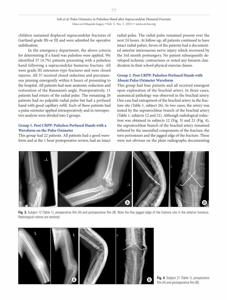
Group 2. Post CRPP: Pulseless Perfused Hands with Absent Pulse Oximeter WaveformThis group had four patients and all received emergent open exploration of the brachial artery. In three cases, anatomical pathology was observed in the brachial artery. One case had entrapment of the brachial artery in the frac-ture site (Table 1, subject 26). In two cases, the artery was tented by the supratrochlear branch of the brachial artery (Table 1, subjects 12 and 21). Although radiological reduc-tion was obtained in subjects 12 (Fig. 3) and 21 (Fig. 4), the supratrochlear branch of the brachial artery remained tethered by the unossified components of the fracture, the torn periosteum and the jagged edge of the fracture. These were not obvious on the plain radiographs documenting
Fig. 3. Subject 12 (Table 1), preoperative film (A) and postoperative film (B). Note the fine jagged edge of the fracture site in the anterior humerus. Radiological indices are restored.
Fig. 4. Subject 21 (Table 1), preoperative film (A) and postoperative film (B).
78
Soh et al. Pulse Oximetry in Pulseless Hand after Supracondylar Humeral FractureClinics in Orthopedic Surgery • Vol. 5, No. 1, 2013 • www.ecios.org
the reduction. The ligation of the binding supratrochlear branch and the freeing of the entrapped brachial artery in these 3 cases led to a prompt return of the radial pulse intraoperatively. The fourth case (Table 1, subject 9), had a large hematoma in the anteromedial cubital fossa but no appreciable pathology over the brachial artery. Following evacuation of the hematoma, radial pulse promptly re-turned. In the early postoperative period, the radial pulse remained present 24 hours postoperative and during all subsequent follow-up visits.
All 4 patients remained well and at 24 months, had a palpable radial pulse with no intervening events of re-stenosis. Of the 4 cases only 1 had concomitant nerve injury affecting the anterior interosseus nerve. The nerve injury recovered at follow-up. All 4 patients did not have any eventual loss of function or forearm claudication. No patients in group 1 or group 2 developed forearm com-partment syndrome.
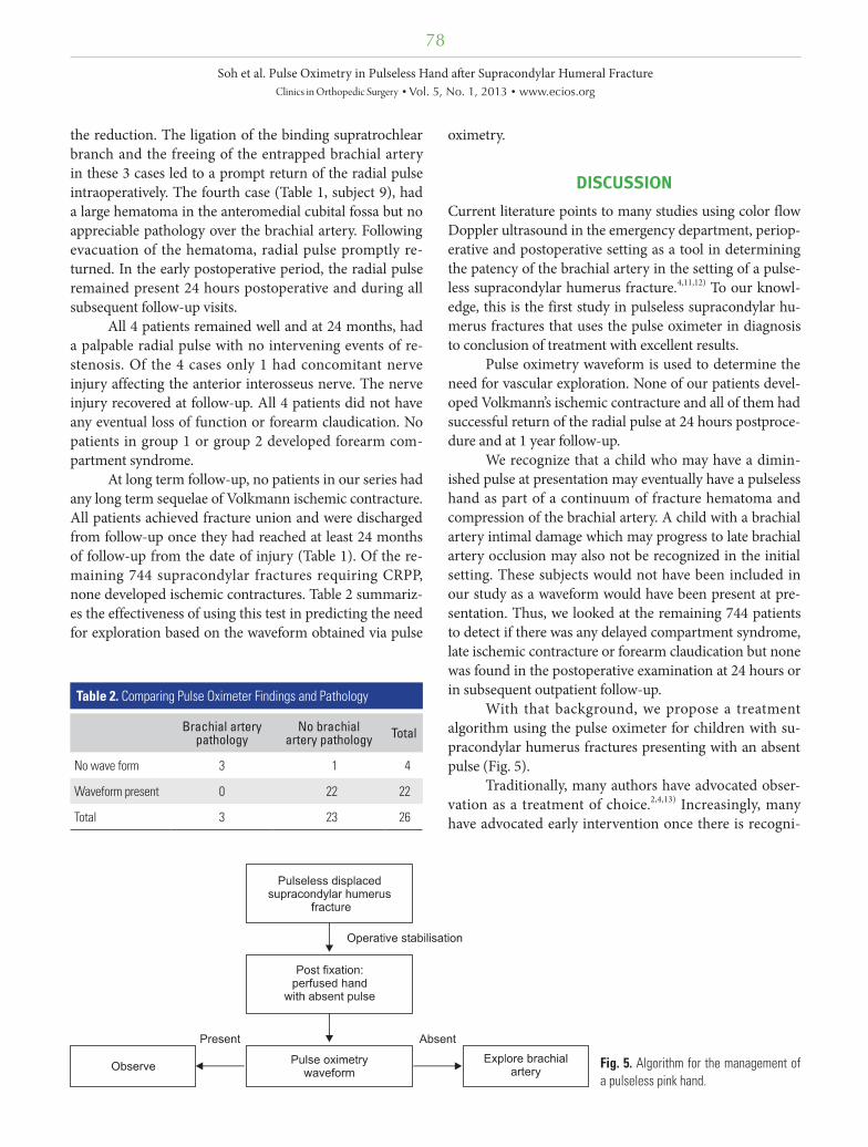
At long term follow-up, no patients in our series had any long term sequelae of Volkmann ischemic contracture. All patients achieved fracture union and were discharged from follow-up once they had reached at least 24 months of follow-up from the date of injury (Table 1). Of the re-maining 744 supracondylar fractures requiring CRPP, none developed ischemic contractures. Table 2 summariz-es the effectiveness of using this test in predicting the need for exploration based on the waveform obtained via pulse
oximetry.
DISCUSSION
Current literature points to many studies using color flow Doppler ultrasound in the emergency department, periop-erative and postoperative setting as a tool in determining the patency of the brachial artery in the setting of a pulse-less supracondylar humerus fracture.4,11,12) To our knowl-edge, this is the first study in pulseless supracondylar hu-merus fractures that uses the pulse oximeter in diagnosis to conclusion of treatment with excellent results.
Pulse oximetry waveform is used to determine the need for vascular exploration. None of our patients devel-oped Volkmann’s ischemic contracture and all of them had successful return of the radial pulse at 24 hours postproce-dure and at 1 year follow-up.
We recognize that a child who may have a dimin-ished pulse at presentation may eventually have a pulseless hand as part of a continuum of fracture hematoma and compression of the brachial artery. A child with a brachial artery intimal damage which may progress to late brachial artery occlusion may also not be recognized in the initial setting. These subjects would not have been included in our study as a waveform would have been present at pre-sentation. Thus, we looked at the remaining 744 patients to detect if there was any delayed compartment syndrome, late ischemic contracture or forearm claudication but none was found in the postoperative examination at 24 hours or in subsequent outpatient follow-up.
With that background, we propose a treatment algorithm using the pulse oximeter for children with su-pracondylar humerus fractures presenting with an absent pulse (Fig. 5).
Traditionally, many authors have advocated obser-vation as a treatment of choice.2,4,13) Increasingly, many have advocated early intervention once there is recogni-
Table 2. Comparing Pulse Oximeter Findings and Pathology
Brachial artery pathology
No brachial artery pathology Total
No wave form 3 1 4
Waveform present 0 22 22
Total 3 23 26
Fig. 5. Algorithm for the management of a pulseless pink hand.
79
Soh et al. Pulse Oximetry in Pulseless Hand after Supracondylar Humeral FractureClinics in Orthopedic Surgery • Vol. 5, No. 1, 2013 • www.ecios.org
tion of vascular injury. Noaman14) explored the brachial artery in 31 children in whom the radial pulse was absent after closed reduction and pinning in a series of 840 grade III fractures. His indications were a pulseless forearm with a pink or cold hand, an absent radial pulse one hour after satisfactory closed reduction and percutaneous pinning and an absent radial pulse associated with an open fracture or signs of tethering of the brachialis muscle. There was arterial damage in 30 children and in one the artery was released from the site of the fracture. Korompilias et al.,15) in his series of 5 children with a pink, pulseless hand also recommended surgical exploration to restore the patency of the brachial artery even in the presence of a viable and well-perfused hand after an attempt at closed reduction.
Most recently, White et al.7) systematically reviewed 19 papers in the English literature where pulseless and perfused supracondylar fractures were managed. A total of 331 such fractures were recorded. Of these, 157 remained pulseless post reduction. Of this group, 90% underwent surgical exploration and 82% was found to have brachial artery injury. This paper also went on to analyze the results of patency of the brachial artery following exploration and in 54 such explorations 91% remained patent at the 1 year follow-up. While the results of this review appear to indicate that brachial artery injury was much higher than our series, we believe that there is some selection bias us-ing pooled retrospective data from 19 such studies. We experienced similar levels of patency of the brachial artery postsurgical exploration at the 1 year follow-up (100%). This systematic review further supports early intervention once there is recognition of brachial artery pathology and challenges early suggestions of ‘watchful waiting’.
In contrast, Ramesh et al.16) reported a series of patients with well perfused hands but absent radial pulse following humerus supracondylar fracture fixation. The author cautioned that these patients did not have excru-ciating pain distal to the elbow that persisted beyond 12 hours after the injury as compared to the series by Blakey et al.9) This demonstrates that the upper limb has an ex-tremely good collateral blood supply.
The abundant collateral supply of the elbow comes from the superior and inferior ulnar collateral artery medially and the profunda brachii artery laterally. These arteries branch from the brachial artery proximal to the olecranon fossa, where supracondylar humerus fractures frequently occur. The profunda brachii artery branches to give a radial recurrent branch and an interosseous recur-rent branch. The superior ulnar collateral artery runs pos-terior to the medial epicondyle to form the posterior ulnar recurrent artery. The inferior ulnar collateral artery runs
anterior to the medial condyle of the humerus to become the anterior ulnar recurrent artery. The radial recurrent and both the ulnar recurrent arteries then rejoin the radial and ulnar artery respectively at the level of the biceps in-sertion on the radial neck.17) This rich collateral network forms the basis for pulseless perfused hands (Table 1, sub-ject 26) despite brachial artery entrapment at the level of the supracondylar humerus.
While we have no patient in our study with both an absent radial pulse and a well perfused limb on long term follow-up, we recognize that it may be possible for a waveform to be present despite brachial artery occlusion due to the rich network of collaterals at the elbow joint. Our study however did not show any patients who did not have return of the radial pulse once intraoperative pulse oximetry showed a good waveform. This may be a limita-tion of our study due to its small sample size or may indi-cate that pulse oximetry is truly objective in determining the eventual return of the radial pulse. At a minimum of 24 months follow-up, all patients had well palpable radial pulses indicating that stenosis or delayed intimal damage resulting in occlusion unlikely occurred.
We are of the opinion that given the readily available resource of the pulse oximeter in the operating room, the challenges of subjectivity in monitoring pain in the post-operative patient and the good results following surgical exploration of the brachial artery that surgical exploration should be undertaken once there is documented injury to the brachial artery. Our study demonstrates that the pulse oximeter is an objective tool to support or reject the deci-sion for surgical exploration.
Mangat et al.18) advocated that the likelihood of vascular injury was high with documented anterior inter-osseous nerve or median nerve injury in his series of 19 patients over a 14-year period. Our study showed different results as only 1 patient who required surgical explora-tion with positive findings of brachial artery pathology had nerve injury at presentation. In the group which had a good waveform and which did not require exploration, 7 patients had nerve injury which recovered during follow-up. None of these patients required surgical exploration. We surmise that nerve injury, while associated with the incidence of brachial artery pathology in a pulseless hand, is not an objective predictor of surgical exploration.
The use of pulse oximetry allows easy access and is a relatively cheap alternative to Doppler ultrasound. Furthermore, it removes the subjectivity in examining the peripheral nerves in an already fretful and irritable child. This allows earlier diagnosis and treatment of the pulseless limb following supracondylar fracture.
80
Soh et al. Pulse Oximetry in Pulseless Hand after Supracondylar Humeral FractureClinics in Orthopedic Surgery • Vol. 5, No. 1, 2013 • www.ecios.org
Previous authors have also suggested the use of angiography to assess the vascular status of the affected limb.3,19) Shaw et al.11) expressed that this may potentially increase the time required before a vascular exploration. Angiography is also invasive and has risks of iatrogenic in-jury to the artery due to its small size.20) In addition, there are also risks of contrast allergy.
We recognize that the use of the pulse oximeter in colder environments may lead to false positives as a result of peripheral vasoconstriction. However, this scenario was avoided by adequately warming the operating theatre.
We acknowledge limitations to our study with re-spect to its limited sample size and the lack of imaging or exploration to confirm the absence of injury to the brachi-al artery. Based on the eventual presence of a radial pulse, this study shows a positive predictive value of 0.75, and a negative predictive value of 1.0. We will need to continue
evaluating this with increasing numbers that are added to our study population over time. We are optimistic that this test has a high specificity of 95.65%, and would want to embark on future studies that test the true effectiveness of the algorithm and to pit the results of the pulse oximeter with that of color flow Doppler in a prospective context to assess the patency of the brachial artery.
In conclusion, the pulse oximeter is an excellent and readily available tool in the emergency department and the operative room to assist diagnosis of the pulseless hand and subsequently, to discern arterial injury in well perfused, postfixation supracondylar humerus fractures.
CONFLICT OF INTEREST
No potential conflict of interest relevant to this article was reported.
REFERENCES1. Green NE. Fractures and dislocations about the elbow. In:
Green NE, Swiontkowski MF, eds. Skeletal trauma in chil-dren. 3rd ed. Philadelphia: Saunders; 2002. 257-321.
2. Garbuz DS, Leitch K, Wright JG. The treatment of supra-condylar fractures in children with an absent radial pulse. J Pediatr Orthop. 1996;16(5):594-6.
3. Shuck JM, Omer GE Jr, Lewis CE Jr. Arterial obstruction due to intimal disruption in extremity fractures. J Trauma. 1972;12(6):481-9.
4. Sabharwal S, Tredwell SJ, Beauchamp RD, et al. Manage-ment of pulseless pink hand in pediatric supracondylar frac-tures of humerus. J Pediatr Orthop. 1997;17(3):303-10.
5. Schoenecker PL, Delgado E, Rotman M, Sicard GA, Capelli AM. Pulseless arm in association with totally displaced su-pracondylar fracture. J Orthop Trauma. 1996;10(6):410-5.
6. Copley LA, Dormans JP, Davidson RS. Vascular injuries and their sequelae in pediatric supracondylar humeral fractures: toward a goal of prevention. J Pediatr Orthop. 1996;16(1):99-103.
7. White L, Mehlman CT, Crawford AH. Perfused, pulseless, and puzzling: a systematic review of vascular injuries in pediatric supracondylar humerus fractures and results of a POSNA questionnaire. J Pediatr Orthop. 2010;30(4):328-35.
8. Reigstad O, Thorkildsen R, Grimsgaard C, Reigstad A, Rokkum M. Supracondylar fractures with circulatory failure after reduction, pinning, and entrapment of the brachial artery: excellent results more than 1 year after open explora-tion and revascularization. J Orthop Trauma. 2011;25(1):26-
30.
9. Blakey CM, Biant LC, Birch R. Ischaemia and the pink, pulseless hand complicating supracondylar fractures of the humerus in childhood: long-term follow-up. J Bone Joint Surg Br. 2009;91(11):1487-92.
10. Beaty JH, Kasser JR. Supracondylar fractures of the dis-tal humerus. In: Beaty JH, Kasser JR, eds. Rockwood and Wilkins’ fractures in children. Philadelphia: Lippincott Wil-liams & Wilkins; 2005. 577-624.
11. Shaw BA, Kasser JR, Emans JB, Rand FF. Management of vascular injuries in displaced supracondylar humerus frac-tures without arteriography. J Orthop Trauma. 1990;4(1):25-9.
12. Fry WR, Smith RS, Sayers DV, et al. The success of duplex ultrasonographic scanning in diagnosis of extremity vascu-lar proximity trauma. Arch Surg. 1993;128(12):1368-72.
13. Malviya A, Simmons D, Vallamshetla R, Bache CE. Pink pulseless hand following supra-condylar fractures: an audit of British practice. J Pediatr Orthop B. 2006;15(1):62-4.
14. Noaman HH. Microsurgical reconstruction of brachial ar-tery injuries in displaced supracondylar fracture humerus in children. Microsurgery. 2006;26(7):498-505.
15. Korompilias AV, Lykissas MG, Mitsionis GI, Kontogeorga-kos VA, Manoudis G, Beris AE. Treatment of pink pulseless hand following supracondylar fractures of the humerus in children. Int Orthop. 2009;33(1):237-41.
16. Ramesh P, Avadhani A, Shetty AP, Dheenadhayalan J, Ra-jasekaran S. Management of acute 'pink pulseless' hand in

81
Soh et al. Pulse Oximetry in Pulseless Hand after Supracondylar Humeral FractureClinics in Orthopedic Surgery • Vol. 5, No. 1, 2013 • www.ecios.org
pediatric supracondylar fractures of the humerus. J Pediatr Orthop B. 2011;20(3):124-8.
17. McMinn RM. Last’s anatomy: regional and applied. 9th ed. New York: Churchill Livingstone; 1994. 78-96.
18. Mangat KS, Martin AG, Bache CE. The 'pulseless pink' hand after supracondylar fracture of the humerus in children: the predictive value of nerve palsy. J Bone Joint Surg Br.
2009;91(11):1521-5.
19. Luria S, Sucar A, Eylon S, et al. Vascular complications of supracondylar humeral fractures in children. J Pediatr Or-thop B. 2007;16(2):133-43.
20. Shaker IJ, White JJ, Signer RD, Golladay ES, Haller JA Jr. Special problems of vascular injuries in children. J Trauma. 1976;16(11):863-7.


![Pediatric Supracondylar Fractures: Are Medial Pins Indicated?are the supracondylar fractures of the humerus that can be managed by both operative and non-operative modalities [1]](https://img.pdfslide.us/doc/110x75/6087220d2ec1ae7c713805b2/pediatric-supracondylar-fractures-are-medial-pins-indicated-are-the-supracondylar.jpg)