Embed Size (px)
Citation preview

Pulmonology
• The medical specialty that studies the anatomy and physiology of the respiratory system and uses diagnostic tests, medical and surgical procedures, and drugs to treat respiratory diseases.

Figure 4-1 Respiratory system

Anatomy and Physiology
• Consists of the right and left lungs and the air passageways that connect the lungs to the outside of the body
• The upper respiratory system is in the head and neck and includes the nose, nasal cavity, and pharynx (throat).
• The upper respiratory system shares structures with the ears, nose, and throat (ENT) system.

Anatomy and Physiology (cont’d)
• The lower respiratory system is in the neck and thoracic cavity and includes the larynx (voice box), trachea (windpipe), bronchi, bronchioles, and alveoli (in the lungs).
• The lungs fill much of the thoracic cavity.• Brings oxygen into the body and expels the
waste product carbon dioxide

Anatomy of the Respiratory System
• Nose and Nasal Cavity– The nose contains the nasal cavity, which is
divided in the center by the nasal septum. – Each side of the nasal septum has three long, bony
projections: the superior, middle, and inferior turbinates, or nasal conchae, which jut into the nasal cavity and slow down inhaled air so that it can be warmed and moistened.

Anatomy of the Respiratory System (cont’d)
• The nasal cavity is lined with mucosa, a mucous membrane that humidifies the air and produces mucus.
• Mucus and hairs in the nose trap inhaled particles of dust, pollen, smoke, and bacteria and keep them from entering the lungs.

Figure 4-2 Nasal cavity

Anatomy of the Respiratory System (cont’d)
• Pharynx– Mucous membranes of the pharynx warm and
moisten inhaled air and trap particles. – A common passageway for inhaled air, exhaled air,
and food.

• Larynx– At its inferior end, the pharynx divides into two
parts: the larynx which leads to the trachea; and the esophagus which leads to the stomach.
– Remains open during respiration and speech, allowing air to pass in and out through the vocal cords.
Anatomy of the Respiratory System (cont’d)
• Larynx (cont’d)– During swallowing, muscles in the neck pull the
larynx up to meet the epiglottis, which seals off the larynx, so that swallowed food moves across the epiglottis and into the esophagus, not into the trachea.
Anatomy of the Respiratory System (cont’d)

Figure 4-3 Larynx

• Trachea– Below the vocal cords, the larynx merges into the
trachea, which is about 1 inch in diameter and 4 inches in length.
– A passageway for inhaled and exhaled air – A column of C-shaped rings of cartilage provide
support to the trachea.
Anatomy of the Respiratory System (cont’d)
• Trachea (cont’d)– On the posterior surface where there is no
cartilage, the trachea is flexible and can flatten to make room when food passes through the esophagus.
Anatomy of the Respiratory System (cont’d)

• Bronchi– The trachea ends at the top of the inverted Y of
the right and left primary bronchi.– The primary bronchi have cartilage rings that
provide support.– Each primary bronchus enters a lung and branches
into smaller bronchioles.
Anatomy of the Respiratory System (cont’d)

• Bronchi (cont’d)– The smallest bronchioles (diameter 1 mm or less)
have smooth muscle around them, but no cartilage.
– The lumen is the central opening in the bronchi and bronchioles through which air passes.
– Bronchopulmonary refers to the bronchi and the lungs.
Anatomy of the Respiratory System (cont’d)

• Bronchi (cont’d)– The bronchial tree includes the trachea, bronchi,
and bronchioles.– The bronchial tree is lined with cilia, small hairs
that move in coordinated waves to carry mucus and trapped particles toward the throat where they can be expelled.
Anatomy of the Respiratory System (cont’d)

Figure 4-4 Trachea, lung, bronchi, bronchioles, and alveoli
• Lungs– Spongy, air-filled structures– Each lung contains lobes, large divisions whose
dividing lines are visible on the outer surface of the lung.
Anatomy of the Respiratory System (cont’d)

• Lungs (cont’d)– The right lung, which is larger, has three lobes:
• The right upper lobe (RUL)• The right middle lobe (RML)• The right lower lobe (RLL)
– The left lung has two lobes:• The left upper lobe (LUL) • The left lower lobe (LLL)
Anatomy of the Respiratory System (cont’d)

• Lungs (cont’d)– The rounded top of each lung is the apex.– The base of each lung lies along the diaphragm.– A bronchus enters the lung at the hilum (an
indentation on the medial surface of the lung).– Inside the lung, the bronchus branches into
bronchioles, which branch into alveoli.
Anatomy of the Respiratory System (cont’d)

• Lungs (cont’d)– Oxygen and carbon dioxide are exchanged
between the alveolus and a nearby small blood vessel (capillary).
– Alveolus secretes surfactant, a protein-fat compound that reduces surface tension and keeps the walls of the alveolus from collapsing with each exhalation.
Anatomy of the Respiratory System (cont’d)

• Lungs (cont’d)– Collectively, the alveoli are the pulmonary
parenchyma, the functional part of the lung, as opposed to the connective tissue framework around them.
Anatomy of the Respiratory System (cont’d)

Figure 4-5 Diaphragm and pleura

• Thoracic Cavity– The thorax is a bony cage that consists of the
sternum (breastbone), the ribs, and the spinal column; it surrounds and protects the thoracic cavity.
– The mediastinum, an irregularly shaped area between the lungs, contains the trachea (and the heart and esophagus).
Anatomy of the Respiratory System (cont’d)

• Thoracic Cavity (cont’d)– The diaphragm, a sheet of skeletal muscle, is
active during normal or forceful inhalation.– Intercostal muscles pull the ribs up and out, or
down and in, during forceful inhalation/exhalation.
– Each lung is surrounded by the pleura, a double-layered serous membrane.
Anatomy of the Respiratory System (cont’d)

• Thoracic Cavity (cont’d) – The visceral pleura is the layer next to the lung
surface, while the parietal pleura is the layer next to the wall of the thorax.
– The pleura secretes pleural fluid, a slippery, watery fluid that allows the two layers to slide smoothly past each other as the lungs expand and contract during respiration.
Anatomy of the Respiratory System (cont’d)

Physiology of Respiration
• Respiration consists of breathing in and breathing out.
• Breathing in is inhalation or inspiration. • Breathing out is exhalation or expiration.

Physiology of Respiration (cont’d)
• Breathing is normally an involuntary process.• The respiratory control centers in the brain
regulate the depth and rate of respiration.• Receptors in large arteries in the chest and
neck send these centers information about the blood level of oxygen; receptors in the brain send information about the blood level of carbon dioxide.

Physiology of Respiration (cont’d)
• The respiratory control centers send nerve impulses to the phrenic nerve, causing the diaphragm to contract and begin inspiration.
• A normal depth and rate of respiration is known as eupnea.

Physiology of Respiration (cont’d)
• Respiration involves five separate processes:– Ventilation―Movement of air in and out of the
lungs – External respiration―Movement of oxygen from
the alveoli into the blood and movement of carbon dioxide from the blood into the alveoli

Physiology of Respiration (cont’d)
– Gas transport―Blood transports oxygen and carbon dioxide; oxygenated blood travels from lungs to heart, where it is pumped throughout the body
– Internal respiration―Movement of oxygen from blood into cells and movement of carbon dioxide from cells into blood
– Cellular respiration―Oxygen is used by the cells to produce energy in the process of metabolism

Figure 4-6 Exhalation(Jim Corwin/Photo Researchers, Inc.)

Figure 4-7 Gas exchange
Diseases and Conditions
• Nose and Pharynx– Upper respiratory infection (URI)
• Trachea, Bronchi, and Bronchioles– Asthma– Bronchitis– Bronchiectasis

Asthma Animation
Click on the screenshot to view an animation on the topic of asthma.
Back to Directory

Figure 4-8 Upper respiratory infection(Mednet/Phototake NYC)
Diseases and Conditions (cont’d)
• Lungs– Abnormal breath sounds– Adult respiratory distress syndrome (ARDS)– Atelectasis – Chronic obstructive pulmonary disease (COPD)

Rhonchi Lung Sounds
Click on the screenshot to view an animation on the topic of lung sounds.
Back to Directory

Stridor Lung Sounds
Click on the screenshot to view an animation on the topic of lung sounds.
Back to Directory

Vesicular Lung Sounds
Click on the screenshot to view an animation on the topic of lung sounds.
Back to Directory

Figure 4-9 Adult respiratory distress syndrome

Diseases and Conditions (cont’d)
• Lungs (cont’d)– Cystic fibrosis (CF)– Empyema– Influenza– Legionnaire’s disease

Figure 4-10 Cystic fibrosis(Pearson Education/PH College)

Figure 4-11 Postural drainage and percussion(Hattie Young/Photo Researchers, Inc.)

Diseases and Conditions (cont’d)
• Lungs (cont’d)– Lung cancer– Occupational lung diseases

Diseases and Conditions (cont’d)
• Lungs (cont’d)– Aspiration pneumonia– Bacterial pneumonia– Bronchopneumonia– Double pneumonia

Figure 4-12 Tar deposits in the lungs(James Stevenson/Photo Researchers, Inc.)
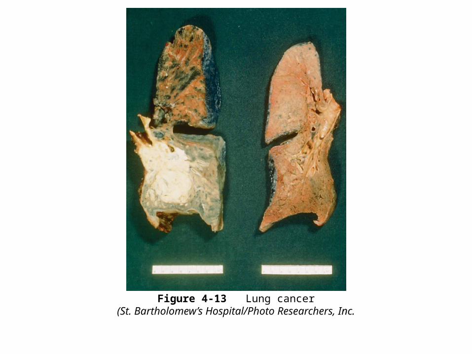
Figure 4-13 Lung cancer(St. Bartholomew’s Hospital/Photo Researchers, Inc.

Diseases and Conditions (cont’d)
• Lungs (cont’d)– Lobar pneumonia– Pneumococcal pneumonia– Pneumocystic jiroveci pneumonia– Viral pneumonia – Walking pneumonia

Figure 4-14 Pneumonia(Custom Medical Stock Photo, Inc.)
Diseases and Conditions (cont’d)
• Lungs (cont’d)– Pulmonary edema– Pulmonary embolism– Severe acute respiratory syndrome (SARS)– Tuberculosis (TB)
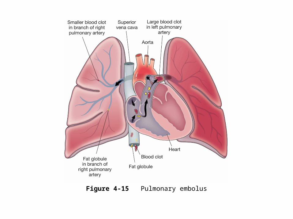
Figure 4-15 Pulmonary embolus
Diseases and Conditions (cont’d)
• Pleura and Thorax– Hemothorax– Pleural effusion– Pleurisy– Pneumothorax

Diseases and Conditions (cont’d)
• Respiration– Apnea– Bradypnea– Cough– Dyspnea– Orthopnea– Tachypnea

Diseases and Conditions (cont’d)
• Oxygen and Carbon Dioxide Levels– Anoxia– Asphyxia– Cyanosis– Hypercapnia– Hypoxemia

Laboratory and Diagnostic Procedures
• Arterial blood gases (ABG)• Carboxyhemoglobin

Laboratory and Diagnostic Procedures (cont’d)
• Oximetry• Pulmonary function test (PFT)

Figure 4-16 Pulse oximeter(O’Brien/Custom Medical Stock Photo, Inc.)

Figure 4-17 Pulmonary function test(BSIP/Phototake NYC)

Laboratory and Diagnostic Procedures (cont’d)
• Sputum culture and sensitivity (C&S)• Tuberculosis tests

Figure 4-18 Culture and sensitivity(Science Heritage/Custom Medical Stock Photo, Inc.)

Laboratory and Diagnostic Procedures (cont’d)
• Radiology and Nuclear Medicine Procedures– Chest radiography – CT scan and MRI scan– Lung scan

Medical and Surgical Procedures
• Medical Procedures– Auscultation and percussion – Cardiopulmonary resuscitation (CPR)– Endotracheal intubation

Figure 4-19 Endotracheal intubation

Medical and Surgical Procedures (cont’d)
• Medical Procedures– Heimlich maneuver– Incentive spirometry– Oxygen therapy – Vital signs

Figure 4-20 Nasal cannula(©Ray Kemp/911 Imaging)

Figure 4-21 Endotracheal tube and Ambu bag(Pearson Education/PH College)

Medical and Surgical Procedures (cont’d)
• Surgical Procedures– Bronchoscopy – Chest tube insertion – Lung resection

Figure 4-22 Lobectomy

Medical and Surgical Procedures (cont’d)
• Surgical Procedures– Thoracocentesis – Thoracotomy – Tracheostomy

Figure 4-23 Tracheostomy(©Jenny Thomas/Pearson Education)

Drug Categories
• These categories of drugs are used to treat respiratory Diseases and Conditions:– Antibiotic drugs – Antitubercular drugs– Antitussive drugs– Bronchodilator drugs– Antiviral drugs
Drug Categories
• These categories of drugs are used to treat respiratory Diseases and Conditions:– Corticosteroid drugs– Expectorant drugs– Leukotriene receptor blocker drugs– Mast cell stabilizer drugs

Figure 4-24 Metered-dose inhaler(Custom Medical Stock Photo, Inc.)

Abbreviations

Abbreviations (cont’d)