Embed Size (px)
Citation preview
Pulmonary Issues in Neuromuscular Diseases
Bassel Salman, MD
Assistant ProfessorOakland UniversityWayne State UniversityDepartment of PediatricsDivision of Hospital MedicineDivision of Pulmonary medicineBeaumont Children Hospital

• Neuromuscular Disorder does NOT mean mental retardation .
• It may OR may NOT be associated with MR.
• It could be congenital or acquired.

Conflict of Interest?

• My life is full of conflicts
• None of which is interesting
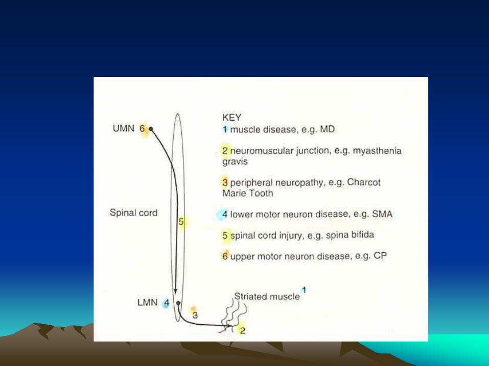
Upper Motor Neuron
• Cerebral Palsy
Lower Motor Neuron
1. Spinal Motor Atrophy (SMA).
2. Peripheral Neuropathy (GBS, CMT).
3. NM Junction (Myasthenia Gravis).
4. Muscular Dystrophies.
Combined Upper and Lower
1. Spinal Cord Injuries.
2. Spina Bifida
Normal Respiratory Muscles Function
Diaphragm
• Acts as a piston that decreases intrapleural pressure.
• Normal motion: BOTH chest wall and abdominal expand “outside”.
• Patients with NMD do NOT report dyspnea until the diaphragm is involved.
Intercostal Muscles
• External: primarily inspiratory.
• Internal: Primarily expiratory.
• Normally: Inspiration is active and expiration is passive.
Abdominal muscles
• Contraction of RA and AO increases intra abdominal pressure and helps active expiration (exercise, Asthma--)
Upper Airway Muscles
• Contract with inspiration, preventing airway collapse.
• Healthy infants: “glottic breaking” is partial ADDUCTION of VC to maintain end exp. Volume .
• “Glottic breaking” is impaired with NMD, resulting in atelectasis
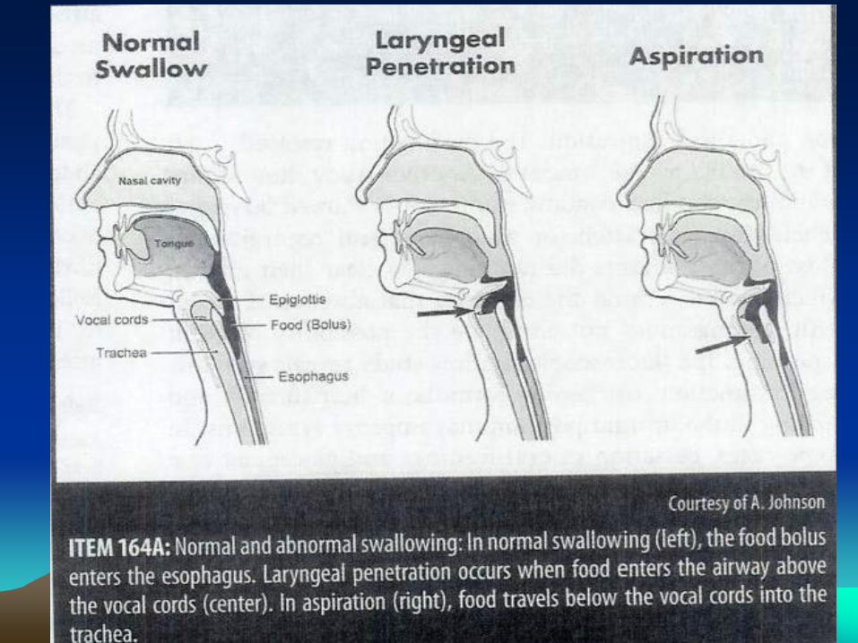
Swallow Function
• Oral phase: voluntary.
• Pharyngeal and esophageal: involuntary.
• Weakness: results in food going thru nose and trachea.
Cough
A. Starts with irritation of receptors of vagus nerve.Inspiration to near TLC.
B. Closure of glottis followed by contraction of abdominal muscle.
C. Glottis suddenly opens resulting in an upward movement in Diaphragm and expulsion of air @ 300 mi/hr.
Cough
• Weakness of the abdominal muscles reduces the effectiveness of cough
SLOW Changes in NMD
• In the early stage little if any paranchymal lung disease is present.
With TIME• Weak muscles and often chest wall
deformity lead to poor lung expansion and ultimately reduced lung growth.
• Weak VC and oropharyngeal muscles lead to recurrent aspiration pneumonias.
• Poor expansion and cough leads to recurrent atelectasis
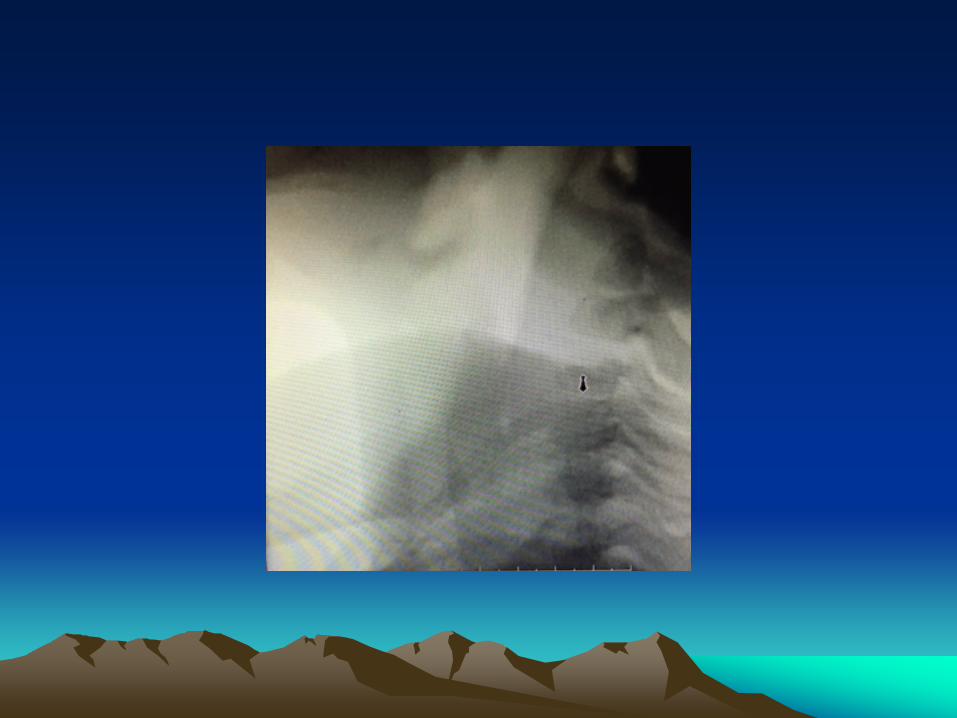
Swallow Study
• Poor swallowing impairs nutrition and growth which worsen the muscle weakness and weakens the immunity.
• Progressive muscle weakness lead to worsen chest deformity and further decrease in lung volumes.
Pulmonary Consequences of NMD
1.Recurrent Pneumonia and aspirations.2.Recurrent Bronchospasm.3.Recurrent Atelectasis.4.OSA.5.Daytime sleepiness and fatigue.6.Drooling, difficulty swallowing.7.Nasal regurgitation.8.Decreased lung volume.9.Death from aspiration pneumonia is the most
common cause of death.
• All leads to Restrictive Lung Disease.
• Often with Obstructive component as well
The GOAL
• Delay/minimize the progression of lung disease.
• Prevent/Treat complications such as Asthma, pneumonia, OSA, --
• Improve the quality of life.
• Lung disease is a “secondary complication/natural progression” of NMD.
• Little if any lung disease is present in the early stages.
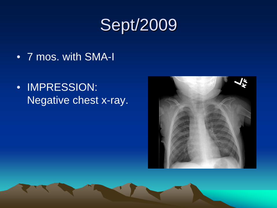
Sept/2009
• 7 mos. with SMA-I
• IMPRESSION:Negative chest x-ray.
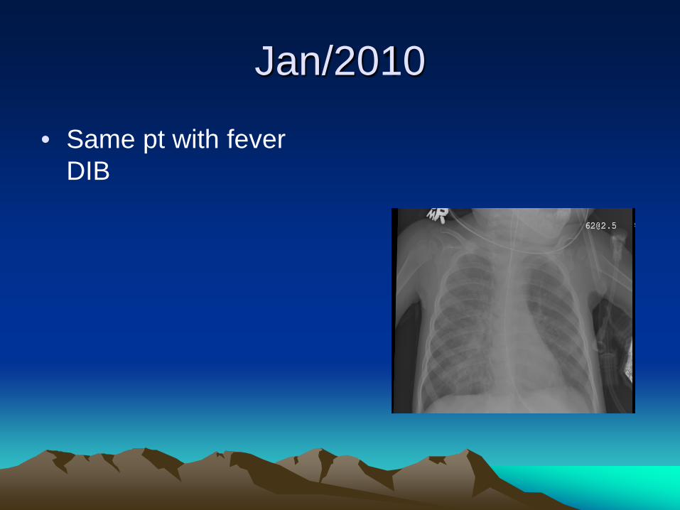
Jan/2010
• Same pt with fever DIB
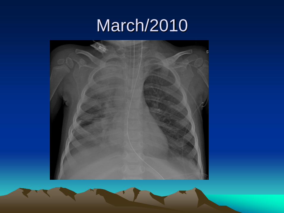
March/2010
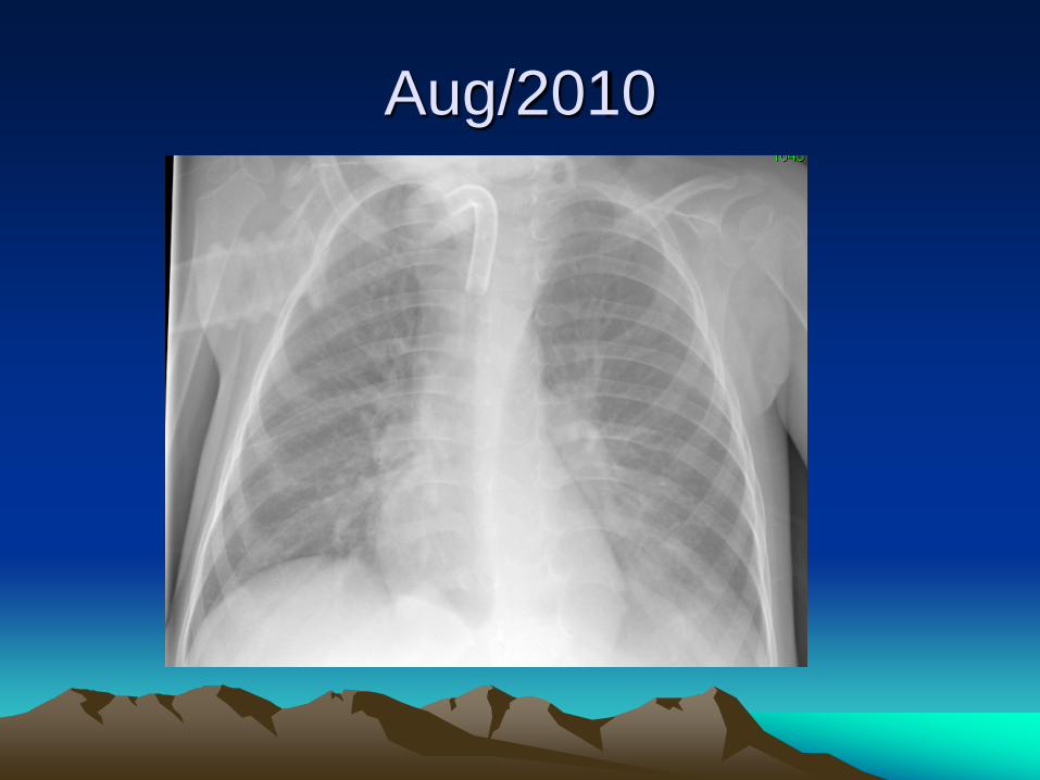
Aug/2010
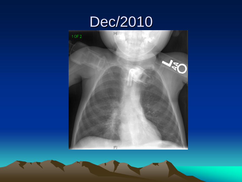
Dec/2010
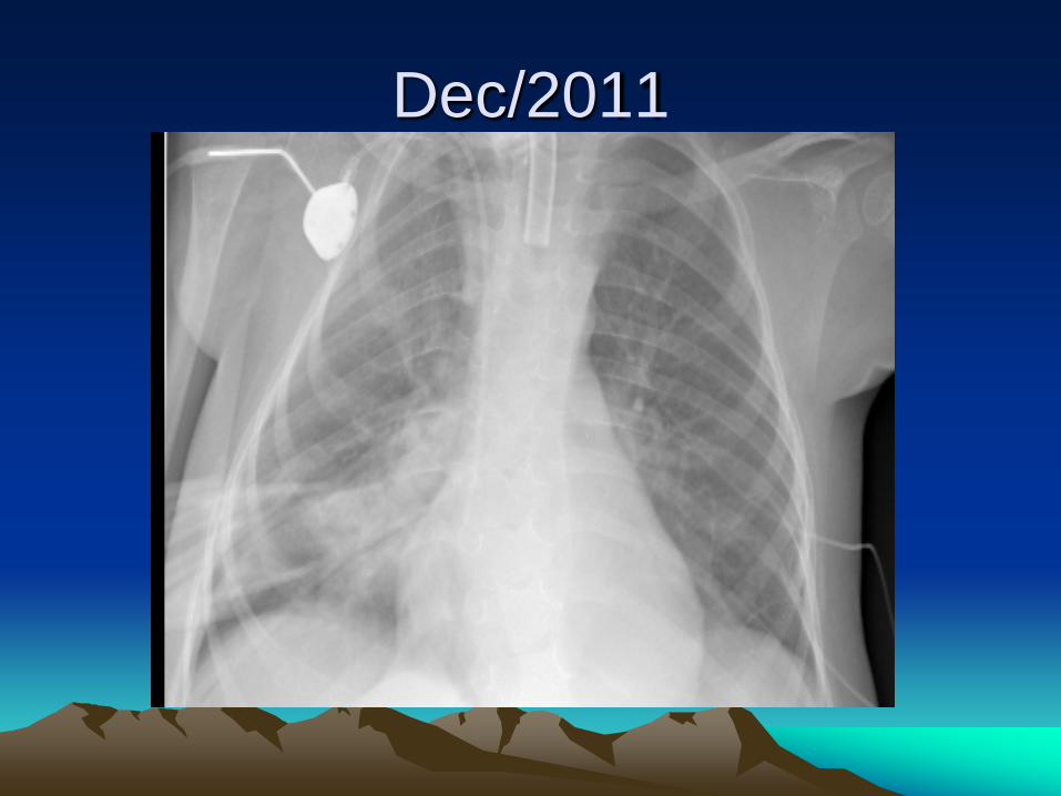
Dec/2011
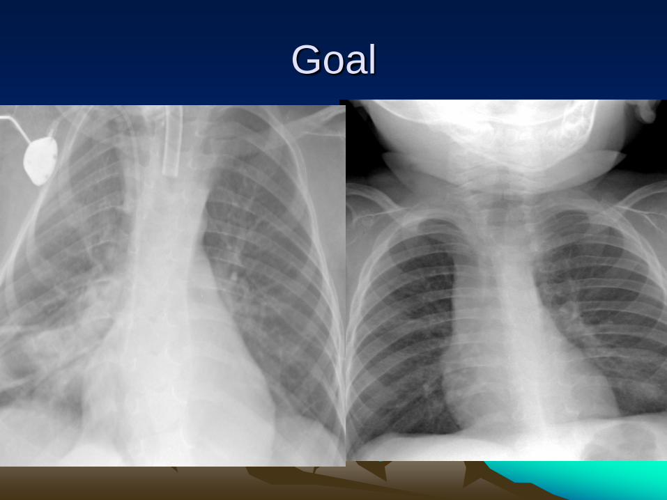
Goal
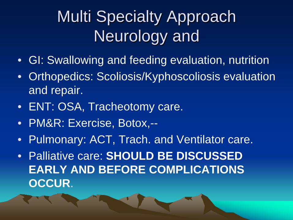
Multi Specialty ApproachNeurology and
• GI: Swallowing and feeding evaluation, nutrition• Orthopedics: Scoliosis/Kyphoscoliosis evaluation
and repair.• ENT: OSA, Tracheotomy care.• PM&R: Exercise, Botox,--• Pulmonary: ACT, Trach. and Ventilator care.• Palliative care: SHOULD BE DISCUSSED
EARLY AND BEFORE COMPLICATIONS OCCUR.
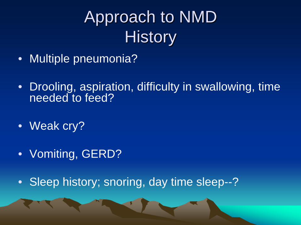
Approach to NMDHistory
• Multiple pneumonia?
• Drooling, aspiration, difficulty in swallowing, time needed to feed?
• Weak cry?
• Vomiting, GERD?
• Sleep history; snoring, day time sleep--?
Physical Examination
• Scoliosis?
• Digital clubbing?
• Cyanosis?
• Gag reflux?
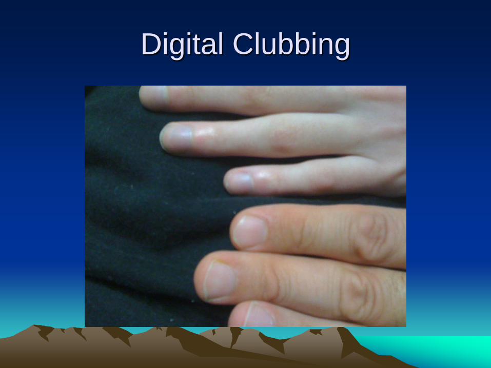
Digital Clubbing
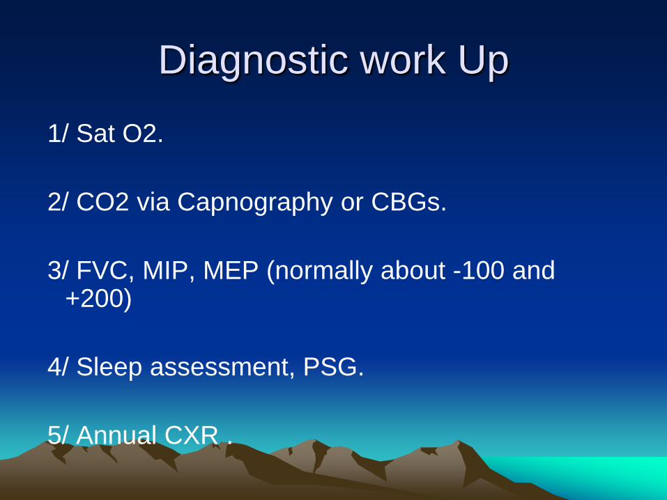
Diagnostic work Up 1/ Sat O2.
2/ CO2 via Capnography or CBGs.
3/ FVC, MIP, MEP (normally about -100 and +200)
4/ Sleep assessment, PSG.
5/ Annual CXR .
Primary Care Visits
Same as per AAP plusFlu vaccine
Pneumococcal Vaccine
Pulmonary F/U
Routine visit is recommended every 3-9 months depends on severity
Pulmonary Management
• Airway Clearance.
• OSA, Assisted Ventilation.
• Treatment of Acute exacerbations.
• Pre-operative evaluation.
Airway Clearance:
1. Essential to prevent PNA, atelectasis, and progressive lung disease
2. Drooling and ineffective cough is a major cause of morbidity and mortality.
3. In cooperative patient, this could be assessed by measuring cough peak flow.
Airway Clearance:
1. CPF< 160 L/min is associated with poor airway clearance.
2. Normal CPF : 147 to 488 L/min in females and from 162 to 728 L/min in males, age range of 4-18 yrs.
3. MEP < 45 cm H2O is also associated with poor airway clearance (normal value is 150-230 cm H2O)
Airway Clearance
1.Manual: CPT with position changes.
2.Vest: uncomfortable due to chest wall deformity and poor cough.
3.Cough assist devices.
4.Medications: DNAse, 3% saline, Robinol
Mucomyst
NAC is NO LONGER used for ACT
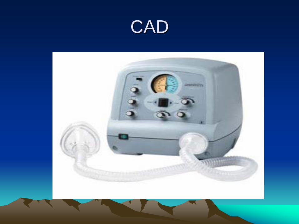
CAD
• Provides a (+30) positive pressure breath.
• Followed by (– 30) negative pressure, air is “sucked out” of the chest.
CAD
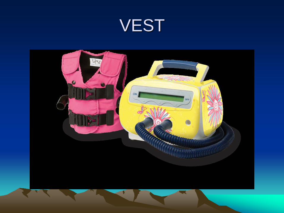
VEST
Vest
Is more of “ secretions mobiliser’ and best use for CF
Assisted Ventilation
• Night time: if a PSG shows significant OSA, Hypoxemia, increased A/HA index
• Continuous: if Sat O2 <92%, PaCO2 is > 55. while awake.
• BiPAP for advanced disease or if CPAP fails, newer recommendations of trying BiPAP 1st

CPAP/BiPAP
• Applied thru a face mask or nasal prongs.
• “Effectiveness” is assessed by number of OSA, DeSats, A/HA index on PSG, and by changes in morning PCO2.
• In severe cases, Tracheostomy and home ventilator is needed
Acute Exacerbations
• Cough.
• Increased secretions.
• Fever.
• Increased WOB.
????
• Is it a PNA/ Trachiatis??
Work Up
• CXR.
• CBGs.
• CRP.
• Respiratory C/S
CBCs
• WBC increased?
• Bands?
• Anemia ?
CBGs
• Always compare with a previous values.
• Many patients with NMD may have a CO2 retention ( PCO2 46-55).
• PH, Bicarb normal or abnormal
Respiratory Culture
Are there any WBCs ?
Most Trached patient are colonized with Staph or Ps.A.
The presence of WBCs on gram stain suggest acute infection
CRP
• Early, non-specific acute phase reactant.
• CRP> 20 “suggests” bacterial pneumonia.
CXR
MUST be compared with previous ones
Antibiotics pending C/S
• Cover MRSA, Anaerobes, Pseudomonas
• Clindamycin, Cefepime,Tobramycine, Cipro
• If resp/tracheal c/s shows little or no PMNs, and CRP is < 15 likely viral and may not need abx
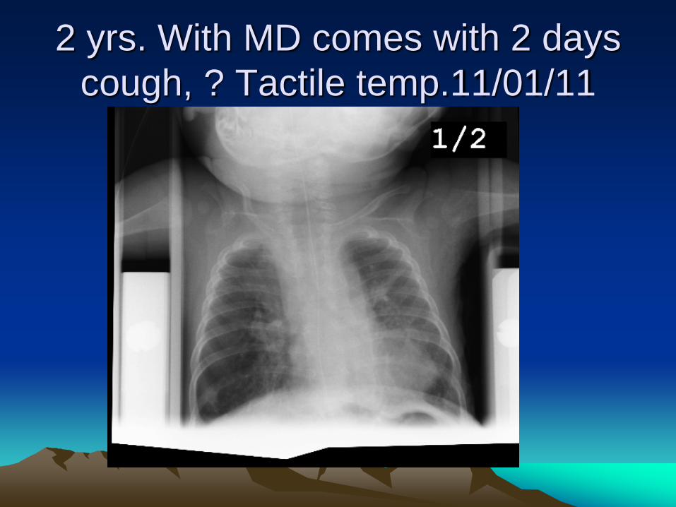
2 yrs. With MD comes with 2 days cough, ? Tactile temp.11/01/11
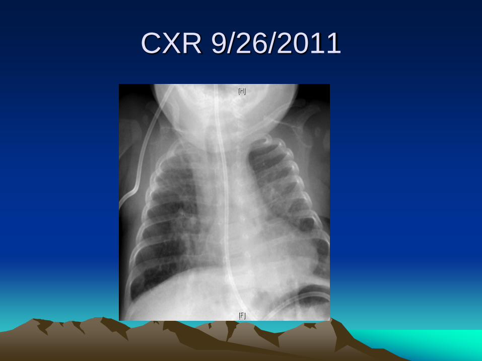
CXR 9/26/2011
• NO changes, Normal CBC, CRP< 4.
• Viral illness. No Abx given
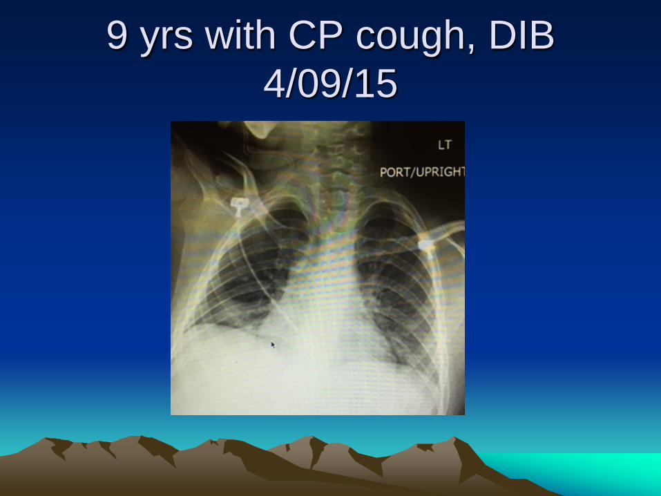
9 yrs with CP cough, DIB4/09/15
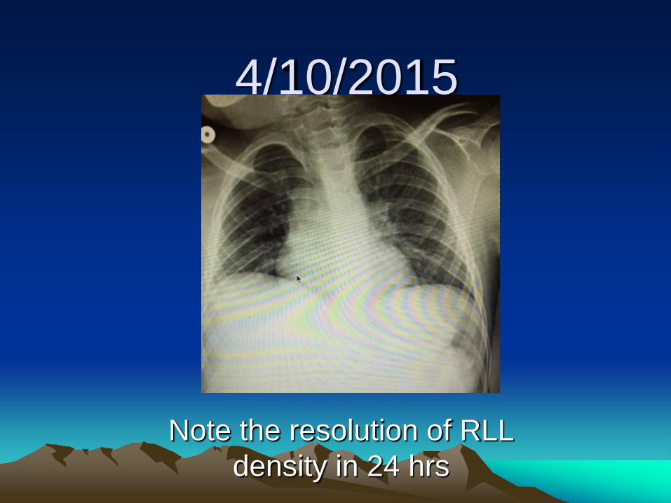
4/10/2015
Note the resolution of RLL density in 24 hrs
When to discharge ?
• Better clinically
• FiO2 back to home dose (usually < 0.4)
• PaCO2 < 55
Scoliosis
• Almost ALL patients with have chest wall deformity of some degree.
• Serial exams (including CXR, PFT )is recommended.
• Orthopedic evaluation is recommended.
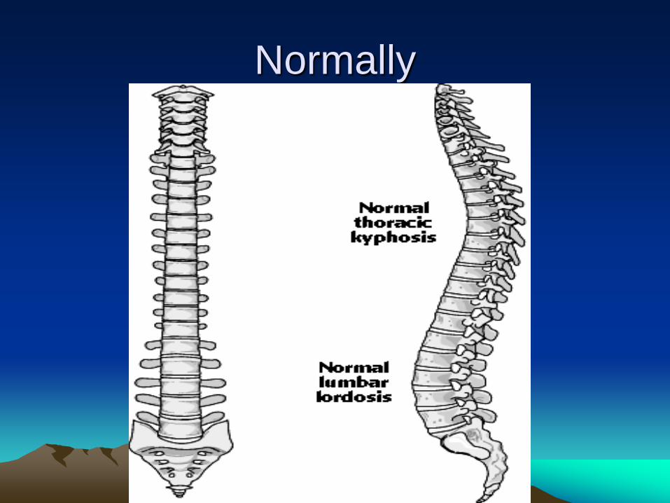
Normally
• Normally there should be no lateral curvature of the spine.
• The normal thoracolumbar spine is relatively straight in the sagittal plane and has a double curve in the coronal plane.
• The thoracic spine in convex posteriorly (kyphosis).
• The lumbar spine is convex anteriorly (lordosis).
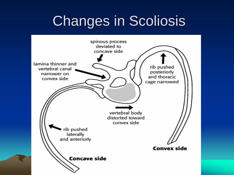
Changes in Scoliosis
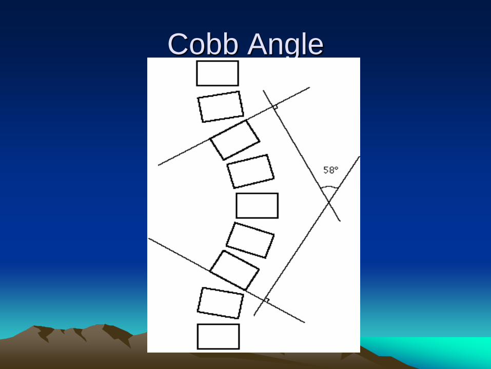
Cobb Angle
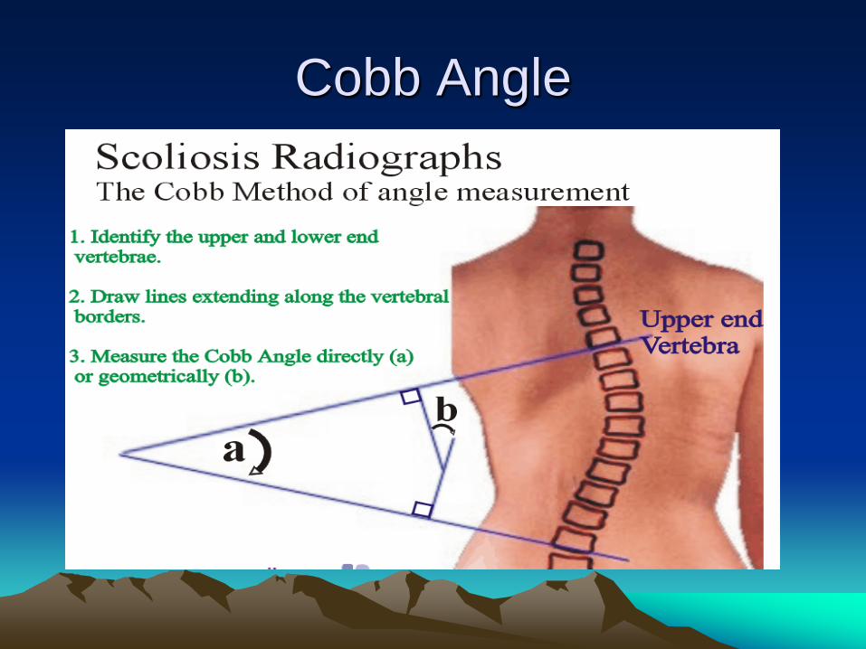
Cobb Angle
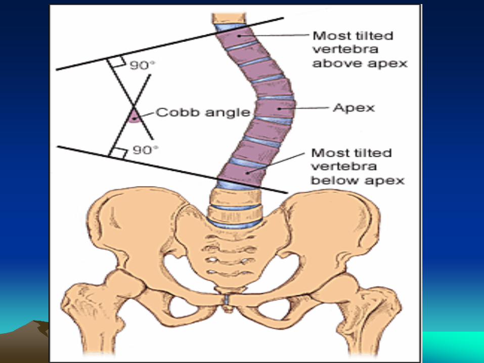
Cobb angle
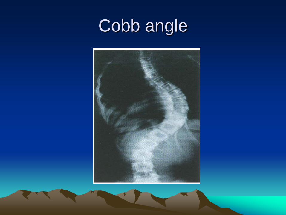
Cobb angle
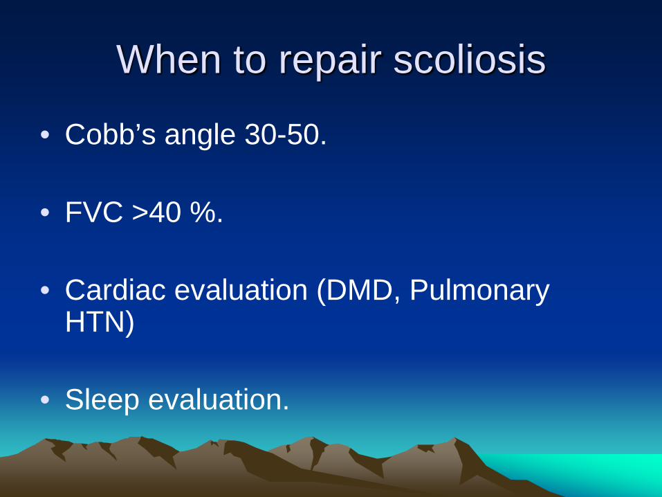
When to repair scoliosis
• Cobb’s angle 30-50.
• FVC >40 %.
• Cardiac evaluation (DMD, Pulmonary HTN)
• Sleep evaluation.
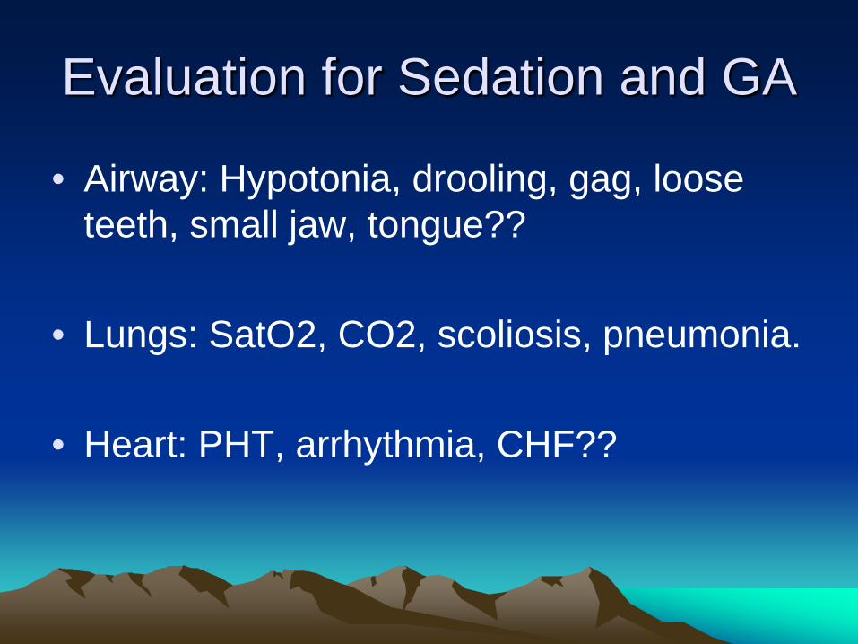
Evaluation for Sedation and GA
• Airway: Hypotonia, drooling, gag, loose teeth, small jaw, tongue??
• Lungs: SatO2, CO2, scoliosis, pneumonia.
• Heart: PHT, arrhythmia, CHF??
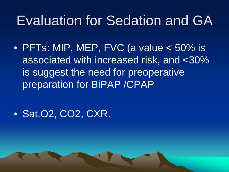
Evaluation for Sedation and GA
• PFTs: MIP, MEP, FVC (a value < 50% is associated with increased risk, and <30% is suggest the need for preoperative preparation for BiPAP /CPAP
• Sat.O2, CO2, CXR.
Evaluation for Sedation and GA
• MUST explain to the parents that in severe cases pts. may be unable to get off the ventilator in the presence of severe lung disease.
• This should be discussed BEFORE sedation/GA.
GI and Nutrition
• Most have poor swallowing.
• GERD is often present.
• Above problems often results in a poor nutrition.
• CLD and chronic increased WOB leads to increased caloric requirments.
GI and Nutrition
• G-tube insertion is the end result to ensure sufficient nutrition.
• High caloric formulas are usually used.

Thank you
References
1.Finder JD, Brinkrant D, Carl J, et al. Respiratory care of the patients with DMD:ATS consensus statement .Am J Respir Crit Care Med.2004; 170(4):456-465
2.Wang CH, Finkkel RS, Bertini ES,et al. Consensus statement for the standard of care in SMA. J Child Neuro. 2007; 22(8): 1027-1049
3.Willig TN, Paulus J, Lacau St Guily, Beon C, Navarro J. Swallowing problems in NMD. Arch Phys Med Rehab. 1994;75(11):1175-1181
4.Brinkrant D, Panitch HB, Benditt JO, et al. American College of Chest Physician statement on respiratory and related management of patients with DMD undergoing Anesthesia and sedation. Chest. 2007;132(6):1977-1986
5.Luna C.C-Reactie Protien in pneumonia,let me try again. CHEST.April
2004;125(4):1192-1195. 6. http://www.ncbi.nlm.nih.gov/pubmed/18496248