Embed Size (px)
Citation preview
PULMONARY FUNCTION TESTS AND THEIR IMPLICATION IN ANAESTHESIA
MODERATOR- PROF SATINDER GOMBARPRESENTER- DR. PREETI SHARMA
INTRODUCTION
Pulmonary function tests is a generic term used to indicate a battery of studies or maneuvers that may be performed using standardized equipment to measure lung function.Evaluates one or more aspects of the respiratory systemRespiratory mechanics Lung parenchymal function/ Gas exchangeCardiopulmonary interaction
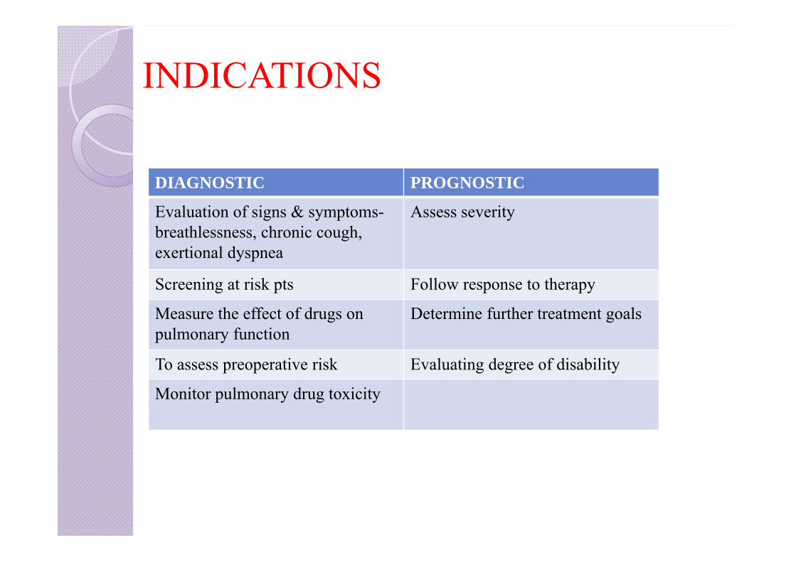
INDICATIONS
DIAGNOSTIC PROGNOSTIC
Evaluation of signs & symptoms-breathlessness, chronic cough, exertional dyspnea
Assess severity
Screening at risk pts Follow response to therapy
Measure the effect of drugs on pulmonary function
Determine further treatment goals
To assess preoperative risk Evaluating degree of disability
Monitor pulmonary drug toxicity
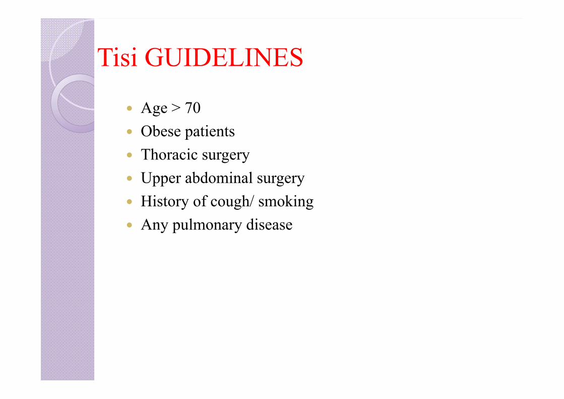
Tisi GUIDELINES
Age > 70 Obese patients Thoracic surgery Upper abdominal surgery History of cough/ smoking Any pulmonary disease
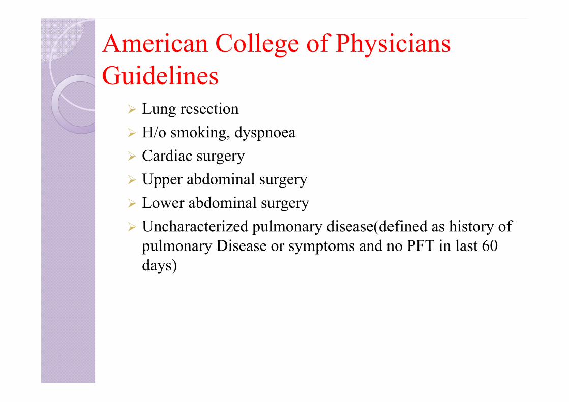
American College of Physicians Guidelines
Lung resection H/o smoking, dyspnoea Cardiac surgery Upper abdominal surgery Lower abdominal surgery Uncharacterized pulmonary disease(defined as history of
pulmonary Disease or symptoms and no PFT in last 60 days)
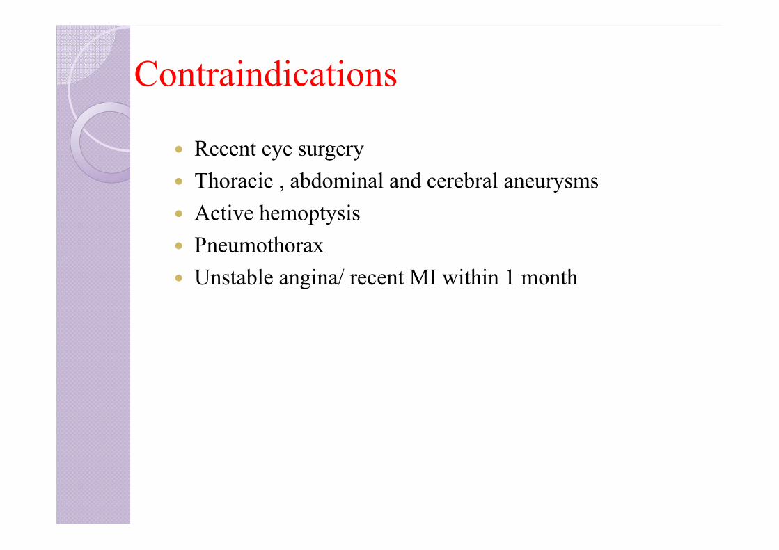
Contraindications
Recent eye surgery Thoracic , abdominal and cerebral aneurysms Active hemoptysis Pneumothorax Unstable angina/ recent MI within 1 month

INDEX
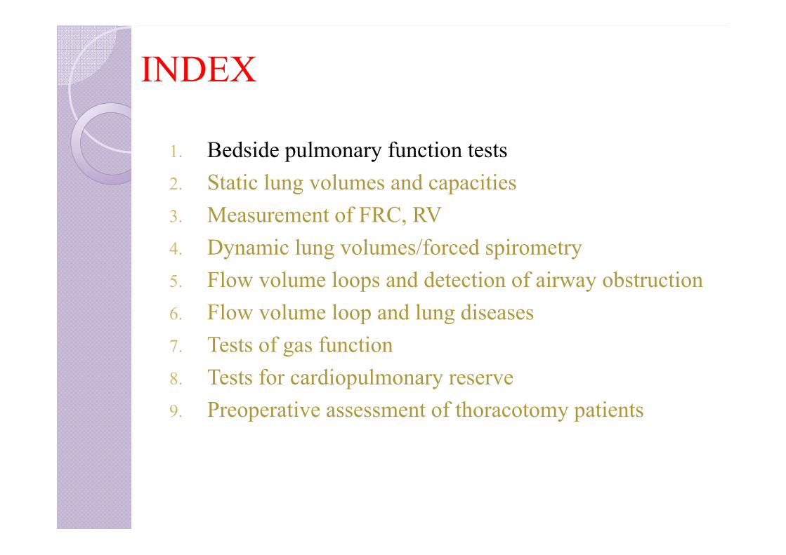
1. Categorization of PFT’s2. Bedside pulmonary function tests3. Static lung volumes and capacities4. Measurement of FRC, RV5. Dynamic lung volumes/forced spirometry6. Flow volume loops and detection of airway obstruction7. Flow volume loop and lung diseases8. Tests of gas exchange function9. Tests for cardiopulmonary reserve10. Preoperative assessment of thoracotomy patients
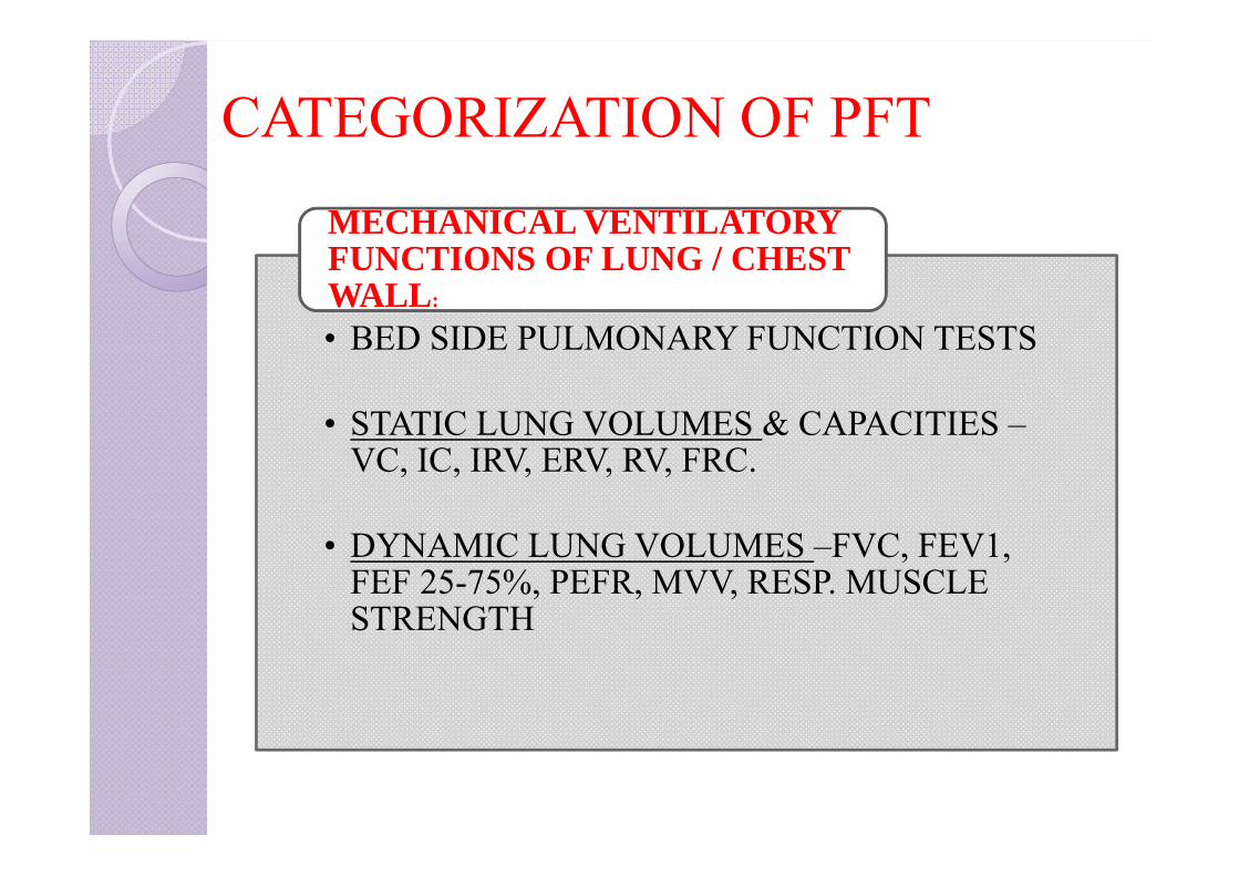
CATEGORIZATION OF PFT
• BED SIDE PULMONARY FUNCTION TESTS
• STATIC LUNG VOLUMES & CAPACITIES –VC, IC, IRV, ERV, RV, FRC.
• DYNAMIC LUNG VOLUMES –FVC, FEV1, FEF 25-75%, PEFR, MVV, RESP. MUSCLE STRENGTH
MECHANICAL VENTILATORY FUNCTIONS OF LUNG / CHEST WALL:
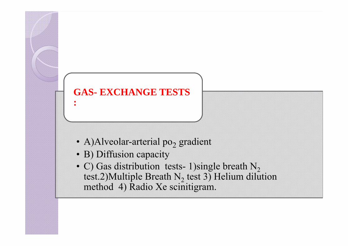
• A)Alveolar-arterial po2 gradient• B) Diffusion capacity• C) Gas distribution tests- 1)single breath N2
test.2)Multiple Breath N2 test 3) Helium dilution method 4) Radio Xe scinitigram.
GAS- EXCHANGE TESTS :
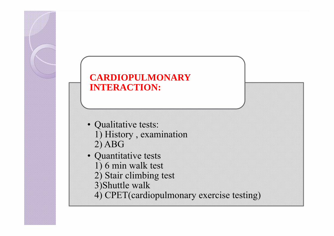
• Qualitative tests: 1) History , examination 2) ABG
• Quantitative tests 1) 6 min walk test 2) Stair climbing test 3)Shuttle walk 4) CPET(cardiopulmonary exercise testing)
CARDIOPULMONARY INTERACTION:
INDEX
1. Bedside pulmonary function tests2. Static lung volumes and capacities3. Measurement of FRC, RV4. Dynamic lung volumes/forced spirometry5. Flow volume loops and detection of airway obstruction6. Flow volume loop and lung diseases7. Tests of gas function8. Tests for cardiopulmonary reserve9. Preoperative assessment of thoracotomy patients
Bed side pulmonary function tests
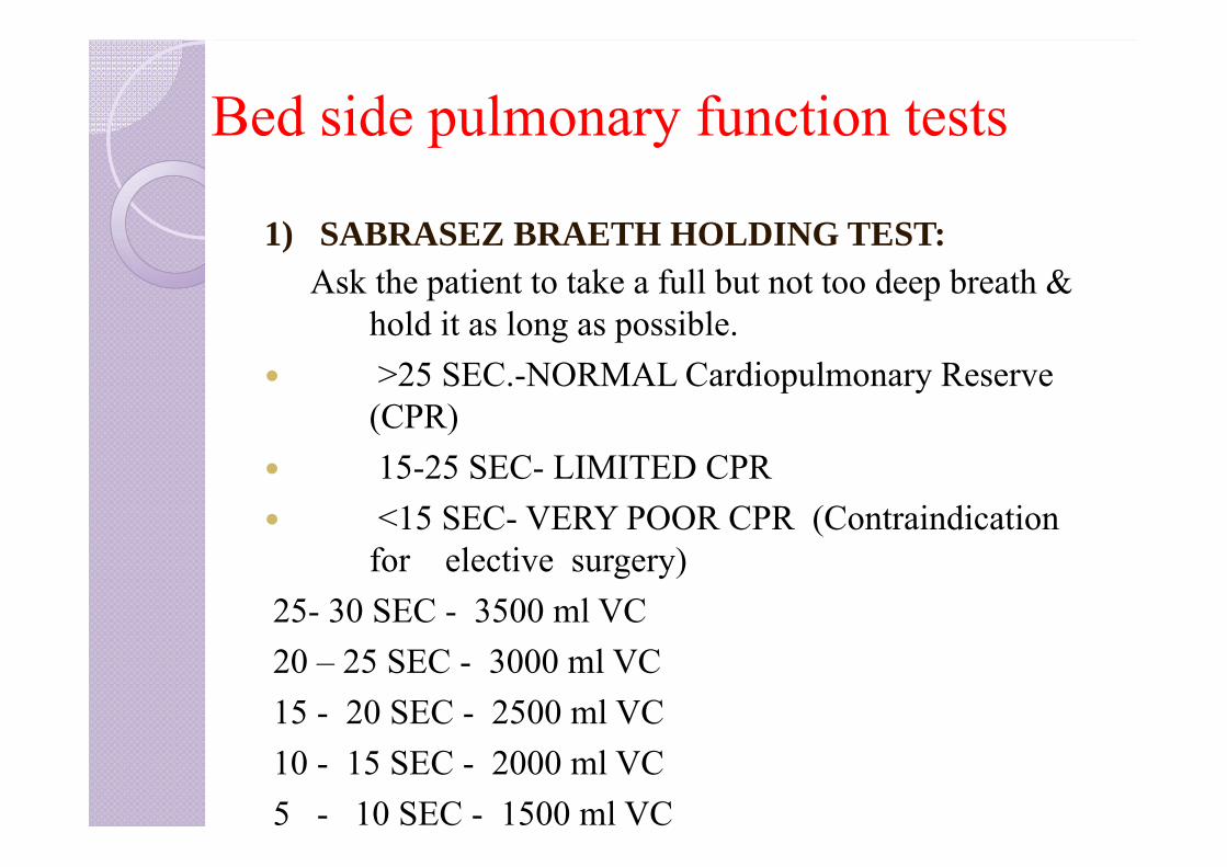
1) SABRASEZ BRAETH HOLDING TEST:Ask the patient to take a full but not too deep breath &
hold it as long as possible. >25 SEC.-NORMAL Cardiopulmonary Reserve
(CPR) 15-25 SEC- LIMITED CPR <15 SEC- VERY POOR CPR (Contraindication
for elective surgery) 25- 30 SEC - 3500 ml VC20 – 25 SEC - 3000 ml VC15 - 20 SEC - 2500 ml VC10 - 15 SEC - 2000 ml VC5 - 10 SEC - 1500 ml VC
Bed side pulmonary function tests
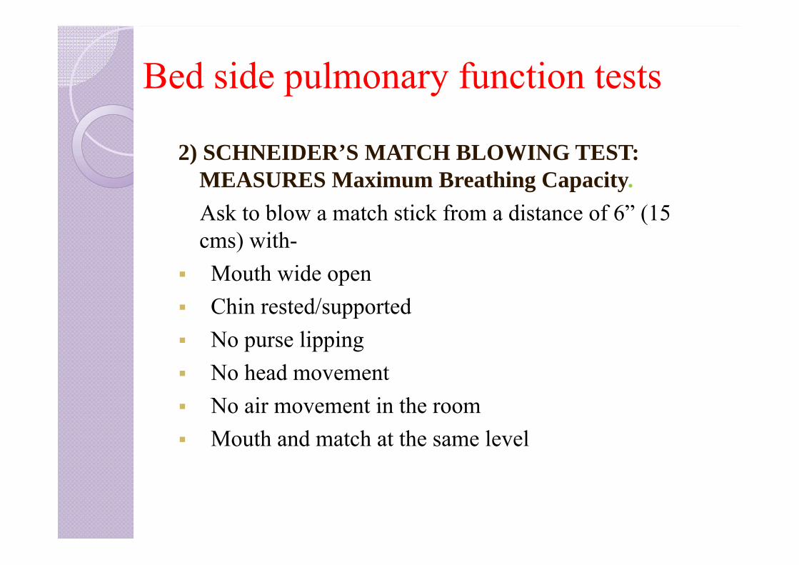
2) SCHNEIDER’S MATCH BLOWING TEST: MEASURES Maximum Breathing Capacity. Ask to blow a match stick from a distance of 6” (15 cms) with-
Mouth wide open Chin rested/supported No purse lipping No head movement No air movement in the room Mouth and match at the same level
Bed side pulmonary function tests
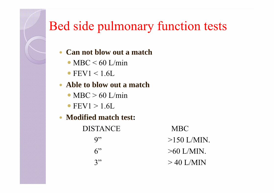
Can not blow out a matchMBC < 60 L/min FEV1 < 1.6L
Able to blow out a matchMBC > 60 L/min FEV1 > 1.6L
Modified match test:DISTANCE MBC
9” >150 L/MIN. 6” >60 L/MIN.3” > 40 L/MIN
Bed side pulmonary function tests
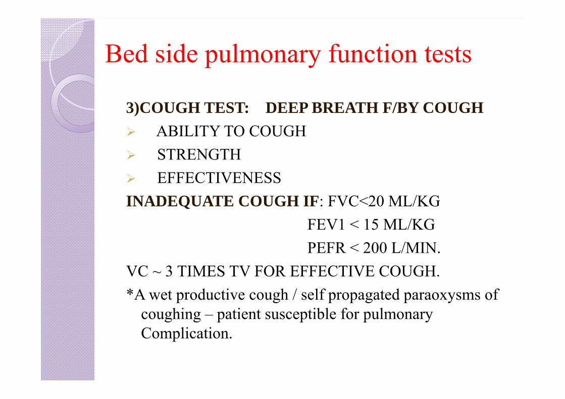
3)COUGH TEST: DEEP BREATH F/BY COUGH ABILITY TO COUGH STRENGTH EFFECTIVENESSINADEQUATE COUGH IF: FVC<20 ML/KG
FEV1 < 15 ML/KGPEFR < 200 L/MIN.
VC ~ 3 TIMES TV FOR EFFECTIVE COUGH.*A wet productive cough / self propagated paraoxysms of
coughing – patient susceptible for pulmonary Complication.
Bed side pulmonary function tests
4)FORCED EXPIRATORY TIME:After deep breath, exhale maximally and forcefully &
keep stethoscope over trachea & listen.N FET – 3-5 SECS.OBS.LUNG DIS. - > 6 SECRES. LUNG DIS.- < 3 SEC
5) SINGLE BREATH COUNT:After deep breath, hold it and start counting till the next
breath. N- 30-40 COUNT Indicates vital capacity
Bed side pulmonary function tests
6) WRIGHT PEAK FLOW METER: Measures PEFR (Peak Expiratory Flow Rate)
N – MALES- 450-700 L/MIN.FEMALES- 350-500 L/MIN.
7) DE-BONO WHISTLE BLOWING TEST: MEASURES PEFR.Patient blows down a wide bore tube at the end of which is a whistle, on the side is a hole with adjustable knob.As subject blows → whistle blows, leak hole is
gradually increased till the intensity of whistle disappears.At the last position at which the whistle can be blown ,
the PEFR can be read off the scale.

DEBONO’S WHISTLE
8) WRIGHT RESPIROMETER : measures TV, MV
Instrument- compact, light and portable. Can be connected to endotracheal tube or face mask MV- instrument record for 1 min. And read directly TV-calculated and dividing MV by counting Respiratory
Rate. Disadvantage: It under- reads at low flow rates and over-
reads at high flow rates.
Bed side pulmonary function tests
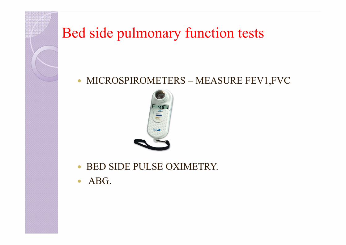
MICROSPIROMETERS – MEASURE FEV1,FVC
BED SIDE PULSE OXIMETRY. ABG.
INDEX
1. Bedside pulmonary function tests2. Static lung volumes and capacities3. Measurement of FRC, RV4. Dynamic lung volumes/forced spirometry5. Flow volume loops and detection of airway obstruction6. Flow volume loop and lung diseases7. Tests of gas function8. Tests for cardiopulmonary reserve9. Preoperative assessment of thoracotomy patients
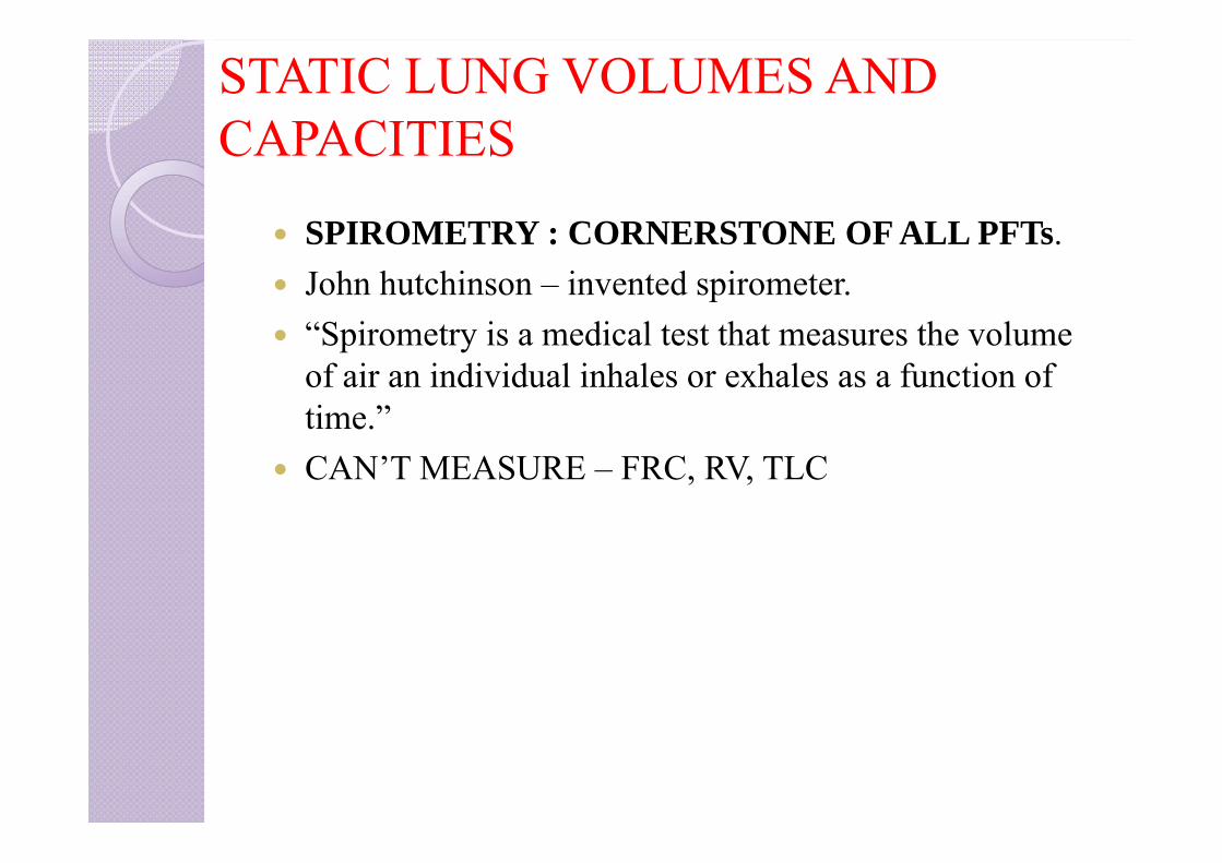
STATIC LUNG VOLUMES AND CAPACITIES
SPIROMETRY : CORNERSTONE OF ALL PFTs. John hutchinson – invented spirometer. “Spirometry is a medical test that measures the volume
of air an individual inhales or exhales as a function of time.”
CAN’T MEASURE – FRC, RV, TLC
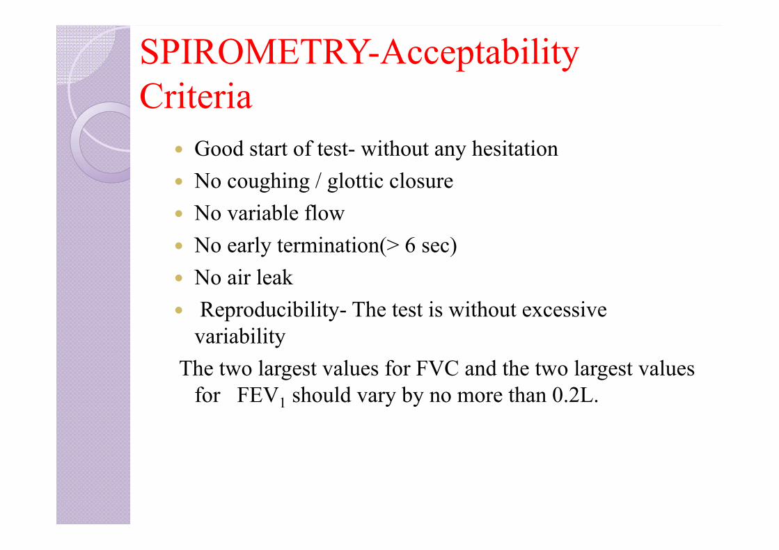
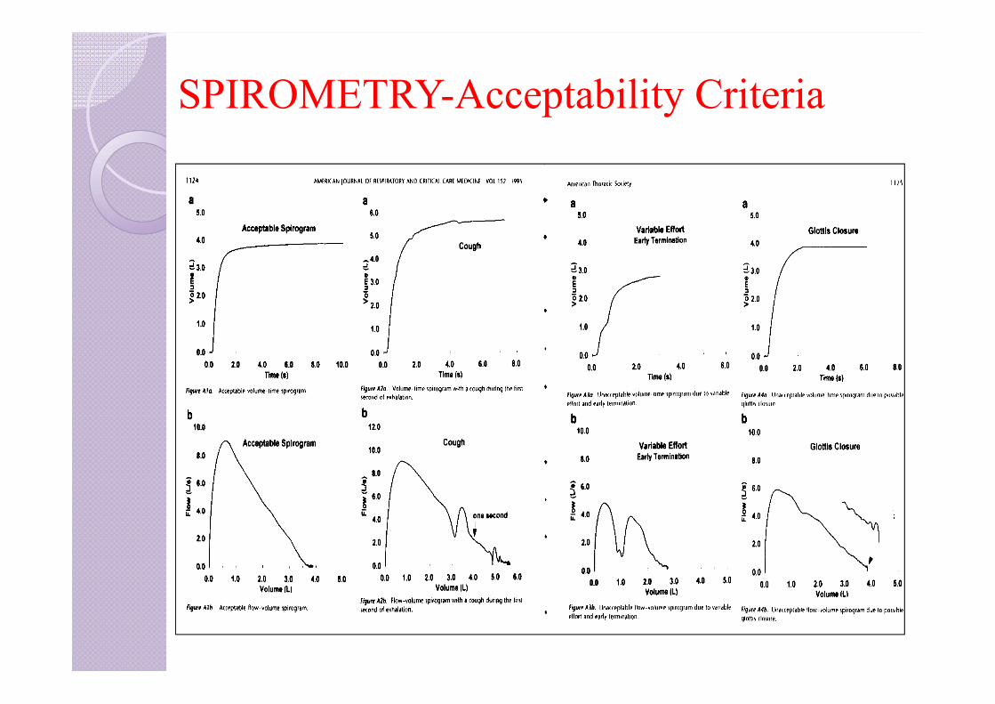
SPIROMETRY-Acceptability Criteria
Good start of test- without any hesitation No coughing / glottic closure No variable flow No early termination(> 6 sec) No air leak Reproducibility- The test is without excessive
variabilityThe two largest values for FVC and the two largest values
for FEV1 should vary by no more than 0.2L.
SPIROMETRY-Acceptability Criteria
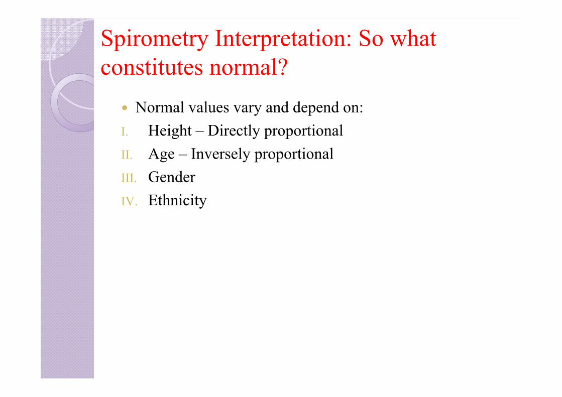
Spirometry Interpretation: So what constitutes normal?
Normal values vary and depend on:I. Height – Directly proportional II. Age – Inversely proportional III. GenderIV. Ethnicity

LUNG VOLUMES AND CAPACITIES
PFT tracings have:Four Lung volumes: tidal volume, inspiratory reserve volume, expiratory reserve volume, and residual volume
Five capacities: inspiratory capacity, expiratory capacity, vital capacity, functional residual capacity, and total lung capacity
Addition of 2 or more volumes comprise a capacity.
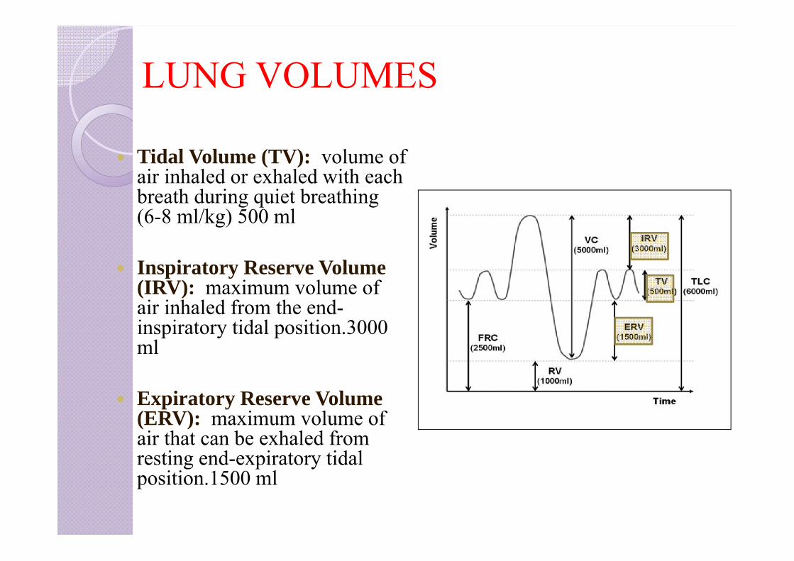
LUNG VOLUMES
Tidal Volume (TV): volume of air inhaled or exhaled with each breath during quiet breathing (6-8 ml/kg) 500 ml
Inspiratory Reserve Volume (IRV): maximum volume of air inhaled from the end-inspiratory tidal position.3000 ml
Expiratory Reserve Volume (ERV): maximum volume of air that can be exhaled from resting end-expiratory tidal position.1500 ml
LUNG VOLUMES
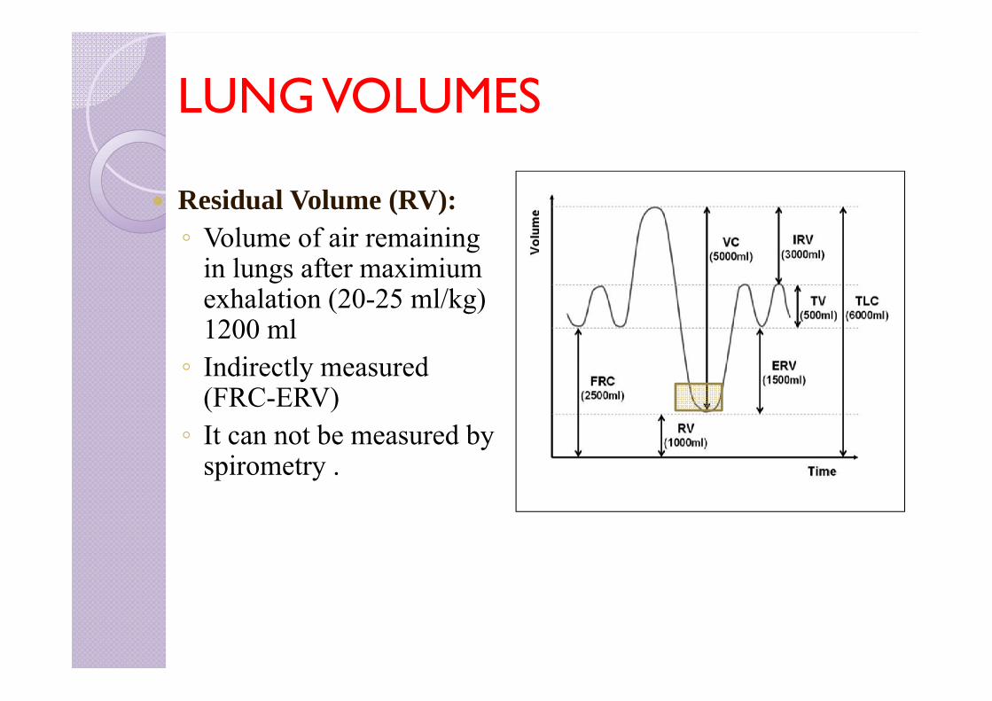
Residual Volume (RV): ◦ Volume of air remaining
in lungs after maximium exhalation (20-25 ml/kg) 1200 ml◦ Indirectly measured
(FRC-ERV)◦ It can not be measured by
spirometry .

LUNG CAPACITIES Total Lung Capacity (TLC):
Sum of all volume compartments or volume of air in lungs after maximum inspiration (4-6 L)
Vital Capacity (VC): TLC minus RV or maximum volume of air exhaled from maximal inspiratory level. (60-70 ml/kg) 5000ml.
Inspiratory Capacity (IC): Sum of IRV and TV or the maximum volume of air that can be inhaled from the end-expiratory tidal position. (2400-3800ml).
Expiratory Capacity (EC): TV+ ERV
LUNG CAPACITIES
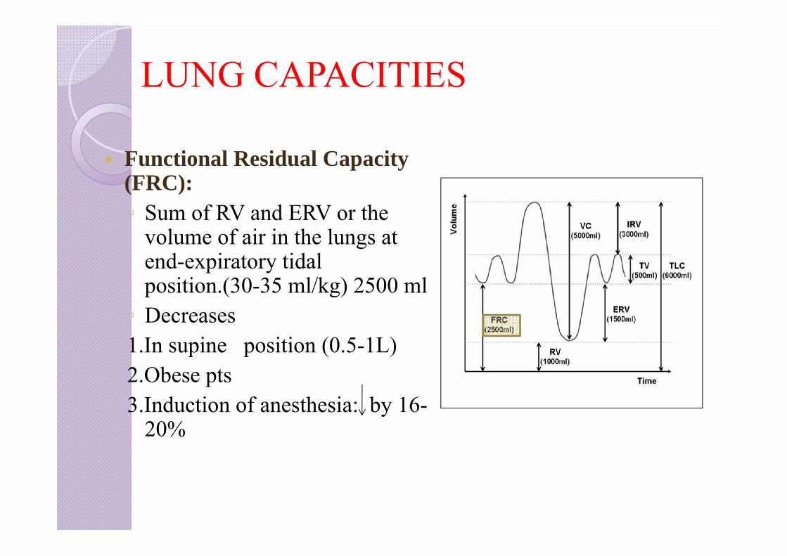
Functional Residual Capacity (FRC): ◦ Sum of RV and ERV or the
volume of air in the lungs at end-expiratory tidal position.(30-35 ml/kg) 2500 ml◦ Decreases 1.In supine position (0.5-1L)2.Obese pts3.Induction of anesthesia: by 16-
20%
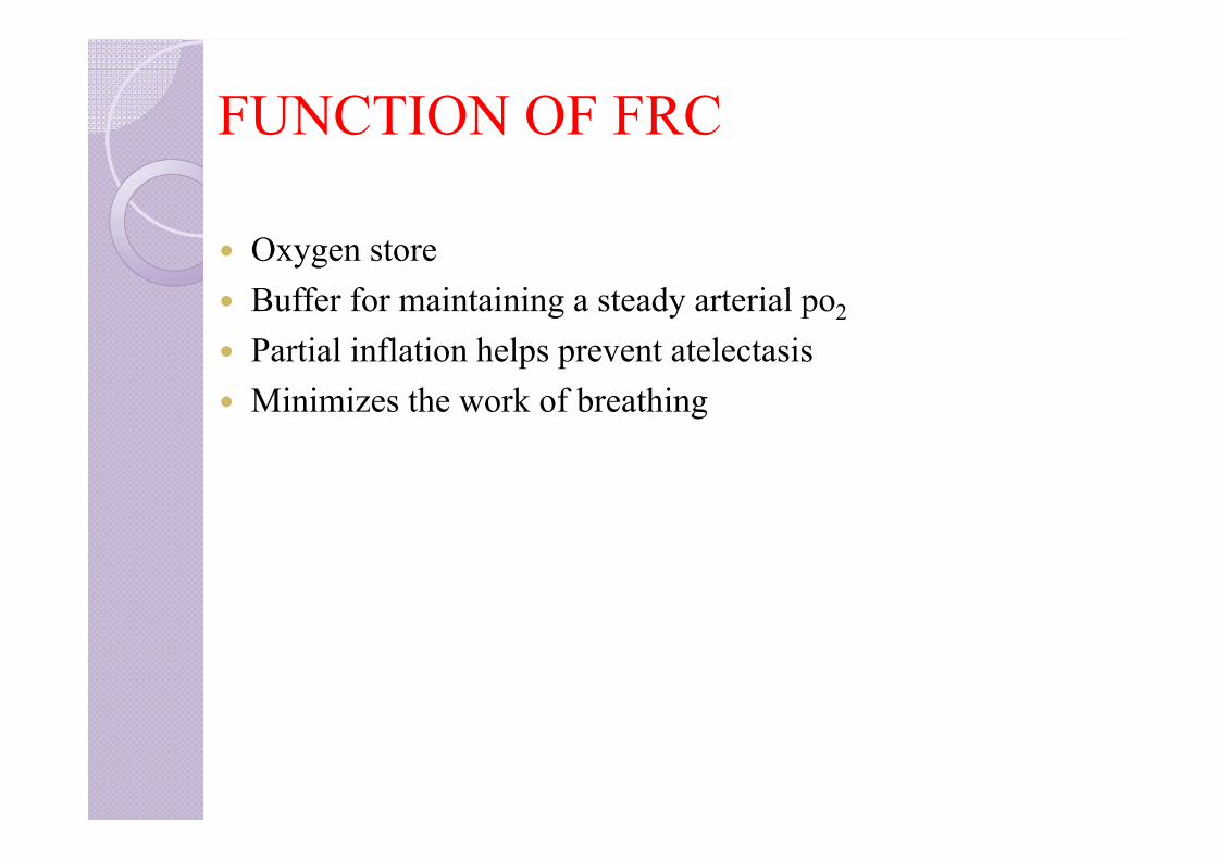
FUNCTION OF FRC
Oxygen store Buffer for maintaining a steady arterial po2
Partial inflation helps prevent atelectasis Minimizes the work of breathing
INDEX
1. Bedside pulmonary function tests2. Static lung volumes and capacities3. Measurement of FRC, RV4. Dynamic lung volumes/forced spirometry5. Flow volume loops and detection of airway obstruction6. Flow volume loop and lung diseases7. Tests of gas function8. Tests for cardiopulmonary reserve9. Preoperative assessment of thoracotomy patients
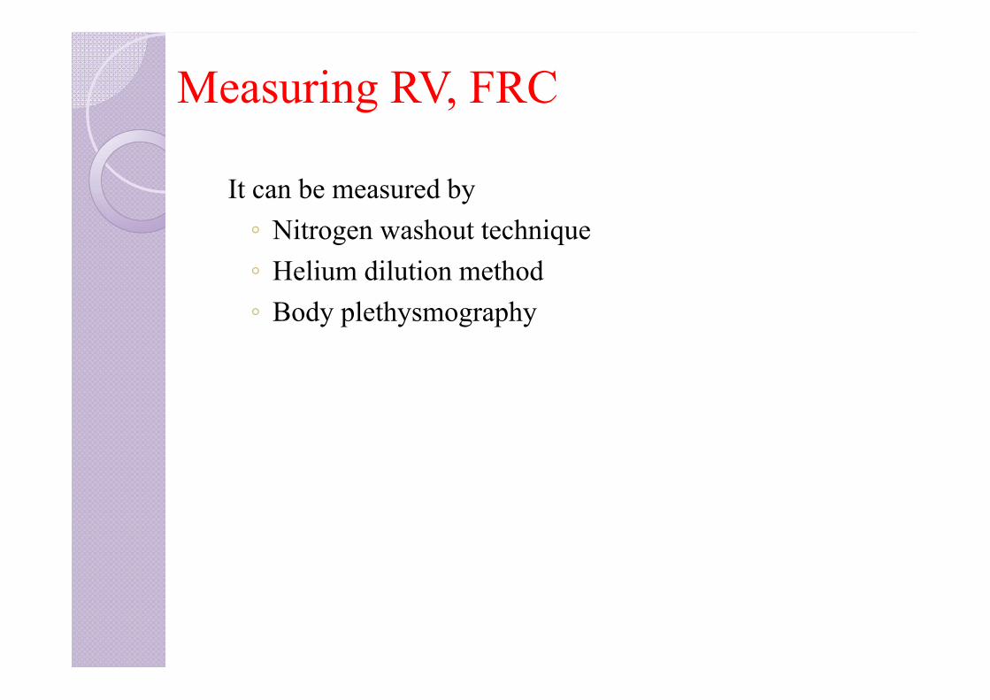
Measuring RV, FRC
It can be measured by◦ Nitrogen washout technique◦ Helium dilution method◦ Body plethysmography
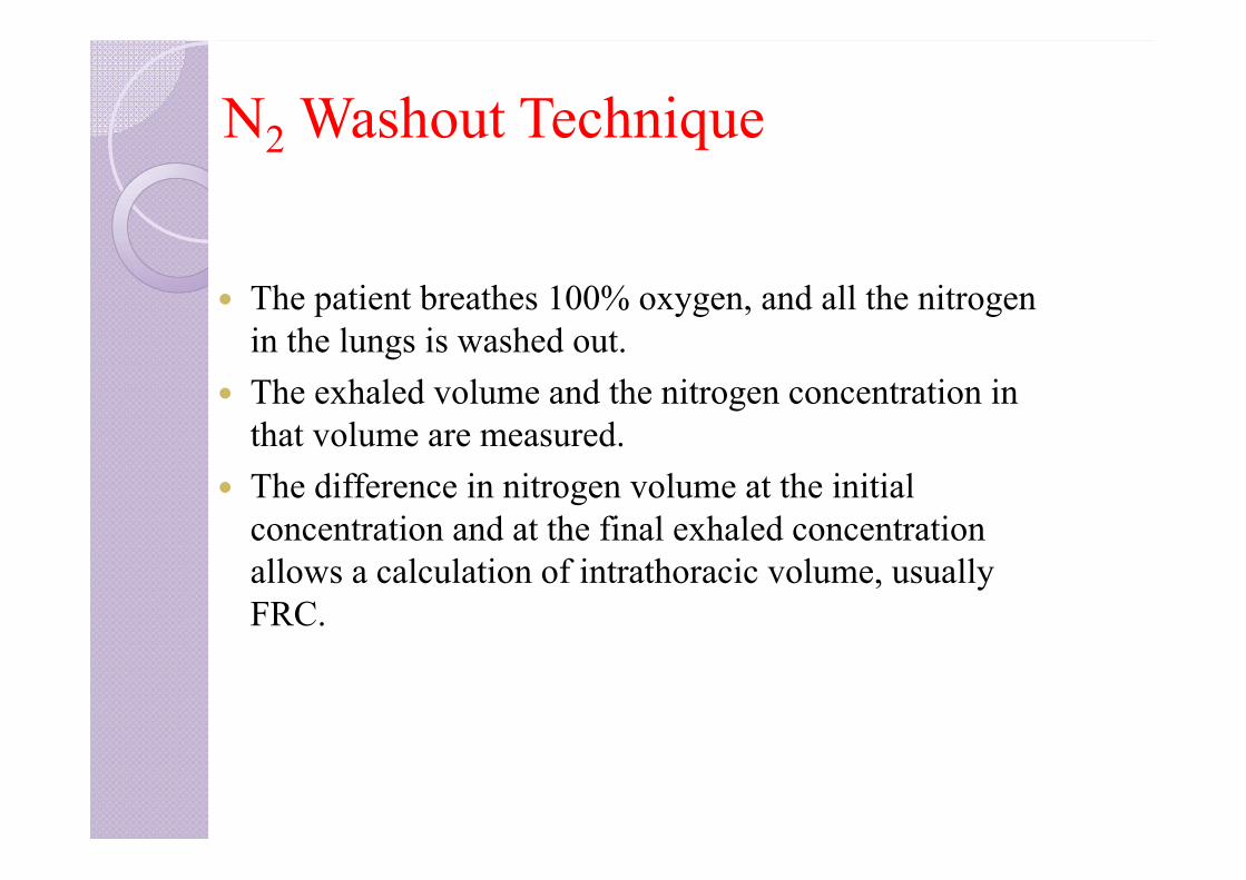
N2 Washout Technique
The patient breathes 100% oxygen, and all the nitrogen in the lungs is washed out.
The exhaled volume and the nitrogen concentration in that volume are measured.
The difference in nitrogen volume at the initial concentration and at the final exhaled concentration allows a calculation of intrathoracic volume, usually FRC.
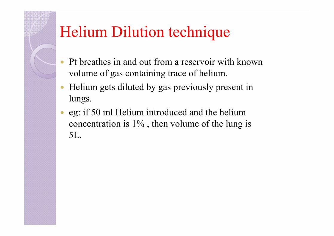
Helium Dilution technique
Pt breathes in and out from a reservoir with known volume of gas containing trace of helium.
Helium gets diluted by gas previously present in lungs.
eg: if 50 ml Helium introduced and the helium concentration is 1% , then volume of the lung is 5L.
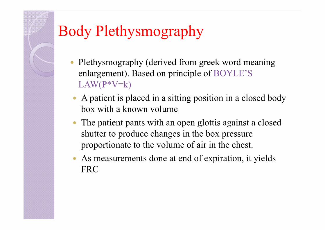
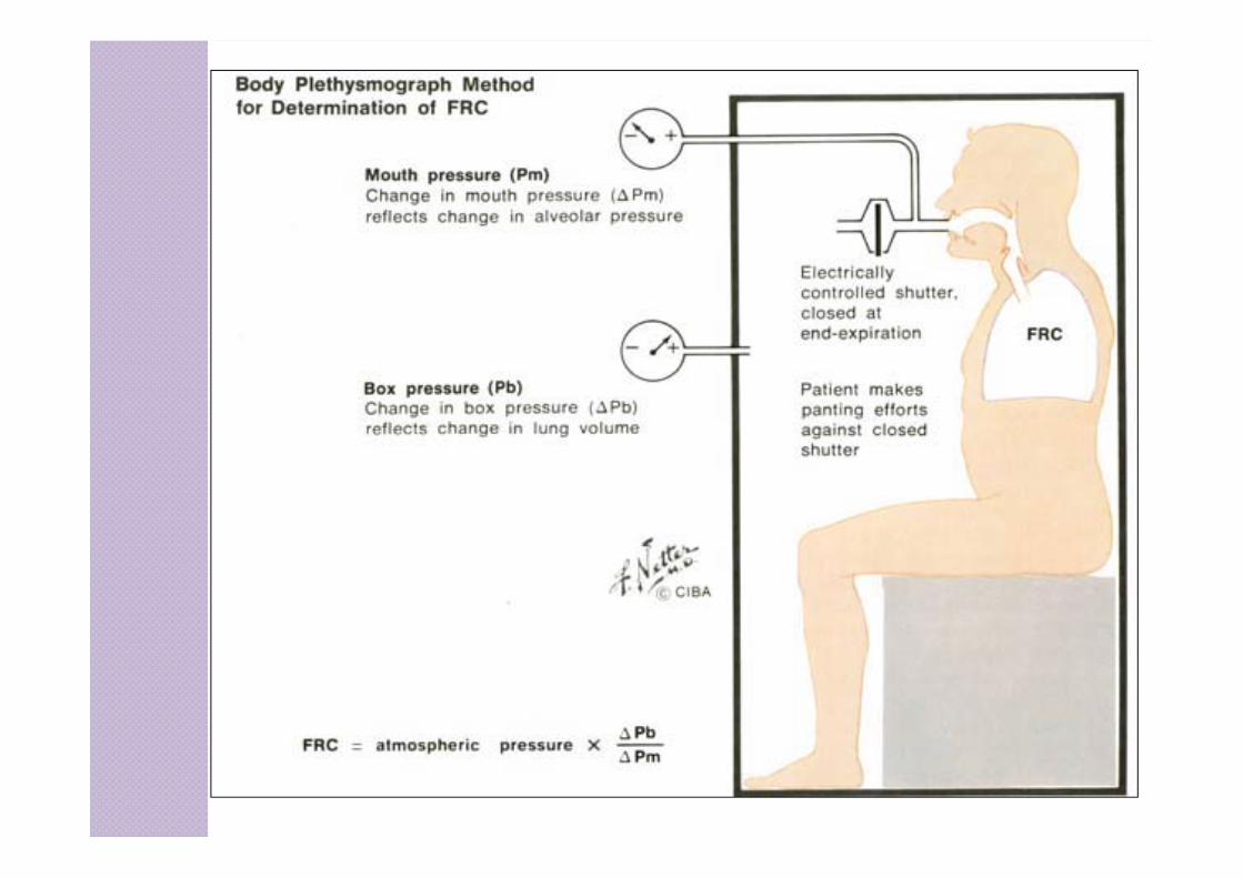
Body Plethysmography
Plethysmography (derived from greek word meaning enlargement). Based on principle of BOYLE’S LAW(P*V=k)
A patient is placed in a sitting position in a closed body box with a known volume
The patient pants with an open glottis against a closed shutter to produce changes in the box pressure proportionate to the volume of air in the chest.
As measurements done at end of expiration, it yields FRC

INDEX
1. Bedside pulmonary function tests2. Static lung volumes and capacities3. Measurement of FRC, RV4. Dynamic lung volumes/forced spirometry5. Flow volume loops and detection of airway obstruction6. Flow volume loop and lung diseases7. Tests of gas function8. Tests for cardiopulmonary reserve9. Preoperative assessment of thoracotomy patients

FORCED SPIROMETRY/TIMED EXPIRATORY SPIROGRAM
Includes measuring:• Pulmonary mechanics – to assess the ability of the lung to move large vol of air quickly through the airways to identify airway obstruction•FVC•FEV1•Several FEF values•Forced inspiratory rates(FIF’s)•MVV
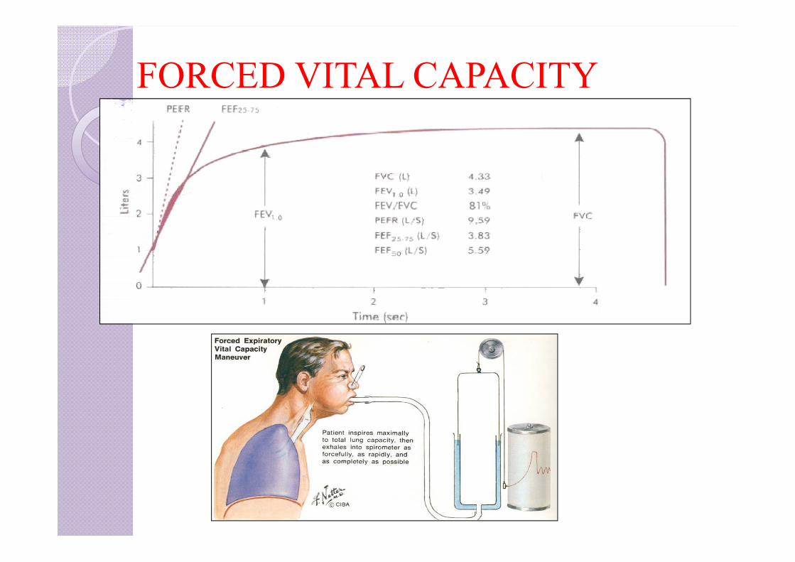
FORCED VITAL CAPACITY
The FVC is the maximum volume of air that can be breathed out as forcefully and rapidly as possible following a maximum inspiration.
Characterized by full inspiration to TLC followed by abrupt onset of expiration to RV
Indirectly reflects flow resistance property of airways.
FORCED VITAL CAPACITY
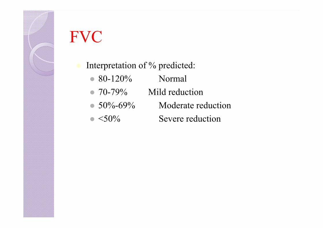
FVC Interpretation of % predicted:
80-120% Normal 70-79% Mild reduction 50%-69% Moderate reduction <50% Severe reduction
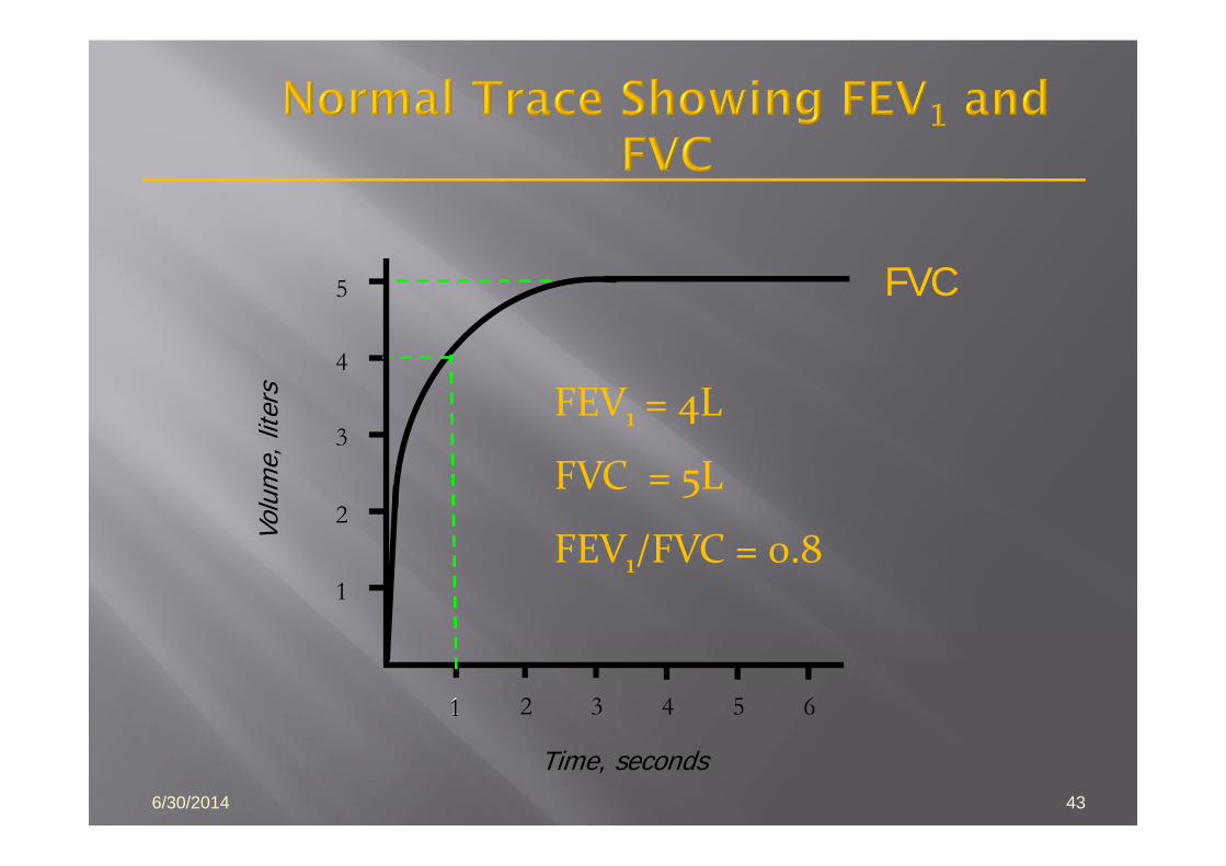
6/30/2014 43
1 2 3 4 5 6
1
2
3
4
Volu
me,
lite
rs
Time, seconds
FVC5
1
FEV1 = 4L
FVC = 5L
FEV1/FVC = 0.8
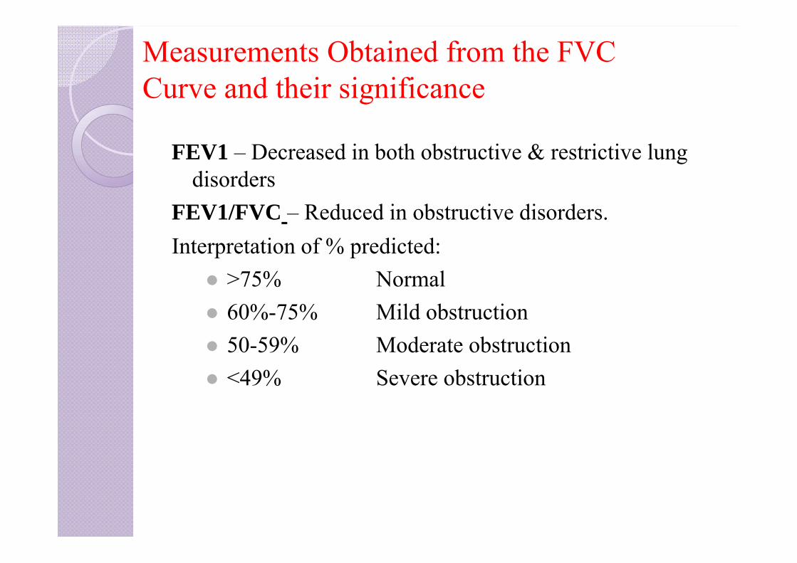
Measurements Obtained from the FVC Curve and their significance
FEV1 – Decreased in both obstructive & restrictive lung disorders
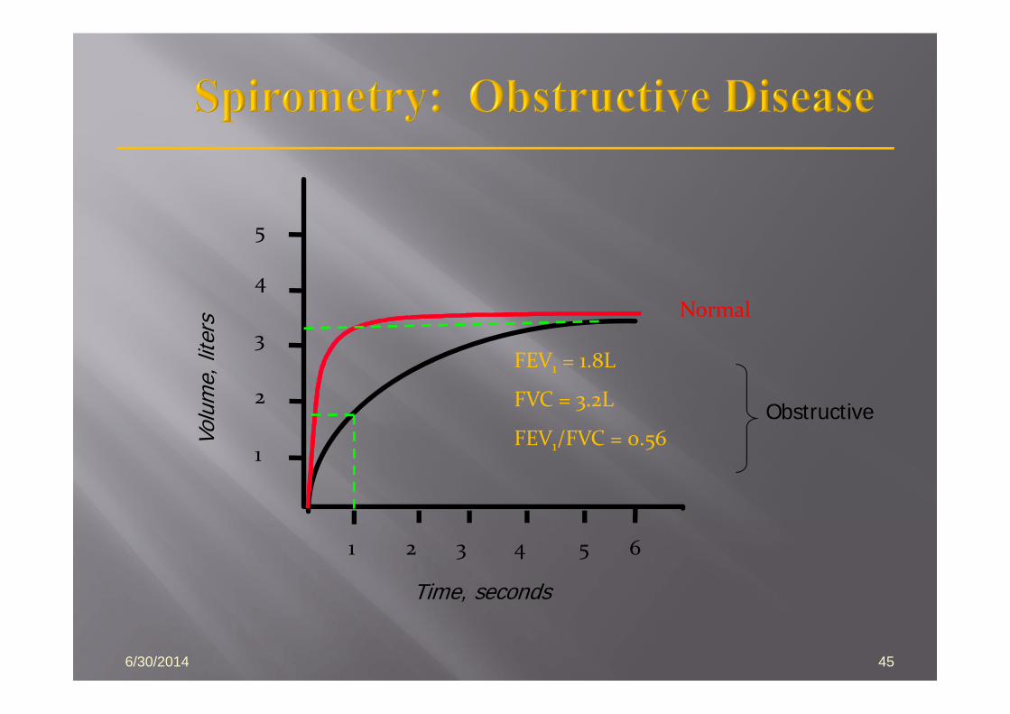
FEV1/FVC – Reduced in obstructive disorders.Interpretation of % predicted:
>75% Normal 60%-75% Mild obstruction 50-59% Moderate obstruction <49% Severe obstruction
6/30/2014 45
Volu
me,
lite
rs
Time, seconds
5
4
3
2
1
1 2 3 4 5 6
FEV1 = 1.8L
FVC = 3.2L
FEV1/FVC = 0.56
Normal
Obstructive
Volu
me,
lite
rs
Time, seconds
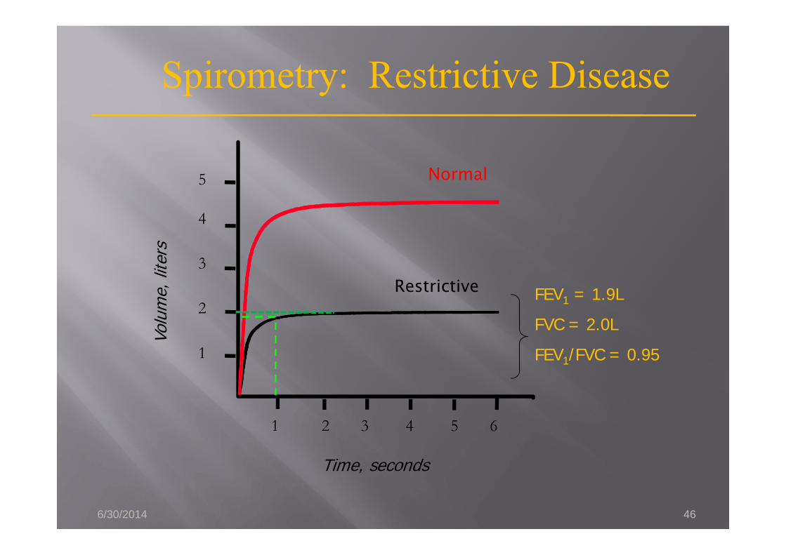
FEV1 = 1.9L
FVC = 2.0L
FEV1/FVC = 0.95
1 2 3 4 5 6
5
4
3
2
1
Spirometry: Restrictive Disease
Normal
Restrictive
6/30/2014 46
Volu
me,
lite
rs
Time, seconds
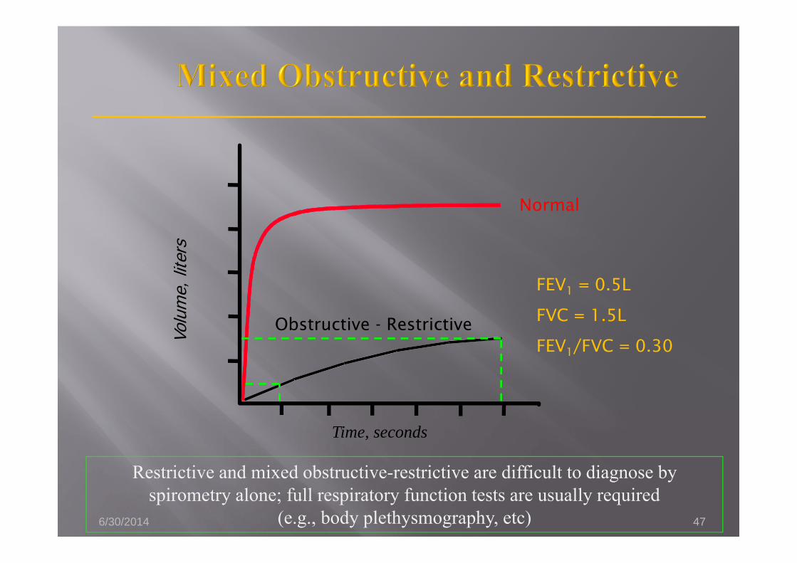
Restrictive and mixed obstructive-restrictive are difficult to diagnose by spirometry alone; full respiratory function tests are usually required
(e.g., body plethysmography, etc)
FEV1 = 0.5L
FVC = 1.5L
FEV1/FVC = 0.30
Normal
Obstructive - Restrictive
6/30/2014 47
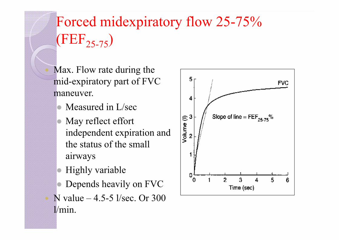
Forced midexpiratory flow 25-75% (FEF25-75)
Max. Flow rate during the mid-expiratory part of FVC maneuver. Measured in L/sec May reflect effort
independent expiration and the status of the small airways
Highly variable Depends heavily on FVC
N value – 4.5-5 l/sec. Or 300 l/min.
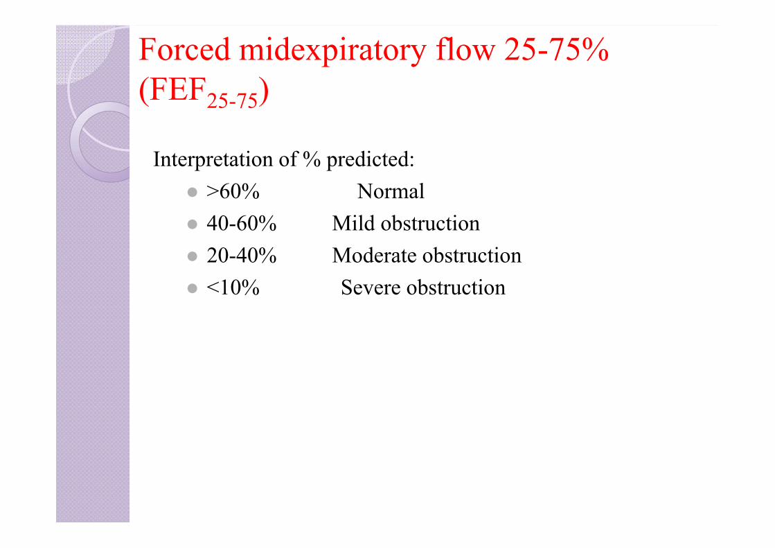
Forced midexpiratory flow 25-75% (FEF25-75)
Interpretation of % predicted: >60% Normal 40-60% Mild obstruction 20-40% Moderate obstruction <10% Severe obstruction
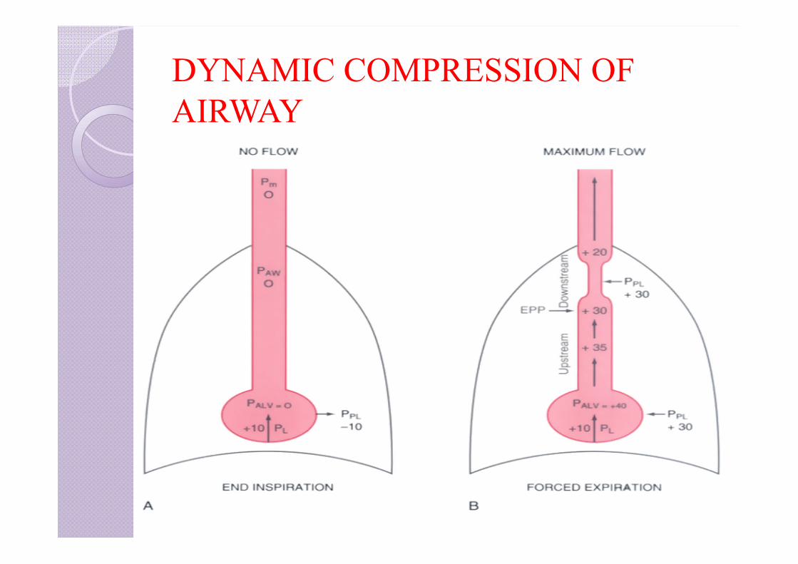
DYNAMIC COMPRESSION OF AIRWAY
EQUAL PRESSURE POINT Slowed expiration reduces the decrease in pressure from the
alveoli toward the mouth because lower flow requires less driving pressure.
By this means the point along the airway tree where pressure inside the airway has dropped to below that outside the airway (equal to pleural pressure) is moved toward the mouth
Thus, slow expiratory flow may make it possible to move the “equal pressure point,” where inside and outside airway pressure is equal, up to the larger airways or the mouth, which will prevent floppy airways from collapsing.
Peak expiratory flow rates
Maximum flow rate during an FVC maneuver occurs in initial 0.1 sec
After a maximal inspiration, the patient expires as forcefully and quickly as he can and the maximum flow rate of air is measured.
It gives a crude estimate of lung function, reflecting larger airway function.
Effort dependent but is highly reproducible.
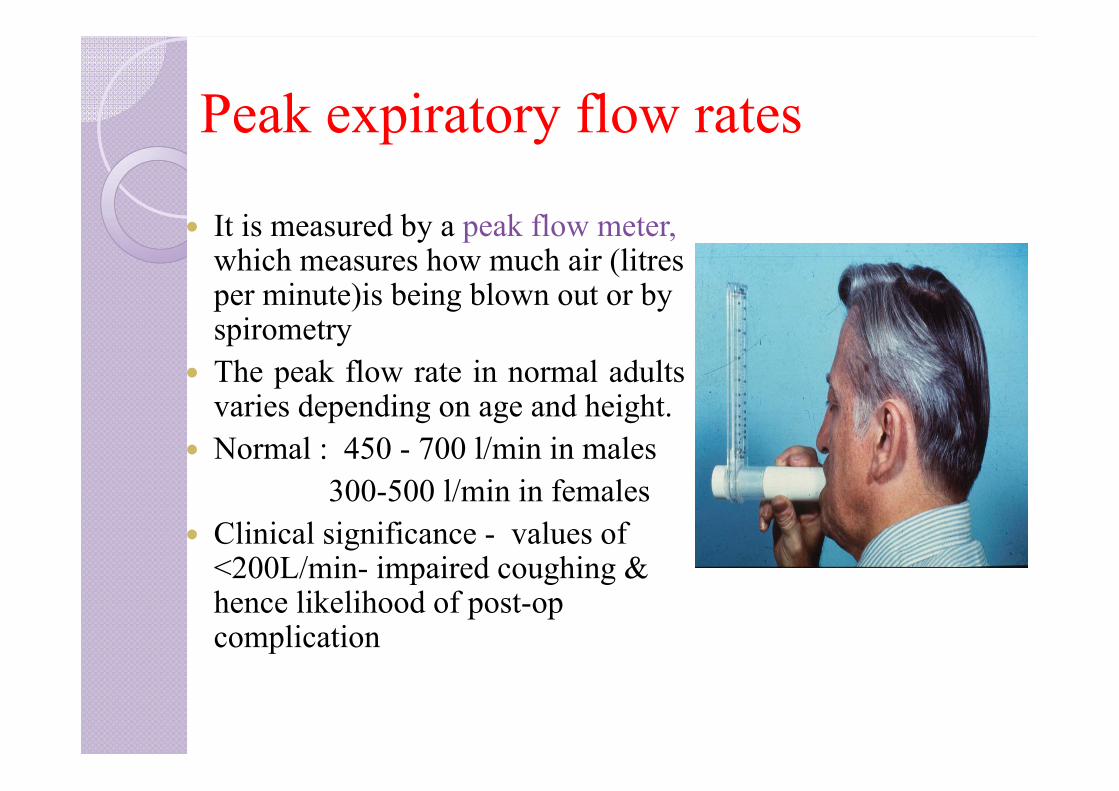
Peak expiratory flow rates
It is measured by a peak flow meter,which measures how much air (litresper minute)is being blown out or by spirometry
The peak flow rate in normal adultsvaries depending on age and height.
Normal : 450 - 700 l/min in males300-500 l/min in females
Clinical significance - values of <200L/min- impaired coughing & hence likelihood of post-op complication
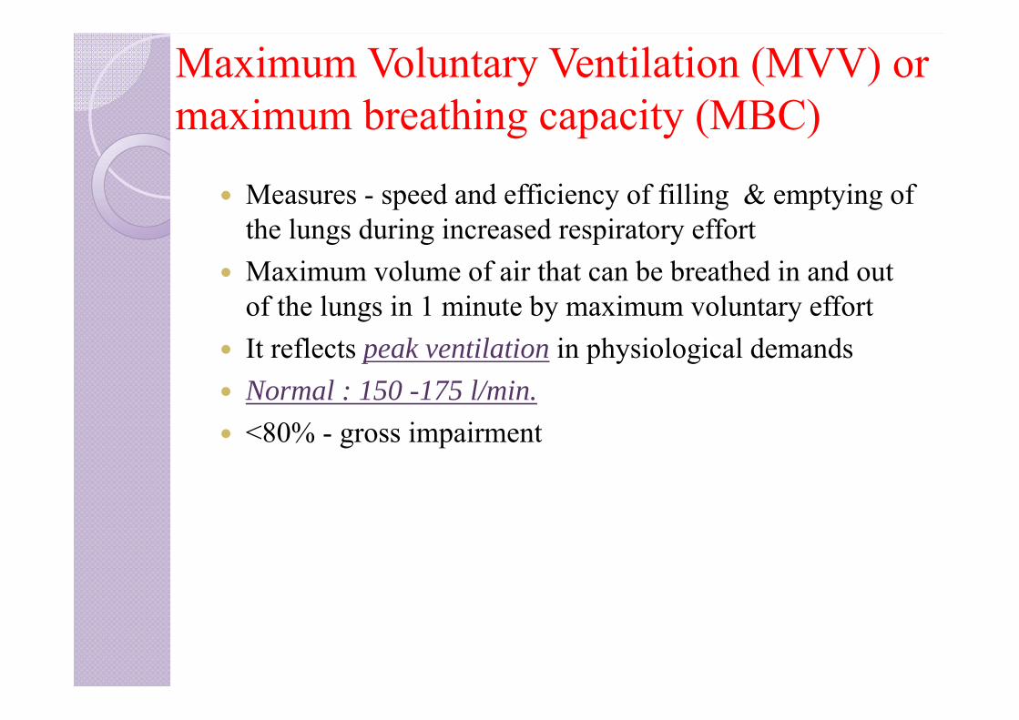
Maximum Voluntary Ventilation (MVV) or maximum breathing capacity (MBC)
Measures - speed and efficiency of filling & emptying of the lungs during increased respiratory effort
Maximum volume of air that can be breathed in and out of the lungs in 1 minute by maximum voluntary effort
It reflects peak ventilation in physiological demands Normal : 150 -175 l/min. <80% - gross impairment
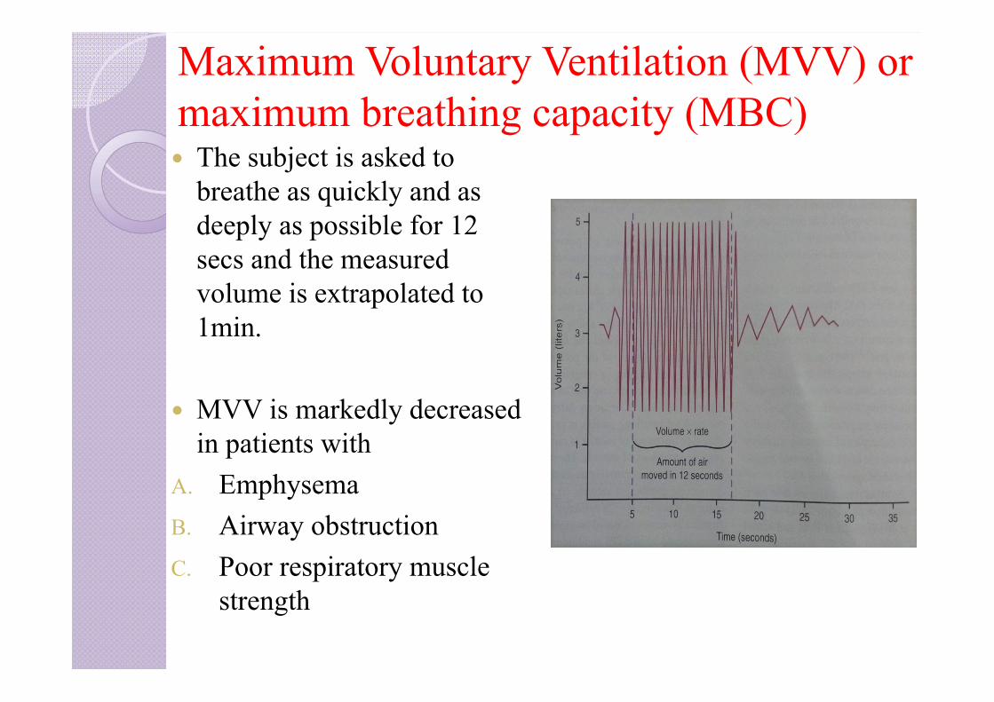
Maximum Voluntary Ventilation (MVV) or maximum breathing capacity (MBC) The subject is asked to
breathe as quickly and as deeply as possible for 12 secs and the measured volume is extrapolated to 1min.
MVV is markedly decreased in patients with
A. EmphysemaB. Airway obstructionC. Poor respiratory muscle
strength

INDEX
1. Bedside pulmonary function tests2. Static lung volumes and capacities3. Measurement of FRC, RV4. Dynamic lung volumes/forced spirometry5. Flow volume loops and detection of airway obstruction6. Flow volume loop and lung diseases7. Tests of gas function8. Tests for cardiopulmonary reserve9. Preoperative assessment of thoracotomy patients
FLOW VOLUME LOOPS “Spirogram” Graphic analysis of flow at various lung
volumes Tracing obtained when a maximal forced expiration from
TLC to RV is followed by maximal forced inspiration back to TLC
Measures forced inspiratory and expiratory flow rate Augments spirometry results Principal advantage of flow volume loops vs. typical
standard spirometric descriptions - identifies the probable obstructive flow anatomical location.
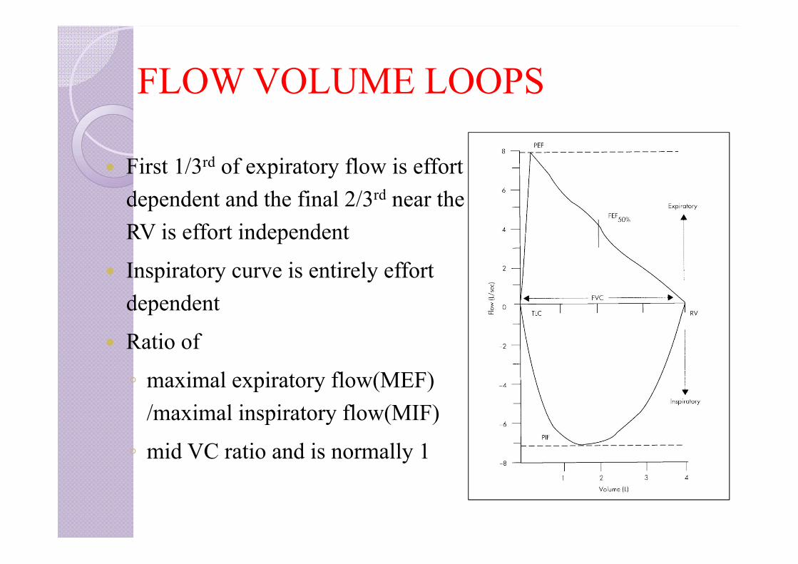
FLOW VOLUME LOOPS
First 1/3rd of expiratory flow is effort dependent and the final 2/3rd near the RV is effort independent
Inspiratory curve is entirely effort dependent
Ratio of
◦ maximal expiratory flow(MEF) /maximal inspiratory flow(MIF)
◦ mid VC ratio and is normally 1

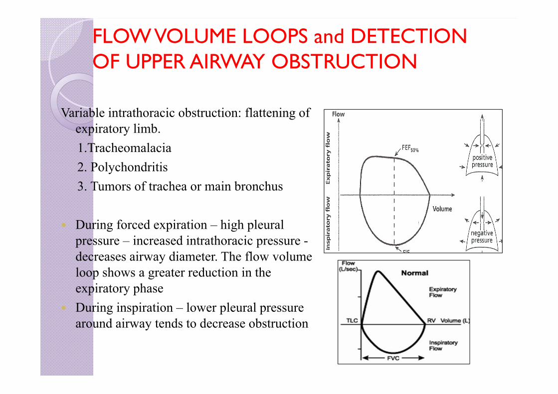
FLOW VOLUME LOOPS and DETECTION OF UPPER AIRWAY OBSTRUCTION
Flow-volume loops provide information on upper airway obstruction:
Fixed obstruction: constant airflow limitation on inspiration and expiration—such as
1. Benign stricture2. Goiter3. Endotracheal neoplasms4. Bronchial stenosis
FLOW VOLUME LOOPS and DETECTION OF UPPER AIRWAY OBSTRUCTION
Variable intrathoracic obstruction: flattening of expiratory limb. 1.Tracheomalacia 2. Polychondritis3. Tumors of trachea or main bronchus
During forced expiration – high pleural pressure – increased intrathoracic pressure -decreases airway diameter. The flow volume loop shows a greater reduction in the expiratory phase
During inspiration – lower pleural pressure around airway tends to decrease obstruction
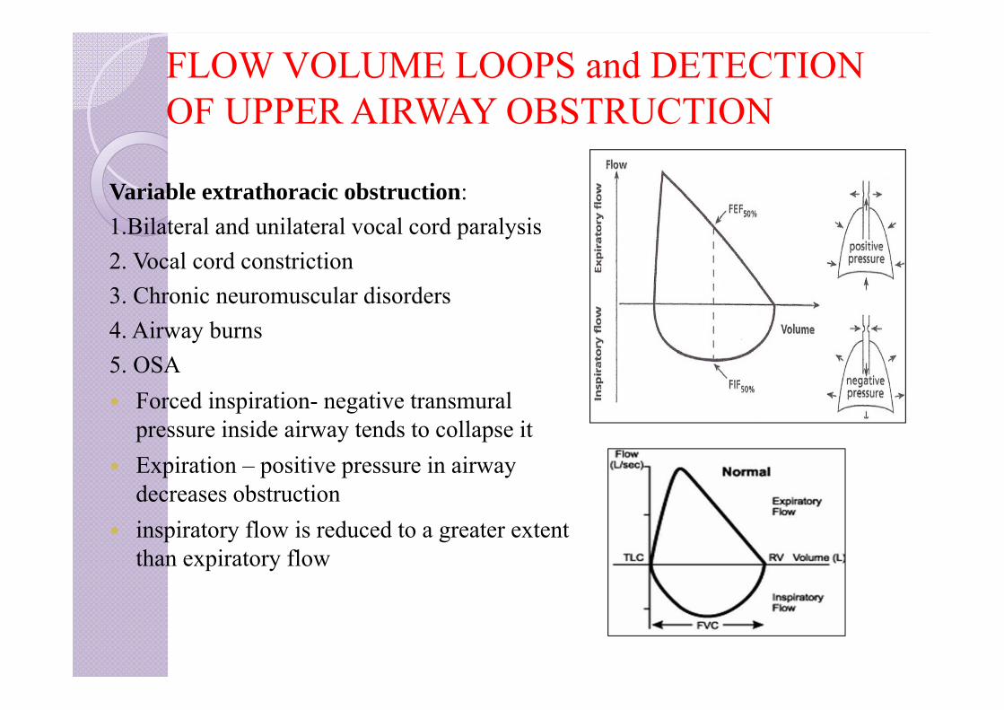
FLOW VOLUME LOOPS and DETECTION OF UPPER AIRWAY OBSTRUCTION
Variable extrathoracic obstruction: 1.Bilateral and unilateral vocal cord paralysis2. Vocal cord constriction 3. Chronic neuromuscular disorders4. Airway burns5. OSA Forced inspiration- negative transmural
pressure inside airway tends to collapse it Expiration – positive pressure in airway
decreases obstruction inspiratory flow is reduced to a greater extent
than expiratory flow
INDEX
1. Bedside pulmonary function tests2. Static lung volumes and capacities3. Measurement of FRC, RV4. Dynamic lung volumes/forced spirometry5. Flow volume loops and detection of airway obstruction6. Flow volume loop and lung diseases7. Tests of gas function8. Tests for cardiopulmonary reserve9. Preoperative assessment of thoracotomy patients
Obstructive Pattern — Evaluation
Common obstructive lung diseases
Asthma COPD (chronic bronchitis, emphysema) Cystic fibrosis.

ASTHMA
Peak expiratory flow reduced so maximum height of the loop is reduced
Airflow reduces rapidly with the reduction in the lung volumes because the airways narrow and the loop become concave
Concavity may be the indicator of airflow obstruction and may present before the change in FEV1 or FEV1/FVC
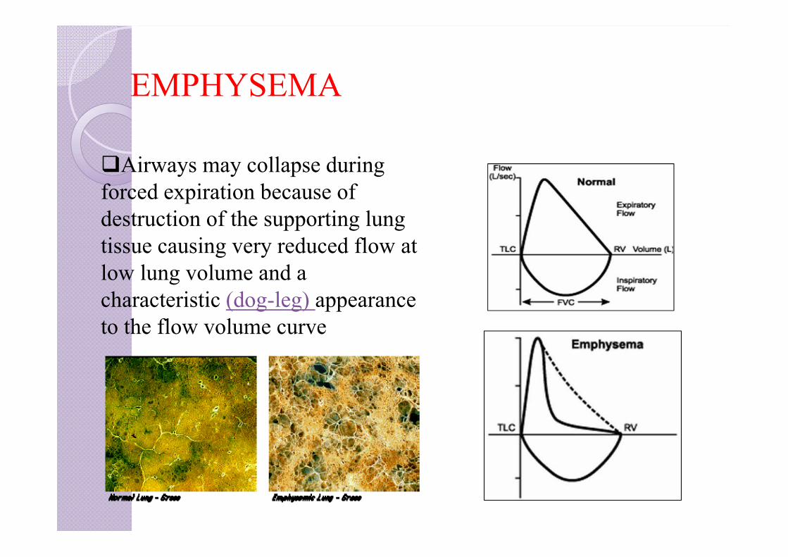
EMPHYSEMA
Airways may collapse during forced expiration because of destruction of the supporting lung tissue causing very reduced flow at low lung volume and a characteristic (dog-leg) appearance to the flow volume curve

REVERSIBILITY
Improvement in FEV1 by 12-15% or 200 ml in repeating spirometry after treatment with Sulbutamol 2.5mg or ipratropium bromide by nebuliser after 15-30 minutes
Reversibility is a characterestic feature of B.Asthma
In chronic asthma there may be only partial reversibility of the airflow obstruction
While in COPD the airflow is irreversible although some cases showed significant improvement

RESTRICTIVE PATTERN
Characterized by reduced lung volumes/decreased lung complianceExamples:•Interstitial Fibrosis•Scoliosis•Obesity•Lung Resection•Neuromuscular diseases•Cystic Fibrosis

RESTRICTIVE PATTERN-flow volume loop low total lung capacity low functional residual capacity low residual volume.
Forced vital capacity (FVC) may be low; however, FEV1/FVC is often normal or greater than
Peak expiratory flow may be preserved or even higher than predicted leads to tall,narrow and steep flow volume loop in expiratory phase.
INDEX
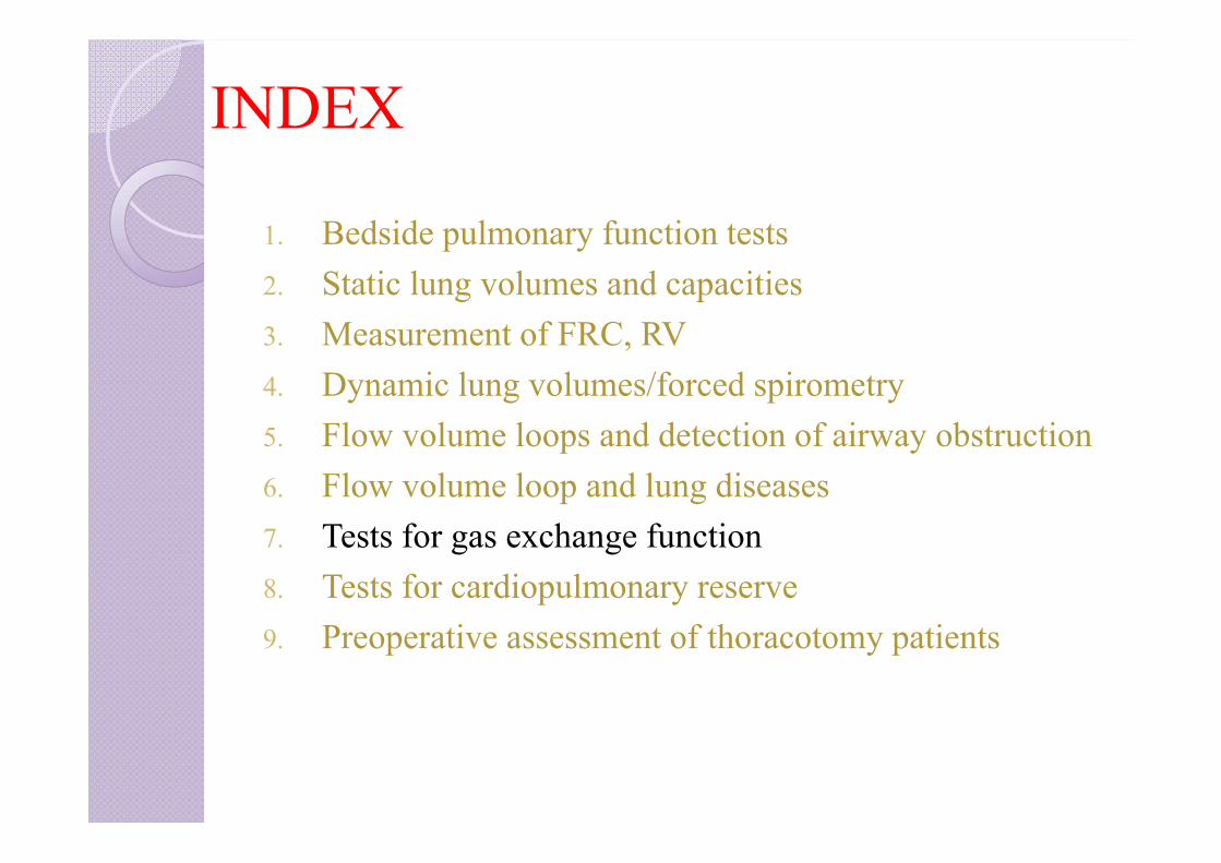
1. Bedside pulmonary function tests2. Static lung volumes and capacities3. Measurement of FRC, RV4. Dynamic lung volumes/forced spirometry5. Flow volume loops and detection of airway obstruction6. Flow volume loop and lung diseases7. Tests for gas exchange function8. Tests for cardiopulmonary reserve9. Preoperative assessment of thoracotomy patients
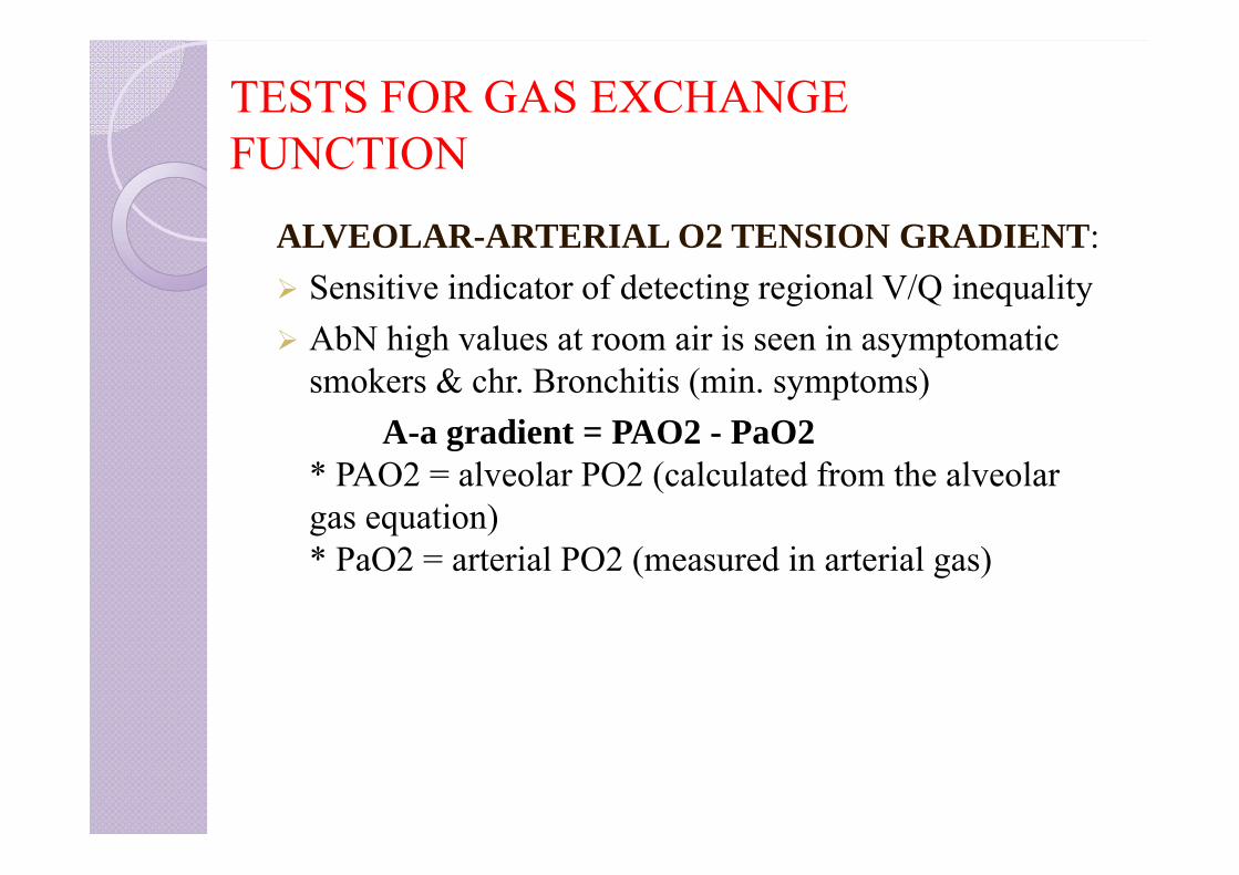
TESTS FOR GAS EXCHANGE FUNCTION
ALVEOLAR-ARTERIAL O2 TENSION GRADIENT: Sensitive indicator of detecting regional V/Q inequality AbN high values at room air is seen in asymptomatic
smokers & chr. Bronchitis (min. symptoms)A-a gradient = PAO2 - PaO2
* PAO2 = alveolar PO2 (calculated from the alveolar gas equation)* PaO2 = arterial PO2 (measured in arterial gas)
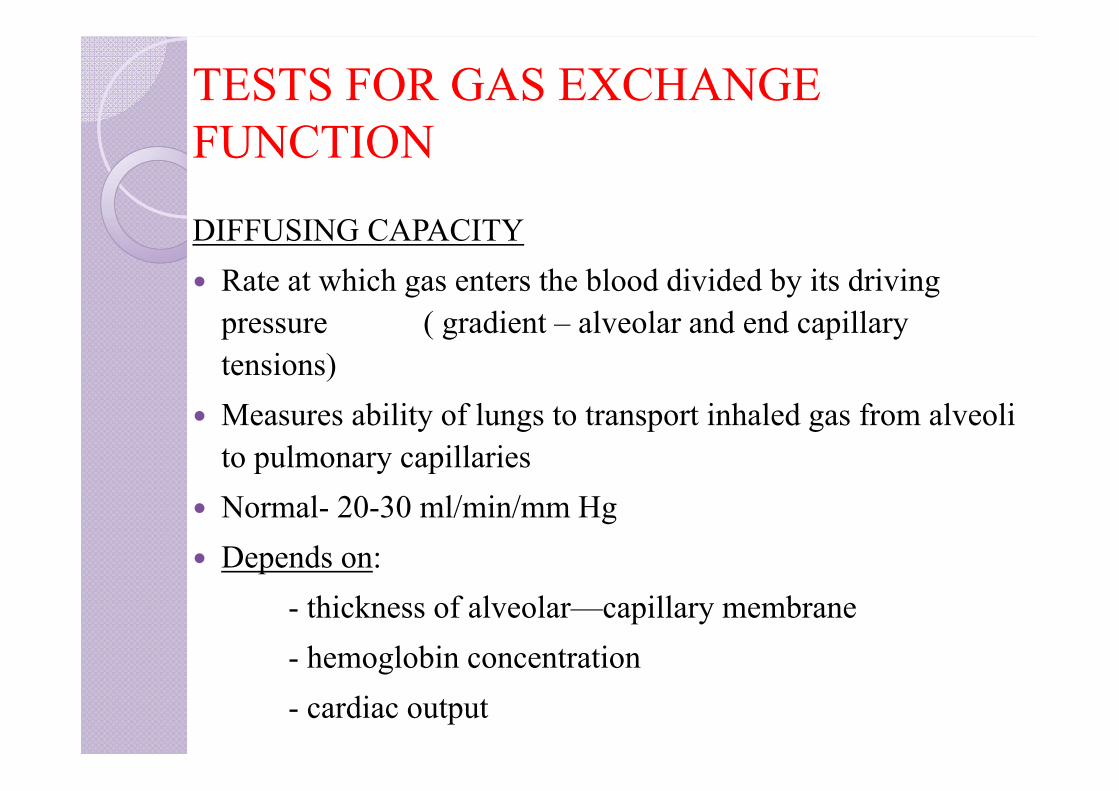
TESTS FOR GAS EXCHANGE FUNCTION
DIFFUSING CAPACITY Rate at which gas enters the blood divided by its driving
pressure ( gradient – alveolar and end capillary tensions)
Measures ability of lungs to transport inhaled gas from alveoli to pulmonary capillaries
Normal- 20-30 ml/min/mm Hg Depends on:
- thickness of alveolar—capillary membrane- hemoglobin concentration- cardiac output
TESTS FOR GAS EXCHANGE FUNCTION
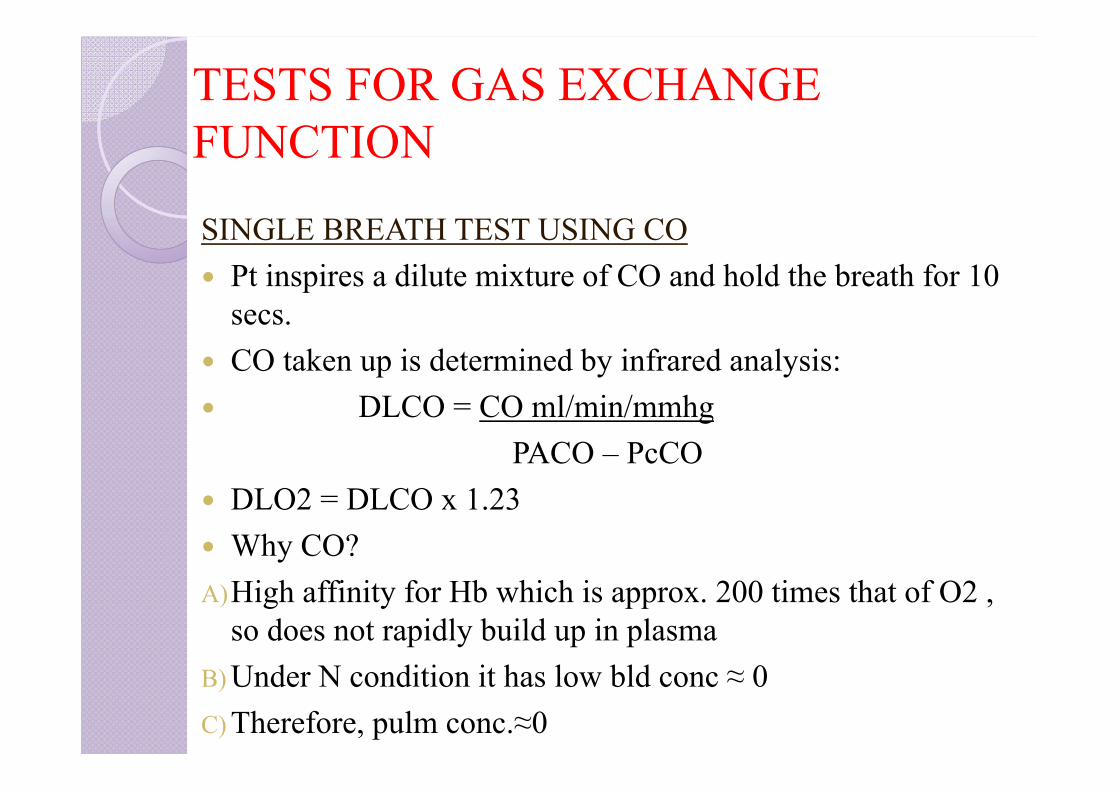
SINGLE BREATH TEST USING CO Pt inspires a dilute mixture of CO and hold the breath for 10
secs. CO taken up is determined by infrared analysis: DLCO = CO ml/min/mmhg
PACO – PcCO DLO2 = DLCO x 1.23 Why CO?A)High affinity for Hb which is approx. 200 times that of O2 ,
so does not rapidly build up in plasmaB) Under N condition it has low bld conc ≈ 0C) Therefore, pulm conc.≈0
The DLCO is low in ILD,but normal in disorders of pleura, chest and neuromuscular disorder causing restrictive lung function.
DLCO is also useful for following the course of or response to therapy in ILD.

FACTORS AFFECTING DLCODECREASE(< 80% predicted)
INCREASE(> 120-140% predicted)
Anemia PolycythemiaCarboxyhemoglobin ExercisePulmonary embolism Congestive heart failureDiffuse pulmonary fibrosisPulmonary emphysema
Predicted DLCO for Hb= Predicted DLCO * (1.7 Hb/10.22 + Hb)
INDEX
1. Bedside pulmonary function tests2. Static lung volumes and capacities3. Measurement of FRC, RV4. Dynamic lung volumes/forced spirometry5. Flow volume loops and detection of airway obstruction6. Flow volume loop and lung diseases7. Tests of gas function8. Tests for cardiopulmonary reserve9. Preoperative assessment of thoracotomy patients
CARDIOPULMONARY INTERACTION
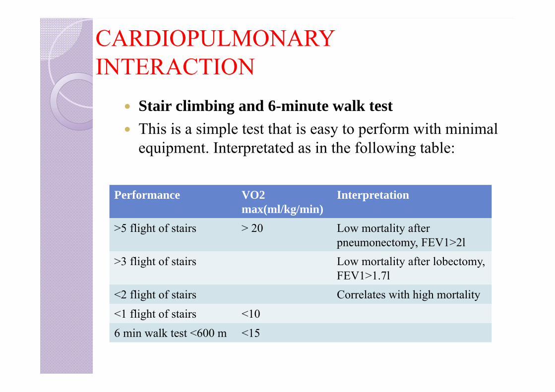
Stair climbing and 6-minute walk test This is a simple test that is easy to perform with minimal
equipment. Interpretated as in the following table:
Performance VO2max(ml/kg/min)
Interpretation
>5 flight of stairs > 20 Low mortality after pneumonectomy, FEV1>2l
>3 flight of stairs Low mortality after lobectomy, FEV1>1.7l
<2 flight of stairs Correlates with high mortality<1 flight of stairs <106 min walk test <600 m <15
CARDIOPULMONARY INTERACTION
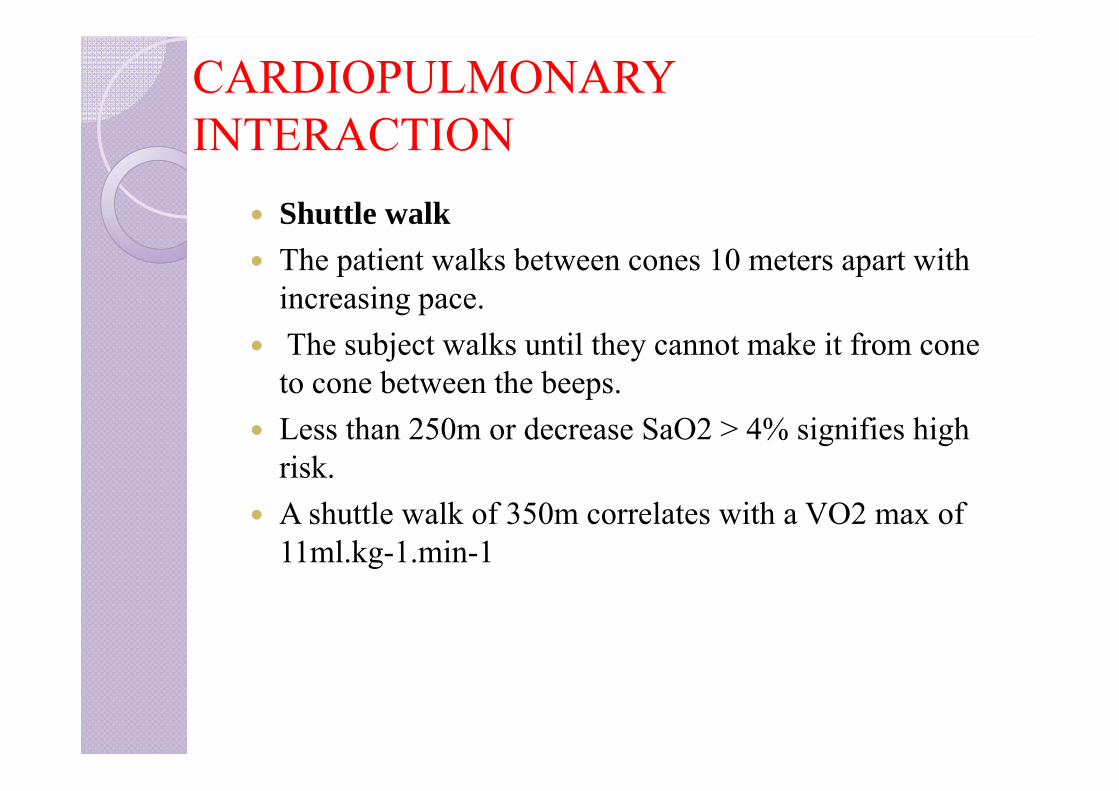
Shuttle walk The patient walks between cones 10 meters apart with
increasing pace. The subject walks until they cannot make it from cone
to cone between the beeps. Less than 250m or decrease SaO2 > 4% signifies high
risk. A shuttle walk of 350m correlates with a VO2 max of
11ml.kg-1.min-1
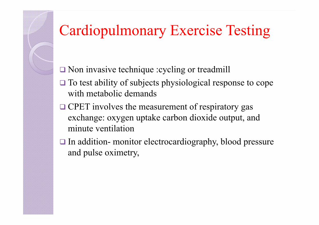
Cardiopulmonary Exercise Testing
Non invasive technique :cycling or treadmill To test ability of subjects physiological response to cope
with metabolic demands CPET involves the measurement of respiratory gas
exchange: oxygen uptake carbon dioxide output, and minute ventilation
In addition- monitor electrocardiography, blood pressure and pulse oximetry,
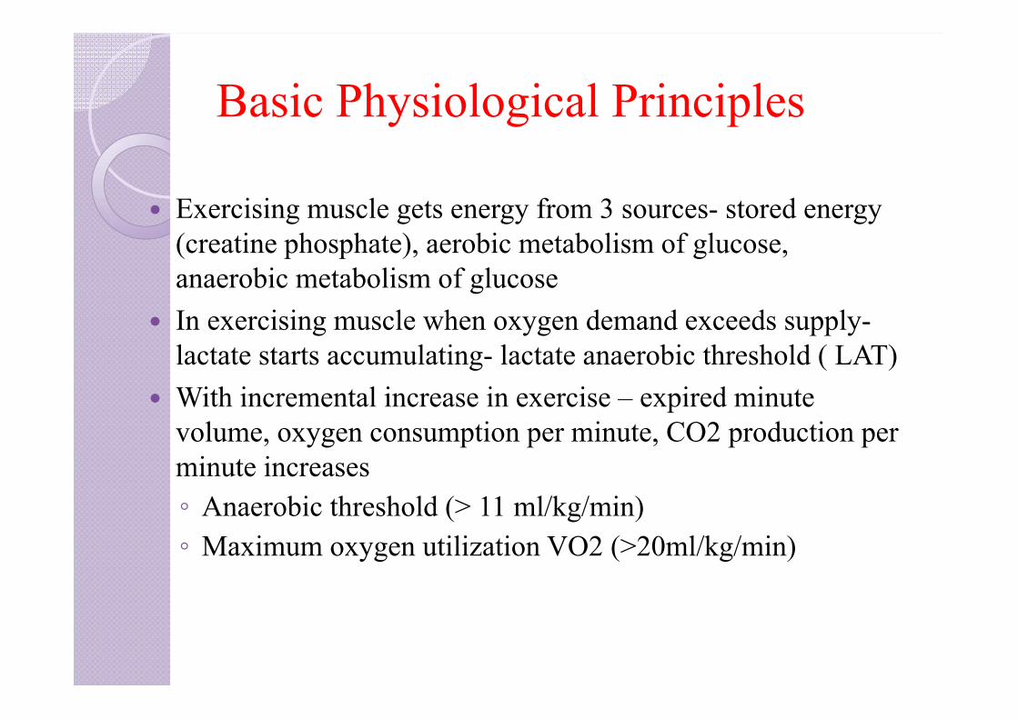
Basic Physiological Principles
Exercising muscle gets energy from 3 sources- stored energy (creatine phosphate), aerobic metabolism of glucose, anaerobic metabolism of glucose
In exercising muscle when oxygen demand exceeds supply-lactate starts accumulating- lactate anaerobic threshold ( LAT)
With incremental increase in exercise – expired minute volume, oxygen consumption per minute, CO2 production per minute increases◦ Anaerobic threshold (> 11 ml/kg/min)◦ Maximum oxygen utilization VO2 (>20ml/kg/min)

INDEX
1. Bedside pulmonary function tests2. Static lung volumes and capacities3. Measurement of FRC, RV4. Dynamic lung volumes/forced spirometry5. Flow volume loops and detection of airway obstruction6. Flow volume loop and lung diseases7. Tests of gas function8. Tests for cardiopulmonary reserve9. Preoperative assessment of thoracotomy patients
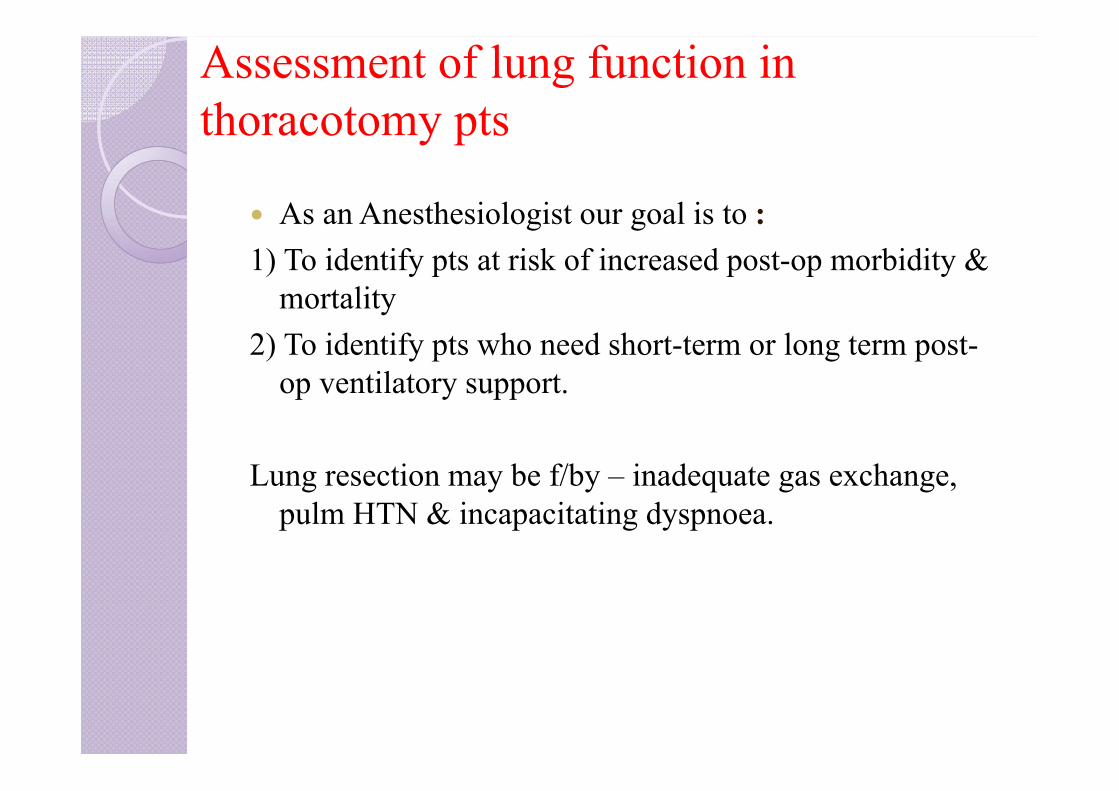
Assessment of lung function in thoracotomy pts
As an Anesthesiologist our goal is to :1) To identify pts at risk of increased post-op morbidity &
mortality2) To identify pts who need short-term or long term post-
op ventilatory support.
Lung resection may be f/by – inadequate gas exchange, pulm HTN & incapacitating dyspnoea.

Assessment of lung function in thoracotomy pts
Calculating the predicted postoperative FEV1 (ppoFEV1) and DLCO (ppoDLCO): There are 5 lung lobes containing19 segments in total with the division of each lobe.
Ppo FEV1=preoperative FEV1 * no. of segments left after resection
19• Can be assessed by ventilation perfusion scan. For eg:
A 57-year-old man is booked for lungresection. His CT chest show a large RUL mass confirmed as carcinoma: ppoFEV1= 50*16/19=42%
Assessment of lung function in thoracotomy pts
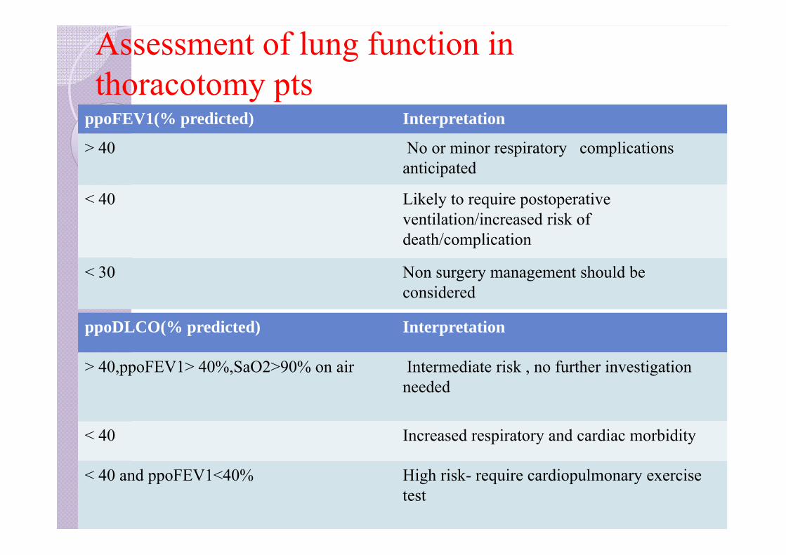
ppoFEV1(% predicted) Interpretation
> 40 No or minor respiratory complicationsanticipated
< 40 Likely to require postoperative ventilation/increased risk of death/complication
< 30 Non surgery management should be considered
ppoDLCO(% predicted) Interpretation
> 40,ppoFEV1> 40%,SaO2>90% on air Intermediate risk , no further investigation needed
< 40 Increased respiratory and cardiac morbidity
< 40 and ppoFEV1<40% High risk- require cardiopulmonary exercise test
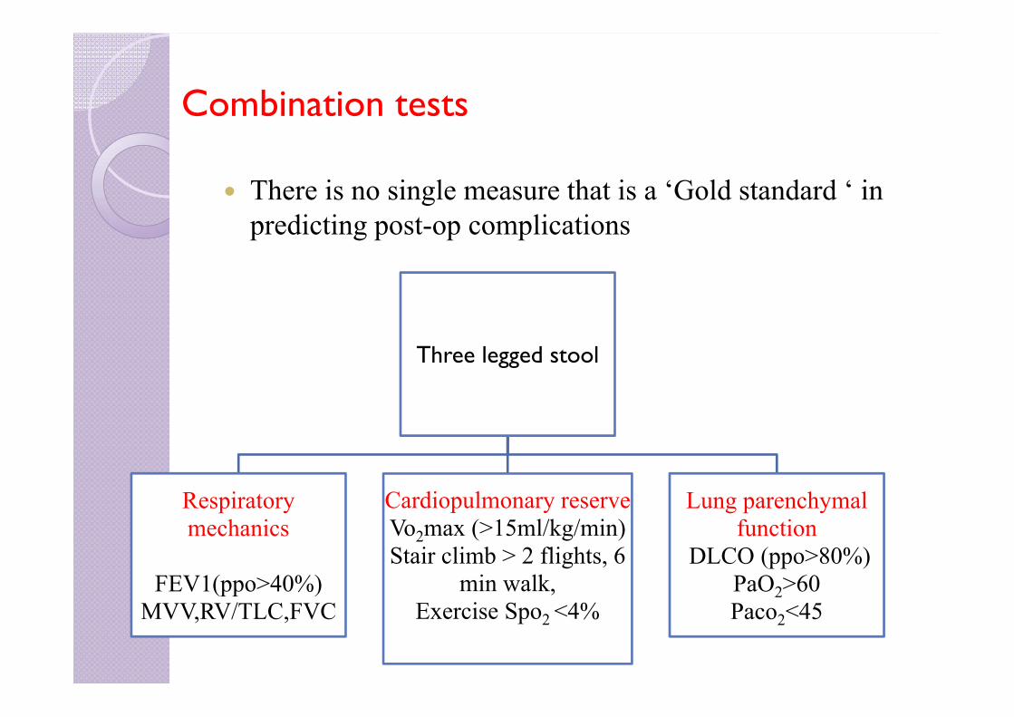
Combination tests
There is no single measure that is a ‘Gold standard ‘ in predicting post-op complications
Three legged stool
Respiratory mechanics
FEV1(ppo>40%)MVV,RV/TLC,FVC
Cardiopulmonary reserveVo2max (>15ml/kg/min) Stair climb > 2 flights, 6
min walk,Exercise Spo2 <4%
Lung parenchymalfunction
DLCO (ppo>80%)PaO2>60Paco2<45
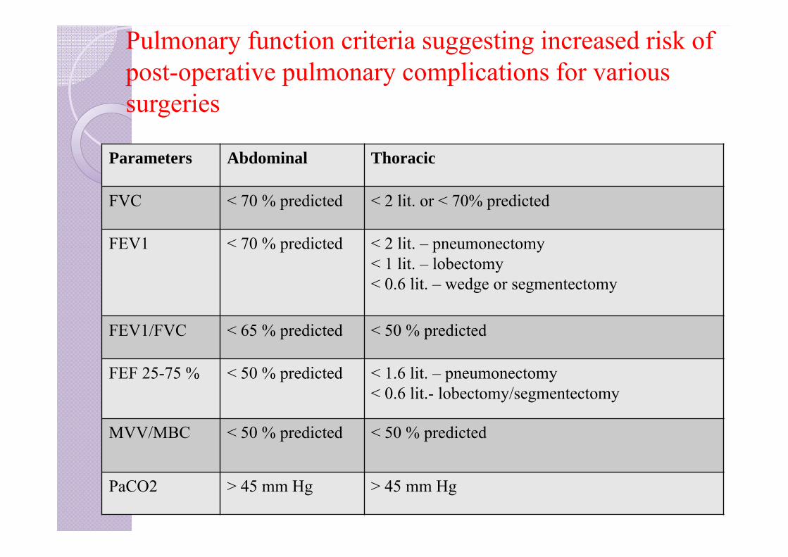
Pulmonary function criteria suggesting increased risk of post-operative pulmonary complications for various surgeries
Parameters Abdominal Thoracic
FVC < 70 % predicted < 2 lit. or < 70% predicted
FEV1 < 70 % predicted < 2 lit. – pneumonectomy< 1 lit. – lobectomy< 0.6 lit. – wedge or segmentectomy
FEV1/FVC < 65 % predicted < 50 % predicted
FEF 25-75 % < 50 % predicted < 1.6 lit. – pneumonectomy< 0.6 lit.- lobectomy/segmentectomy
MVV/MBC < 50 % predicted < 50 % predicted
PaCO2 > 45 mm Hg > 45 mm Hg
They act only to support or exclude a diagnosis. A combination of a thorough history and physical exam,
as well as supporting laboratory data and imaging is helpful in developing a anaesthetic plan for pt with pulmonary dysfunction.

THANK YOU