Embed Size (px)
Citation preview
Samokhin et al., Sci. Transl. Med. 10, eaap7294 (2018) 13 June 2018
S C I E N C E T R A N S L A T I O N A L M E D I C I N E | R E S E A R C H A R T I C L E
1 of 12
P U L M O N A R Y A R T E R I A L H Y P E R T E N S I O N
NEDD9 targets COL3A1 to promote endothelial fibrosis and pulmonary arterial hypertensionAndriy O. Samokhin1, Thomas Stephens1, Bradley M. Wertheim1,2, Rui-Sheng Wang3, Sara O. Vargas4, Lai-Ming Yung5, Minwei Cao1, Marcel Brown1, Elena Arons1, Paul B. Dieffenbach2, Jason G. Fewell6, Majed Matar6, Frederick P. Bowman1, Kathleen J. Haley2, George A. Alba7, Stefano M. Marino8,9, Rahul Kumar10, Ivan O. Rosas2, Aaron B. Waxman2, William M. Oldham2, Dinesh Khanna11, Brian B. Graham10, Sachiko Seo12, Vadim N. Gladyshev8, Paul B. Yu5, Laura E. Fredenburgh2, Joseph Loscalzo1,5, Jane A. Leopold5, Bradley A. Maron5*
Germline mutations involving small mothers against decapentaplegic–transforming growth factor– (SMAD–TGF-) signaling are an important but rare cause of pulmonary arterial hypertension (PAH), which is a disease characterized, in part, by vascular fibrosis and hyperaldosteronism (ALDO). We developed and analyzed a fibrosis protein-protein network (fibrosome) in silico, which predicted that the SMAD3 target neural precursor cell ex-pressed developmentally down-regulated 9 (NEDD9) is a critical ALDO-regulated node underpinning pathogenic vascular fibrosis. Bioinformatics and microscale thermophoresis demonstrated that oxidation of Cys18 in the SMAD3 docking region of NEDD9 impairs SMAD3-NEDD9 protein-protein interactions in vitro. This effect was re-produced by ALDO-induced oxidant stress in cultured human pulmonary artery endothelial cells (HPAECs), resulting in impaired NEDD9 proteolytic degradation, increased NEDD9 complex formation with Nk2 homeobox 5 (NKX2-5), and increased NKX2-5 binding to COL3A1. Up-regulation of NEDD9-dependent collagen III expression corre-sponded to changes in cell stiffness measured by atomic force microscopy. HPAEC-derived exosomal signaling targeted NEDD9 to increase collagen I/III expression in human pulmonary artery smooth muscle cells, identifying a second endothelial mechanism regulating vascular fibrosis. ALDO-NEDD9 signaling was not affected by treat-ment with a TGF- ligand trap and, thus, was not contingent on TGF- signaling. Colocalization of NEDD9 with collagen III in HPAECs was observed in fibrotic pulmonary arterioles from PAH patients. Furthermore, NEDD9 ab-lation or inhibition prevented fibrotic vascular remodeling and pulmonary hypertension in animal models of PAH in vivo. These data identify a critical TGF-–independent posttranslational modification that impairs SMAD3-NEDD9 binding in HPAECs to modulate vascular fibrosis and promote PAH.
INTRODUCTIONActivation of small mothers against decapentaplegic 2/3 (SMAD2/3) signal transduction through stimulation of the transforming growth factor– receptor II (TGF--RII) complex is a principal mechanism underlying transcriptional regulation of genes coding for collagen (1). However, perturbations to TGF- biofunctionality are associated with collagen deposition patterns that vary across cell types (2) and within patients who share circumspect clinical phenotypes (3). This heterogeneity suggests that the TGF- “master switch” theory may be an insufficient construct by which to understand fibrosis globally. Nonetheless, little is established on TGF-–independent and cell- specific molecular mechanisms that control collagen synthesis to promote fibrosis in human disease.
Pulmonary arterial hypertension (PAH) is characterized, in part, by a fibrotic vasculopathy in pulmonary arterioles, which promotes early right heart failure and death (4). In PAH, endothelial dysfunc-tion and vascular fibrosis are key pathogenic events that occur con-currently with changes in the normal cellular redox potential. Previous association studies imply a positive correlation between oxidant stress and collagen I/III protein synthesis via TGF- in pulmonary endo-thelial cells in vitro (5) or up-regulated endothelial mesenchymal transition (EndMT) in subpopulations of cells in remodeled PAH vessels in vivo (6). However, transgenic experimental PAH models or PAH patients harboring germline mutations that exaggerate TGF- signaling express an angioproliferative-dominant phenotype with inconsistent vascular fibrosis patterns (7). Collectively, these data suggest that there may be alternative or parallel redox-sensitive molec-ular mechanisms in endothelial cells that contribute to fibrosis in PAH.
In PAH patients, overactivation of the renin-angiotensin axis is associated with elevated concentrations of the mineralocorticoid hormone aldosterone (ALDO) (8), which correlate positively with hemodynamic measures of vascular remodeling (9). Pathophysiolog-ical ALDO concentrations observed in PAH are sufficient to perturb the normal redox potential of cultured pulmonary artery endothelial cells (PAECs) by direct activation of the reactive oxygen species–generating enzyme NOX4 (10). However, stimulation of the miner-alocorticoid receptor by ALDO is implicated in both adaptive wound healing in skin and pathogenic vascular fibrosis in PAH (5, 11). This observation suggests that there is substantial overlap among ALDO
1Department of Medicine, Brigham and Women’s Hospital, Boston, MA 02115, USA. 2Division of Pulmonary and Critical Care Medicine, Brigham and Women’s Hospital, Boston, MA 02115, USA. 3Channing Division of Network Medicine, Brigham and Women’s Hospital, Boston, MA 02115, USA. 4Department of Pathology, Boston Children’s Hospital, Boston, MA 02115, USA. 5Division of Cardiovascular Medicine, Department of Medicine, Brigham and Women’s Hospital, Boston, MA 02115, USA. 6Celsion Corporation, Lawrenceville, NJ 08648, USA. 7Department of Pulmonary and Critical Care Medicine, Massachusetts General Hospital, Boston, MA 02114, USA. 8Division of Genetics, Department of Medicine, Brigham and Women’s Hospital, Boston, MA 02115, USA. 9Department of Biotechnology, Akdeniz University, Konyaaltı, Antalya 07058, Turkey. 10Program in Translational Lung Research, Department of Medicine, University of Colorado Anschutz Medical Campus, Aurora, CO 80045, USA. 11Division of Rheumatology, University of Michigan Scleroderma Program, Ann Arbor, MI 48109, USA. 12Department of Hematology and Oncology, National Cancer Research Center East, Kashiwa-shi, Chiba-ken 277-8577, Japan.*Corresponding author. Email: [email protected]
Copyright © 2018 The Authors, some rights reserved; exclusive licensee American Association for the Advancement of Science. No claim to original U.S. Government Works
by guest on June 9, 2020http://stm
.sciencemag.org/
Dow
nloaded from
Samokhin et al., Sci. Transl. Med. 10, eaap7294 (2018) 13 June 2018
S C I E N C E T R A N S L A T I O N A L M E D I C I N E | R E S E A R C H A R T I C L E
2 of 12
signaling pathways irrespective of fibrosis subtype. To address this knowledge gap, we hypothesized that applying Bayesian (condi-tional probability) analytical methods to ALDO gene targets segre-gated by distinct fibrosis functionalities would enable the discovery of unknown PAH-specific fibrogenic mechanisms, which may be independent of TGF- signaling.
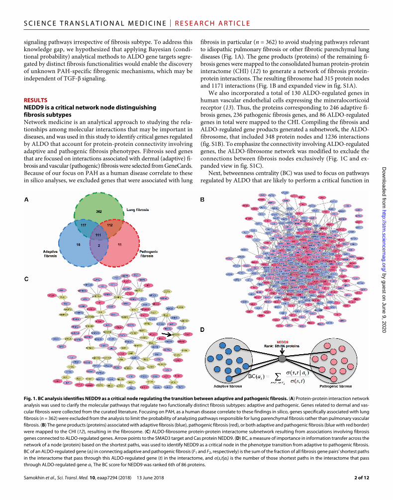
RESULTSNEDD9 is a critical network node distinguishing fibrosis subtypesNetwork medicine is an analytical approach to studying the rela-tionships among molecular interactions that may be important in diseases, and was used in this study to identify critical genes regulated by ALDO that account for protein-protein connectivity involving adaptive and pathogenic fibrosis phenotypes. Fibrosis seed genes that are focused on interactions associated with dermal (adaptive) fi-brosis and vascular (pathogenic) fibrosis were selected from GeneCards. Because of our focus on PAH as a human disease correlate to these in silico analyses, we excluded genes that were associated with lung
fibrosis in particular (n = 362) to avoid studying pathways relevant to idiopathic pulmonary fibrosis or other fibrotic parenchymal lung diseases (Fig. 1A). The gene products (proteins) of the remaining fi-brosis genes were mapped to the consolidated human protein-protein interactome (CHI) (12) to generate a network of fibrosis protein- protein interactions. The resulting fibrosome had 315 protein nodes and 1171 interactions (Fig. 1B and expanded view in fig. S1A).
We also incorporated a total of 130 ALDO-regulated genes in human vascular endothelial cells expressing the mineralocorticoid receptor (13). Thus, the proteins corresponding to 246 adaptive fi-brosis genes, 236 pathogenic fibrosis genes, and 86 ALDO-regulated genes in total were mapped to the CHI. Compiling the fibrosis and ALDO-regulated gene products generated a subnetwork, the ALDO- fibrosome, that included 348 protein nodes and 1236 interactions (fig. S1B). To emphasize the connectivity involving ALDO-regulated genes, the ALDO-fibrosome network was modified to exclude the connections between fibrosis nodes exclusively (Fig. 1C and ex-panded view in fig. S1C).
Next, betweenness centrality (BC) was used to focus on pathways regulated by ALDO that are likely to perform a critical function in
Fig. 1. BC analysis identifies NEDD9 as a critical node regulating the transition between adaptive and pathogenic fibrosis. (A) Protein-protein interaction network analysis was used to clarify the molecular pathways that regulate two functionally distinct fibrosis subtypes: adaptive and pathogenic. Genes related to dermal and vas-cular fibrosis were collected from the curated literature. Focusing on PAH, as a human disease correlate to these findings in silico, genes specifically associated with lung fibrosis (n = 362) were excluded from the analysis to limit the probability of analyzing pathways responsible for lung parenchymal fibrosis rather than pulmonary vascular fibrosis. (B) The gene products (proteins) associated with adaptive fibrosis (blue), pathogenic fibrosis (red), or both adaptive and pathogenic fibrosis (blue with red border) were mapped to the CHI (12), resulting in the fibrosome. (C) ALDO-fibrosome protein-protein interactome subnetwork resulting from associations involving fibrosis genes connected to ALDO-regulated genes. Arrow points to the SMAD3 target and Cas protein NEDD9. (D) BC, a measure of importance in information transfer across the network of a node (protein) based on the shortest paths, was used to identify NEDD9 as a critical node in the phenotype transition from adaptive to pathogenic fibrosis. BC of an ALDO-regulated gene (ai) in connecting adaptive and pathogenic fibrosis (F1 and F2, respectively) is the sum of the fraction of all fibrosis gene pairs’ shortest paths in the interactome that pass through this ALDO-regulated gene (t) in the interactome, and (s,t|ai) is the number of those shortest paths in the interactome that pass through ALDO-regulated gene ai. The BC score for NEDD9 was ranked 6th of 86 proteins.
by guest on June 9, 2020http://stm
.sciencemag.org/
Dow
nloaded from
Samokhin et al., Sci. Transl. Med. 10, eaap7294 (2018) 13 June 2018
S C I E N C E T R A N S L A T I O N A L M E D I C I N E | R E S E A R C H A R T I C L E
3 of 12
the fibrosis phenotype transition and thus permit identification of critical nodes dis-tinguishing adaptive from pathogenic fibrosis in silico. Using this approach, neural precursor cell expressed develop-mentally down-regulated 9 (NEDD9) emerged from our analysis. Specifically, NEDD9 was assigned a top BC score (P = 0.004 versus other ALDO-regulated genes; P = 7.5 × 10⁻⁷ versus random network) (Fig. 1D). Of the proteins with high BC scores, NEDD9 was selected for further analysis based on its previously reported role in clinical phenotypes that overlap with PAH, such as connective tissue di-sease (14), converging lines of evidence in tumorigenic cells lines that have indi-rectly implicated negative regulation of NEDD9 by SMAD3 via proteolytic deg-radation (15), and lack of data on the role of NEDD9 in primary cardiopulmonary diseases.
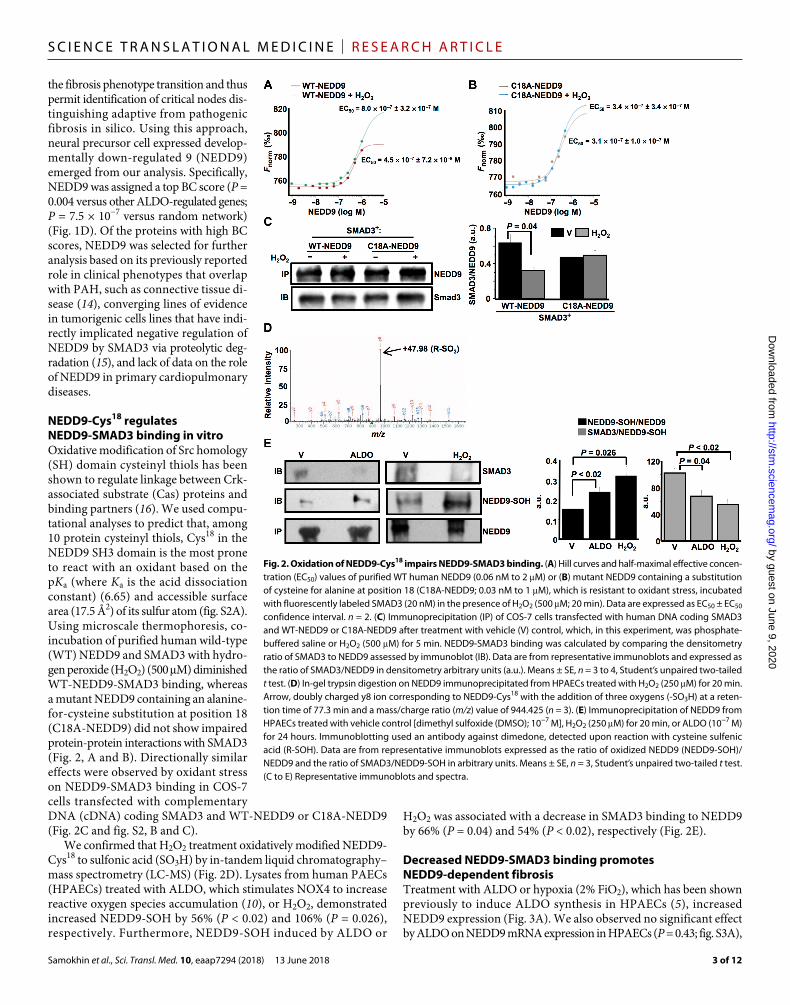
NEDD9-Cys18 regulates NEDD9-SMAD3 binding in vitroOxidative modification of Src homology (SH) domain cysteinyl thiols has been shown to regulate linkage between Crk- associated substrate (Cas) proteins and binding partners (16). We used compu-tational analyses to predict that, among 10 protein cysteinyl thiols, Cys18 in the NEDD9 SH3 domain is the most prone to react with an oxidant based on the pKa (where Ka is the acid dissociation constant) (6.65) and accessible surface area (17.5 Å2) of its sulfur atom (fig. S2A). Using microscale thermophoresis, co- incubation of purified human wild-type (WT) NEDD9 and SMAD3 with hydro-gen peroxide (H2O2) (500 M) diminished WT-NEDD9-SMAD3 binding, whereas a mutant NEDD9 containing an alanine- for-cysteine substitution at position 18 (C18A-NEDD9) did not show impaired protein-protein interactions with SMAD3 (Fig. 2, A and B). Directionally similar effects were observed by oxidant stress on NEDD9-SMAD3 binding in COS-7 cells transfected with complementary DNA (cDNA) coding SMAD3 and WT-NEDD9 or C18A-NEDD9 (Fig. 2C and fig. S2, B and C).
We confirmed that H2O2 treatment oxidatively modified NEDD9- Cys18 to sulfonic acid (SO3H) by in-tandem liquid chromatography– mass spectrometry (LC-MS) (Fig. 2D). Lysates from human PAECs (HPAECs) treated with ALDO, which stimulates NOX4 to increase reactive oxygen species accumulation (10), or H2O2, demonstrated increased NEDD9-SOH by 56% (P < 0.02) and 106% (P = 0.026), respectively. Furthermore, NEDD9-SOH induced by ALDO or
H2O2 was associated with a decrease in SMAD3 binding to NEDD9 by 66% (P = 0.04) and 54% (P < 0.02), respectively (Fig. 2E).
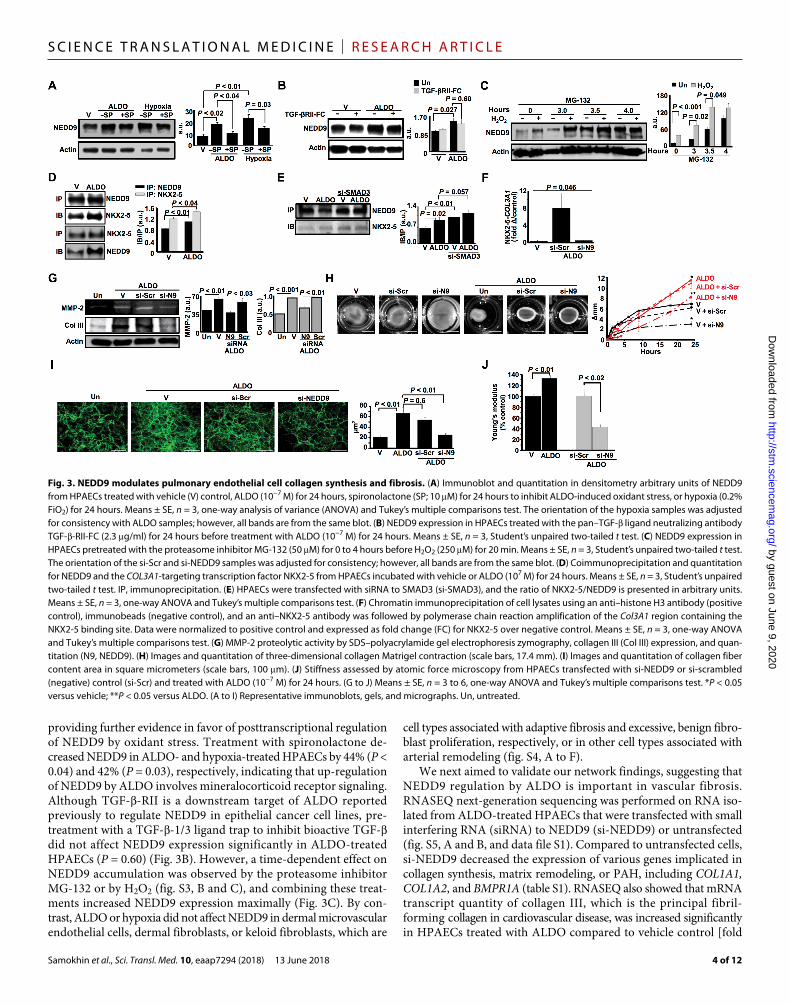
Decreased NEDD9-SMAD3 binding promotes NEDD9-dependent fibrosisTreatment with ALDO or hypoxia (2% FiO2), which has been shown previously to induce ALDO synthesis in HPAECs (5), increased NEDD9 expression (Fig. 3A). We also observed no significant effect by ALDO on NEDD9 mRNA expression in HPAECs (P = 0.43; fig. S3A),
Fig. 2. Oxidation of NEDD9-Cys18 impairs NEDD9-SMAD3 binding. (A) Hill curves and half-maximal effective concen-tration (EC50) values of purified WT human NEDD9 (0.06 nM to 2 M) or (B) mutant NEDD9 containing a substitution of cysteine for alanine at position 18 (C18A-NEDD9; 0.03 nM to 1 M), which is resistant to oxidant stress, incubated with fluorescently labeled SMAD3 (20 nM) in the presence of H2O2 (500 M; 20 min). Data are expressed as EC50 ± EC50 confidence interval. n = 2. (C) Immunoprecipitation (IP) of COS-7 cells transfected with human DNA coding SMAD3 and WT-NEDD9 or C18A-NEDD9 after treatment with vehicle (V) control, which, in this experiment, was phosphate- buffered saline or H2O2 (500 M) for 5 min. NEDD9-SMAD3 binding was calculated by comparing the densitometry ratio of SMAD3 to NEDD9 assessed by immunoblot (IB). Data are from representative immunoblots and expressed as the ratio of SMAD3/NEDD9 in densitometry arbitrary units (a.u.). Means ± SE, n = 3 to 4, Student’s unpaired two-tailed t test. (D) In-gel trypsin digestion on NEDD9 immunoprecipitated from HPAECs treated with H2O2 (250 M) for 20 min. Arrow, doubly charged y8 ion corresponding to NEDD9-Cys18 with the addition of three oxygens (-SO3H) at a reten-tion time of 77.3 min and a mass/charge ratio (m/z) value of 944.425 (n = 3). (E) Immunoprecipitation of NEDD9 from HPAECs treated with vehicle control [dimethyl sulfoxide (DMSO); 10−7 M], H2O2 (250 M) for 20 min, or ALDO (10−7 M) for 24 hours. Immunoblotting used an antibody against dimedone, detected upon reaction with cysteine sulfenic acid (R-SOH). Data are from representative immunoblots expressed as the ratio of oxidized NEDD9 (NEDD9-SOH)/NEDD9 and the ratio of SMAD3/NEDD9-SOH in arbitrary units. Means ± SE, n = 3, Student’s unpaired two-tailed t test. (C to E) Representative immunoblots and spectra.
by guest on June 9, 2020http://stm
.sciencemag.org/
Dow
nloaded from
Samokhin et al., Sci. Transl. Med. 10, eaap7294 (2018) 13 June 2018
S C I E N C E T R A N S L A T I O N A L M E D I C I N E | R E S E A R C H A R T I C L E
4 of 12
providing further evidence in favor of posttranscriptional regulation of NEDD9 by oxidant stress. Treatment with spironolactone de-creased NEDD9 in ALDO- and hypoxia-treated HPAECs by 44% (P < 0.04) and 42% (P = 0.03), respectively, indicating that up-regulation of NEDD9 by ALDO involves mineralocorticoid receptor signaling. Although TGF--RII is a downstream target of ALDO reported previously to regulate NEDD9 in epithelial cancer cell lines, pre-treatment with a TGF--1/3 ligand trap to inhibit bioactive TGF- did not affect NEDD9 expression significantly in ALDO-treated HPAECs (P = 0.60) (Fig. 3B). However, a time-dependent effect on NEDD9 accumulation was observed by the proteasome inhibitor MG-132 or by H2O2 (fig. S3, B and C), and combining these treat-ments increased NEDD9 expression maximally (Fig. 3C). By con-trast, ALDO or hypoxia did not affect NEDD9 in dermal microvascular endothelial cells, dermal fibroblasts, or keloid fibroblasts, which are
cell types associated with adaptive fibrosis and excessive, benign fibro-blast proliferation, respectively, or in other cell types associated with arterial remodeling (fig. S4, A to F).
We next aimed to validate our network findings, suggesting that NEDD9 regulation by ALDO is important in vascular fibrosis. RNASEQ next-generation sequencing was performed on RNA iso-lated from ALDO-treated HPAECs that were transfected with small interfering RNA (siRNA) to NEDD9 (si-NEDD9) or untransfected (fig. S5, A and B, and data file S1). Compared to untransfected cells, si-NEDD9 decreased the expression of various genes implicated in collagen synthesis, matrix remodeling, or PAH, including COL1A1, COL1A2, and BMPR1A (table S1). RNASEQ also showed that mRNA transcript quantity of collagen III, which is the principal fibril- forming collagen in cardiovascular disease, was increased significantly in HPAECs treated with ALDO compared to vehicle control [fold
Fig. 3. NEDD9 modulates pulmonary endothelial cell collagen synthesis and fibrosis. (A) Immunoblot and quantitation in densitometry arbitrary units of NEDD9 from HPAECs treated with vehicle (V) control, ALDO (10−7 M) for 24 hours, spironolactone (SP; 10 M) for 24 hours to inhibit ALDO-induced oxidant stress, or hypoxia (0.2% FiO2) for 24 hours. Means ± SE, n = 3, one-way analysis of variance (ANOVA) and Tukey’s multiple comparisons test. The orientation of the hypoxia samples was adjusted for consistency with ALDO samples; however, all bands are from the same blot. (B) NEDD9 expression in HPAECs treated with the pan–TGF- ligand neutralizing antibody TGF--RII-FC (2.3 g/ml) for 24 hours before treatment with ALDO (10−7 M) for 24 hours. Means ± SE, n = 3, Student’s unpaired two-tailed t test. (C) NEDD9 expression in HPAECs pretreated with the proteasome inhibitor MG-132 (50 M) for 0 to 4 hours before H2O2 (250 M) for 20 min. Means ± SE, n = 3, Student’s unpaired two-tailed t test. The orientation of the si-Scr and si-NEDD9 samples was adjusted for consistency; however, all bands are from the same blot. (D) Coimmunoprecipitation and quantitation for NEDD9 and the COL3A1-targeting transcription factor NKX2-5 from HPAECs incubated with vehicle or ALDO (107 M) for 24 hours. Means ± SE, n = 3, Student’s unpaired two-tailed t test. IP, immunoprecipitation. (E) HPAECs were transfected with siRNA to SMAD3 (si-SMAD3), and the ratio of NKX2-5/NEDD9 is presented in arbitrary units. Means ± SE, n = 3, one-way ANOVA and Tukey’s multiple comparisons test. (F) Chromatin immunoprecipitation of cell lysates using an anti–histone H3 antibody (positive control), immunobeads (negative control), and an anti–NKX2-5 antibody was followed by polymerase chain reaction amplification of the Col3A1 region containing the NKX2-5 binding site. Data were normalized to positive control and expressed as fold change (FC) for NKX2-5 over negative control. Means ± SE, n = 3, one-way ANOVA and Tukey’s multiple comparisons test. (G) MMP-2 proteolytic activity by SDS–polyacrylamide gel electrophoresis zymography, collagen III (Col III) expression, and quan-titation (N9, NEDD9). (H) Images and quantitation of three-dimensional collagen Matrigel contraction (scale bars, 17.4 mm). (I) Images and quantitation of collagen fiber content area in square micrometers (scale bars, 100 m). (J) Stiffness assessed by atomic force microscopy from HPAECs transfected with si-NEDD9 or si-scrambled (negative) control (si-Scr) and treated with ALDO (10−7 M) for 24 hours. (G to J) Means ± SE, n = 3 to 6, one-way ANOVA and Tukey’s multiple comparisons test. *P < 0.05 versus vehicle; **P < 0.05 versus ALDO. (A to I) Representative immunoblots, gels, and micrographs. Un, untreated.
by guest on June 9, 2020http://stm
.sciencemag.org/
Dow
nloaded from
Samokhin et al., Sci. Transl. Med. 10, eaap7294 (2018) 13 June 2018
S C I E N C E T R A N S L A T I O N A L M E D I C I N E | R E S E A R C H A R T I C L E
5 of 12
change (FC) = +2.47; n = 3 to 6 samples from female and male donors, multiple test– corrected P value; false discovery rate (FDR) = 0.016; P = 0.001] (data file S2). However, this effect was inhibited by transfection with si-NEDD9 (FC = +1.78 in si-NEDD9 + ALDO versus vehicle; n = 3 to 6 samples from female and male donors, multiple test–corrected P value; FDR = 0.250; P = 0.07) (data file S3).
The transcription factor Nk2 homeobox 5 (NKX2-5) is predicted to target COL3A1, and we observed NEDD9 expression in the nu-cleus of HPAECs with or without ALDO treatment and in the nu-cleus of HPAECs isolated from PAH patients (PAH-HPAECs) (fig. S5, C and D, and movies S1 to S3). ALDO increased nuclear colocaliza-tion (fig. S6A) and coimmunoprecipitation of NKX2-5 with NEDD9 (Fig. 3D). We also observed an inverse relationship between the amount of the NEDD9-SMAD3 complex and that of NEDD9–NKX2-5 complex (r = −0.91, P < 0.01), as well as a positive correlation between the amount of NEDD9 and that of the NEDD9–NKX2-5 complex (r = +0.86, P = 0.03) (fig. S6B). These findings were consistent with our observation that si-NEDD9 decreased NKX2-5 without significantly influencing SMAD3 expression (P = 0.75) (fig. S6C) and, overall, show that impaired SMAD3 association with NEDD9 pro-motes NEDD9 stability, which, in turn, permits greater NEDD9–NKX2-5 interaction. Transfection with si-SMAD3 (fig. S6D) increased NEDD9–NKX2-5 coimmunoprecipitation under basal condi tions and in ALDO-treated HPAECs (Fig. 3E). ALDO increased NKX2-5 binding to the COL3A1 gene as well as COL3A1 mRNA transcript quantity, which was inhibited significantly by si-NEDD9 (P = 0.046) and si-NKX2-5 (P < 0.001), respectively (Fig. 3F and fig. S6, E and F).
We next analyzed the functional consequences of increased NEDD9 on COL3A1 gene transcription in ALDO-treated HPAECs. Molecular inhibition of NEDD9 attenuated the ALDO-induced in-creased collagen III expression, matrix metalloproteinase-2 (MMP-2) proteolytic activity, three-dimensional collagen Matrigel contraction, and cell stiffness (Fig. 3, G to J). In contrast, overexpression of NEDD9 correlated positively with collagen III expression in HPAECs (fig. S7A). Profibrotic changes to HPAECs by ALDO (fig. S7B) included a dis-tinctive “net” pattern of collagen stranding in HPAECs that was not evident in human pulmonary artery smooth muscle cells (HPASMCs) or normal human lung fibroblasts (NHLFs) (fig. S7C). In addition, an ALDO-induced fibrotic phenotype in HPAECs was observed with-out differences in the expression of von Willebrand factor (vWF), vascular endothelial cadherin (VE-cadherin), platelet endothelial cell adhesion molecule-1 (PECAM-1), or the EndMT markers twist- related protein 1 (TWIST) and -smooth muscle actin (-SMA) (fig. S7, C and D).
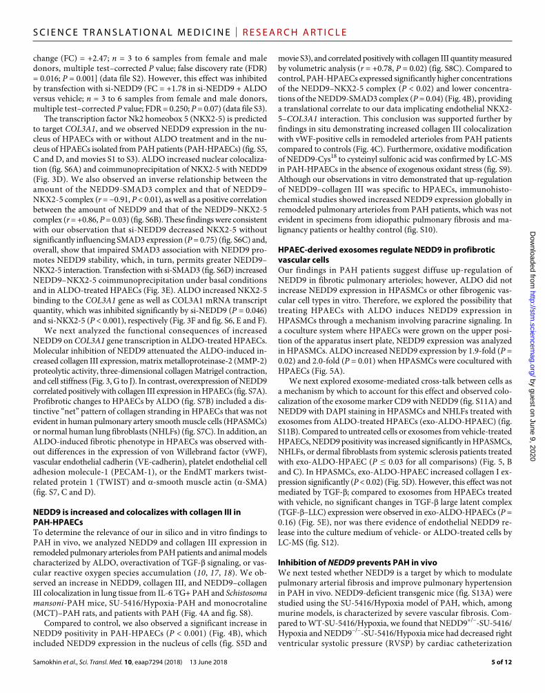
NEDD9 is increased and colocalizes with collagen III in PAH-HPAECsTo determine the relevance of our in silico and in vitro findings to PAH in vivo, we analyzed NEDD9 and collagen III expression in remodeled pulmonary arterioles from PAH patients and animal models characterized by ALDO, overactivation of TGF- signaling, or vas-cular reactive oxygen species accumulation (10, 17, 18). We ob-served an increase in NEDD9, collagen III, and NEDD9–collagen III colocalization in lung tissue from IL-6 TG+ PAH and Schistosoma mansoni-PAH mice, SU-5416/Hypoxia-PAH and monocrotaline (MCT)–PAH rats, and patients with PAH (Fig. 4A and fig. S8).
Compared to control, we also observed a significant increase in NEDD9 positivity in PAH-HPAECs (P < 0.001) (Fig. 4B), which included NEDD9 expression in the nucleus of cells (fig. S5D and
movie S3), and correlated positively with collagen III quantity measured by volumetric analysis (r = +0.78, P = 0.02) (fig. S8C). Compared to control, PAH-HPAECs expressed significantly higher concentrations of the NEDD9–NKX2-5 complex (P < 0.02) and lower concentra-tions of the NEDD9-SMAD3 complex (P = 0.04) (Fig. 4B), providing a translational correlate to our data implicating endothelial NKX2-5–COL3A1 interaction. This conclusion was supported further by findings in situ demonstrating increased collagen III colocalization with vWF-positive cells in remodeled arterioles from PAH patients compared to controls (Fig. 4C). Furthermore, oxidative modification of NEDD9-Cys18 to cysteinyl sulfonic acid was confirmed by LC-MS in PAH-HPAECs in the absence of exogenous oxidant stress (fig. S9). Although our observations in vitro demonstrated that up-regulation of NEDD9–collagen III was specific to HPAECs, immunohisto-chemical studies showed increased NEDD9 expression globally in remodeled pulmonary arterioles from PAH patients, which was not evident in specimens from idiopathic pulmonary fibrosis and ma-lignancy patients or healthy control (fig. S10).
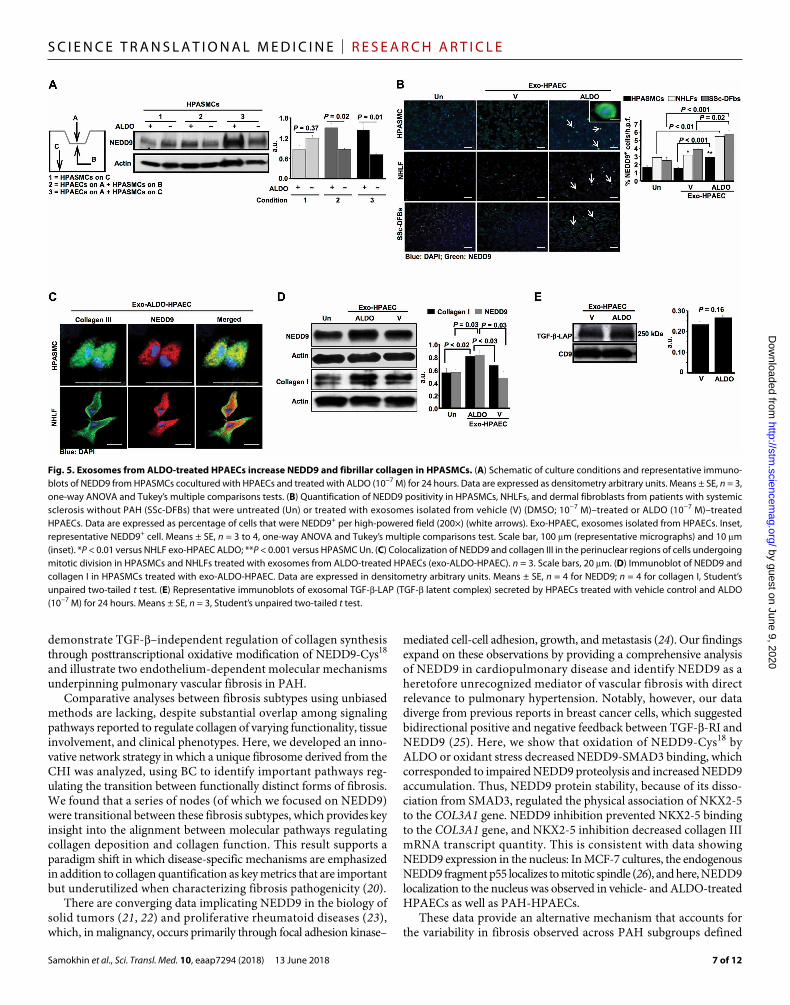
HPAEC-derived exosomes regulate NEDD9 in profibrotic vascular cellsOur findings in PAH patients suggest diffuse up-regulation of NEDD9 in fibrotic pulmonary arterioles; however, ALDO did not increase NEDD9 expression in HPASMCs or other fibrogenic vas-cular cell types in vitro. Therefore, we explored the possibility that treating HPAECs with ALDO induces NEDD9 expression in HPASMCs through a mechanism involving paracrine signaling. In a coculture system where HPAECs were grown on the upper posi-tion of the apparatus insert plate, NEDD9 expression was analyzed in HPASMCs. ALDO increased NEDD9 expression by 1.9-fold (P = 0.02) and 2.0-fold (P = 0.01) when HPASMCs were cocultured with HPAECs (Fig. 5A).
We next explored exosome-mediated cross-talk between cells as a mechanism by which to account for this effect and observed colo-calization of the exosome marker CD9 with NEDD9 (fig. S11A) and NEDD9 with DAPI staining in HPASMCs and NHLFs treated with exosomes from ALDO-treated HPAECs (exo-ALDO-HPAEC) (fig. S11B). Compared to untreated cells or exosomes from vehicle-treated HPAECs, NEDD9 positivity was increased significantly in HPASMCs, NHLFs, or dermal fibroblasts from systemic sclerosis patients treated with exo-ALDO-HPAEC (P ≤ 0.03 for all comparisons) (Fig. 5, B and C). In HPASMCs, exo-ALDO-HPAEC increased collagen I ex-pression significantly (P < 0.02) (Fig. 5D). However, this effect was not mediated by TGF-; compared to exosomes from HPAECs treated with vehicle, no significant changes in TGF- large latent complex (TGF-–LLC) expression were observed in exo-ALDO-HPAECs (P = 0.16) (Fig. 5E), nor was there evidence of endothelial NEDD9 re-lease into the culture medium of vehicle- or ALDO-treated cells by LC-MS (fig. S12).
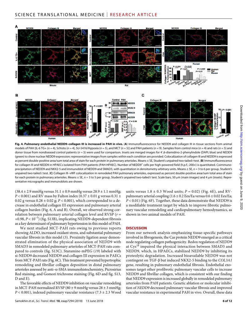
Inhibition of NEDD9 prevents PAH in vivoWe next tested whether NEDD9 is a target by which to modulate pulmonary arterial fibrosis and improve pulmonary hypertension in PAH in vivo. NEDD9-deficient transgenic mice (fig. S13A) were studied using the SU-5416/Hypoxia model of PAH, which, among murine models, is characterized by severe vascular fibrosis. Com-pared to WT-SU-5416/Hypoxia, we found that NEDD9+/−-SU-5416/Hypoxia and NEDD9−/−-SU-5416/Hypoxia mice had decreased right ventricular systolic pressure (RVSP) by cardiac catheterization
by guest on June 9, 2020http://stm
.sciencemag.org/
Dow
nloaded from
Samokhin et al., Sci. Transl. Med. 10, eaap7294 (2018) 13 June 2018
S C I E N C E T R A N S L A T I O N A L M E D I C I N E | R E S E A R C H A R T I C L E
6 of 12
(38.4 ± 2.9 mmHg versus 31.1 ± 0.9 mmHg versus 28.9 ± 1.1 mmHg; P < 0.001) and RV mass by Fulton index (0.37 ± 0.01 g versus 0.31 ± 0.02 g versus 0.28 ± 0.02 g; P < 0.001), which corresponded to a de-crease in endothelial collagen III expression and pulmonary arterial collagen burden (Fig. 6, A and B). Overall, we observed strong cor-relation between pulmonary arterial collagen level and RVSP (r = +0.98, P < 10−5) (fig. S13B), implicating NEDD9-dependent fibrosis as a key determinant of pulmonary hypertension in this model of PAH.
We next studied MCT-PAH rats owing to previous reports showing ALDO, increased oxidant stress, and substantial pulmonary vascular fibrosis in this model (5). Proximity ligation assay demon-strated elimination of the physical association of NEDD9 with SMAD3 in remodeled pulmonary arterioles of MCT-PAH rats com-pared to controls (fig. S13C). Staramine-mPEG (19) labeled with si-NEDD9 decreased NEDD9 and collagen III expression in PAECs from MCT-PAH rats (Fig. 6C). This treatment prevented hypertrophic remodeling and fibrillar collagen deposition in distal pulmonary arterioles assessed by anti–-SMA immunohistochemistry, Picrosirius Red staining, and Gomori trichrome staining (Fig. 6D and fig. S13, D and E).
The favorable effects of NEDD9 inhibition on vascular remodeling in MCT-PAH normalized RVSP (80 ± 9 mmHg versus 28 ± 3 mmHg; P < 0.001), indexed pulmonary vascular resistance (7.3 ± 2.3 Wood
units versus 1.8 ± 0.3 Wood units; P = 0.02) (Fig. 6E), and RV- pulmonary arterial coupling (1.6 ± 0.2 Ees/Ea versus 0.6 ± 0.02 Ees/Ea; P < 0.01) (Fig. 6F). Together, these data demonstrate that NEDD9 is a modifiable treatment target by which to improve fibrotic pulmo-nary vascular remodeling and cardiopulmonary hemodynamics, as shown in two animal models of PAH.
DISCUSSIONFrom our network analysis emphasizing tissue-specific pathways involved in fibrogenesis, the Cas protein NEDD9 emerged as a critical node regulating collagen pathogenicity. Redox regulation of NEDD9 at Cys18 impaired the physical interaction between SMAD3 and NEDD9, which, in HPAECs, stabilized NEDD9 by inhibiting its proteolytic degradation. Increased bioavailable NEDD9 was not contingent on TGF- but induced NKX2-5 binding to the COL3A1 gene, resulting in pulmonary endothelial fibrosis. Endothelial exo-somes target other profibrotic pulmonary vascular cells to increase NEDD9 and fibrillar collagen, which is consistent with our finding that NEDD9 expression is increased globally in remodeled pulmonary arterioles from PAH patients. Genetic ablation or molecular inhibi-tion of NEDD9 decreased pulmonary vascular fibrosis and improved vascular resistance in experimental PAH in vivo. Overall, these data
Fig. 4. Pulmonary endothelial NEDD9–collagen III is increased in PAH in vivo. (A) Immunofluorescence for NEDD9 and collagen III in tissue sections from animal models of PAH [IL-6 TG+ (n = 4), Schisto (n = 4), SU-5416/Hypoxia (n = 5), and MCT (n = 5)] and PAH patients (n = 9). Samples from control mice (n = 4) and rats (n = 5) and donor tissue from nondiseased control patients (n = 5) were used for comparison. Insets are merged images for 4′,6-diamidino-2-phenylindole (DAPI; blue) and NEDD9 (green) to show nuclear NEDD9 expression; representative images from samples within each condition are provided. Colocalization of collagen III and NEDD9 is expressed as percent double-positive area/sum total area of stain for each protein in pulmonary arterioles. Means ± SE, Student’s unpaired two-tailed t test. (B) Immunofluorescence for collagen III and NEDD9 in HPAECs isolated from PAH patients (PAH-HPAEC). Number of NEDD9+ cells per high-powered field (h.p.f.; 200×) is quantitated. Coimmuno-precipitation of NEDD9 and NKX2-5 and immunoblot of NEDD9 and SMAD3, with quantitation in densitometry arbitrary units. Means ± SE, n = 3 to 6 per group, Student’s unpaired two-tailed t test. (C) Collagen III–vWF colocalization in remodeled PAH pulmonary arterioles, expressed as percent double-positive area/sum total area of stain for each protein in pulmonary arterioles. Means ± SE, n = 3 to 5 per group, Student’s unpaired two-tailed t test. Scale bars, 50 m (main images) and 4 m (insets). Repre-sentative micrographs and immunoblots are shown.
by guest on June 9, 2020http://stm
.sciencemag.org/
Dow
nloaded from
Samokhin et al., Sci. Transl. Med. 10, eaap7294 (2018) 13 June 2018
S C I E N C E T R A N S L A T I O N A L M E D I C I N E | R E S E A R C H A R T I C L E
7 of 12
demonstrate TGF-–independent regulation of collagen synthesis through posttranscriptional oxidative modification of NEDD9-Cys18 and illustrate two endothelium-dependent molecular mechanisms underpinning pulmonary vascular fibrosis in PAH.
Comparative analyses between fibrosis subtypes using unbiased methods are lacking, despite substantial overlap among signaling pathways reported to regulate collagen of varying functionality, tissue involvement, and clinical phenotypes. Here, we developed an inno-vative network strategy in which a unique fibrosome derived from the CHI was analyzed, using BC to identify important pathways reg-ulating the transition between functionally distinct forms of fibrosis. We found that a series of nodes (of which we focused on NEDD9) were transitional between these fibrosis subtypes, which provides key insight into the alignment between molecular pathways regulating collagen deposition and collagen function. This result supports a paradigm shift in which disease-specific mechanisms are emphasized in addition to collagen quantification as key metrics that are important but underutilized when characterizing fibrosis pathogenicity (20).
There are converging data implicating NEDD9 in the biology of solid tumors (21, 22) and proliferative rheumatoid diseases (23), which, in malignancy, occurs primarily through focal adhesion kinase–
mediated cell-cell adhesion, growth, and metastasis (24). Our findings expand on these observations by providing a comprehensive analysis of NEDD9 in cardiopulmonary disease and identify NEDD9 as a heretofore unrecognized mediator of vascular fibrosis with direct relevance to pulmonary hypertension. Notably, however, our data diverge from previous reports in breast cancer cells, which suggested bidirectional positive and negative feedback between TGF--RI and NEDD9 (25). Here, we show that oxidation of NEDD9-Cys18 by ALDO or oxidant stress decreased NEDD9-SMAD3 binding, which corresponded to impaired NEDD9 proteolysis and increased NEDD9 accumulation. Thus, NEDD9 protein stability, because of its disso-ciation from SMAD3, regulated the physical association of NKX2-5 to the COL3A1 gene. NEDD9 inhibition prevented NKX2-5 binding to the COL3A1 gene, and NKX2-5 inhibition decreased collagen III mRNA transcript quantity. This is consistent with data showing NEDD9 expression in the nucleus: In MCF-7 cultures, the endogenous NEDD9 fragment p55 localizes to mitotic spindle (26), and here, NEDD9 localization to the nucleus was observed in vehicle- and ALDO-treated HPAECs as well as PAH-HPAECs.
These data provide an alternative mechanism that accounts for the variability in fibrosis observed across PAH subgroups defined
Fig. 5. Exosomes from ALDO-treated HPAECs increase NEDD9 and fibrillar collagen in HPASMCs. (A) Schematic of culture conditions and representative immuno-blots of NEDD9 from HPASMCs cocultured with HPAECs and treated with ALDO (10−7 M) for 24 hours. Data are expressed as densitometry arbitrary units. Means ± SE, n = 3, one-way ANOVA and Tukey’s multiple comparisons tests. (B) Quantification of NEDD9 positivity in HPASMCs, NHLFs, and dermal fibroblasts from patients with systemic sclerosis without PAH (SSc-DFBs) that were untreated (Un) or treated with exosomes isolated from vehicle (V) (DMSO; 10−7 M)–treated or ALDO (10−7 M)–treated HPAECs. Data are expressed as percentage of cells that were NEDD9+ per high-powered field (200×) (white arrows). Exo-HPAEC, exosomes isolated from HPAECs. Inset, representative NEDD9+ cell. Means ± SE, n = 3 to 4, one-way ANOVA and Tukey’s multiple comparisons test. Scale bar, 100 m (representative micrographs) and 10 m (inset). *P < 0.01 versus NHLF exo-HPAEC ALDO; **P < 0.001 versus HPASMC Un. (C) Colocalization of NEDD9 and collagen III in the perinuclear regions of cells undergoing mitotic division in HPASMCs and NHLFs treated with exosomes from ALDO-treated HPAECs (exo-ALDO-HPAEC). n = 3. Scale bars, 20 m. (D) Immunoblot of NEDD9 and collagen I in HPASMCs treated with exo-ALDO-HPAEC. Data are expressed in densitometry arbitrary units. Means ± SE, n = 4 for NEDD9; n = 4 for collagen I, Student’s unpaired two-tailed t test. (E) Representative immunoblots of exosomal TGF--LAP (TGF- latent complex) secreted by HPAECs treated with vehicle control and ALDO (10−7 M) for 24 hours. Means ± SE, n = 3, Student’s unpaired two-tailed t test.
by guest on June 9, 2020http://stm
.sciencemag.org/
Dow
nloaded from
Samokhin et al., Sci. Transl. Med. 10, eaap7294 (2018) 13 June 2018
S C I E N C E T R A N S L A T I O N A L M E D I C I N E | R E S E A R C H A R T I C L E
8 of 12
commonly by overactivation of TGF-, including hereditable PAH from bone morphogenetic protein receptor-2 (BMPR-2) mutation or Schistosoma mansoni-PAH, which was a model that was included in the current study. Our findings demonstrate that endothelial exosomes transduce the remodeling effect of ALDO on different vascular cell types. This, in turn, has important implications on un-derstanding diffuse patterns of fibrosis in cardiac tissues and other targets of end-organ damage mediated by ALDO in many cardio-pulmonary diseases, including the pulmonary circulation in PAH (5, 27).
Previous reports have identified vascular smooth muscle cells, adventitial fibroblasts, and endothelial cells via EndMT in the patho-genesis of vascular fibrosis in PAH (28, 29). On the basis of a pub-lished work indicating that collagen synthesis in stimulated HPAECs occurs at a time point before EndMT onset (5), we focused on alter-native (or parallel) mechanisms by which endothelial cells may con-tribute to fibrosis in the absence of phenotype switching. Here, up-regulation of NEDD9 by oxidant stress controlled HPAEC col-
lagen net formation to increase cell stiffness without altering ex-pression of EndMT proteins. This finding suggests that endothelial cells are independent contributors to collagen synthesis, resulting in increased cell stiffness through NEDD9, and thus identifies en-dothelial fibrosis as a critical PAH pathophenotype. Furthermore, endothelial exosomes from ALDO-treated HPAECs increased NEDD9 and collagen in other cell types associated with vascular fibrosis, in-cluding HPASMCs. This mechanism may also have direct relevance to other profibrotic cell types, particularly adventitial fibroblasts, which are sensitive to paracrine signaling and regulate collagen depo-sition in PAH (30). Further investigations focusing on endothelial NEDD9 in early-stage disease, which is characterized by increased oxidant stress, endothelial injury, and vascular fibrosis (31), or as a target of circulating exosomes are warranted (32).
Epigenetic and posttranscriptional regulation of signaling path-ways that control vascular structure and function is central to PAH (33). In HPAECs, ALDO increases reactive oxygen species accumula-tion, including H2O2, which is generated via the reduction of superoxide
Fig. 6. Gene ablation or molecular inhibi-tion of NEDD9 prevented vascular fibrosis and PAH in vivo. (A) RVSP and RV mass by Fulton index (RV/LV + S) in male and female C57BL (WT), C57BL/NEDD9+/−, and C57BL/NEDD9−/− mice injected with Sugen-5416 (20 mg/kg) every 7 days during a 3-week period of hypoxia treatment (10% FiO2). Means ± SE, n = 6 to 9 mice per condition for RVSP; n = 5 to 10 mice per condition for heart weight, one-way ANOVA and Tukey’s multiple comparisons test. (B) Representative images of Gomori trichrome staining to assess vascular fibrosis burden and vWF–collagen III colocalization, expressed as per-cent double-positive area/sum total area of stain for each protein, of pulmonary arterioles from WT, NEDD9+/−, and NEDD9−/− mice treated with or without SU-5416/Hypoxia. Means ± SE, n = 6 to 10 mice per condition for trichrome; n = 4 to 7 mice per condition for vWF–collagen III, one-way ANOVA and Tukey’s multiple comparisons test. Scale bar, 50 m. (C) NEDD9–collagen III and NEDD9-vWF colocalization in lung tissue from male Sprague-Dawley rats adminis-
trated MCT (50 mg/kg) and treated with Staramine-mPEG (1.5 mg/kg) formulated with NEDD9 siRNA (si-NEDD9). Means ± SE, n = 4 to 5 rats per condition, one-way ANOVA and Tukey’s multiple comparisons test. Scale bars, 50 m. (D) The number of hypertrophic vessels/high-powered field (red arrow) and percent vascular fibrillar collagen in paraffin-embedded lung sections was analyzed using anti–-SMA immunohistochemistry and Picrosirius Red staining, respectively, from vehicle (V)–, MCT-, and MCT + si-NEDD9–treated rats. Means ± SE, n = 4 to 5 rats per condition, one-way ANOVA and Tukey’s multiple comparisons test. Scale bars, 100 m (main images) and 50 m (insets, representative hypertrophic vessel magnified). (E) Table of phenotypic changes in vehicle-, MCT-, and MCT + si-NEDD9–treated rats. Means ± SE, n = 5 to 7 rats per condition. P values in column represent one-way ANOVA. *P < 0.05 versus vehicle; **P < 0.001 versus MCT; ***P = 0.02 versus MCT by Tukey’s multiple comparisons test. (F) Representative RV pressure-volume loops are shown to quantify RV-pulmonary artery coupling, assessed by the ratio of end-systolic elastance (Ees)/pulmonary vascular elastance (Ea) in MCT- and MCT + si-NEDD9–treated rats. Means ± SE, n = 3 rats per condition, Student’s unpaired two-tailed t test. HR, heart rate; MAP, mean arterial pressure; RAP, right atrial pressure; CI, cardiac index; CO, cardiac output; PVRi, indexed pulmonary vascular resistance (in Wood units).
by guest on June 9, 2020http://stm
.sciencemag.org/
Dow
nloaded from
Samokhin et al., Sci. Transl. Med. 10, eaap7294 (2018) 13 June 2018
S C I E N C E T R A N S L A T I O N A L M E D I C I N E | R E S E A R C H A R T I C L E
9 of 12
anion spontaneously or via superoxide dismutases, or directly through a two-electron reduction of molecular oxygen catalyzed by NOX4. The sulfur atom of cysteinyl thiols may react with partially reduced oxygen species to form higher oxidative intermediaries that modify the conformational structure of proteins, impair G protein (hetero-trimeric guanine nucleotide–binding protein)–coupled receptor sig-naling, or inhibit S-nitrosylation to modulate cardiovascular disease (34). However, previous reports in PAH have studied thiols with known biofunctionality and tested the consequences of thiol oxida-tion indirectly (10). By contrast, computational modeling was used in this study to rank thiol reactivity based on pKa and access to oxi-dative insults, as predicted when analyzing proteins with NEDD9 structural homology. From this approach, Cys18 was identified as a redox-sensitive regulatory protein cysteinyl thiol, which was not based on a priori understanding of thiol biochemistry in NEDD9. Microscale thermophoresis provided definitive evidence on NEDD9-SMAD3 binding affinity and the contribution of Cys18 to the dynamics of this protein-protein interaction. Our observations in HPAECs show a time course–dependent increase in NEDD9 accumulation induced by H2O2 and that oxidation of NEDD9-Cys18 included the forma-tion of cysteine sulfenic acid, which is a reversible modification. However, further experimentation is needed to determine whether the redox switch at NEDD9-Cys18 is reversible and whether this bears on the therapeutic relevance of attenuating vascular oxidant stress in PAH.
Characterizing the redox regulation of NEDD9-SMAD3 may be relevant to a range of functionally essential pathways in PAH, in-cluding BMPR-2 signaling. Our findings have implications for he-reditary PAH, because vascular cells expressing a germline mutation in the BMPR2 gene are predisposed to both increased oxidant stress (35) and increased SMAD3 (36). Under these circumstances, two parallel but independent profibrotic signaling pathways relevant to PAH emerge through (i) oxidized NEDD9 and (ii) increased bioactive SMAD3 that may occur due to the direct effects of BMPR2 muta-tion or impaired NEDD9-SMAD3 binding.
Apoptosis resistance, dysregulated cell growth, and unopposed vascular proliferation contribute to the vasculopathy of PAH (37). Although NEDD9 has been linked to these processes in other cell types, and its inhibition improved hypertrophic remodeling in exper-imental PAH, the direct effect of NEDD9 on mitosis or cell survival was not investigated specifically in our study. Our data quantifying the EC50 for NEDD9-SMAD3 binding were limited by the maximum concentration of protein tested, and it is also possible that other NEDD9 thiols not analyzed in this study could exert functional effects on endothelial phenotype. Along these lines, alternative NEDD9- Cys18 posttranslational modifications relevant to ALDO and PAH-HPAEC biology, including via cysteine reactivity with peroxynitrite and other reactive nitrogen species or formation of S-nitrosothiols, could affect NEDD9 stability and its bioactivity and merit further investigation.
We focused on NEDD9-dependent signaling involving SMAD3; however, cross-talk between SMAD3 and other SMAD isoforms, as well as TGF-–connective tissue growth factor (CTGF) signaling, has been implicated in the pathobiology of pulmonary vascular fibrosis and PAH (37). Thus, additional studies in HPAECs are required to understand the effect of SMAD-SMAD interactions on proteolytic regulation of NEDD9, the effect of impaired NEDD9-SMAD3 binding on SMAD bioactivity, and NEDD9-independent pathways regulating collagen. Further investigation is also needed to clarify the precise
mechanism by which to account for redox regulation of NEDD9 selectively in HPAECs. This finding may be consistent with vari-ability across cell types reported for intracellular reductive potential (38), subcellular compartmentalization of proteins (NEDD9) that affect exposure to oxidants (39, 40), and signaling responses to ALDO (5).
Although our seed genes were selected from the curated data-base, it was not possible to confirm that adaptive fibrosis genes were not also implicated in maladaptive processes, such as hypertrophic scar. In turn, including such pathways could have affected the validity and topology of our networks. In addition, plasma or vascular ALDO concentrations were not available from the PAH patients, which could affect the translational relevance of our findings implicating ALDO-NEDD9-fibrosis signaling in vivo; however, the adverse effects of oxidant stress in PAH are well established, and this was a central mechanistic focus of this study. Experiments analyzing the effect of si-NEDD9 on PAH used a disease prevention experimental design, and therefore, the therapeutic implications of our in vivo data require further investigation.
In summary, we identify NEDD9 as a contributor to pulmonary arterial fibrosis, vascular remodeling, and pulmonary hypertension in PAH. Our observations illustrate two previously unrecognized mech-anisms underpinning endothelial regulation of vascular fibrosis: oxidative modification of NEDD9-Cys18 to increase COL3A1 gene transcription in HPAECs and paracrine signaling mediated by HPAEC- exosomes to increase fibrillar collagen expression in profibrotic target cells. Our findings on NEDD9-SMAD3 binding affinity also suggest that opportunity may exist to maintain normal NEDD9 proteolysis by inhibiting Cys18 oxidation and prevent fibrotic vascular remod-eling. Collectively, these data suggest that NEDD9 mediates fibrotic vascular remodeling, with potential therapeutic relevance for PAH patients.
MATERIALS AND METHODSStudy designWe hypothesized that systems biology could be used to identify disease- specific mechanisms underpinning pathogenic fibrosis, in-cluding vascular fibrotic remodeling in PAH. All data included in the study were reproduced across multiple iterations of the same experiment, which were often performed on different days, replicated by different investigators involved in the project, and confirmed using multiple approaches. Cell culture experiments were performed at least three times and at least in triplicate for each replicate when possible. For studies involving rats and mice, animals were randomly assigned to a treatment intervention. Power calculations for these studies were performed based on previous work (10, 17, 19). The number of animals needed in each treatment group was calculated to detect a 40% difference in pulmonary pressures with a 5% -error, and 80% power was used for performing experiments. For experi-ments involving human lung tissue, the number of samples included in each experiment was based largely on availability. Once conditions for an experiment were optimized, all data were included in the ab-sence of a specific technical or procedural reason that confounded the interpretation of a finding or whether the data met the prespecified, standard outlier definition of |±|2 SD of the mean. When possible for experiments involving biochemistry, cell biology, and other in vitro assays, the senior author (B.A.M.) and another experienced inves-tigator (J.A.L.) involved in the project were blinded to the treatment
by guest on June 9, 2020http://stm
.sciencemag.org/
Dow
nloaded from
Samokhin et al., Sci. Transl. Med. 10, eaap7294 (2018) 13 June 2018
S C I E N C E T R A N S L A T I O N A L M E D I C I N E | R E S E A R C H A R T I C L E
10 of 12
conditions for initial data review/interpretation. A similar policy was used for blinding with respect to animal experiments; however, owing to the severity of vascular remodeling and pulmonary hyper-tension in experimental PAH and patients with PAH, this was not possible uniformly.
Human lung samplesThe experimental protocol included only discarded lung samples and was approved by the institutional review board at Brigham and Women’s Hospital (IRB #2015P001526, #2011P002419, and #2001P000876) and Boston Children’s Hospital (IRB #P00010717). Hematoxylin and eosin, elastin, and Gomori trichrome staining, as well immuno-fluorescence (described in detail in Supplementary Materials and Methods), were performed on distal pulmonary arterioles from formalin- fixed, paraffin-embedded archival lung specimens acquired by clinical experts in the field that confirmed accurate patient diagnosis as follows: lung explant for PAH (S.O.V.) or idiopathic pulmonary fibrosis (I.O.R.), and open lung biopsy to diagnose malignancy (S.O.V.) (table S2). Samples from controls without lung or pulmonary vascular dis-ease were fully deidentified discarded donor lung samples provided by the New England Organ Bank (K.J.H.).
Animal models of PAHAll animals were handled in accordance with National Institutes of Health guidelines, and all procedures were approved by the local committees at Brigham and Women’s Hospital and the University of Colorado. All animals were fed standard chow. Animal treat-ments and analyses were controlled and blinded, whenever possible. Additional details can be found in Supplementary Materials and Methods.
Statistical analysisAll analyses were performed using Origin 9.01 or OriginPro, GraphPad Prism v7.03, and Cytoscape 3.5.1. Data are means ± SEM. Comparison between two groups was performed by the Student’s unpaired two-tailed t test. For the analyses involving the proximity ligation assay and assessment of NEDD9 versus p130Cas expression using anti- NEDD9 Ab2, a signal was not detectable in the control group, and therefore, a one-sample t test was used to determine whether results for the comparator group were statistically significant. One-way ANOVA was used to examine differences in response to treatments between groups. An analysis of covariance (ANCOVA) was used to examine differences between groups and controls for the effect of continuous variables. Post hoc analysis was performed by the meth-od of Tukey. P < 0.05 was considered significant. For experiments with n < 20, individual data points are presented in the figures or are included in data file S4.
SUPPLEMENTARY MATERIALSwww.sciencetranslationalmedicine.org/cgi/content/full/10/445/eaap7294/DC1Materials and MethodsFig. S1. Fibrosome and ALDO-fibrosome networks.Fig. S2. Computational modeling predicts that NEDD9-Cys18 is highly prone to oxidant stress.Fig. S3. Proteasomal inhibition or oxidant stress increases NEDD9 accumulation in HPAECs.Fig. S4. ALDO does not affect NEDD9 expression in cell types associated with adaptive fibrosis.Fig. S5. NEDD9 is expressed in the nucleus of HPAECs and PAH-HPAECs.Fig. S6. NKX2-5 and NEDD9 colocalize to the HPAEC nucleus, and inhibition of NKX2-5 prevents COL3A1 mRNA transcription mediated by ALDO.Fig. S7. ALDO increases NEDD9-dependent fibrosis in HPAECs without changes to EndMT marker expression.Fig. S8. Nuclear NEDD9 expression in vascular cells from pulmonary arterioles in human PAH.Fig. S9. The cysteine sulfonic acid modification at NEDD9-Cys18 is observed in PAH-HPAECs.
Fig. S10. NEDD9 expression is increased globally in remodeled pulmonary arterioles from patients with PAH.Fig. S11. The exosome marker CD9 in cells treated with exosomes from HPAECs.Fig. S12. NEDD9 was not detected by LC-MS in the culture medium of ALDO-treated cells.Fig. S13. Inhibition of NEDD9 improves vascular fibrosis in PAH in vivo.Table S1. The effect of NEDD9 inhibition on differential expression of genes related to fibrosis in HPAECs.Table S2. Clinical and demographic information for biological samples used in this study.Movie S1. Rotational three-dimensional videos acquired by confocal microscopy demonstrate nuclear NEDD9 expression in vehicle-treated HPAECs.Movie S2. Rotational three-dimensional videos acquired by confocal microscopy demonstrate nuclear NEDD9 expression in ALDO-treated HPAECs.Movie S3. Rotational three-dimensional videos acquired by confocal microscopy demonstrate nuclear NEDD9 expression in PAH-HPAECs.Data file S1. Differentially expressed genes from RNASEQ analysis of HPAECs treated with ALDO (10−7 M) for 24 hours versus ALDO for 24 hours after transfection with si-NEDD9.Data file S2. Differentially expressed genes from RNASEQ analysis of HPAECs treated with vehicle control or ALDO (10−7 M) for 24 hours.Data file S3. Differentially expressed genes from RNASEQ analysis of HPAECs treated with vehicle control or ALDO (10−7 M) for 24 hours after transfection with si-NEDD9.Data file S4. Primary data.References (41–65)
REFERENCES AND NOTES 1. C. E. Daniels, M. C. Wilkes, M. Edens, T. J. Kottom, S. J. Murphy, A. H. Limper, E. B. Leof,
Imatinib mesylate inhibits the profibrogenic activity of TGF- and prevents bleomycin-mediated lung fibrosis. J. Clin. Invest. 114, 1308–1316 (2004).
2. I. Grafe, T. Yang, S. Alexander, E. P. Homan, C. Lietman, M. M. Jiang, T. Bertin, E. Munivez, Y. Chen, B. Dawson, Y. Ishikawa, M. A. Weis, T. K. Sampath, C. Ambrose, D. Eyre, H. P. Bächinger, B. Lee, Excessive transforming growth factor- signaling is a common mechanism in osteogenesis imperfecta. Nat. Med. 20, 670–675 (2014).
3. H. Wei, J. H. Hu, S. N. Angelov, K. Fox, J. Yan, R. Enstrom, A. Smith, D. A. Dichek, Aortopathy in a mouse model of Marfan syndrome is not mediated by altered transforming growth factor- signaling. J. Am. Heart Assoc. 6, e004968 (2017).
4. B. A. Maron, N. Galiè, Diagnosis, treatment, and clinical management of pulmonary arterial hypertension in the contemporary era: A review. JAMA Cardiol. 1, 1056–1065 (2016).
5. B. A. Maron, W. M. Oldham, S. Y. Chan, S. O. Vargas, E. Arons, Y.-Y. Zhang, J. Loscalzo, J. A. Leopold, Upregulation of steroidogenic acute regulatory protein by hypoxia stimulates aldosterone synthesis in pulmonary artery endothelial cells to promote pulmonary vascular fibrosis. Circulation 130, 168–179 (2014).
6. B. Ranchoux, F. Antigny, C. Rucker-Martin, A. Hautefort, C. Péchoux, H. J. Bogaard, P. Dorfmüller, S. Remy, F. Lecerf, S. Planté, S. Chat, E. Fadel, A. Houssaini, I. Anegon, S. Adnot, G. Simonneau, M. Humbert, S. Cohen-Kaminsky, F. Perros, Endothelial-to-mesenchymal transition in pulmonary hypertension. Circulation 131, 1006–1018 (2015).
7. K. M. Drake, B. J. Dunmore, L. N. McNelly, N. W. Morrell, M. A. Aldred, Correction of nonsense BMPR2 and SMAD9 mutations by ataluren in pulmonary arterial hypertension. Am. J. Respir. Cell Mol. Biol. 49, 403–409 (2013).
8. L. Calvier, E. Legchenko, L. Grimm, H. Sallmon, A. Hatch, B. D. Plouffe, C. Schroeder, J. Bauersachs, S. K. Murthy, G. Hansmann, Galectin-3 and aldosterone as potential tandem biomarkers in pulmonary arterial hypertension. Heart 102, 390–396 (2016).
9. B. A. Maron, A. R. Opotowsky, M. J. Landzberg, J. Loscalzo, A. B. Waxman, J. A. Leopold, Plasma aldosterone levels are elevated in patients with pulmonary arterial hypertension in the absence of left ventricular heart failure: A pilot study. Eur. J. Heart Fail. 15, 277–283 (2013).
10. B. A. Maron, Y.-Y. Zhang, K. White, S. Y. Chan, D. E. Handy, C. E. Mahoney, J. Loscalzo, J. A. Leopold, Aldosterone inactivates the endothelin-B receptor via a cysteinyl thiol redox switch to decrease pulmonary endothelial nitric oxide levels and modulate pulmonary arterial hypertension. Circulation 126, 963–974 (2012).
11. T. F. Mitts, S. Bunda, Y. Wang, A. Hinek, Aldosterone and mineralocorticoid receptor antagonists modulate elastin and collagen deposition in human skin. J. Invest. Dermatol. 130, 2396–2406 (2010).
12. J. Menche, A. Sharma, M. Kitsak, S. D. Ghiassian, M. Vidal, J. Loscalzo, A.-L. Barabási, Disease networks. Uncovering disease-disease relationships through the incomplete interactome. Science 347, 1257601 (2015).
13. N. Sekizawa, T. Yoshimoto, E. Hayakawa, N. Suzuki, T. Sugiyama, Y. Hirata, Transcriptome analysis of aldosterone-regulated genes in human vascular endothelial cell lines stably expressing mineralocorticoid receptor. Mol. Cell. Endocrinol. 341, 78–88 (2011).
14. T. Katayose, S. Iwata, N. Oyaizu, O. Hosono, T. Yamada, N. H. Dang, R. Hatano, H. Tanaka, K. Ohnuma, C. Morimoto, The role of Cas-L/NEDD9 as a regulator of collagen-induced arthritis in a murine model. Biochem. Biophys. Res. Commun. 460, 1069–1075 (2015).
by guest on June 9, 2020http://stm
.sciencemag.org/
Dow
nloaded from
Samokhin et al., Sci. Transl. Med. 10, eaap7294 (2018) 13 June 2018
S C I E N C E T R A N S L A T I O N A L M E D I C I N E | R E S E A R C H A R T I C L E
11 of 12
15. Y. Omata, S. Nakamura, T. Koyama, T. Yasui, J. Hirose, N. Izawa, T. Matsumoto, Y. Imai, S. Seo, M. Kurokawa, S. Tsutsumi, Y. Kadono, C. Morimoto, H. Aburatani, T. Miyamoto, S. Tanaka, Identification of Nedd9 as a TGF--Smad2/3 target gene involved in RANKL-induced osteoclastogenesis by comprehensive analysis. PLOS ONE 11, e0157992 (2016).
16. J. V. Evans, A. G. Ammer, J. E. Jett, C. A. Bolcato, J. C. Breaux, K. H. Martin, M. V. Culp, P. M. Gannett, S. A. Weed, Src binds cortactin through an SH2 domain cystine-mediated linkage. J. Cell Sci. 125, 6185–6197 (2012).
17. M. K. Steiner, O. L. Syrkina, N. Kolliputi, E. J. Mark, C. A. Hales, A. B. Waxman, Interleukin-6 overexpression induces pulmonary hypertension. Circ. Res. 104, 236–244 (2009).
18. R. Kumar, C. Mickael, B. Kassa, L. Gebreab, J. C. Robinson, D. E. Koyanagi, L. Sanders, L. Barthel, C. Meadows, D. Fox, D. Irwin, M. Li, B. A. McKeon, S. Riddle, R. Dale Brown, L. E. Morgan, C. M. Evans, D. Hernandez-Saavedra, A. Bandeira, J. P. Maloney, T. M. Bull, W. J. Janssen, K. R. Stenmark, R. M. Tuder, B. B. Graham, TGF- activation by bone marrow-derived thrombospondin-1 causes Schistosoma- and hypoxia-induced pulmonary hypertension. Nat. Commun. 8, 15494 (2017).
19. R. Aghamohammadzadeh, Y.-Y. Zhang, T. E. Stephens, E. Arons, P. Zaman, K. J. Polach, M. Matar, L.-M. Yung, P. B. Yu, F. P. Bowman, A. R. Opotowsky, A. B. Waxman, J. Loscalzo, J. A. Leopold, B. A. Maron, Up-regulation of the mammalian target of rapamycin complex 1 subunit Raptor by aldosterone induces abnormal pulmonary artery smooth muscle cell survival patterns to promote pulmonary arterial hypertension. FASEB J. 30, 2511–2527 (2016).
20. J. C. Moon, B. Sachdev, A. G. Elkington, W. J. McKenna, A. Mehta, D. J. Pennell, P. J. Leed, P. M. Elliott, Gadolinium enhanced cardiovascular magnetic resonance in Anderson-Fabry disease. Evidence for a disease specific abnormality of the myocardial interstitium. Eur. Heart J. 24, 2151–2155 (2003).
21. H. Ji, M. R. Ramsey, D. N. Hayes, C. Fan, K. McNamara, P. Kozlowski, C. Torrice, M. C. Wu, T. Shimamura, S. A. Perera, M.-C. Liang, D. Cai, G. N. Naumov, L. Bao, C. M. Contreras, D. Li, L. Chen, J. Krishnamurthy, J. Koivunen, L. R. Chirieac, R. F. Padera, R. T. Bronson, N. I. Lindeman, D. C. Christiani, X. Lin, G. I. Shapiro, P. A. Jänne, B. E. Johnson, M. Meyerson, D. J. Kwiatkowski, D. H. Castrillon, N. Bardeesy, N. E. Sharpless, K.-K. Wong, LKB1 modulates lung cancer differentiation and metastasis. Nature 448, 807–810 (2007).
22. R. T. Zhou, M. He, Z. Yu, Y. Liang, Y. Nie, S. Tai, C. B. Teng, Baicalein inhibits pancreatic cancer cell proliferation and invasion via suppression of NEDD9 expression and its downstream Akt and ERK signaling pathways. Oncotarget 8, 56351–56363 (2017).
23. R. Miyake-Nishijima, S. Iwata, S. Saijo, H. Kobayashi, S. Kobayashi, A. Souta-Kuribara, O. Hosono, H. Kawasaki, H. Tanaka, E. Ikeda, Y. Okada, Y. Iwakura, C. Morimoto, Role of Crk-associated substrate lymphocyte type in the pathophysiology of rheumatoid arthritis in tax transgenic mice and in humans. Arthritis Rheum. 48, 1890–1900 (2003).
24. P. Bradbury, C. T. Bach, A. Paul, G. M. O’Neill, Src kinase determines the dynamic exchange of the docking protein NEDD9 (neural precursor cell expressed developmentally down-regulated gene 9) at focal adhesions. J. Biol. Chem. 289, 24792–24800 (2014).
25. M. Zheng, P. J. McKeown-Longo, Regulation of HEF1 expression and phosphorylation by TGF-1 and cell adhesion. J. Biol. Chem. 277, 39599–39608 (2002).
26. S. F. Law, Y.-Z. Zhang, A. J. P. Klein-Szanto, E. A. Golemis, Cell cycle-regulated processing of HEF1 to multiple protein forms differentially targeted to multiple subcellular compartments. Mol. Cell. Biol. 18, 3540–3551 (1998).
27. J. A. Leopold, A. Dam, B. A. Maron, A. W. Scribner, R. Liao, D. E. Handy, R. C. Stanton, B. Pitt, J. Loscalzo, Aldosterone impairs vascular reactivity by decreasing glucose-6-phosphate dehydrogenase activity. Nat. Med. 13, 189–197 (2007).
28. M. Das, E. C. Dempsey, J. T. Reeves, K. R. Stenmark, Selective expansion of fibroblast subpopulations from pulmonary artery adventitia in response to hypoxia. Am. J. Physiol. Lung Cell. Mol. Physiol. 282, L976–L986 (2002).
29. R. K. Hopper, J.-R. A. J. Moonen, I. Diebold, A. Cao, C. J. Rhodes, N. F. Tojais, J. K. Hennigs, M. Gu, L. Wang, M. Rabinovitch, In pulmonary arterial hypertension, reduced BMPR2 promotes endothelial-to-mesenchymal transition via HMGA1 and its target slug. Circulation 133, 1783–1794 (2016).
30. K. C. El Kasmi, S. C. Pugliese, S. R. Riddle, J. M. Roth, A. L. Anderson, M. G. Frid, M. Li, S. S. Pullamsetti, R. Savai, M. A. Nagel, M. A. Fini, B. B. Graham, R. M. Tuder, J. E. Friedman, H. K. Eltzschig, R. J. Sokol, K. R. Stenmark, Adventitial fibroblasts induce a distinct proinflammatory/profibrotic macrophage phenotype in pulmonary hypertension. J. Immunol. 193, 597–609 (2014).
31. F. Liu, C. M. Haeger, P. B. Dieffenbach, D. Sicard, I. Chrobak, A. M. F. Coronata, M. M. Suarez Velandia, S. Vitali, R. A. Colas, P. C. Norris, A. Marinkovi, X. Liu, J. Ma, C. D. Rose, S. J. Lee, S. A. Comhair, S. C. Erzurum, J. D. McDonald, C. N. Serhan, S. R. Walsh, D. J. Tschumperlin, L. E. Fredenburgh, Distal vessel stiffening is an early and pivotal mechanobiological regulator of vascular remodeling and pulmonary hypertension. JCI Insight 1, e86987 (2016).
32. J. M. Aliotta, M. Pereira, S. Wen, M. S. Dooner, M. Del Tatto, E. Papa, L. R. Goldberg, G. L. Baird, C. E. Ventetuolo, P. J. Quesenberry, J. R. Klinger, Exosomes induce and reverse
monocrotaline-induced pulmonary hypertension in mice. Cardiovasc. Res. 110, 319–330 (2016).
33. F. Chen, X. Li, E. Aquadro, S. Haigh, J. Zhou, D. W. Stepp, N. L. Weintraub, S. A. Barman, D. J. R. Fulton, Inhibition of histone deacetylase reduces transcription of NADPH oxidases and ROS production and ameliorates pulmonary arterial hypertension. Free Radic. Biol. Med. 99, 167–178 (2016).
34. A. Zhou, R. W. Carrell, M. P. Murphy, Z. Wei, Y. Yan, P. L. D. Stanley, P. E. Stein, F. Broughton Pipkin, R. J. Read, A redox switch in angiotensinogen modulates angiotensin release. Nature 468, 108–111 (2010).
35. J. P. Fessel, C. R. Flynn, L. J. Robinson, N. L. Penner, S. Gladson, C. J. Kang, D. H. Wasserman, A. R. Hemnes, J. D. West, Hyperoxia synergizes with mutant bone morphogenetic protein receptor 2 to cause metabolic, oxidant injury, and pulmonary hypertension. Am. J. Respir. Cell Mol. Biol. 49, 778–787 (2013).
36. P. D. Upton, R. J. Davies, T. Tamara, N. W. Morrell, Transforming growth factor-1 represses bone morphogenetic protein-mediated Smad signaling in pulmonary artery smooth muscle cells via Smad3. Am. J. Respir. Cell Mol. Biol. 49, 1135–1145 (2013).
37. R. M. Tuder, S. L. Archer, P. Dorfmuller, S. C. Erzurum, C. Guignabert, E. Michelakis, M. Rabinovitch, R. Schermuly, K. R. Stenmark, N. W. Morrell, Relevant issues in the pathology and pathobiology of pulmonary hypertension. J. Am. Coll. Cardiol. 62, D4–D12 (2013).
38. D. E. Handy, J. Loscalzo, Responses to reductive stress in the cardiovascular system. Free Radic. Biol. Med. 109, 114–124 (2017).
39. B. A. Maron, T. Michel, Subcellular localization of oxidants and redox modulation of endothelial nitric oxide synthase. Circ. J. 76, 2497–2512 (2012).
40. S. F. Law, J. Estojak, B. Wang, T. Mysliwiec, G. Kruh, E. A. Golemis, Human enhancer of filamentation 1, a novel p130cas-like docking protein, associates with focal adhesion kinase and induces pseudohyphal growth in Saccharomyces cerevisiae. Mol. Cell. Biol. 16, 3327–3337 (1996).
41. I. R. Preston, K. D. Sagliani, R. R. Warburton, N. S. Hill, B. L. Fanburg, I. Z. Jaffe, Mineralocorticoid receptor antagonism attenuates experimental pulmonary hypertension. Am. J. Physiol. Lung Cell. Mol. Physiol. 304, L678–L688 (2013).
42. L. C. Freeman, A set of measures of centrality based on betweenness. Sociometry 40, 35–41 (1977).
43. M. Biasini, S. Bienert, A. Waterhouse, K. Arnold, G. Studer, T. Schmidt, F. Kiefer, T. Gallo Cassarino, M. Bertoni, L. Bordoli, T. Schwede, SWISS-MODEL: Modelling protein tertiary and quaternary structure using evolutionary information. Nucleic Acids Res. 42, W252–W258 (2014).
44. I. Soylu, S. M. Marino, Cy-preds: An algorithm and a web service for the analysis and prediction of cysteine reactivity. Proteins 84, 278–291 (2016).
45. P. Bradbury, M. Mahmassani, J. Zhong, K. Turner, A. Paul, N. M. Verrills, G. M. O’Neill, PP2A phosphatase suppresses function of the mesenchymal invasion regulator NEDD9. Biochim. Biophys. Acta 1823, 290–297 (2012).
46. L.-M. Yung, I. Nikolic, S. D. Paskin-Flerlage, R. S. Pearsall, R. Kumar, P. B. Yu, A selective transforming growth factor- ligand trap attenuates pulmonary hypertension. Am. J. Respir. Crit. Care Med. 194, 1140–1151 (2016).
47. D. A. Murphy, S. A. Courtneidge, The ‘ins’ and ‘outs’ of podosomes and invadopodia: Characteristics, formation and function. Nat. Rev. Mol. Cell Biol. 12, 413–426 (2011).
48. P.-S. Tsou, B. Balogh, A. J. Pinney, G. Zakhem, A. Lozier, M. A. Amin, W. A. Stinson, E. Schiopu, D. Khanna, D. A. Fox, A. E. Koch, Lipoic acid plays a role in scleroderma: Insights obtained from scleroderma dermal fibroblasts. Arthritis Res. Ther. 16, 411 (2014).
49. A. Shevchenko, H. Tomas, J. Havlis, J. V. Olsen, M. Mann, In-gel digestion for mass spectrometric characterization of proteins and proteomes. Nat. Protoc. 1, 2856–2860 (2006).
50. B. A. Maron, Y.-Y. Zhang, D. E. Handy, A. Beuve, S.-S. Tang, J. Loscalzo, J. A. Leopold, Aldosterone increases oxidant stress to impair guanylyl cyclase activity by cysteinyl thiol oxidation in vascular smooth muscle cells. J. Biol. Chem. 284, 7665–7672 (2009).
51. J. Peng, S. P. Gygi, Proteomics: The move to mixtures. J. Mass Spectrom. 36, 1083–1091 (2001).
52. N. E. Saidu, G. Noé, O. Cerles, L. Cabel, N. Kavian-Tessler, S. Chouzenoux, M. Bahuaud, C. Chéreau, C. Nicco, K. Leroy, B. Borghese, F. Goldwasser, F. Batteux, J. Alexandre, Dimethyl fumarate controls the NRF2/DJ-1 axis in cancer cells: Therapeutic applications. Mol. Cancer Ther. 16, 529–539 (2017).
53. M. T. Tran, Z. K. Zsengeller, A. H. Berg, E. V. Khankin, M. K. Bhasin, W. Kim, C. B. Clish, I. E. Stillman, S. A. Karumanchi, E. P. Rhee, S. M. Parikh, PGC1 drives NAD biosynthesis linking oxidative metabolism to renal protection. Nature 531, 528–532 (2016).
54. N. A. Raof, D. Rajamani, H. C. Chu, A. Gurav, J. M. Johnson, F. W. LoGerfo, L. Pradhan-Nabzdyk, M. Bhasin, The effects of transfection reagent polyethyleneimine (PEI) and non-targeting control siRNAs on global gene expression in human aortic smooth muscle cells. BMC Genomics 17, 20 (2016).
55. R. L. Strausberg, E. A. Feingold, L. H. Grouse, J. G. Derge, R. D. Klausner, F. S. Collins, L. Wagner, C. M. Shenmen, G. D. Schuler, S. F. Altschul, B. Zeeberg, K. H. Buetow, C. F. Schaefer, N. K. Bhat, R. F. Hopkins, H. Jordan, T. Moore, S. I. Max, J. Wang, F. Hsieh,
by guest on June 9, 2020http://stm
.sciencemag.org/
Dow
nloaded from
Samokhin et al., Sci. Transl. Med. 10, eaap7294 (2018) 13 June 2018
S C I E N C E T R A N S L A T I O N A L M E D I C I N E | R E S E A R C H A R T I C L E
12 of 12
L. Diatchenko, K. Marusina, A. A. Farmer, G. M. Rubin, L. Hong, M. Stapleton, M. B. Soares, M. F. Bonaldo, T. L. Casavant, T. E. Scheetz, M. J. Brownstein, T. B. Usdin, S. Toshiyuki, P. Carninci, C. Prange, S. S. Raha, N. A. Loquellano, G. J. Peters, R. D. Abramson, S. J. Mullahy, S. A. Bosak, P. J. McEwan, K. J. McKernan, J. A. Malek, P. H. Gunaratne, S. Richards, K. C. Worley, S. Hale, A. M. Garcia, L. J. Gay, S. W. Hulyk, D. K. Villalon, D. M. Muzny, E. J. Sodergren, X. Lu, R. A. Gibbs, J. Fahey, E. Helton, M. Ketteman, A. Madan, S. Rodrigues, A. Sanchez, M. Whiting, A. Madan, A. C. Young, Y. Shevchenko, G. G. Bouffard, R. W. Blakesley, J. W. Touchman, E. D. Green, M. C. Dickson, A. C. Rodriguez, J. Grimwood, J. Schmutz, R. M. Myers, Y. S. Butterfield, M. I. Krzywinski, U. Skalska, D. E. Smailus, A. Schnerch, J. E. Schein, S. J. Jones, M. A. Marra; Mammalian Gene Collection Program Team, Generation and initial analysis of more than 15,000 full-length human and mouse cDNA sequences. Proc. Natl. Acad. Sci. U.S.A. 99, 16899–16903 (2002).
56. M. Ponticos, T. Partridge, C. M. Black, D. J. Abraham, G. Bou-Gharios, Regulation of collagen type I in vascular smooth muscle cells by competition between Nkx2.5 and EF1/ZEB1. Mol. Cell Biol. 24, 6151–6161 (2004).
57. Y.-S. Chu, S. Dufour, J. P. Thiery, E. Perez, F. Pincet, Johnson-Kendall-Roberts theory applied to living cells. Phys. Rev. Lett. 94, 028102 (2005).
58. O. G. de Jong, M. C. Verhaar, Y. Chen, P. Vader, H. Gremmels, G. Posthuma, R. M. Schiffelers, M. Gucek, B. W. van Balkom, Cellular stress conditions are reflected in the protein and RNA content of endothelial cell-derived exosomes. J. Extracell. Vesicles 1, 1 (2012).
59. B. B. Graham, J. Chabon, L. Gebreab, J. Poole, E. Debella, L. Davis, T. Tanaka, L. Sanders, N. Dropcho, A. Bandeira, R. W. Vandivier, H. C. Champion, G. Butrous, X.-J. Wang, T. A. Wynn, R. M. Tuder, Transforming growth factor- signaling promotes pulmonary hypertension caused by Schistosoma mansoni. Circulation 128, 1354–1364 (2013).
60. S. Seo, T. Asai, T. Saito, T. Suzuki, Y. Morishita, T. Nakamoto, M. Ichikawa, G. Yamamoto, M. Kawazu, T. Yamagata, R. Sakai, K. Mitani, S. Ogawa, M. Kurokawa, S. Chiba, H. Hirai, Crk-associated substrate lymphocyte type is required for lymphocyte trafficking and marginal zone B cell maintenance. J. Immunol. 175, 3492–3501 (2005).
61. J. M. McLendon, S. R. Joshi, J. Sparks, M. Matar, J. G. Fewell, K. Abe, M. Oka, I. F. McMurtry, W. T. Gerthoffer, Lipid nanoparticle delivery of a microRNA-145 inhibitor improves experimental pulmonary hypertension. J. Control. Release 210, 67–75 (2015).
62. Y. Song, L. Coleman, J. Shi, H. Beppu, K. Sato, K. Walsh, J. Loscalzo, Y.-Y. Zhang, Inflammation, endothelial injury, and persistent pulmonary hypertension in heterozygous BMPR2-mutant mice. Am. J. Physiol. Heart Circ. Physiol. 295, H677–H690 (2008).
63. F. S. de Man, M. L. Handoko, J. J. M. van Ballegoij, I. Schalij, S. J. P. Bogaards, P. E. Postmus, J. van der Velden, N. Westerhof, W. J. Paulus, A. Vonk-Noordegraaf, Bisoprolol delays progression towards right heart failure in experimental pulmonary hypertension. Circ. Heart Fail. 5, 97–105 (2012).
64. K. Sagawa, L. Maughan, H. Suga, K. Sunagawa, Effects of growth and aging of organisms on ESPVR: Normalization of Ees for heart size, in Cardiac Contraction and the Pressure-Volume Relationship, K. Sagawa, L. Maughan, Eds. (Oxford Univ. Press, 1988), pp. 352–353.
65. O. Söderberg, M. Gullberg, M. Jarvius, K. Ridderstråle, K.-J. Leuchowius, J. Jarvius, K. Wester, P. Hydbring, F. Bahram, L.-G. Larsson, U. Landegren, Direct observation of individual endogenous protein complexes PLA by proximity ligation. Nat. Methods 3, 995–1000 (2006).
Acknowledgments: We would like to acknowledge the Pulmonary Hypertension Breakthrough Initiative (PHBI) for access to cells, which is supported by the National Heart, Lung, and Blood Institute (NHLBI #R24HL123767) and the Cardiovascular Medical Research and Education Fund (CMREF). We also wish to acknowledge R. Liao (Brigham and Women’s Hospital) for assistance in acquiring confocal microscopy images, R. Tomaino (Harvard Medical School) for assistance with LC-MS, E. Tsou (University of Michigan) for assistance with
harvesting dermal fibroblasts from systemic sclerosis patients, K. Arnett (Center for Macromolecular Interactions, Harvard Medical School) for assistance with the microscale thermophoresis experiments, M. Bhasin for assistance with RNASEQ experiments, and C. Haeger for assistance with the atomic force microscopy experiments. This work was performed, in part, at the Center for Nanoscale Systems (CNS), a member of the National Nanotechnology Coordinated Infrastructure Network, which is supported by the NSF under award no. 1541959. CNS is part of Harvard University. The following reagent was provided by the National Institute of Allergy and Infectious Diseases (NIAID) Schistosomiasis Resource Center for distribution through BEI Resources (NIH-NIAID contract HHSN272201000005I) (NIH: Schistosoma mansoni, strain NMRI, Exposed Swiss Webster Mice, NR-21963). Funding: Relevant funding sources are as follows: B.A.M.: NIH 1K08HL11207-01A1, NIH R56HL131787, NIH 1R01HL139613-01, American Heart Association (AHA 15GRNT25080016), Pulmonary Hypertension Association, CMREF, Klarman Foundation at Brigham and Women’s Hospital, and National Scleroderma Foundation; W.M.O.: NIH K08HL128802; G.A.A.: NIH 1F32HL139019-01; B.M.W.: NIH 5T32HL007633-32; J.A.L: NIH/NHLBI 1U01HL125215-01; J.L.: R37 HL061795, HL108630 (MAPGEN), U54 HL119145, and PPG HL048743; P.B.D.: NHLBI F32HL131228; D.K.: NIH/MIAMS K24 AR063120 and R01 ARO70470; P.B.Y.: NIH 5R01-HL131910; V.N.G.: NIH GM065204; L.E.F.: NIH R01HL114839. Author contributions: Experiments involving cell biology or other in vitro analyses were performed by A.O.S., T.S., M.C., M.B., E.A., F.P.B., G.A.A., and B.A.M. Experiments involving computational analyses of NEDD9 cysteines were performed by S.M.M., V.N.G., and B.A.M. Experiments involving microscale thermophoresis were performed or reviewed by A.O.S., W.M.O., J.L., and B.A.M. Experiments involving analysis of samples from animals or humans were performed, supervised, or supported by A.O.S., B.M.W., S.O.V., L.-M.Y., E.A., K.J.H., R.K., I.O.R., A.B.W., P.B.Y., D.K., B.B.G., and B.A.M. Staramine reagent was purified and validated by J.G.F. and M.M. Transgenic NEDD9 embryos were supplied by S.S. Experiments involving systems biology and network analyses were performed by R.-S.W., B.A.M., and J.L. Experiments involving atomic force microscopy were performed or supervised by A.O.S., P.B.D., L.E.F., and B.A.M. The manuscript was drafted, reviewed, and revised by all authors, particularly A.O.S., J.A.L., J.L., and B.A.M. Competing interests: There are no patents to disclose that are relevant to this work. B.A.M. received investigator-initiated grant support by Gilead Sciences Inc. D.K. received investigator-initiated grants from Bristol-Myers Squibb (BMS), Bayer, and Pfizer and acts as a consultant to Actelion, Boehringer Ingelheim, BMS, Bayer, Corbus, CSL Behring, Cytori, Eicos, GlaxoSmithKline, Genentech/Roche, Sanofi, and Union Chimique Belge. Data and materials availability: HPAECs from patients with PAH are available from the PHBI to qualified applicants under a material transfer agreement with the PHBI. Dermal fibroblasts from patients with systemic sclerosis are available from D. Khanna under a material transfer agreement with the University of Michigan. The RNSASEQ data discussed in this publication have been deposited in National Center for Biotechnology Information’s Gene Expression Omnibus (GEO) and are accessible through GEO Series accession number GSE113659 (www.ncbi.nlm.nih.gov/geo/query/acc.cgi?acc=GSE113659). Inquiries for access or review of the primary data should be addressed to the corresponding author using the contact information provided within the manuscript.
Submitted 21 August 2017Accepted 23 May 2018Published 13 June 201810.1126/scitranslmed.aap7294
Citation: A. O. Samokhin, T. Stephens, B. M. Wertheim, R.-S. Wang, S. O. Vargas, L.-M. Yung, M. Cao, M. Brown, E. Arons, P. B. Dieffenbach, J. G. Fewell, M. Matar, F. P. Bowman, K. J. Haley, G. A. Alba, S. M. Marino, R. Kumar, I. O. Rosas, A. B. Waxman, W. M. Oldham, D. Khanna, B. B. Graham, S. Seo, V. N. Gladyshev, P. B. Yu, L. E. Fredenburgh, J. Loscalzo, J. A. Leopold, B. A. Maron, NEDD9 targets COL3A1 to promote endothelial fibrosis and pulmonary arterial hypertension. Sci. Transl. Med. 10, eaap7294 (2018).
by guest on June 9, 2020http://stm
.sciencemag.org/
Dow
nloaded from
hypertension to promote endothelial fibrosis and pulmonary arterialCOL3A1NEDD9 targets
Joseph Loscalzo, Jane A. Leopold and Bradley A. MaronOldham, Dinesh Khanna, Brian B. Graham, Sachiko Seo, Vadim N. Gladyshev, Paul B. Yu, Laura E. Fredenburgh, Kathleen J. Haley, George A. Alba, Stefano M. Marino, Rahul Kumar, Ivan O. Rosas, Aaron B. Waxman, William M.Minwei Cao, Marcel Brown, Elena Arons, Paul B. Dieffenbach, Jason G. Fewell, Majed Matar, Frederick P. Bowman, Andriy O. Samokhin, Thomas Stephens, Bradley M. Wertheim, Rui-Sheng Wang, Sara O. Vargas, Lai-Ming Yung,
DOI: 10.1126/scitranslmed.aap7294, eaap7294.10Sci Transl Med
therapeutic targeting of NEDD9 may be effective for combating vascular fibrosis in PAH.metrics were normalized in a rat model of PAH treated with siRNA targeting NEDD9. This work suggests thatprotected from pulmonary hypertension, and collagen deposition, vascular remodeling, and cardiopulmonary modification of NEDD9 impaired its degradation and promoted collagen production. Mice lacking NEDD9 wereNEDD9 as a critical protein involved in vascular remodeling, fibrosis, and pulmonary hypertension. Oxidative
used in silico analysis to identifyet al.generation, wound healing, and fibrosis, is also increased in PAH. Samokhin during pulmonary arterial hypertension (PAH). Aldosterone, a hormone involved in reactive oxygen species
Cell proliferation and fibrosis (the accumulation of excess connective tissue) cause arterioles to thickenIn silico sleuthing for pulmonary hypertension
ARTICLE TOOLS http://stm.sciencemag.org/content/10/445/eaap7294
MATERIALSSUPPLEMENTARY http://stm.sciencemag.org/content/suppl/2018/06/11/10.445.eaap7294.DC1
CONTENTRELATED
http://stm.sciencemag.org/content/scitransmed/12/543/eaaz5660.fullhttp://stke.sciencemag.org/content/sigtrans/12/584/eaar2668.fullhttp://stm.sciencemag.org/content/scitransmed/10/456/eaam7598.fullhttp://stm.sciencemag.org/content/scitransmed/5/208/208sr5.fullhttp://stm.sciencemag.org/content/scitransmed/5/200/200ra117.fullhttp://stm.sciencemag.org/content/scitransmed/9/413/eaao4583.fullhttp://stm.sciencemag.org/content/scitransmed/10/438/eaao0303.full
REFERENCES
http://stm.sciencemag.org/content/10/445/eaap7294#BIBLThis article cites 64 articles, 22 of which you can access for free
PERMISSIONS http://www.sciencemag.org/help/reprints-and-permissions
Terms of ServiceUse of this article is subject to the
registered trademark of AAAS. is aScience Translational MedicineScience, 1200 New York Avenue NW, Washington, DC 20005. The title
(ISSN 1946-6242) is published by the American Association for the Advancement ofScience Translational Medicine
of Science. No claim to original U.S. Government WorksCopyright © 2018 The Authors, some rights reserved; exclusive licensee American Association for the Advancement
by guest on June 9, 2020http://stm
.sciencemag.org/
Dow
nloaded from