Embed Size (px)
Citation preview

Publication V
Reetta Nylund, Niels Kuster, and Dariusz Leszczynski. 2010. Analysis of proteome response to the mobile phone radiation in two types of human primary endothelial cells. Proteome Science, volume 8, article 52, 7 pages.
© 2010 by authors
RESEARCH Open Access
Analysis of proteome response to the mobilephone radiation in two types of human primaryendothelial cellsReetta Nylund1, Niels Kuster2, Dariusz Leszczynski1*
Abstract
Background: Use of mobile phones has widely increased over the past decade. However, in spite of the extensiveresearch, the question of potential health effects of the mobile phone radiation remains unanswered. We haveearlier proposed, and applied, proteomics as a tool to study biological effects of the mobile phone radiation, usingas a model human endothelial cell line EA.hy926. Exposure of EA.hy926 cells to 900 MHz GSM radiation has causedstatistically significant changes in expression of numerous proteins. However, exposure of EA.hy926 cells to 1800MHz GSM signal had only very small effect on cell proteome, as compared with 900 MHz GSM exposure. In thepresent study, using as model human primary endothelial cells, we have examined whether exposure to 1800 MHzGSM mobile phone radiation can affect cell proteome.
Results: Primary human umbilical vein endothelial cells and primary human brain microvascular endothelial cellswere exposed for 1 hour to 1800 MHz GSM mobile phone radiation at an average specific absorption rate of2.0 W/kg. The cells were harvested immediately after the exposure and the protein expression patterns of thesham-exposed and radiation-exposed cells were examined using two dimensional difference gel electrophoresis-based proteomics (2DE-DIGE). There were observed numerous differences between the proteomes of humanumbilical vein endothelial cells and human brain microvascular endothelial cells (both sham-exposed). Thesedifferences are most likely representing physiological differences between endothelia in different vascular beds.However, the exposure of both types of primary endothelial cells to mobile phone radiation did not cause anystatistically significant changes in protein expression.
Conclusions: Exposure of primary human endothelial cells to the mobile phone radiation, 1800 MHz GSM signalfor 1 hour at an average specific absorption rate of 2.0 W/kg, does not affect protein expression, when theproteomes were examined immediately after the end of the exposure and when the false discovery rate correctionwas applied to analysis. This observation agrees with our earlier study showing that the 1800 MHz GSM radiationexposure had only very limited effect on the proteome of human endothelial cell line EA.hy926, as compared withthe effect of 900 MHz GSM radiation.
BackgroundThe use of mobile phones has widely increased over thepast decade. In spite of the extensive research, the ques-tion of the possible health effects of the mobile phoneradiation remains open. In 2001 we have proposed [1]and subsequently demonstrated [2] that proteomicscould be used as a tool to find the protein targets that areaffected by the mobile phone radiation. Based on the
knowledge which proteins respond to the mobile phoneradiation, new hypotheses about the possible biologicaleffects might be put forward for testing. So far, the pro-teomics approach has been used only in a very few stu-dies examining effects of the mobile phone radiation[2-10]. Therefore, based on this very limited material, itis not yet possible to draw any general conclusions aboutthe effects of this radiation on cell proteome or on thephysiological processes regulated by the affected proteins.In our earlier studies we have determined that the 900
MHz GSM mobile phone radiation induces proteome* Correspondence: [email protected] - Radiation and Nuclear Safety Authority, Helsinki, FinlandFull list of author information is available at the end of the article
Nylund et al. Proteome Science 2010, 8:52http://www.proteomesci.com/content/8/1/52
© 2010 Nylund et al; licensee BioMed Central Ltd. This is an Open Access article distributed under the terms of the Creative CommonsAttribution License (http://creativecommons.org/licenses/by/2.0), which permits unrestricted use, distribution, and reproduction inany medium, provided the original work is properly cited.
changes in human endothelial cell line EA.hy926 [2-5].Furthermore, it appears that cell response to this radia-tion might depend on the transcriptome and proteomeexpressed by the cells at the time of exposure [5,11].Using two variants of the EA.hy926 cell line, wehave observed that the variants responded differently,on transcriptome and proteome level, to the same900 MHz GSM signal [5]. On the other hand, we haveobserved that exposure of EA.hy926 cells to 1800 MHzGSM radiation had very low, if at all, statistically signifi-cant effect on cell proteome [8]. Therefore, it is unclearwhether 900 MHz and 1800 MHz GSM radiation differin their ability to induce biological effects.In the present study, using primary human endothelial
cells derived from two different vascular beds, we haveexamined cell responses to 1800 MHz GSM signal ofmobile phone radiation. The examined cells were pri-mary human umbilical vein endothelial cells (HUVEC)and primary human brain microvasculature endothelialcells (HBMEC). Both of the primary endothelial celltypes were exposed for 1 hour to the 1800 MHz GSMmobile phone radiation at an average specific absorptionrate (SAR) of 2.0 W/kg and harvested, as in our earlierstudies [2-5,8], immediately after the end of exposure.The protein expression patterns in both cell types wereexamined using two dimensional difference gel electro-phoresis (2D DIGE) -based proteomics [12].
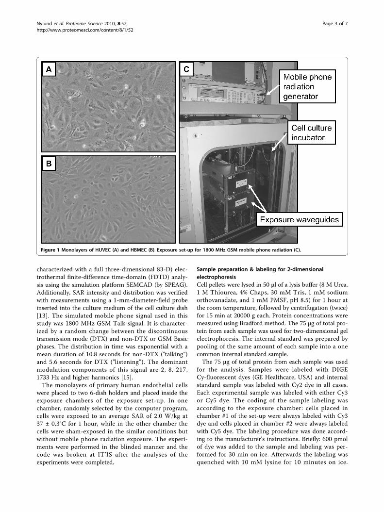
Materials and methodsCell culture and conditionsPrimary human umbilical vein endothelial cells (HUVEC)were purchased form Lonza, Switzerland and cultivatedaccording to manufacturer’s instructions. The purchasedHUVEC were a pool of cells from several donors. Formobile phone radiation experiments, cells were removedfrom culture flasks by brief trypsinization, washed in cellculture medium and seeded in the 35 mm-diameter“CellBIND” Petri dishes (Corning, USA). After overnightincubation the medium in the dishes was replaced with afresh one and the monolayers of HUVEC (Figure 1A)were exposed to the mobile phone radiation in a specialexposure chamber. The sham samples (unexposed con-trol) were produced simultaneously in an identical shamexposure chamber (see below description of the system).Immediately after the end of exposure the cells werequickly washed with warm (37°C) PBS and harvestedwith warm versene solution. In total, 13 independentsham and exposed samples were generated from HUVECin 13 different exposure experiments.Primary human brain microvascular endothelial cells
(HBMEC) were purchased from ScienCell ResearchLaboratories, USA and cultivated according to manufac-turer’s instructions. The purchased HBMEC were froma single donor and all cells used for the experiments
were from the same batch. Before experiments cellswere grown to confluency, detached with trypsin andseeded in the 35 mm-diameter “CellBIND” Petri dishes(Corning, USA) that were additionally coated with fibro-nectin (1.5%, overnight at 37°C) (Sigma, USA). Seventy-two hours after seeding the medium was replaced with afresh one and the monolayers of HBMEC (Figure 1B)were exposed to the mobile phone radiation using thesame exposure chamber as for HUVEC. Immediatelyafter the end of exposure the cells were rinsed withwarm PBS and harvested with trypsin. In total, 11 inde-pendent sham and exposed samples were generatedfrom HBMEC in 11 different exposure experiments.
Mobile phone radiation exposureThe sXc-1800 exposure system, developed and providedby the IT’IS Foundation (Zurich, Switzerland) was usedfor exposing cells to 1800 MHz GSM signal (Figure 1C).The detailed description of the system and the dosime-try of it have been presented elsewhere [13]. Briefly: Thesystem consists of two identical exposure chambersmounted inside the same cell culture incubator (NuAireUS Autoflow CO2 Water-Jacketed Incubator, NuAire,USA). One of the chambers acted as a sham control (noradiation) and the other as an experimental (with radia-tion). Sham exposure chamber and RF exposure cham-ber were randomly assigned by the computer programthat controlled exposures. This computer program hasgenerated during the experiment encrypted files withinformation in which of the two chambers was radiationand which acted as sham-control. These encrypted fileswere decoded after the experiment by chamber manu-facturer, IT’IS, Zurich, Switzerland. This set-up per-mitted blinded execution of experiments. The exposuresystem is fully automated and enables controlled expo-sures of cells (H-polarization or at H-field maximum ofthe standing wave [14]) at freely programmable ampli-tude modulations. Identical environmental conditionsexisted in both chambers (sham and experimental) sincethey were both located in the same cell culture incuba-tor and the inlets of the airflow through them are at thesame location. At 10 seconds intervals the system hasmonitored the incident field strengths, the proper func-tioning of the ventilators, the outlet air temperaturesand the state of all equipment. The Pt100 temperaturesensors (accuracy ± 0.1 °C) have been calibrated prior tothe installation and the recorded differences in tempera-ture are well within the specified long-term stability ofthe calibration. The induced temperature load, due toradiation absorption, has been characterized as a func-tion of SAR (t) for different signals and volumes of med-ium. This enables a reliable estimate of the maximumtemperature rise as a function of the exposure [13].SAR distribution within the cell culture dish was
Nylund et al. Proteome Science 2010, 8:52http://www.proteomesci.com/content/8/1/52
Page 2 of 7
characterized with a full three-dimensional 83-D) elec-trothermal finite-difference time-domain (FDTD) analy-sis using the simulation platform SEMCAD (by SPEAG).Additionally, SAR intensity and distribution was verifiedwith measurements using a 1-mm-diameter-field probeinserted into the culture medium of the cell culture dish[13]. The simulated mobile phone signal used in thisstudy was 1800 MHz GSM Talk-signal. It is character-ized by a random change between the discontinuoustransmission mode (DTX) and non-DTX or GSM Basicphases. The distribution in time was exponential with amean duration of 10.8 seconds for non-DTX ("talking”)and 5.6 seconds for DTX ("listening”). The dominantmodulation components of this signal are 2, 8, 217,1733 Hz and higher harmonics [15].The monolayers of primary human endothelial cells
were placed to two 6-dish holders and placed inside theexposure chambers of the exposure set-up. In onechamber, randomly selected by the computer program,cells were exposed to an average SAR of 2.0 W/kg at37 ± 0.3°C for 1 hour, while in the other chamber thecells were sham-exposed in the similar conditions butwithout mobile phone radiation exposure. The experi-ments were performed in the blinded manner and thecode was broken at IT’IS after the analyses of theexperiments were completed.
Sample preparation & labeling for 2-dimensionalelectrophoresisCell pellets were lysed in 50 μl of a lysis buffer (8 M Urea,1 M Thiourea, 4% Chaps, 30 mM Tris, 1 mM sodiumorthovanadate, and 1 mM PMSF, pH 8.5) for 1 hour atthe room temperature, followed by centrifugation (twice)for 15 min at 20000 g each. Protein concentrations weremeasured using Bradford method. The 75 μg of total pro-tein from each sample was used for two-dimensional gelelectrophoresis. The internal standard was prepared bypooling of the same amount of each sample into a onecommon internal standard sample.The 75 μg of total protein from each sample was used
for the analysis. Samples were labeled with DIGECy-fluorescent dyes (GE Healthcare, USA) and internalstandard sample was labeled with Cy2 dye in all cases.Each experimental sample was labeled with either Cy3or Cy5 dye. The coding of the sample labeling wasaccording to the exposure chamber: cells placed inchamber #1 of the set-up were always labeled with Cy3dye and cells placed in chamber #2 were always labeledwith Cy5 dye. The labeling procedure was done accord-ing to the manufacturer’s instructions. Briefly: 600 pmolof dye was added to the sample and labeling was per-formed for 30 min on ice. Afterwards the labeling wasquenched with 10 mM lysine for 10 minutes on ice.
Figure 1 Monolayers of HUVEC (A) and HBMEC (B). Exposure set-up for 1800 MHz GSM mobile phone radiation (C).
Nylund et al. Proteome Science 2010, 8:52http://www.proteomesci.com/content/8/1/52
Page 3 of 7
Samples deriving from the same exposure (Cy3 and Cy5labeled) were pooled together with Cy2 labeled internalstandard and separated all together in a single 2DE gel.
2-dimensional electrophoresisThe isoelectric focusing was performed using an IPGphor3apparatus (GE Healthcare) and 24 cm long IEF strips pH 4-7 (GE Healthcare). The samples were loaded using in-gelrehydration loading in a buffer containing 9 M Urea, 2%Chaps, 0.5% IPG buffer pH 4-7, and 65 mM DTT for 5 h.IEF was run with 50 μA/strip at 20°C using step-and-holdmethod as follows: 50 V 8 h; 100 V 1 h; 500 V 1 h; 1000 V1 h; 2000 V 1 h; 5000 V 1 h, 10000 V until 95000 Vhrswere achieved. After the end of IEF run the strips wereequilibrated for 15 min with 6 M urea, 30% glycerol, 50mM Tris-HCl, 2% SDS, and 10 mg/mL DTT for 15 minand then for another 15 min in the same buffer, in which25 mg/mL iodoacetamide (IAA) has replaced DTT. SDS-PAGE was run in 10% gel using Ettan DALTsix Electro-phoresis system (GE Healthcare) using 1 mm low fluores-cent glass plates with the constant settings of 10 mA/1W/gel for the first hour and then 12 mA/1.5W/gel overnightat 20°C. After the electrophoresis the gels were scannedbetween the glass plates with Typhoon Trio scanner (GEHealthcare) with the appropriate excitation and emissionwavelengths for Cy2, Cy3, and Cy5 dyes. The PMT voltageswere optimized in such manner that the maximum signalintensity was approximately on the same level for all dyes.
Data acquiring and analysisThe images were acquired with Typhoon Trio scanner(GE Healthcare). The datasets containing images fromCy3, and Cy5 labeled samples and Cy2 labeled internalstandard were cropped with ImageQuant tool-software(GE Healthcare). The datasets were cropped to containthe same pattern of proteins in all cases. The datasetswere then imported to DeCyder 6.5 software (GE Health-care), in which the batch processor was used to detectand to match the spots. The 10000 spots were assumedto be found in the spot detection, and the volume of30000 was used as a cut-off filter. After a brief manualvisual check of the matched spots the workspace wasimported to DeCyder Extended Data Analysis module(EDA) for statistical analysis. For EDA analysis proteinspots, which were found at least in 70% of gels, wereincluded. The student t-test was used to find differen-tially expressed protein spots. False discovery rate correc-tion (FDR) was applied when t-test was performed inEDA module. Also principal component analysis (PCA)was performed in EDA for the spot maps. The lists of thestatistically significantly affected spots were importedback to DeCyder Biological Variation Analysis module(BVA) in which the results were filtered on the basis ofthe average ratio between the samples.
ResultsTwo different types of primary human endothelial cellswere used in this study, the HUVEC and the HBMEC.The protein expression patterns of these cells were exam-ined using 2DE-based proteomics with DIGE-technique.In total, 13 separate replicates of sham and exposed pro-teomes were generated from HUVEC and 11 separatereplicates of sham and exposed proteomes from HBMEC.The same internal standard was used for all samplesallowing better technical quality and less variancebetween the gels. All gel images were analyzed togetherin DeCyder 6.5. In total, 2863 protein spots were detectedin the master gel. Protein spots which were detected in70% of spot maps were included in the EDA analysis(total of 1746 spots).The proteome analysis has shown differences in 2D pro-
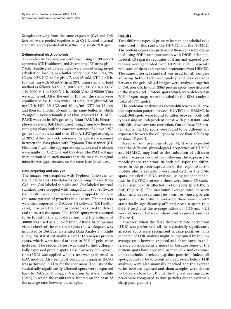
tein expression pattern between HUVEC and HBMEC. Intotal, 368 spots were found to differ between both celltypes using an independent t-test with p ≤ 0.0001 andwith false discovery rate correction. Out of these 368 pro-tein spots, the 145 spots were found to be differentiallyexpressed between the cell types by more than 2-folds upor down (Figure 2).Based on our previous study [4], it was expected
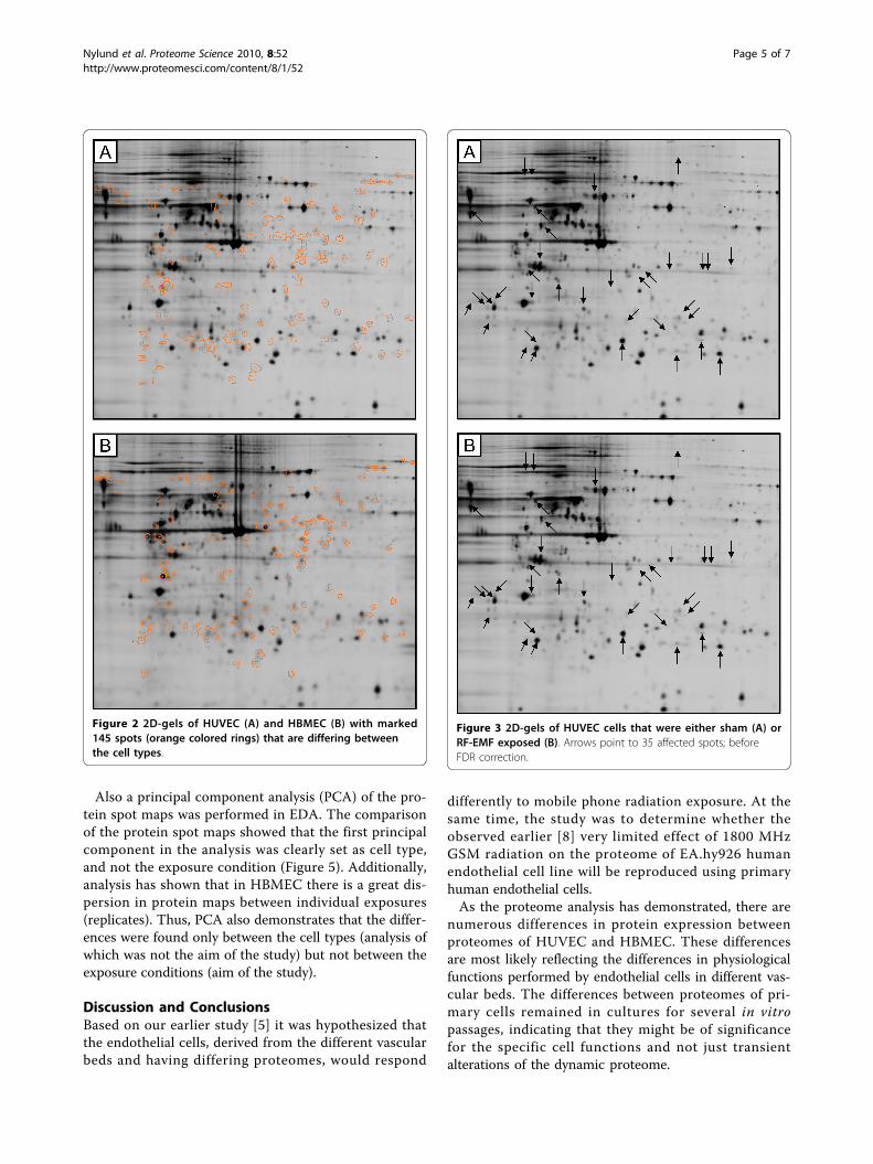
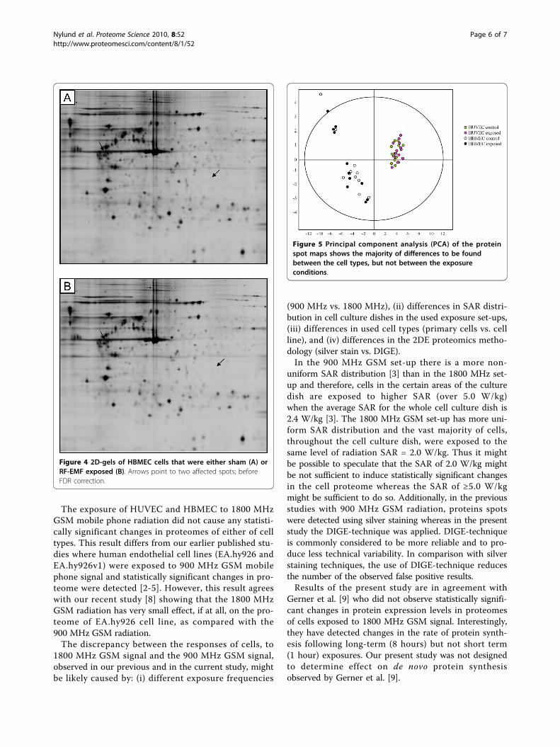
that the different physiological properties of HUVECand HBMEC, may lead to the induction of differentprotein expression profiles following the exposure tomobile phone radiation. In both cell types the differ-ences in the protein expression in the response to themobile phone radiation were analyzed for the 1746spots included in EDA analysis, using independent t-test. In HUVEC proteome there were found 35 statis-tically significantly affected protein spots (p ≤ 0.05; t-test) (Figure 3). The maximum average ratio, betweensham and exposed samples, was for these proteinspots = 1.33. In HBMEC proteome there were found 2statistically significantly affected protein spots (p ≤0.05; t-test) and the average ratios of -1.16 and +1.1were observed between sham and exposed samples(Figure 4).However, when the false discovery rate correction
(FDR) was performed, all the statistically significantlyaffected spots were recognized as false positives. Thisoutcome of FDR analysis might be explained by the lowaverage ratio between exposed and sham samples (dif-ference considered as a noise) or because some of theprotein spots have appeared in manual visual examina-tion as technical artefacts (e.g. dust particles). Indeed, allspots, found to be differentially expressed before FDRanalysis, were also manually checked and the averageratios between exposed and sham samples were shownto be very close to 1.0 and the highest average ratiopeaks were recognized as dust particles due to extremelysharp peak geometry.
Nylund et al. Proteome Science 2010, 8:52http://www.proteomesci.com/content/8/1/52
Page 4 of 7
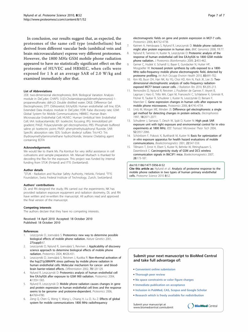
Also a principal component analysis (PCA) of the pro-tein spot maps was performed in EDA. The comparisonof the protein spot maps showed that the first principalcomponent in the analysis was clearly set as cell type,and not the exposure condition (Figure 5). Additionally,analysis has shown that in HBMEC there is a great dis-persion in protein maps between individual exposures(replicates). Thus, PCA also demonstrates that the differ-ences were found only between the cell types (analysis ofwhich was not the aim of the study) but not between theexposure conditions (aim of the study).
Discussion and ConclusionsBased on our earlier study [5] it was hypothesized thatthe endothelial cells, derived from the different vascularbeds and having differing proteomes, would respond
differently to mobile phone radiation exposure. At thesame time, the study was to determine whether theobserved earlier [8] very limited effect of 1800 MHzGSM radiation on the proteome of EA.hy926 humanendothelial cell line will be reproduced using primaryhuman endothelial cells.As the proteome analysis has demonstrated, there are
numerous differences in protein expression betweenproteomes of HUVEC and HBMEC. These differencesare most likely reflecting the differences in physiologicalfunctions performed by endothelial cells in different vas-cular beds. The differences between proteomes of pri-mary cells remained in cultures for several in vitropassages, indicating that they might be of significancefor the specific cell functions and not just transientalterations of the dynamic proteome.
Figure 2 2D-gels of HUVEC (A) and HBMEC (B) with marked145 spots (orange colored rings) that are differing betweenthe cell types.
Figure 3 2D-gels of HUVEC cells that were either sham (A) orRF-EMF exposed (B). Arrows point to 35 affected spots; beforeFDR correction.
Nylund et al. Proteome Science 2010, 8:52http://www.proteomesci.com/content/8/1/52
Page 5 of 7
The exposure of HUVEC and HBMEC to 1800 MHzGSM mobile phone radiation did not cause any statisti-cally significant changes in proteomes of either of celltypes. This result differs from our earlier published stu-dies where human endothelial cell lines (EA.hy926 andEA.hy926v1) were exposed to 900 MHz GSM mobilephone signal and statistically significant changes in pro-teome were detected [2-5]. However, this result agreeswith our recent study [8] showing that the 1800 MHzGSM radiation has very small effect, if at all, on the pro-teome of EA.hy926 cell line, as compared with the900 MHz GSM radiation.The discrepancy between the responses of cells, to
1800 MHz GSM signal and the 900 MHz GSM signal,observed in our previous and in the current study, mightbe likely caused by: (i) different exposure frequencies
(900 MHz vs. 1800 MHz), (ii) differences in SAR distri-bution in cell culture dishes in the used exposure set-ups,(iii) differences in used cell types (primary cells vs. cellline), and (iv) differences in the 2DE proteomics metho-dology (silver stain vs. DIGE).In the 900 MHz GSM set-up there is a more non-
uniform SAR distribution [3] than in the 1800 MHz set-up and therefore, cells in the certain areas of the culturedish are exposed to higher SAR (over 5.0 W/kg)when the average SAR for the whole cell culture dish is2.4 W/kg [3]. The 1800 MHz GSM set-up has more uni-form SAR distribution and the vast majority of cells,throughout the cell culture dish, were exposed to thesame level of radiation SAR = 2.0 W/kg. Thus it mightbe possible to speculate that the SAR of 2.0 W/kg mightbe not sufficient to induce statistically significant changesin the cell proteome whereas the SAR of ≥5.0 W/kgmight be sufficient to do so. Additionally, in the previousstudies with 900 MHz GSM radiation, proteins spotswere detected using silver staining whereas in the presentstudy the DIGE-technique was applied. DIGE-techniqueis commonly considered to be more reliable and to pro-duce less technical variability. In comparison with silverstaining techniques, the use of DIGE-technique reducesthe number of the observed false positive results.Results of the present study are in agreement with
Gerner et al. [9] who did not observe statistically signifi-cant changes in protein expression levels in proteomesof cells exposed to 1800 MHz GSM signal. Interestingly,they have detected changes in the rate of protein synth-esis following long-term (8 hours) but not short term(1 hour) exposures. Our present study was not designedto determine effect on de novo protein synthesisobserved by Gerner et al. [9].
Figure 4 2D-gels of HBMEC cells that were either sham (A) orRF-EMF exposed (B). Arrows point to two affected spots; beforeFDR correction.
Figure 5 Principal component analysis (PCA) of the proteinspot maps shows the majority of differences to be foundbetween the cell types, but not between the exposureconditions.
Nylund et al. Proteome Science 2010, 8:52http://www.proteomesci.com/content/8/1/52
Page 6 of 7
In conclusion, our results suggest that, as expected, theproteomes of the same cell type (endothelium) butderived from different vascular beds (umbilical vein andbrain microvasculature) express very different proteomes.However, the 1800 MHz GSM mobile phone radiationappeared to have no statistically significant effect on theproteome of HUVEC and HBMEC, when cells wereexposed for 1 h at an average SAR of 2.0 W/kg andexamined immediately after that.
List of Abbreviations2DE: two-dimensional electrophoresis; BVA: Biological Variation Analysismodule in DeCyder; CHAPS: 3-[(3-Cholamidopropyl)dimethylammonio]-1-propanesulfonate; ddH2O: Double distilled water; DIGE: Difference GelElectrophoresis; DTT: Dithioreitol; EA.hy926: Human endothelial cell line; EDA:Extended Data Analysis module in DeCyder; FDR: False discovery rate; GSM:Global System for Mobile Communications; HBMEC: Human BrainMicrovascular Endothelial Cell; HUVEC: Human Umbilical Vein EndothelialCell; IAA: Iodoacetamide; IEF: Isoelectric focusing; IPG: Immobilized pHgradient; PAGE: Polyacrylamide gel electrophoresis; PBS: Phosphate bufferedsaline; pI: Isoelectric point; PMSF: phenylmethylsulphonyl fluoride; SAR:Specific absorption rate; SDS: Sodium dodecyl sulfate; Tris-HCl: Tris(hydroxymethyl)aminomethane hydrochloride, Versene Chelating agentcontaining EDTA
AcknowledgementsWe would like to thank Ms. Pia Kontturi for very skilful assistance in cellcultivations and sample preparation. Mr. Manuel Murbach is thanked fordecoding the files for the exposure. This project was funded by internalfunding from STUK (Finland) and IT’IS (Switzerland).
Author details1STUK - Radiation and Nuclear Safety Authority, Helsinki, Finland. 2IT’ISFoundation, Swiss Federal Institute of Technology, Zurich, Switzerland.
Authors’ contributionsDL and RN designed the study; RN carried out the experiments; NK hasprovided radiation exposure equipment and radiation dosimetry, DL and RNhave written and re-written the manuscript. All authors read and approvedthe final version of the manuscript.
Competing interestsThe authors declare that they have no competing interests.
Received: 14 April 2010 Accepted: 18 October 2010Published: 18 October 2010
References1. Leszczynski D, Joenväärä S: Proteomics: new way to determine possible
biological effects of mobile phone radiation. Nature Genetics 2001,27(suppl):67.
2. Leszczynski D, Nylund R, Joenväärä S, Reivinen J: Applicability of discoveryscience approach to determine biological effects of mobile phoneradiation. Proteomics 2004, 4:426-431.
3. Leszczynski D, Joenväärä S, Reivinen J, Kuokka R: Non-thermal activation ofthe hsp27/p38MAPK stress pathway by mobile phone radiation inhuman endothelial cells: Molecular mechanism for cancer- and blood-brain barrier-related effects. Differentiation 2002, 70:120-129.
4. Nylund R, Leszczynski D: Proteomics analysis of human endothelial cellline EA.hy926 after exposure to GSM 900 radiation. Proteomics 2004,4:1359-1365.
5. Nylund R, Leszczynski D: Mobile phone radiation causes changes in geneand protein expression in human endothelial cell lines and the responseseems to be genome- and proteome-dependent. Proteomics 2006,6:4769-4780.
6. Zeng Q, Chen G, Weng Y, Wang L, Chiang H, Lu D, Xu Z: Effects of globalsystem for mobile communications 1800 MHz radiofrequency
electromagnetic fields on gene and protein expression in MCF-7 cells.Proteomics 2006, 6:4732-4738.
7. Karinen A, Heinävaara S, Nylund R, Leszczynski D: Mobile phone radiationmight alter protein expression in human skin. BMC Genomics 2008, 11:77.
8. Nylund R, Tammio H, Kuster N, Leszczynski D: Proteomic analysis of theresponse of human endothelial cell line EA.hy926 to 1800 GSM mobilephone radiation. J Proteomics Bioinformatics 2009, 2:455-462.
9. Gerner C, Hudek V, Schandl U, Bayer E, Gundacker N, Hutter HP,Mosgoeller W: Increased protein synthesis by cells exposed to a 1800-MHz radio-frequency mobile phone electromagnetic field, detected byproteome profiling. Int Arch Occupp Environ Health 2010, 83:691-702.
10. Kim KB, Byun OH, Han NK, Ko YG, Choi HD, Kim N, Pack JK, Lee JS: Two-dimensional electrophoretic analysis of radio frequency radiation-exposed MCF7 breast cancer cells. J Radiation Res 2010, 51:205-213.
11. Remondini D, Nylund R, Reivinen J, Poulletier de Gannes F, Veyret B,Lagroye I, Haro E, Trillo MA, Capri M, Franceschi C, Schlatterer K, Gminski R,Fitzner R, Tauber R, Schuderer J, Kuster N, Leszczynski D, Bersani F,Maercker C: Gene expression changes in human cells after exposure tomobile phone microwaves. Proteomics 2006, 6:4745-4754.
12. Unlü M, Morgan ME, Minden JS: Difference gel electrophoresis: a singlegel method for detecting changes in protein extracts. Electrophoresis1997, 18:2071-2077.
13. Schuderer J, Samaras T, Oesch W, Spät D, Kuster N: High peak SARexposure unit with tight exposure and environmental control for in vitroexperiments at 1800 MHz. IEEE Transact Microwave Theor Tech 2004,52:2057-2066.
14. Schönborn F, Pokovic K, Burkhardt M, Kuster N: Basis for optimization ofin vitro exposure apparatus for health hazard evaluations of mobilecommunications. Bioelectromagnetics 2001, 22:547-559.
15. Tillmann T, Ernst H, Ebert S, Kuster N, Behnke W, Rittinghausen S,Dasenbrock C: Carcinogenicity study of GSM and DCS wirelesscommunication signals in B6C3F1 mice. Bioelectromagnetics 2007,28:173-187.
doi:10.1186/1477-5956-8-52Cite this article as: Nylund et al.: Analysis of proteome response to themobile phone radiation in two types of human primary endothelialcells. Proteome Science 2010 8:52.
Submit your next manuscript to BioMed Centraland take full advantage of:
• Convenient online submission
• Thorough peer review
• No space constraints or color figure charges
• Immediate publication on acceptance
• Inclusion in PubMed, CAS, Scopus and Google Scholar
• Research which is freely available for redistribution
Submit your manuscript at www.biomedcentral.com/submit
Nylund et al. Proteome Science 2010, 8:52http://www.proteomesci.com/content/8/1/52
Page 7 of 7