Embed Size (px)
Citation preview

OFFICE OF ENVIRONMENTAL HEALTH HAZARD ASSESSMENT
Public Health Goal
Antimony in Drinking Water
September 2016
Pesticide and Environmental Toxicology Branch Office of Environmental Health Hazard Assessment California Environmental Protection Agency

Public Health Goal for Antimony
in Drinking Water
Prepared by
Pesticide and Environmental Toxicology Branch Office of Environmental Health Hazard Assessment
California Environmental Protection Agency
September 2016

Public Health Goal for ii September 2016 Antimony in Drinking Water
LIST OF CONTRIBUTORS
Public Health Goals for Chemicals in California Drinking Water Pesticide and Environmental Toxicology Branch
Office of Environmental Health Hazard Assessment California Environmental Protection Agency
Author Dan Qiao, M.D., Ph.D.
Reviewers
Lori Lim, Ph.D.
Meng Sun, Ph.D.
Rose Cendak, M.S.
Rajpal Tomar, Ph.D.
Final Reviewers
Elaine M. Khan, Ph.D.
Melanie Marty, Ph.D.
David Siegel, Ph.D.
David Ting, Ph.D.
Acting Director Lauren Zeise, Ph.D.

Public Health Goal for iii September 2016 Antimony in Drinking Water
PREFACE
This Public Health Goal (PHG) technical support document provides information on health effects from contaminants in California drinking water. PHGs are developed for chemical contaminants based on the best available data in the scientific literature and using the most current principles, practices, and methods used by public health professionals. These documents and the analyses contained therein provide estimates of the levels of contaminants in drinking water that would pose no significant health risk to individuals consuming the water on a daily basis over a lifetime.
Under the California Safe Drinking Water Act of 1996 (Health and Safety Code section 116365), the Office of Environmental Health Hazard Assessment (OEHHA) develops PHGs for drinking water contaminants in California based exclusively on public health considerations. OEHHA periodically reviews PHGs and revises them as necessary based on the availability of new scientific data. This document presents an update for antimony for which a PHG was published in 1997. PHGs published by OEHHA are for use by the State Water Resources Control Board (SWRCB) in establishing primary drinking water standards (California Maximum Contaminant Levels, or MCLs). Whereas PHGs are based solely on scientific and public health considerations without regard to economic cost considerations, drinking water standards adopted by SWRCB consider economic factors and technological feasibility. Each standard adopted shall be set at a level that is as close as feasible to the corresponding PHG, placing emphasis on the protection of public health. PHGs established by OEHHA are not regulatory and represent only non-mandatory goals. Under federal law, MCLs established by SWRCB must be at least as stringent as the corresponding federal MCL if one exists.
In July 2014, responsibility for the state’s drinking water regulatory program was transferred to SWRCB from the California Department of Public Health. References in this document to drinking water monitoring and regulation may cite either or both entities as appropriate.

Public Health Goal for iv September 2016 Antimony in Drinking Water
TABLE OF CONTENTS
PREFACE-------------------------------------------------------------------------------------------- iii
SUMMARY ------------------------------------------------------------------------------------------- 1
INTRODUCTION ------------------------------------------------------------------------------------ 1
BASIS FOR THE 1997 PHG --------------------------------------------------------------------- 4
UPDATED TOXICOLOGICAL REVIEW ------------------------------------------------------- 6
Pharmacokinetics ------------------------------------------------------------------------------------------------------------ 6
Non-Carcinogenic Toxicity --------------------------------------------------------------------------------------------- 8
Carcinogenicity -------------------------------------------------------------------------------------------------------------- 13
Genotoxicity ------------------------------------------------------------------------------------------------------------------- 14
Mode of Action --------------------------------------------------------------------------------------------------------------- 16
PHG DERIVATION ------------------------------------------------------------------------------- 18
RISK CHARACTERIZATION ------------------------------------------------------------------ 23
REFERENCES ------------------------------------------------------------------------------------ 25
APPENDIX I: HISTOPATHOLOGICAL GRADING --------------------------------------- 35
APPENDIX II: BMD MODELING -------------------------------------------------------------- 37
APPENDIX III: NTP (1992) STUDY ---------------------------------------------------------- 40

Public Health Goal for 1 September 2016 Antimony in Drinking Water
SUMMARY
This document presents an update of the public health goal (PHG) for antimony, as total antimony ion. The previous PHG (OEHHA, 1997) of 20 micrograms per liter (µg/L) or 20 parts per billion (ppb) was based on decreased longevity observed in rats in a chronic toxicity study (Schroeder et al., 1970). The updated PHG of 1 ppb is based on liver histopathological changes, and supported by the blood glucose levels and the tissue retention of antimony in rats in a 90-day oral toxicity study (Poon et al., 1998). Updated dose-response assessment and drinking water ingestion rates are incorporated into the derivation of this updated PHG.
INTRODUCTION
The Office of Environmental Health Hazard Assessment (OEHHA) performs health risk assessments and develops public health goals (PHGs) for drinking water contaminants in California. A PHG is the concentration of a contaminant in drinking water that is estimated to pose no significant health risk to individuals consuming the water on a daily basis over a lifetime. This document presents a PHG update for antimony. This update incorporates a thorough review of the current scientific literature and the most current risk assessment practices and methods, as well as relevant chemical-specific toxicity data.
Chemical Identity and Properties
Antimony (Sb) is an element present in relatively small amounts in the earth’s crust. It is a metalloid and has four oxidation states (Sb(-3), (0), (+3), (+5)), and two stable isotopes of atomic weights 121 (57 percent) and 123 (43 percent). Antimony compounds are soluble in very strong acid and basic solutions. Under neutral conditions, the predominant species is Sb(OH)6- for pentavalent forms and Sb(OH)3 for trivalent forms.
Some notable antimony compounds are diantimony trioxide (Sb2O3, also called antimony trioxide), which is slightly soluble in water (2.76 mg/L), and antimony potassium tartrate (Sb2K2(C4H2O6)2 3H2O, also called APT), which is very soluble in water (83 g/L).
Primary Uses
Antimony has been used since antiquity as a medicine, to induce emesis and to treat other conditions. It has also been used in cosmetics. It is rarely found in pure form in nature. Although not used in large quantities, antimony is used extensively for many purposes, including being alloyed with a number of metals to improve their properties. Antimony alloyed with lead is used in batteries to increase hardness. A significant use of antimony is for the production of antimony trioxide as a fire retardant (ATSDR, 1992; Butterman and Carlin, 2004). Antimony is also used as a catalyst in the manufacture of plastics, including

Public Health Goal for 2 September 2016 Antimony in Drinking Water
polyethylene terephthalate (PET), a polyester of terephthalic acid and ethylene glycol, which is used in bottled water containers.
Pentavalent antimonials are used to treat leishmaniasis, although the parasites appear to have developed resistance to the antimonials (Croft et al., 2006). Antiparasitic pentavalent antimonials, such as sodium stibogluconate (Pentostam®) or meglumine antimoniate (Glucantim®), are the mainstays of therapy. Until recently, sodium stibogluconate was the only recommended treatment in the United States and was available only through the Centers for Disease Control and Prevention (CDC, 2013). The non-antimonial drug amphotericin B in its liposomal form (as opposed to amphotericin B deoxycholate) has recently been approved and is now considered to be the drug of choice for visceral leishmaniasis because of its shorter course and lower toxicity. However, cost issues prevent the use of liposomal drugs in most countries, where the mainstay of treatment is still prolonged intravenous treatment with antimonials, despite ever-increasing patterns of resistance and treatment failures.
APT was once used in the treatment of schistosomiasis (Cleve, et al. 1955) but, due to some severe side effects, has been replaced by non-antimonial drugs like praziquantel (WHO, 2006).
Environmental Releases
Releases of antimony and its compounds to the environment occur from natural discharges such as windblown dust, volcanic eruption, sea spray, forest fires and other natural processes. Anthropogenic sources include mining and processing of ores and the production of antimony metal, alloys, antimony oxide, and compounds containing antimony, and recycling and incineration of antimony containing products. According to the U.S. Environmental Protection Agency’s (US EPA) Toxics Release Inventory, an estimated 7,621,131 pounds of antimony and antimony compounds were released to the environment in 2012.1
Studies of antimony in ice cores in Canada and in lake sediments in Sweden showed that antimony levels have increased significantly since the industrial revolution, presumably from dust deposition (Krachler et al., 2005; Grahn et al., 2006). However, Reimann et al. (2010) pointed out that the wide variability in antimony at different locations makes it difficult to distinguish among the various natural and anthropogenic sources.
Soil methylation of antimony by microbes has been reported, with the resultant volatile antimony products being made airborne (Bentley and Chasteen, 2002). This appears to be a very minor process compared to the amount of methylation
1 Accessed at: http://iaspub.epa.gov/triexplorer/tri_release.chemical

Public Health Goal for 3 September 2016 Antimony in Drinking Water
that occurs with arsenic. Stibine (SbH3), a very toxic gaseous compound of antimony, can also be released by the action of microorganisms.
Occurrence in Drinking Water
Very low concentrations of antimony are found in pristine ambient waters. Antimony levels were above the detection limit in six wells among the 7,174 public water supply wells tested in California during the three-year period of 2012, 2013, and 2014, with the highest level at 28 ppb.2 The detection limit for purposes of reporting is 6 ppb,3 the same as California’s maximum contaminant level (MCL) for antimony.
Antimony use in plumbing materials and fittings is thought to be a significant source of antimony content in tap water (WHO, 2003). Antimony was found to leach from copper pipes with solder joints made from 95% tin and 5% antimony in soft water (Subramanian, 1991). In contrast to high-purity water (pH of 6.8) and well water (pH 8.1; hardness 155 mg/L as CaCO3), significant leaching of antimony occurred into tap water (pH 7.8, hardness 30 mg/L as CaCO3), but only after 7 days of contact time. At the end of 7, 28 and 90 days of contact time, the amount of antimony leached into tap water resulted in final concentrations of 2.0, 3.7 and 7.3 µg/L, respectively.
Filella and co-workers (Filella et al., 2002a,b) reviewed reports of antimony levels and its different oxidation states, or speciation, in ambient waters. Antimony can be found in the dissolved phase regardless of its oxidation state and in the absence of sulfur (Filella et al., 2002b). According to thermodynamics, antimony should be present as Sb(V) (the pentavalent form) in oxic systems, and as Sb(III) (the trivalent form) in anoxic systems. However, Sb(III) has been detected in oxic waters, which leads to the question of how Sb(III) is stabilized in oxic systems. Filella and colleagues suggested that there is likely a role for organic matter, particularly organic acids, in stabilizing Sb(III). Both Sb(III) and Sb(V) ions hydrolyze easily; Sb(III) is present as the neutral species Sb(OH)3, and Sb(V) as the anion Sb(OH)6- (also referred to as SbO3-) (Filella et al., 2002a). Both forms exhibit low reactivity in ocean water. There is limited information on the speciation and transformation of Sb in natural waters. The ratio of Sb(V) to Sb(III) in a water body depends on the oxidative potential of the water. For some rivers in the United States, the ratio can range from less than 10 to over 100 (Filella et al., 2002a).
2 Data accessed with GeoTracker GAMA: http://geotracker.waterboards.ca.gov/gama/. The data for public water supply wells accessed with GeoTracker GAMA do not indicate whether the source is raw (untreated) water or treated water; therefore, the results in the dataset may not be representative of the water delivered to customers. 3 Accessed at: http://www.waterboards.ca.gov/drinking_water/certlic/drinkingwater/MCLsandPHGs.shtml

Public Health Goal for 4 September 2016 Antimony in Drinking Water
The European Union’s risk assessment report for diantimony trioxide (EU, 2008) indicated that although diantimony trioxide has low solubility in water (2.76 mg Sb/L), antimony ions will nonetheless be liberated from dissolved diantimony trioxide over time, albeit at a slower rate than the more soluble antimony compounds such as antimony chlorides. The report notes that the toxicity of antimony is expected to be exerted through its ions and, “In the environment, diantimony trioxide will dissolve to the trivalent and predominantly to the pentavalent forms of antimony. …As a consequence, diantimony trioxide, originating from production and use, will be present as antimony in drinking water, food and breast milk.” In terms of the antimony species in water, the report states that, “In natural waters antimony exists almost exclusively in the dissolved phase in the two valency states +3 and +5,” and “antimony should almost exclusively be present as Sb(V) in oxic systems, and as Sb(III) in anoxic systems.” The report also emphasizes the limited knowledge on antimony species due to the lack of analytical techniques and the “difficult chemistry” of antimony.
Ninety percent of the worldwide manufacture of polyethylene terephthalate (PET), a polyester of terephthalic acid and ethylene glycol, employs antimony trioxide as a catalyst (Shotyk, 2006). Antimony levels in pristine Canadian ground water, averaging 0.002 ppb Sb, were compared against 12 brands of Canadian water bottled in PET. The bottled waters averaged 0.2 ppb antimony. Levels were higher in imported German bottled water. A single lot of German bottled water, originally 0.36 ppb antimony, rose to 0.63 ppb after three months of storage at room temperature (Shotyk, 2006). Westerhoff et al. (2008) showed that Sb leaching from PET containers was time and temperature dependent. Nine commercial bottled waters collected in Arizona ranged from 0.1 to 0.5 ppb (mean 0.2 ppb) at the start of their study, and after 3 months of storage at 22 oC the mean Sb levels had increased to only 0.23 ppb. However, storage of the water in PET bottles at 60 to 85 oC resulted in an increasing rate of Sb release into the water (Westerhoff et al., 2008). At 60 oC, the federal antimony MCL of 6 ppb was achieved in the water stored in plastic bottles in 187 days, while at 85 oC, only 1.3 days were required to reach this level. These authors noted that Arizona summer temperatures inside cars, garages, and enclosed storage areas can exceed 65 oC.
BASIS FOR THE 1997 PHG
Critical Study and Endpoint
In 1997, OEHHA published a PHG of 20 ppb for antimony in drinking water. The PHG was based on a chronic drinking water study in rats by Schroeder et al. (1970). Fifty male and 50 female Long-Evans rats were exposed to 0 or 5 mg/L (5 ppm) of antimony in the form of APT, the trivalent form, in drinking water from weaning until death (more than 1,000 days). An average daily dose of 0.43 mg per kilogram of body weight per day (mg/kg-day) antimony was estimated by US

Public Health Goal for 5 September 2016 Antimony in Drinking Water
EPA (1992b) based on a mean body weight of 0.35 kg and a water consumption rate of 30 milliliters (mL)/day. There was no significant effect on body weight gain, but there was a significant decrease (by chi-square analysis) in longevity. In males, mean longevity was 1,160 ± 27.8 (standard error) days for control animals, compared to 999 ± 7.8 days for the treated group. In females it was 1,304 ± 36 days for controls, compared to 1,092 ± 30 days for the treated group. Serum cholesterol levels were increased in the males but decreased in females. Fasting glucose levels were not significantly affected by treatment in either males or females but non-fasting glucose levels were lower in exposed males and females.
Schroeder et al. (1970) also observed deposition of antimony in the kidney, liver, heart, lung, and spleen, which increased with age. No increase in tumor incidence was noted. An epidemic of viral pneumonia during the experiment was reported for this colony of rats, but the investigators noted that enough of the animals survived such that the results were valid. Based on the observed decrease in longevity and altered blood glucose and serum cholesterol levels, a lowest-observed-adverse-effect level (LOAEL) of 0.43 mg/kg-day was identified.
Derivation of the 1997 PHG
The exposure evaluation assumed a 70 kg adult body weight, water ingestion rate of 2 liters per day, and a relative source contribution (RSC) of 40%. There are small amounts of antimony in food. The average concentration of antimony in meat, vegetables, and seafood has been estimated to range from 0.2 to 1.1 ppb (ATSDR, 1992). For the purpose of PHG development, it was estimated that 40% of the total daily intake of antimony could come from drinking water. The remaining 60% could come from ingestion of food, inhalation of dust, and contact with soil. An uncertainty factor of 300 (10 for intraspecies variability, 10 for interspecies extrapolation, and 3 for LOAEL to no-observed-adverse-effect level (NOAEL) extrapolation) was applied to the LOAEL of 0.43 mg/kg-day and a PHG of 20 ppb was developed. There was inadequate evidence of antimony-induced carcinogenicity through oral ingestion, thus the PHG was based on non-carcinogenic effects (decrease in longevity and altered blood glucose and serum cholesterol levels). Updated Literature Review
Since the publication of the antimony PHG in 1997, two antimony studies and three reports from government agencies have been published that are relevant to the development of the PHG. These publications are:
• Hext PM, Pinto PJ, Rimmel BA (1999). Subchronic feeding study of antimony trioxide in rats.

Public Health Goal for 6 September 2016 Antimony in Drinking Water
• Poon R, Chu I, Lecavalier P, Valli VE, Foster W, Gupta S, Thomas B (1998). Effects of antimony on rats following 90-day exposure via drinking water.
• EU (2008). European Union Risk Assessment Report: Diantimony Trioxide.
• IPCS (1998). Antimony trioxide and antimony trisulfide. (last updated 1/21/98) International Agency for Research on Cancer – Summaries and Evaluations.
• WHO (2003). Antimony in drinking water: Background document for development of WHO guidelines for drinking-water quality.
UPDATED TOXICOLOGICAL REVIEW
Pharmacokinetics
Antimony absorption from the gastrointestinal system is relatively low. A number of factors are likely to affect the absorption of antimony, including chemical form, particle size and solubility, species, age, and diet. The nearly insoluble diantimony trioxide form is proposed to have an oral absorption of 1% in humans (EU, 2008). Fifteen to 20% of 124SbCl3 was absorbed by cows (Van Bruwaene, 1982). Approximately 15% of APT was orally absorbed in rats (Moskalev, 1959). In a repeated oral dosing protocol with SbCl3, BALB/c mice were estimated to absorb approximately 7% (Gerber, 1982). It is worth noting that the pharmacokinetics database is quite limited, and the relative absorption of the forms of antimony found in water is not well known. Antimony was found to be excreted mainly through the bile in cows. Cows administered single oral doses of 21.1 mg of antimony (as 124SbCl3) excreted 82% of the dose in feces and 1% of the dose in urine. The antimony excreted in milk was less than 0.01% of the dose (Bruwaene et al., 1982).
Few studies were located regarding dermal penetration of antimony compounds. Antimony or its compounds are generally not sufficiently water or lipid soluble to make this a significant route of exposure. However, contact with antimony trioxide dust has been reported to produce a condition called “antimony spots,” which are dermal papules and pustules around the sweat and sebaceous glands (IPCS, 1998). Roper and Stupart (2006) used six samples (1 abdominal and 5 breast samples) obtained from women to study dermal penetration of a ‘low’ dose and a ‘high’ dose of diantimony trioxide, 100 μg/cm2 and 300 μg/cm2, respectively. The total dermal absorption was estimated to be 0.26% and 0.14%, respectively, for the two doses following 24 hours of exposure.
Animal studies showed that antimony (Sb(III) or Sb(V)) is bound to red blood cells after absorption (Molokhia and Smith, 1969; Felicetti et al., 1974; Gerber et al., 1982; Dieter et al., 1991). It is then transported to the spleen, liver, and bone (Casals, 1972) and to a lesser extent into skin and hair (Felicetti et al., 1974; Berman et al., 1988). The major metabolic pathway of antimony in humans and

Public Health Goal for 7 September 2016 Antimony in Drinking Water
rats is the oxidation of Sb(III) to Sb(V) (Ogra, 2009; Kobayashi and Ogra, 2009). On the other hand, some studies have indicated that when pentavalent antimonials are administered, Sb(V) may be reduced to Sb(III) both in vitro and in vivo (Frezard et al., 2009; Ferreira et al., 2003; Frezard et al., 2001; Petit de Pena et al., 1990).
Chulay et al. (1988) examined the pharmacokinetics of antimony in 5 patients being treated for leishmaniasis (10 mg Sb/kg-day as sodium stibogluconate (2 patients) or meglumine antimoniate (3 patients) by intramuscular (i.m.) injection for 30 days). The authors found the data were best described by a “two compartment, three term model.” The first compartment includes the blood or plasma volume into which the drug is absorbed after injection, and the second compartment may represent a peripheral compartment into which the drug is distributed or may be related to in vivo conversion of Sb(V) to Sb(III). The three terms were said to represent an initial absorption phase with a mean half-life of 0.85 hr, a rapid elimination phase with a mean half-life of 2.02 hrs, and a slow elimination phase with a mean half-life of 76 hrs. Notably, there was a 4-fold increase in nadir blood antimony levels during treatment, from an average of 0.6% of peak concentrations after the first dose to an average of 2.5% of peak concentrations after the 30th dose, indicating a gradual accumulation of the drug over the course of treatment (Chulay et al., 1988). Similarly, Miekeley et al. (2002) monitored total antimony in biological samples (whole blood, plasma, urine, and hair) from patients treated with meglumine antimoniate (5 mg Sb/kg-day by i.m. injection) for 30 days and reported a half-life of 24-72 hrs for the rapid excretion phase and a half-life of >50 days for the slow elimination phase.
Friedrich et al. (2012) examined the disposition of antimony in rhesus monkeys treated with meglumine antimoniate at 5 or 20 mg Sb(V)/kg-day by i.m. injection for 21 days. As in human whole blood, nadir plasma antimony levels in rhesus monkeys rose steadily from 19.6 ± 4 and 65.1 ± 17.4 ng/g (24 hrs after first injection), up to 27.4 ± 5.8 and 95.7 ± 6.6 ng/g (24 hr after 21st injection) in the 5 and 20 mg/kg-day dose groups, respectively. There was a gradual decline in plasma antimony levels, with a terminal elimination phase half-life of 35.8 days. The proportion of Sb(III) rose from 5% on posttreatment day 1 to 50% on posttreatment day 9. Liver concentrations of antimony (posttreatment days 55 and 95) were >1,000 ng/g wet weight.
Antimony levels in select tissues from rats treated with 300 mg Sb(V)/kg-day as meglumine antimoniate subcutaneously for 21 days include:
• Spleen: 148 ± 14.0 µ/g at posttreatment day 1 and 81.9 ± 4.6 µ/g at posttreatment day 21.
• Liver: 13.8 ± 1.3 µg/g at posttreatment day 1 and 3.2 ± 0.2 µg/g at posttreatment day 21.
• The levels in the spleen were considerably higher than the levels in the bone, thyroid and kidneys, which in turn were greater than the levels in the liver.

Public Health Goal for 8 September 2016 Antimony in Drinking Water
The authors noted that in rats, as in humans, the antimony blood levels after meglumine antimoniate treatment can be described by a two-compartment model with a fast (t1/2 = 0.6 h) and a slow (t1/2 = >>24 h) elimination phase (Coelho et al., 2014).
In a comparison of children versus adults treated with meglumine antimoniate (20 mg Sb/kg-day i.m. injection for 20 days), children had a 42% lower area under the 24-hr time-concentration curve (AUC0-24), a 16% lower peak concentration, and a 75% higher weight-adjusted clearance than adults. The authors concluded that antimony exposure is significantly lower in children than in adults given the same weight-adjusted dose, primarily due to a higher antimony clearance rate (Cruz et al., 2007).
Lyon et al. (2002) investigated the age dependence of the liver content of selected elements, including antimony, in children as an index of internal exposure. In autopsy liver samples collected from 157 subjects ages <1 to 6 years, they found that antimony levels did not vary much between four age categories: fetal, neonate, infant, young child. Median levels were 0.9, 0.8, 2.0, 2.3 ng/g wet weight, respectively, although medians for fetuses and neonates were slightly lower than for infants and young children.
Non-Carcinogenic Toxicity
This section reviews the toxicology data on antimony, focusing on the studies published since the 1997 PHG. The reader is referred to the 1997 antimony PHG for a detailed review of the studies published before 1997.
As discussed in the 1997 PHG, antimony is an emetic and humans who ingest antimony can have severe stomach upset, resulting in vomiting. Sundar and Chakravarty (2010) report that acute exposures to antimony trioxide as low as 0.5 mg/kg have resulted in vomiting. Exposure to antimony fumes and dusts by workers can cause dermatitis and, less commonly, effects on the heart and kidneys (OEHHA 1997, Sundar and Chakravarty, 2010).
A critical concern in the use of antimonials for treatment of human parasite infections has been heart and liver toxicity (Khayyal et al. 1967, 1973). Cerebellar ataxia has been observed with long-term treatment with sodium stibogluconate, a pentavalent antimonial used for visceral leishmaniasis. Two Sudanese patients who were treated with Pentostam® (sodium stibogluconate) exhibited signs of this condition, which subsided when the drug was withdrawn (Khalil et al., 2006).
While a number of studies report on side-effects of antimonial parasite treatments, the available human data are not suitable for deriving a PHG because of the exposure routes, durations, limited numbers of subjects, or uncertainties in exposure estimates. Therefore the updated PHG is based on animal data.

Public Health Goal for 9 September 2016 Antimony in Drinking Water
In the Schroeder et al. (1970) drinking water study, the basis for the 1997 PHG, 0.43 mg/kg-day of antimony (as APT) reduced the longevity of male and female rats. While deposition of antimony in liver, kidney, heart and other tissues was noted, there was limited reporting of organ toxicity, although a decrease in heart weight was noted for males. Four subsequent toxicity studies provide additional data on the toxicity of antimony compounds (Coelho et al., 2014; Miranda et al., 2006; Hext et al., 1999; Poon et al., 1998). In addition, the toxicity information reported by the National Toxicology Program (NTP, 1992) is critically evaluated and compared with those reported by Schroeder et al. (1970) and Poon et al. (1998). Coelho et al. (2014) Developmental and Reproductive Toxicity Study
Coelho et al. (2014) exposed pregnant Wistar rats to meglumine antimoniate (Glucantime®) via subcutaneous injections at doses of 0, 75, 150, and 300 mg Sb(V)/kg-day for 42 consecutive days, starting from gestation day (GD) 0 through lactation, to post-natal day (PND) 21. At the highest dose, there was a nearly 30% reduction in the number of live pups per litter on PND 1 and a decrease of pup weight at birth of approximately 13%. Statistically significant decreases in maternal body weight gain during gestation were reported at 150 and 300 mg/kg-day. Development and reproductive performance were not affected in the offspring except for a minor effect on body weight gain and vertical exploration in the open field for female offspring. The study also demonstrated that antimony is transferred to the fetus and newborn via the placenta and maternal milk. Antimony levels remained high in the liver and blood of offspring 2 months after the end of exposure. A previous study by the same research group, using the same rat strain and exposure, reported embryotoxicity, embryolethality, reduced fetal weight, and increased occurrence of some soft tissue and skeletal variations (Miranda et al., 2006). It is worth noting that the blood Sb concentration in the highest dose group is at least 100 times higher than the blood levels reported in patients treated with Sb(V) drugs (Chulay et al, 1988). While the results of these studies are notable, the route of exposure limits its applicability for PHG derivation.
Hext et al. (1999) 90-Day Oral Rat Study
Hext et al. (1999) studied the effects of diantimony trioxide in rats in a 90-day study commissioned by the Associated Plastic Manufacturers of Europe. Alpk:APfSD (Wistar-derived) rats (12/sex/dose) were fed diets containing 0, 1,000, 5,000 or 20,000 ppm Sb2O3 (diantimony trioxide) for 90 days. Mean doses for males were 84.2, 421.2 and 1,686 mg Sb2O3/kg-day (equivalent to 70.3, 351.8, and 1,408.4 mg antimony/kg-day, respectively). Female mean doses were 97.1, 484.1 and 1,879 mg Sb2O3/kg-day (equivalent to 81.1, 404.4, and 1,569.6 mg antimony/kg-day, respectively). At the highest dose, elevated levels of alanine aminotransferase, aspartate aminotransferase, creatine kinase, plasma triglyceride (males), and plasma cholesterol were noted. Absolute and

Public Health Goal for 10 September 2016 Antimony in Drinking Water
relative liver weights and the number of pituitary cysts were increased in both sexes at the high dose. Based upon the above information, OEHHA identified a NOAEL of 421.2 mg Sb2O3/kg-day or 351.8 mg antimony/kg-day.
Poon et al. (1998) 90-day Oral Rat Study
Poon et al. (1998) treated male and female Sprague Dawley rats (15/sex/dose) with APT (Sb(III)) in drinking water at concentrations of 0, 0.5, 5, 50 or 500 ppm antimony for 90 days. The doses calculated by the authors were 0, 0.06, 0.56, 5.58 and 42.17 mg of antimony/kg-day in males and 0, 0.06, 0.64, 6.13 and 45.69 mg of antimony/kg-day in females. Additional groups of 10 rats were added to the control and 500 ppm groups and held for an additional four-week recovery period after the dosing. During treatment, the highest dose group had decreased food and water intake and showed decreased body weight gain. The food and water intake and body weight gain resumed during the recovery period. In the highest-dose males, one rat had a cirrhotic liver and three rats had gross hematuria. The authors reported a dose-dependent drop in the blood glucose levels for both males and females, with changes in females occurring as low as 5 ppm. However, this result appears questionable, as the control group glucose levels dropped during the recovery period to the same level as that of the highest dose group during both treatment and recovery periods. Cholesterol levels for the highest-dose females were significantly lower than controls. Alkaline phosphatase and creatinine were decreased in rats of both sexes in the highest dose group. Hematological parameters (red blood cell counts, mean corpuscular volume, platelets) were significantly different from controls for high-dose males, while for high-dose females the only significant hematological difference was a depression of monocyte counts.
Histopathological changes were noted in the thyroid, spleen, liver, thymus and pituitary gland. Changes with an apparent dose trend included: reduced follicular size, increased epithelial height and nuclear vesiculation in the thyroid; and anisokaryosis, hyperchromaticity, increased portal density, and increased perivenous homogeneity in the liver. Some histopathological changes such as nuclear anisokaryosis in the liver persisted during the recovery period, though with a decreased degree of severity.
The authors also followed the retention of antimony in tissues after sacrifice. Antimony was detected in the spleen and red blood cells of male and female rats at the lowest exposure concentration of 0.5 ppm. At the next higher concentration, 5 ppm, antimony residues were also detected in the liver and kidney; at 50 ppm antimony was detected also in abdominal fat, brain and serum. Deposition of antimony in these tissues increased with dose and residues persisted in the tissues of high-dose animals after a 4-week recovery period, though at lower levels than animals sacrificed at the end of the treatment period.
Based on the histopathological changes and the marked accumulation of antimony in red blood cells at the 5 ppm level, and the persistence of antimony in

Public Health Goal for 11 September 2016 Antimony in Drinking Water
the spleen, along with a decrease in the glucose levels in females at the same level, the authors selected 0.5 ppm or 0.06 mg/kg-day as the NOAEL for this study. Selected effects observed in the Poon et al. (1998) study are presented in Table 1.
Table 1. Summary of selected effects observed in rats following oral exposure to antimony potassium tartrate for 90 days, data from Poon et al. (1998)
Antimony in drinking
water (ppm)
Serum glucose (mg/dL)
n = 15
Liver nuclear anisokaryosis
(number of animals with effecta/total number of
animals examined)
Antimony residue in tissues (µg/g wet weight)
n = 5
Spleen Red blood cells Liver
Male
0
0.5
5
50
500
255 ± 67
235 ± 43
211 ± 49
215 ± 35
204 ± 40
0/17b
1/15
3/15
14/15d
15/15d
<0.1
0.17 ± 0.06
1.25 ± 0.12
9.98 ± 0.81
17.86 ± 3.75
0.1
0.37 ± 0.12
11.48 ± 1.26
139.75 ± 22.88 238.74 ± 31.37
<0.1
<0.1
0.23 ± 0.05
3.87 ± 0.47
25.22 ± 8.29
Female
0
0.5
5
50
500
242 ± 55
217 ± 22
200 ± 25d
207 ± 27d
198 ± 25d
3/17c
9/15d
14/15d
15/15d
15/17d
<0.1
0.21 ± 0.06
1.40 ± 0.14
14.16 ± 1.82
18.98 ± 2.96
<0.1
0.41 ± 0.10
12.94 ± 1.86
144.86 ± 32.29 199.89 ± 29.11
<0.1
<0.1
0.29 ± 0.03
4.14 ± 0.62
19.52 ± 2.15
a OEHHA classified “effect” as histological changes that were graded by the study authors as mild to moderate. Minimal changes were classified as no effect (Appendix I). b Trend: p-value < 0.0001 c Trend: p-value = 0.0131 d Statistically different from control, p < 0.05, calculated by OEHHA using Fisher’s exact test NTP Studies
14-Day Rat and Mouse Oral Studies
NTP (1992) studied the effects of drinking APT solution for fourteen days in rats (10/sex/dose group) and mice (10/sex/dose group). Doses determined by the study authors, based on average water consumption and body weights of male and female groups combined, were approximately 0, 16, 28, 59, 94 or 168 mg/kg-day of APT for rats and 0, 59, 98, 174, 273, or 407 mg/kg-day of APT for mice. Increases in relative liver weight were noted in male and female rats at the

Public Health Goal for 12 September 2016 Antimony in Drinking Water
highest dose, however, details were not provided. Accumulation of antimony in the rat liver at the 59 and 94 mg/kg-day doses was approximately 5-6 µg Sb/g tissue (estimated from a bar graph, highest dose not shown), while blood levels of antimony ranged between 15 and 20 µg Sb/g. In mice, dose-related increases in relative liver weight were also noted, however, details on weights and doses were not provided. Liver concentrations of approximately 24 µg Sb/g tissue were detected at the 273 mg/kg-day dose. Stomach lesions and cytoplasmic vacuolization of hepatocytes were observed in male and female mice at the highest dose of 407 mg/kg-day.
16-Day Rat and Mouse Intraperitoneal Studies
NTP (1992) also administered APT via intraperitoneal (i.p.) injection twelve times over the course of 16 days to groups of rats and mice (10/sex/dose group) at doses of 0, 1.5, 3, 6, 11, or 22 mg/kg of APT for rats and 0, 6, 13, 25, 50, or 100 mg/kg of APT for mice. Liver lesions (mild to marked hepatocellular necrosis in the periportal portion of the lobule) were observed in one male and two female rats from the 22 mg/kg group that died on the second day of the study. Kidney degeneration was also observed in one male and one female rat in the highest dose group. A dose-related increase in antimony accumulation occurred in the blood, liver, spleen, heart, and kidney in rats. All mice died in the highest dose group (100 mg/kg), with additional deaths occurring at doses as low as 6 mg/kg. Liver lesions (necrosis, inflammation/fibrosis) were observed in both male and female mice at the 50 mg/kg dose, with liver concentrations measured at approximately 24 µg Sb/g tissue.
13-Week Rat and Mouse Intraperitoneal Studies
In addition, groups of rats and mice (10/sex/dose group) were given 0, 1.5, 3, 6, 12, or 24 mg/kg doses of APT 3 times per week for 13 weeks by i.p. injection. Rats were more sensitive to antimony compared to mice. In rats, hepatocellular degeneration and necrosis were observed. Histopathological lesions consisted of inflammation of the liver capsule and multiple foci of hepatocellular degeneration, or necrosis. Most histopathological lesions were observed at or above 6 mg/kg except inflammation of the capsule, which was observed at the 3 mg/kg dose level (Table 2). Dose-related elevations in activities of the liver specific serum enzymes, sorbitol dehydrogenase and alanine aminotransferase were also noted. The highest levels of antimony detected in the rat liver were approximately 30 µg Sb/g tissue. The study authors identified the liver as the most sensitive target organ for APT toxicity (NTP, 1992).
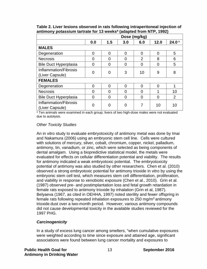
Public Health Goal for 13 September 2016 Antimony in Drinking Water
Table 2. Liver lesions observed in rats following intraperitoneal injection of antimony potassium tartrate for 13 weeksa (adapted from NTP, 1992)
Dose (mg/kg) 0.0 1.5 3.0 6.0 12.0 24.0 a
MALES Degeneration 0 0 0 0 0 5 Necrosis 0 0 0 2 8 6 Bile Duct Hyperplasia 0 0 0 0 0 5 Inflammation/Fibrosis (Liver Capsule) 0 0 3 10 9 8
FEMALES Degeneration 0 0 0 0 0 1 Necrosis 0 0 0 0 1 10 Bile Duct Hyperplasia 0 0 0 0 0 2 Inflammation/Fibrosis (Liver Capsule) 0 0 0 7 10 10
a Ten animals were examined in each group; livers of two high-dose males were not evaluated due to autolysis.
Other Toxicity Studies
An in vitro study to evaluate embryotoxicity of antimony metal was done by Imai and Nakamura (2006) using an embryonic stem cell line. Cells were cultured with solutions of mercury, silver, cobalt, chromium, copper, nickel, palladium, antimony, tin, vanadium, or zinc, which were selected as being components of dental amalgam. Using a biopredictive statistical model, the metals were evaluated for effects on cellular differentiation potential and viability. The results for antimony indicated a weak embryotoxic potential. The embryotoxicity potential of antimony was also studied by other researchers. Chen et al. (2010) observed a strong embryotoxic potential for antimony trioxide in vitro by using the embryonic stem cell test, which measures stem cell differentiation, proliferation, and viability in response to xenobiotic exposure (Chen et al., 2010). Grin et al. (1987) observed pre- and postimplantation loss and fetal growth retardation in female rats exposed to antimony trioxide by inhalation (Grin et al, 1987). Belyaeva (1967, as cited in OEHHA, 1997) noted sterility and fewer offspring in female rats following repeated inhalation exposures to 250 mg/m3 antimony trioxide
dust over a two-month period. However, various antimony compounds
did not cause developmental toxicity in the available studies reviewed for the 1997 PHG.
Carcinogenicity
In a study of excess lung cancer among smelters, “when cumulative exposures were weighted according to time since exposure and attained age, significant associations were found between lung cancer mortality and exposures to

Public Health Goal for 14 September 2016 Antimony in Drinking Water
arsenic, lead and antimony” (Jones et al., 2007). The concomitant exposure to arsenic complicates the interpretation of this study. In a hypothesis generating study, Keshavarzi et al. (2012) noted the presence of high levels of antimony and strontium in soils in the Golestan province of Iran where levels of esophageal cancer is high. Since this is an ecological study, no inference can be drawn between carcinogenicity and antimony in the soil. There are no substantial new human data for judging the carcinogenicity of antimony or antimony compounds since the publication of the PHG. The previous PHG report noted a single mortality study in which lung cancer was elevated in smelter workers exposed to antimony (Schnorr et al., 1995).
The International Agency for Research on Cancer (IARC, 1989) found that there is sufficient evidence for the carcinogenicity of antimony trioxide in experimental animals and classified it as possibly carcinogenic to humans (Group 2B). The studies providing the basis for the classification were conducted via the inhalation route. Antimony trioxide is on California’s Proposition 65 list as a carcinogen on this basis. The European Union (EU, 2008) indicated that there is no evidence that antimony compounds are associated with the development of tumors when ingested. While there are no carcinogenicity studies of the relatively insoluble antimony trioxide via the oral route, limited information from chronic studies in mice and rats of the more soluble APT did not find increased tumor occurrence (Schroeder et al., 1970; Kanisawa and Schroeder, 1969). However, in light of the findings in the “Genotoxicity” section (below), and the carcinogenicity determination for antimony trioxide via the inhalation route, the potential for carcinogenicity of soluble antimony via the oral route cannot be ruled out. Nonetheless, antimony trioxide is not expected to be in water as it is relatively insoluble. The small fraction that dissolves is no longer antimony trioxide but becomes the antimony cation, which is covered in the PHG. As such, OEHHA is developing the PHG based on a noncancer toxicological endpoint.
Genotoxicity
Genotoxicity and mutagenicity testing of antimony-containing compounds has yielded highly variable results. This can be expected because of the wide range of solubility and reactivity of antimony-containing compounds; the results seem characteristic of many other metals and metalloid-salts in such tests. There appears to be some potential for chromosomal aberrations and clastogenicity, particularly for the more soluble salts, as described below.
The potential for five antimony compounds – stibine (SbH3), trimethylstibine (Sb(CH₃)₃), APT (K2Sb2(C4H2O6)2), potassium hydroxyantimonate (K[Sb(ОН)6]) and trimethyl antimony dichloride (C3H9Cl2Sb) – to nick plasmid DNA from pBR 322 was evaluated (Andrewes et al., 2004). Trimethylstibine and stibine were found to be equipotent to trimethylarsine in their ability to nick DNA. The others were inert. The authors speculated that damage done to DNA by stibine or trimethylstibine likely proceeds from the generation of reactive oxygen intermediates, as it is presumed to do with trimethylarsine. In another in vitro

Public Health Goal for 15 September 2016 Antimony in Drinking Water
study, antimony trichloride and APT were evaluated for their potential to prevent DNA repair of γ-irradiation damage in Chinese hamster ovary cells (CHO) (Takahashi et al., 2002). Both the trichloride and tartrate entities inhibited DNA repair in a dose-related fashion. A micronucleus assay alone and in combination with fluorescence in situ hybridization (FISH) technique was performed on human lymphocytes exposed to several metals including potassium antimonate (Sb(V)) (Migliore et al., 1999). Two donor cell lines were used. In both cases there was some variability in the dose-related increases in induction of micronuclei. With the FISH technique it is possible to discriminate clastogenic activity from whole chromosome loss. With potassium antimonate, both clastogenic and aneuploidogenic events occurred.
In mouse bone marrow micronucleus tests with antimony trioxide, no clastogenic effects were observed either with one dose of 5,000 mg/kg or with repeated dosing at 400, 668, and 1,000 mg/kg-day for 7, 14, or 21 days by oral gavage (Elliott et al., 1998). In another in vivo study of clastogenic effects (Kirkland et al., 2007), rats were given antimony trioxide orally for 21 days at doses of 250, 500 and 1,000 mg/kg-day. The only clinical sign was a loss of body weight at the high dose. Chromosomal aberrations and micronuclei were scored from blood cells and compared with controls (positive and negative). No significant (at p<0.05) differences were observed between treated groups and controls, and there was no dose-related trend.
Gebel and colleagues investigated the mechanism of arsenic genotoxicity by comparing its interaction with other substances including antimony (Gebel, 1997, 1998, 2001; Gebel et al., 1996, 1997; Hasgekar et al., 2006). Gebel (2001) noted although antimony is not a point mutagen, its role could be similar to that reported for arsenic, being involved in modulating DNA repair. Takahashi et al. (2002) showed that antimony trichloride and APT were able to inhibit the repair of double-stranded DNA breaks, perhaps like arsenic, through the inhibition of the incision step in the nucleotide excision repair process.
An association between exposure to antimony and oxidative DNA damage was noted in workers (Cavallo et al., 2002). Twenty-three workers assigned to activities involving antimony trioxide (Sb2O3) as a textile fire-retardant were assessed for antimony exposure with personal monitoring devices. The exposed group was split into two groups based on job function; one group had substantially more direct exposure to antimony than the other. The concentration of Sb2O3 was 0.12 ± 0.11 µg Sb/m3 for the high-exposure group and 0.052 ± 0.038 µg Sb/m3 for the low-exposure group. These groups were tested and compared with controls (selected and matched according to smoking and age). Blood was tested for sister chromatid exchange and micronuclei, and in an FPG (formamido-pyrimidine-glycosylase) enzyme-modified comet assay. While no differences from controls were noted in the sister chromatid exchange and micronucleus assay, the FPG enzyme-modified comet assay showed a significant difference between the high exposure group and controls (p = 0.002).

Public Health Goal for 16 September 2016 Antimony in Drinking Water
No significant difference was noted between the lower exposure group and controls. Since the FPG comet assay provides a specific measure of DNA damage, the authors concluded that antimony appeared to have the potential to damage DNA.
Mode of Action
The mode of action for toxicity remains largely unknown for antimony compounds. Some studies have indicated that apoptosis, oxidative stress, disruption of intracellular Ca2+, and mitochondrial dysfunction may be involved.
Huang et al. (1998) noted DNA fragmentation as a marker of apoptosis in human fibroblasts, CHO cells, and human bronchial epithelial cells in a four-hour exposure to SbCl3. In a separate study, APT induced reactive oxygen intermediates, which appeared to mediate apoptosis in a human promyelocytic leukemia (HL-60) cell line (Lecureur et al., 2002a). Lecureur et al. (2002b) noted that APT increased caspase and reactive oxygen intermediates in apoptotic human lymphoma cells (Daudi and Jurkat cells). Apoptotic activity was diminished by addition of the antioxidant N-acetylcysteine, with which antimony probably reacts directly, therefore indicating that the toxicity of antimony is possibly modulated by the redox status of the cells. A study by Hashemzaei and colleagues (2015) suggests that Sb-induced liver cell lysis is mediated by reactive oxygen species formation, lipid peroxidation and decline of mitochondrial membrane potential. Bento et al. (2013) treated CF-1 mice with meglumine antimoniate (Sb(V)) by subcutaneous injections at doses of 0, 20, 60, or 120 mg Sb/kg-day for three days. The results further demonstrated that Sb(V) causes oxidative stress in organs of CF-1 mice by inducing protein carbonylation, lipoperoxidation, and imbalance between superoxide dismutase and catalase activities.
Mann et al. (2006) studied signaling pathways associated with antimony-induced apoptosis in acute promyelocytic leukemia cell lines. Such apoptosis is associated with the production of reactive oxygen intermediates and induction of caspases, and the cascade of reactions mediating apoptosis appear to be associated with c-jun kinase (JNK) and its upstream regulator SEK1. Antimony-induced apoptosis was increased by buthione sulfoxime treatment, which decreases intracellular glutathione. A leukemic cell line that is resistant to arsenic because of a presumed increase in the glutathione content was shown to be resistant to the effects of antimony. The authors showed that antimony trioxide increased the activity of JNK in a dose-dependent manner, which enhances the SEK1-JNK cascade thought to be mediated by reactive oxygen intermediates and is apparently involved in antimony-induced apoptosis.
Similarly, Sereno et al. (2001) studied the effects of pentavalent and trivalent antimonials on the immature, or amastigote, form of Leishmania infantum. They concluded that trivalent antimonials were exceedingly effective (at the low concentration of 10 μg/mL) in inducing DNA fragmentation, leading to cell death.

Public Health Goal for 17 September 2016 Antimony in Drinking Water
They observed that although certain aspects of antimonial action appeared, like late-stage apoptosis, to involve endonuclease activity, this was not due to the activation of caspase-1, caspase-3, calpain, cysteine protease or to proteasomal activation. More study appears to be needed to determine if a different set of caspases/enzymes are involved in generating DNA fragmentation in this organism.
Tirmenstein et al. (1995) attempted to elucidate the nature of antimony’s cardiotoxic effects through experiments on cardiomyocytes. APT induced formation of reactive oxygen intermediates that could harm neonatal cardiomyocytes. However, it was thought that neonatal cardiomyocytes might be more susceptible to reactive intermediates, perhaps due to low glutathione (Tirmenstein et al., 1995, 1997). Using sublethal concentrations of antimony, they also noted that intracellular calcium mobilization could be inhibited during the excitation/contraction response.
Carrying this further, Wey et al. (1997) showed that antimony treatment increased intracellular Ca+2 in the myocytes. Intracellular Ca+2 is involved in chemical-induced oxidative stress and cell toxicity. Addition of a calcium chelator to the culture inhibited the cell toxicity. This work appears to support the hypothesis that antimony, unlike other metals such as iron, does not directly produce oxygen radicals, but rather produces them through stimulation of existing oxidative processes in the cell (probably at the mitochondrial level). Further work by the same group (Snawder et al., 1999) showed that sublethal levels of antimony increased glutathione and heme oxygenase activity. Furthermore, induction of the heat stress proteins HO-1, HSP70, and HSP25/27 was noted, but with no increase in HSP60. After an 18-hour period of exposure to sublethal levels of APT, the cardiomyocytes were able to withstand an otherwise lethal concentration of the same agent. The authors reason that this protection was afforded by induction of the stress proteins and increased glutathione concentration. This protection was removed upon addition of protein synthesis inhibitors.
The role of iron, reactive oxygen species, glutathione and Ca2+ were investigated in the antimony- and arsenic-induced death of Leishmania (Mehta and Shaha, 2006). Both metalloids can induce cell death through apoptosis, with an increase in reactive oxygen species. Both cause mitochondrial dysfunction with loss of membrane potential. In this study, arsenic increased intracellular levels of Ca2+, while antimony did not. Cellular glutathione levels were reduced by antimony, but not by arsenic, and addition of glutathione rescued cells treated with antimony, but not arsenic. Finally, iron depletion increased cell survival despite exposure to antimony or arsenic.
One of the critical issues for understanding antimony toxicity remains the importance of speciation, which has not been well studied in biological systems. Arsenite (trivalent As) has been proven to be more toxic than arsenate (pentavalent As), particularly after being methylated, and this relationship was

Public Health Goal for 18 September 2016 Antimony in Drinking Water
thought to be true for antimony (Patterson et al., 2003). Using cultured human keratinocytes, Patterson et al. (2003) showed that pentavalent As or Sb added to the cell cultures resulted in little reduction to the trivalent forms. Trivalent Sb was similar to trivalent As in its toxicity to keratinocytes, while pentavalent Sb was virtually without effect. However, Hansen and colleagues (2011) exposed a human macrophage cell line (Mono Mac 6) to sodium stibogluconate (Sb(V)) and found that up to 23% of the intracellular Sb was Sb(III). Furthermore, Lopez et al. (2015) demonstrated that glutathione was the reducing agent responsible for Sb(V) to Sb(III) transformation in human whole blood cells and Sun et al. (2000) reported the existence of an Sb(III)-glutathione complex in red blood cells.
For the differences in toxicity, the EU (2008) report states that “any firm conclusion on differences in toxicity between the two valences is difficult to make,” and since “there [is] no conclusive evidence supporting a significant difference in toxicity between the two valences, it is decided not to differentiate between relevant and reliable toxicity results originating from tri- or pentavalent antimony studies.”
Metalloid transport is another aspect of interaction of antimony ions with cells that is apparently significant to toxic mechanisms. Uptake of antimony and arsenic depends upon transporter proteins (Tamas and Wysocki, 2001; Bentley and Chasteen, 2002). In bacteria, resistance to the toxic effects of arsenic and antimony has been found to involve induction of specific operons that confer resistance by inhibiting production of these transporter proteins. However, it is currently unknown whether higher organisms may also have the ability to develop tolerance to antimony based on alteration of cellular uptake resulting from similar mechanisms.
PHG DERIVATION
In the 1997 PHG, the most relevant studies for risk extrapolation for drinking water exposures were judged to be the chronic drinking water studies in mice and rats conducted by Schroeder and colleagues (1968, 1970). Only one dose level was applied in each of these studies. Because the estimated dose to rats was lower than to mice (0.43 mg/kg-day for rats versus 0.83 mg/kg-day for mice) and the observed effect (shorter lifespan) was significant for both male and female rats, the rat study was selected as the most sensitive indicator of antimony toxicity. In this study (Schroeder et al., 1970), Long-Evans rats (at least 50/sex/dose) were exposed to 0 or 5 mg/L of APT (Sb(III)) in drinking water from weaning until death (over 1,000 days). Based on the observed decrease in longevity and altered blood glucose and serum cholesterol levels, a LOAEL of 0.43 mg/kg-day was identified. Limitations of this study are: (1) single dose level, (2) loss of animals due to infection, and (3) limited toxicity evaluation. However, the Schroeder et al. (1968, 1970) studies remain the only chronic oral toxicity studies conducted on antimony compounds and they provide valuable information on the long-term effects of chronic antimony ingestion.

Public Health Goal for 19 September 2016 Antimony in Drinking Water
After the PHG was published, the Poon et al. (1998) subchronic exposure study became available. The Poon et al. (1998) study used multiple doses of APT (Sb(III)), monitored more parameters than the Schroeder et al. (1968, 1970) studies, and effects were reported at lower levels. Thus, OEHHA is selecting the Poon et al. (1998) study as the critical study for updating the antimony PHG. Benchmark dose (BMD) modeling (US EPA BMDS, Version 2.4) is conducted for dose-response characterization of the critical endpoint - liver nuclear anisokaryosis.
Several researchers and regulatory agencies have associated liver anisokaryosis with chemical exposure. Although anisokaryosis can be found in the liver of aging rodents, it can also be induced in response to toxic insult and has been well documented as a treatment-related lesion induced by xenobiotics (Chu et al., 1990; Chu et al., 1980; Besteman et al., 2007; Takasawa et al., 2013; Hirata-Koizumi et al., 2008; Walter et al., 2000; Moir et al., 1997; Poon et al., 1997; Kari et al., 1992; Junge and Thornburg, 1989; Siu et al., 1983; Bird et al., 1982). Liver anisokaryosis was listed as one of the “compound-related lesions” observed in male mice exposed to hydroquinone (NTP, 1989). Furthermore, in developing the intermediate-duration Minimal Risk Level (MRL) for toxaphene, the Agency for Toxic Substances and Disease Registry (ATSDR) identified a lowest-observed-adverse-effect level based on liver anisokaryosis as a histopathologic lesion (ATSDR, 2010).
Antimony has been associated with hepatocellular damage and impaired liver metabolism as demonstrated in human studies when used in the treatment for leishmaniasis. Cutaneous leishmaniasis patients treated with antimony have been shown to have alterations in liver enzymes such as alanine aminotransferase (ALT) and glutathione S-transferase B1 (GST), markers for liver damage and impairment of liver metabolism (Hepburn et al., 1993; Hepburn et al., 1994; Oliveira et al., 2011; Andersen et al., 2005). For visceral leishmaniasis, impaired peroxisomal function, hepatitis, and hepatic failure were observed in patients treated with antimony (Gupta et al., 2009; Oliveira et al., 2009). Patients with mucosal leishmaniasis also showed elevations in liver enzymes along with electrocardiogram abnormalities or musculoskeletal pain when treated with antimony (Saenz et al., 1991; Franke et al., 1990).
Grimaldi et al. (2010) noted there was a clear correlation (r = 0.94; P = 0.001) between liver antimony levels and the extent to which hepatocytes were affected in rhesus monkeys receiving i.m. injections of 5 or 20 mg Sb(V)/kg-day as meglumine antimoniate for 21 days. Histopathology of liver samples showed focal hepatocellular necrosis, fatty changes in stellate cells, and hypotrophy of the hepatic parenchyma at the center of the lobules. The authors also noted that the plasma pharmacokinetic profile of antimony in L. braziliensis-infected macaques treated with meglumine antimoniate was similar to that reported for human cases receiving Sb(V) drugs (Grimaldi et al., 2010).

Public Health Goal for 20 September 2016 Antimony in Drinking Water
BALB/c mice receiving i.p. injections of 80 mg Sb/kg-day as meglumine antimoniate for 20 days exhibited vacuolization and granulosity in hepatocytes. Further evidence of hepatotoxicity included an increase in TUNEL (Terminal deoxynucleotidyl transferase (TdT) dUTP Nick-End Labeling)-positive hepatocytes and increased apoptotic index. The level of antimony in the livers of mice after treatment was 4.2 ± 1.0 µg/g of wet liver; liver antimony levels were not reported for control animals (Kato et al., 2014).
Accumulation of antimony in the liver and thyroid was observed in rats receiving 2% diantimony trioxide in their diet for eight months, with considerable amounts of antimony remaining in these organs 40 days after diantimony trioxide administration had ceased (Gross et al., 1955, as cited by EU, 2008). Furthermore, Poon et al. (1998) observed an accumulation of antimony in the liver of exposed rats that increased with the dose and persisted, though at lower levels, during the four-week recovery period. Schroeder et al. (1970) also noted an accumulation of antimony with age in the rat liver at the same concentration, 5 ppm, at which antimony accumulation was observed in the Poon et al. (1998) study.
Selection of the liver as the target organ for antimony is also supported by two repeated dose oral studies using diantimony trioxide. Sunagawa (1981) observed increased levels of aspartate aminotransferase and alkaline phosphatase in rats in a 24-week diantimony trioxide feeding study. Cloudy swelling in hepatic cords was noted in histopathological examinations of the liver. Hext et al. (1999) administered diantimony trioxide in the diet to rats for 90 days. Changes in relative liver weights were noted in both sexes, along with alterations in liver enzymes. Despite its insolubility and poor oral absorption, liver effects were observed following diantimony trioxide administration.
For BMD modeling, histopathological changes observed in the Poon et al. (1998) study were graded based on the histological grading system developed by the authors (Chu et al., 1995; Poon et al., 1998; personal communication with Health Canada, 2012). The histological grading system and the grading results used for the BMD modeling are presented in Appendix I. All models were run with default parameters and a benchmark response of a 10% increase in response over background for dichotomous data. A benchmark response of 10% instead of 5% was used for the histopathological endpoints because the effects observed may be considered minimally biologically significant, or a mild effect. The model selected for point of departure (POD) consideration is presented in Table 3 and model outputs are presented in Appendix II; poorly fitting models are not presented. When using BMD modeling, the BMDL, which is the lower limit of the 95 percent confidence interval of the BMD resulting in the benchmark response, is selected as the POD. The lowest BMDL of 0.14 mg/kg-day based on liver nuclear anisokaryosis is selected as the POD. This BMDL of 0.14 mg/kg-day is supported by the NOAEL of 0.06 mg/kg-day determined by Poon et al. (1998) based on the observed changes in blood glucose levels, histological changes,

Public Health Goal for 21 September 2016 Antimony in Drinking Water
and the tissue retention of antimony. The POD of 0.14 mg of antimony/kg-day is therefore selected for the PHG calculation.
Table 3. Benchmark dose modeling in rats following oral exposure to antimony potassium tartrate for 90 days, data from Poon et al. (1998)
Endpoint Model Name p-valuea BMD10 (mg/kg-day)
BMDL10b (mg/kg-day)
Liver nuclear anisokaryosis in males
Gammac 0.83 0.22 0.14
a p-values ≥ 0.05 indicate the model adequately fits the data. b The BMDL is the lower limit of the 95% confidence interval of the BMD resulting in a 10% increase in response over background. c The Weibull and Quantal-linear models had the same fit as the Gamma model.
Methodology
For estimation of a health-protective concentration of a chemical in drinking water, an acceptable daily dose (ADD) of the chemical from all sources will first be calculated. This involves incorporation of appropriate estimates of uncertainty in the extrapolation of the critical toxic dose from human or animal studies to the estimation of a lifetime ADD that is unlikely to result in any toxic effects. For this purpose, the following equation will be used:
ADD = POD UF
where, ADD = acceptable daily dose, an estimate of the maximum daily
dose that can be consumed by humans for an entire lifetime without adverse health effects;
POD = point of departure, in units of milligrams per kilogram of body weight per day (mg/kg-day); this can be the no-observed-adverse-effect level (NOAEL), lowest-observed-adverse-effect level (LOAEL), or lower limit of the 95% confidence interval of the benchmark dose estimated from the critical study (BMDL);
UF = uncertainty factor(s); for a list of default uncertainty factors, see OEHHA (2008).
Calculation of a public health-protective concentration (C, in mg/L) for a chemical in drinking water uses the following equation for non-carcinogenic endpoints: C = ADD (mg/kg-day) × RSC
DWI

Public Health Goal for 22 September 2016 Antimony in Drinking Water
where, RSC = relative source contribution (usually 20% to 80%,
expressed as 0.20 to 0.80); DWI = daily water intake rate expressed as liters per kilogram of
body weight per day; time-weighed lifetime average drinking water consumption rate for the general population = 0.053 L/kg-day (calculated from lifestage-specific water consumption rates in OEHHA, 2012, as discussed below).
For oral ingestion rates, the OEHHA PHG program uses age-specific water intake rate estimates (OEHHA, 2012) derived from a nationwide survey of food and beverage intake from approximately 20,000 individuals (U.S. Department of Agriculture’s Continuing Survey of Food Intake of Individuals 1994-1996, 1998 dataset). These age-specific intake rates are normalized by body weight and expressed as liters of water ingested per kilogram of body weight per day (L/kg-day) as shown in Table 4 below. The updated water ingestion rates indicate that drinking water intake per unit body weight is higher in infants than in adults. Previous PHGs using intake rates of 2 liters per day for adults and 1 liter per day for a 10 kg child are being updated with these more refined estimates. Table 4. Time-weighed lifetime average drinking water intake rate for the general population
Life stage Age range (years)
Oral ingestion (L/kg-day)
Infant 0 to <2 0.196 Child 2 to <16 0.061 Adult 16-70 0.045
Time-weighted average over lifetime 0.053 (2/70*0.196+14/70*0.061+54/70*0.045)
Dermal and inhalation exposure during household uses of tap water are not expected to be significant due to the low dermal absorption and low volatility of antimony and its compounds (HSDB, 2005). Therefore, the drinking water intake by the oral route is assumed to cover the total intake resulting from antimony in tap water.
Calculation based on the Poon et al. (1998) study
As discussed earlier, the BMDL10 of 0.14 mg/kg-day based on the Poon et al. (1998) study in rats is identified as the POD and is used for the calculation of the ADD. A total UF of 1,000 is applied, which includes a factor of 10 for interspecies extrapolation, 30 for variation in the human population (10 for pharmacokinetics and √10 for pharmacodynamics), and √10 for extrapolating

Public Health Goal for 23 September 2016 Antimony in Drinking Water
from subchronic to lifetime exposure. The full 10-fold default UF for pharmacokinetics is applied due to concerns regarding variability in the human population for absorption, distribution, tissue accumulation, excretion, and conversion of Sb(V) to Sb(III). The √10 for subchronic to lifetime exposure is based on the study’s duration of 8-12% of estimated lifetime (OEHHA, 2008). Thus,
ADD = 0.14 mg/kg-day = 1.4 x 10⁻⁴ mg/kg-day = 0.14 µg/kg-day 1,000
Calculation of the human health-protective concentration, C, from the ADD must account for exposure of humans to other sources of antimony, such as from food or ambient air. In the absence of data to indicate otherwise, the RSC value of 0.40 used in the 1997 antimony PHG is retained. Incorporating the time-weighted average lifetime drinking water consumption rate (DWI) for the general population with this RSC and the ADD, the health-protective concentration is:
C = ADD (mg/kg-day) × RSC DWI
= 0.14 µg/kg-day × 0.40 = 1.1 µg/L = 1 ppb (rounded) 0.053 L/kg-day
Thus, the updated Public Health Goal for antimony is 1 ppb.
Consideration of the NTP (1992) study
NTP (1992) is a well-conducted study on the effects of APT. Despite the fact that i.p. injection is not a natural route of exposure, and the difficulty in converting the i.p. dose to an oral dose, the NTP (1992) study provided useful information regarding the potential liver toxicity of antimony, and can be used as a comparison and support for the current PHG calculation. A detailed discussion of the evaluation of the data from the NTP (1992) study is presented in Appendix III.
RISK CHARACTERIZATION
According to the World Health Organization, Sb(III) may be more toxic than Sb(V) and the inorganic compounds are generally more toxic than the organic compounds (WHO, 2003). APT, which is highly water soluble and contains the trivalent form of antimony (Sb(III)), may have greater oral toxicity than some other compounds of antimony. Because both the Poon et al. (1998) study and the WHO (2003) guidelines are based on APT, they acknowledged that their results would probably overestimate the risk from the predominant antimony species in drinking water, the pentavalent form of antimony (Sb(V)). However, as indicated in the EU (2008) report, “[T]here [is] no conclusive evidence supporting a significant difference in toxicity between the two valences.” Thus, it is

Public Health Goal for 24 September 2016 Antimony in Drinking Water
reasoned that the determination of a drinking water level based on the Poon et al. (1998) study would likely be health protective.
For comparison with the current PHG, the US federal, Canadian and WHO drinking water guidance levels for antimony are shown in Table 5, with the toxicological basis for the values.
Table 5. Non-cancer oral guidance values for antimony
Guidance Critical Study
Species and
Duration of Study
Endpoint Point of
Departure (mg/kg-
day)
Uncertainty Factor
ADD (mg/kg-
day)
Water Guidance
Value
Updated OEHHA
PHG (2016)
Poon et al.
(1998)
Rat, 90 days
oral
Histopatho-logical
changes in liver
0.14 (BMDL)
1,000 (30 for
intraspecies, 10 for
interspecies, 3 for
subchronic to chronic)
1.4 x 10⁻⁴ 1 ppb
USEPA (IRIS) Oral RfD (1991)
& MCL
Schroeder et al.
(1970)
Rat, chronic
oral
Decreased longevity,
blood glucose and cholesterol
0.4 (LOAEL)
1,000 (10 for
intraspecies, 10 for
interspecies, 10 for
LOAEL to NOAEL)
4 x 10⁻⁴ 6 ppb
WHO Guidelines for Drinking
Water Quality (2003)
Poon et al.
(1998)
Rat, 90 days
oral
Decreased body weight
gain, reduced food and
water intake
6.0 (NOAEL)
1,000 (10 for
intraspecies, 10 for
interspecies, 10 for
subchronic to chronic)
6 x 10⁻³ 20 ppb
Health Canada
Maximum Acceptable Concentra-tion (2008)
Poon et al.
(1998)
Rat, 90 days
oral
Histopatho-logical
changes 0.06
(NOAEL)
300 (10 for
intraspecies, 10 for
interspecies, 3 for
subchronic to chronic)
2 x 10⁻⁴ 4 ppba
a Although Health Canada’s calculated maximum acceptable concentration (MAC) for antimony in drinking water is 0.004 mg/L (4 ppb), the MAC was set at the practical quantitation level of 0.006 mg/L (6 ppb).

Public Health Goal for 25 September 2016 Antimony in Drinking Water
The US EPA maximum contaminant level goal (MCLG) for antimony of 6 ppb is based on the Schroeder et al. (1970) study in rats, as shown in Table 5 (US EPA, 1992a,b, 1995). Like the 1997 PHG, US EPA used the LOAEL of 0.43 mg/kg-day based on reduced longevity observed in the study; however, a greater uncertainty factor was applied. A 70 kg adult drinking 2 liters of water per day and a RSC of 40 percent from drinking water was assumed by US EPA. US EPA did not consider the Poon et al. (1998) study, which was released after the establishment of the MCLG. The MCLG was adopted as the federal MCL.
The World Health Organization (WHO) guideline value of 20 ppb was developed based on an assumption of a 60 kg adult drinking 2 liters of water per day and an RSC of 10 percent from drinking water. Also, like OEHHA, WHO used the Poon et al. (1998) study, though the WHO value is derived from a NOAEL of 6.0 mg/kg-day that is based on some effects of antimony such as reduced body weight gain and reduced food and water intake observed in the study (WHO, 2003). However, OEHHA determined there are other adverse effects at lower doses and developed a PHG of 1 ppb.
Health Canada’s calculated Maximum Acceptable Concentration (MAC) for antimony in drinking water is 4 ppb (Health Canada, 2008). However, the final MAC was based on the limit of detection (6 ppb). The calculated MAC is based on a NOAEL of 0.06 mg/kg-day from Poon et al. (1998) study, an estimated water consumption rate of 1.5 L/day, an average adult body weight of 70 kg, and a relative source contribution of 38%.
REFERENCES
Andersen EM, Cruz-Saldarriaga M, Llanos-Cuentas A, Luz-Cjuno M, Echevarria J, Miranda-Verastegui C, Colina O, Berman JD (2005). Comparison of meglumine antimoniate and pentamidine for peruvian cutaneous leishmaniasis. Am J Trop Med Hyg 72, 133-137. Andrewes P, Kitchin KT, Wallace K (2004). Plasmid DNA damage caused by stibine and trimethylstibine. Toxicol Appl Pharmacol 194:41-8. ATSDR (1992). Toxicological profile for antimony. Agency for Toxic Substances and Disease Registry, U.S. Public Health Service. TP-91/02. September, 1992. ATSDR (2010). Draft toxicological profile for toxaphene. Agency for Toxic Substances and Disease Registry, U.S. Public Health Service. September, 2010. Bentley R, Chasteen TG (2002). Microbial methylation of metalloids: Arsenic, antimony, and bismuth. Microbiol Mol Biol Rev 66:250-71.
Bento DB, de Souza B, Steckert AV, Dias RO, Leffa DD, Moreno SE, Petronilho F, de Andrade VM, Dal-Pizzol F, Romão PR (2013). Oxidative stress in mice

Public Health Goal for 26 September 2016 Antimony in Drinking Water
treated with antileishmanial meglumine antimoniate. Res in Vet Sci 95(3):1134-41.
Berman JD, Gallalee JF, Gallalee JV (1988). Pharmacokinetics of pentavalent antimony (Pentostam) in hamsters. Am J Trop Med Hyg 39, 41-45.
Besteman EG, Zimmerman KL, Huckle WR, Prater MR, Gogal RM, Jr, Holladay SD (2007). 2,3,7,8-tetrachlorodibenzo-p-dioxin (TCDD) or diethylstilbestrol (DES) cause similar hematopoietic hypocellularity and hepatocellular changes in murine fetal liver, but differentially affect gene expression. Toxicol Patho 35, 788-794. Bird RP, Draper HH, Valli VE (1982). Toxicological evaluation of malonaldehyde: a 12-month study of mice. J Toxicol Environ Health 10, 897-905. Butterman WC, Carlin JF, Jr. (2004). Mineral Commodity Profiles: Antimony. U.S. Department of the Interior, U.S. Geological Survey Open-File Report 03-019. Accessed at: http://pubs.usgs.gov/of/2003/of03-019/of03-019.pdf. Casals J (1972). Pharmacokinetic and toxicological studies of antimony dextran glucoside (RL-712). Br J Pharmacol 46:281-8. Cavallo D, Iavicoli I, Setini A, Marinaccio A, Perniconi B, Carelli G. Lavicoli S. (2002). Genotoxic risk and oxidative DNA damage in workers exposed to antimony trioxide. Environ Mol Mutagen 40:184-9. CDC (2013). Fourth National Report on Human Exposure to Environmental Chemicals. Centers for Disease Control and Prevention (CDC), March 2013. http://www.cdc.gov/exposurereport/pdf/FourthReport_UpdatedTables_Mar2013.pdf Chen R, Chen J, Cheng S, Qin J, Li W, Zhang L, Jiao H, Yu X, Zhang X, Lahn BT, Xiang AP (2010). Assessment of embryotoxicity of compounds in cosmetics by the embryonic stem cell test. Toxicol Mech Methods 20(3), 112-118. Chu I, Villeneuve DC, Becking GC, Iverson F, Ritter L, Valli VE, Reynolds LM (1980). Short-term study of the combined effects of mirex, photomirex, and kepone with halogenated biphenyls in rats. J Toxicol Environ Health 6, 421-432. Chu I, Villeneuve DC, Valli VE, Black WD, Robinson K, Beyrouty P (1990). A teratological assessment of coal liquefaction products in the rat. J Appl Toxicol 10, 411-416. Chu I, Villeneuve DC, Yagminas A, Lecavalier P, Hakansson H, Ahlborg UG, Valli VE, Kennedy SW, Bergman A, Seegal RF, et al. (1995). Toxicity of PCB 77 (3,3',4,4'-tetrachlorobiphenyl) and PCB 118 (2,3',4,4'5-pentachlorobiphenyl) in the rat following subchronic dietary exposure. Fundam Appl Toxicol 26, 282-292. Chulay JD, Fleckenstein L, Smith DH (1988). Pharmacokinetics of antimony treatment of visceral leishmaniasis with sodium stibogluconate or meglumine antimoniate. Transact of the Royal Soc of Tropic Med and Hygiene 82:69-72.

Public Health Goal for 27 September 2016 Antimony in Drinking Water
Cleve EA, Langsjoen PH, Hensler NM (1955). The Toxic effect of tartar emetic in treatment of schistosomiasis. Am J Med Sci 229 (1), 74-80. Coelho DR, De-Carvalho RR, Rocha RC, Saint’Pierre TD, Paumgartten FJ (2014). Effects of in utero and lactational exposure to Sb V on rat neurobehavioral development and fertility. Reprod Toxicol 50:98-107. Croft SL, Shyam S Fairlamb AH (2006). Drug Resistance in Leishmaniasis. Clinical Microbiology Reviews 19(1): 111-126. Cruz A, Rainey PM, Herwaldt BL, Stagni G, Plalcios R, Trujillo R, Saravia NG (2007). Pharmacokinetics of antimony in children treated for leishmaniasis with meglumine antimoniate. J Infect Disease 195:602-608. Dieter MP, Jameson CW, Elwell MR, Lodge JW, Hejtmancik M, Grumbein SL, Ryan M, Peters AC (1991). Comparative toxicity and tissue distribution of antimony potassium tartrate in rats and mice dosed by drinking water or intraperitoneal injection. J Toxicol Environ Health 34, 51-82. Elliott BM, Mackay JM, Clay P, Ashby J (1998). An assessment of the genetic toxicology of antimony trioxide. Mutat Res 415:109-17. EU (2008). European Union Risk Assessment Report: Diantimony Trioxide. Accessed at: http://esis.jrc.ec.europa.eu/doc/risk_assessment/REPORT/datreport415.pdf Felicetti SA, Thomas RG, McClellan RO (1974). Metabolism of two valence states of inhaled antimony in hamsters. Am Ind Hyg Assoc J 35:292-300. Ferreira Cdos S, Martins PS, Demicheli C, Brochu C, Ouellette M, Frezard F (2003). Thiol-induced reduction of antimony(V) into antimony(III): a comparative study with trypanothione, cysteinyl-glycine, cysteine and glutathione. Biometals 16, 441-446. Filella M, Belzile N, Chen Y-W (2002a). Antimony in the environment: a review focused on natural waters - I. Occurrence. Earth-Sci Rev 57: 125-176. Filella M, Belzile N, Chen Y-W (2002b). Antimony in the environment: a review focused on natural waters II. Relevant solution chemistry. Earth-Sci Rev 59: 265-285. Franke ED, Wignall FS, Cruz ME, Rosales E, Tovar AA, Lucas CM, Llanos-Cuentas A, Berman JD (1990). Efficacy and toxicity of sodium stibogluconate for mucosal leishmaniasis. Ann Inter Med 113, 934-940. Frezard F, Demicheli C, Ferreira CS, Costa MA (2001). Glutathione-induced conversion of pentavalent antimony to trivalent antimony in meglumine antimoniate. Antimicrob Agents Chemother 45, 913-916. Frezard F, Demicheli C, Ribeiro RR (2009). Pentavalent antimonials: new perspectives for old drugs. Molecules 14, 2317-2336. Friedrich K, Vieira FA, Porrozzi R, Marchevsky RS, Miekeley N, Grimaldi G, Paumgartten FJR (2012). Disposition of antimony in rhesus monkeys infected

Public Health Goal for 28 September 2016 Antimony in Drinking Water
with leishmanial braziliensis and treated with meglumine antimoniate. J Tox Envron Health 75:63-75. Gebel T, Kevekordes S, Schaefer J, von Platen H, Dunkelberg H (1996). Assessment of a possible genotoxic environmental risk in sheep bred on grounds with strongly elevated contents of mercury, arsenic and antimony. Mutat Res 368:267-74. Gebel T, Christensen S, Dunkelberg H (1997). Comparative and environmental genotoxicity of antimony and arsenic. Anticancer Res 17:2603-7. Gebel T (1997). Arsenic and antimony: Comparative approach on mechanistic toxicology. Chem Biol Interact 107:131-44. Gebel T (1998). Suppression of arsenic-induced chromosome mutagenicity by antimony. Mutat Res 412:213-8. Gebel T (2001). Unanswered questions in arsenic toxicology. J Environ Pathol Toxicol Oncol 20:299-309. Gerber GB, Maes J, Eykens B (1982). Transfer of antimony and arsenic to the developing organism. Arch Toxicol 49:159-68. Grahn E, Karlsson S, Karlsson U, Düker A (2006). Historical pollution of seldom monitored trace elements in Sweden--Part B: sediment analysis of silver, antimony, thallium and indium. J Environ Monit 8(7):732-44. Grin NV, Govorunova NN, Bessemmyl AN, Pavlovich LV (1987). Embryotoxic action of antimony oxide in an experiment. Gig Sanit 10, 85-86. Gross P, Brown JHU, Westrick ML, Srsic RP, Butler NL, Hatch TF (1955). Toxicologic study of calcium halophosphate phosphors and antimony trioxide I. Acute and chronic toxicity and some pharmacologic aspects. In A. M. A. Archives of Industrial Health. Edited by Drinker P 1955; 11, pp 473-478. American Medical Association, Chicago. Gupta S, Raychaudhury B, Datta SC (2009). Host peroxisomal properties are not restored to normal after treatment of visceral leishmaniasis with sodium antimony gluconate. Exp Parasitol 123, 140-145. Hansen C, Hansen EW, Hansen HR, Gammelgaard B, Stiirup S (2011). Reduction of Sb(V) in a human macrophage cell line measured by HPLC-ICP-MS. Biol Trace Elem Res 144:234-243. Hasgekar N, Beck JP, Dunkelberg H, Hirsch-Ernst KI, Gebel TW (2006). Influence of antimonite, selenite, and mercury on the toxicity of arsenite in primary rat hepatocytes. Biol Trace Elem Res 111:167-83. Hashemzaei M, Pourahmad J, Safaeinejad F, Tabrizian K, Akbari F, Bagheri G, Hosseini MJ, Shahraki J (2015). Antimony induces oxidative stress and cell death in normal hepatocytes. Toxicol & Environ Chem. 97(2):256-265.

Public Health Goal for 29 September 2016 Antimony in Drinking Water
Health Canada (2008). Water quality and health – antimony. Accessed at: http://www.hc-sc.gc.ca/ewh-semt/pubs/water-eau/antimony-antimoine/index-eng.php Hepburn NC, Siddique I, Howie AF, Beckett GJ, Hayes PC (1993). Hepatotoxicity of sodium stibogluconate in leishmaniasis. Lancet 342, 238-239. Hepburn NC, Siddique I, Howie AF, Beckett GJ, Hayes PC (1994). Hepatotoxicity of sodium stibogluconate therapy for American cutaneous leishmaniasis. Trans R Soc Trop Med Hyg 88, 453-455. Hext PM, Pinto PJ, Rimmel BA (1999). Subchronic feeding study of antimony trioxide in rats. J Appl Toxicol 19:205-9. Hirata-Koizumi M, Matsuyama T, Imai T, Hirose A, Kamata E, Ema M (2008). Lack of gender-related difference in the toxicity of 2-(2'-hydroxy-3',5'-di-tert-butylphenyl)benzotriazole in preweaning rats. Drug Chem Toxicol 31, 275-287. HSDB (2005). Antimony compounds. Hazardous Substances Data Bank, National Library of Medicine. Accessed at: http://toxnet.nlm.nih.gov/cgi-bin/sis/search/f?./temp/~UV7Ho7:2. Huang H, Shu SC, Shih JH, Kuo CJ, Chiu ID (1998). Antimony trichloride induces DNA damage and apoptosis in mammalian cells. Toxicology 129:113-23. IARC (1989). Monographs on the Evaluation of the Carcinogenic Risk of Chemicals to Man. Vol. 47. p. 291. International Agency for Cancer Research, World Health Organization, Lyon, France. Imai K and Nakamura M (2006). In vitro embryotoxicity testing of metals for dental use by differentiation of embryonic stem cell test. Congenit Anom (Kyoto) 46:34-8. IPCS (1998). Antimony trioxide. (last updated 1/28/98) International Programme on Chemical Safety. Accessed at: http://www.inchem.org/documents/ukpids/ukpids/ukpid38.htm. Jones SR, Atkin P, Holroyd C, Lutman E, Vives i Batlle J, Wakeford R, Walker P (2007). Lung cancer mortality at a UK tin smelter. Occup Med 57(4), 238-245. Accessed at http://occmed.oxfordjournals.org/content/57/4/238.long Junge RE, Thornburg L (1989). Copper poisoning in four llamas. J Am Vet Med Assoc 195, 987-989. Kanisawa M and Schroeder HA (1969). Life term studies on the effect of trace elements on spontaneous tumors in mice and rats. Cancer Res 29, 892-895. Kari FW, Bucher J, Eustis SL, Haseman JK, Huff JE (1992). Toxicity and carcinogenicity of hydroquinone in F344/N rats and B6C3F1 mice. Food Chem Toxicol 30, 737-747.

Public Health Goal for 30 September 2016 Antimony in Drinking Water
Keshavarzi B, Moore F, Naimeddin A, Rahmani F (2012). The role of selenium and selected trace elements in the etiology of esophageal cancer in high risk Golestan province of Iran. Sci Total Environ. 433, 89-97. Khalil EA, Ahmed AE, Musa AM, Hussein MH (2006). Antimony-induced cerebellar ataxia. Saudi Med J 27:90-2. Khayyal MT, Girgis NI, McConnell E (1967). The use of Penicillamine as an adjuvant to tartar emetic in the treatment of experimental schistosomiasis. Bull Wld Hlth Org 37, 387-392. Accessed at http://www.ncbi.nlm.nih.gov/pmc/articles/PMC2554273/?page=1 Khayyal MT, Saleh S, El Masri AM (1973). Penicillamine as an adjuvant to antimonial therapy of schistosomiasis: effect on liver function tests in rabbits and on antischistosomal activity. Bull Wld Hlth Org 48(4), 415-420. Accessed at http://www.ncbi.nlm.nih.gov/pmc/articles/PMC2481109/?page=5 Kirkland D, Whitwell J, Deyo J, Serex T (2007). Failure of antimony trioxide to induce micronuclei or chromosomal aberrations in rat bone-marrow after sub-chronic oral dosing. Mutat Res 627:119-28. Kobayashi A and Ogra Y (2009). Metabolism of tellurium, antimony and germanium simultaneously administered to rats. The J Toxicol Sci 34, 295-303. Krachler M, Zheng J, Koerner R, Zdanowicz C, Fisher D, Shotyk W (2005). Increasing atmospheric antimony contamination in the northern hemisphere: snow and ice evidence from Devon Island, Arctic Canada. J Environ Monit 7(12):1169-76. Lecureur V, Lagadic-Gossmann D, Fardel O (2002a). Potassium antimonyl tartrate induces reactive oxygen species-related apoptosis in human myeloid leukemic HL60 cells. Int J Oncol 20:1071-6. Lecureur V, Le Thiec A, Le Meur A, Amiot L, Drenou B, et al. (2002b). Potassium antimonyl tartrate induces caspase- and reactive oxygen species-dependent apoptosis in lymphoid tumoral cells. Br J Haematol 119:608-15. Lòpez S, Aguilar L, Mercado L, Bravo M, Quiroz W (2015). Sb(V) reactivity with human blood components: redox effects. PLoS One 10: e0114796. Lyon TDB, Patriarca M, Howatson AG, Fleming PJ, Blair PS, Fell GS (2002). Age dependence of potentially toxic elements (Sb, Cd, Pb, Ag) in human liver tissue from paediatric subjects. J Environ Monit 4:1034-1039. Mann KK, Davison K, Colombo M, Colosimo AL, Diaz Z, et al. (2006). Antimony trioxide-induced apoptosis is dependent on SEK1/JNK signaling. Toxicol Lett 160:158-70. Mehta A, Shaha C (2006). Mechanism of metalloid-induced death in Leishmania spp.: Role of iron, reactive oxygen species, Ca2+ and glutathione. Free Rad Biol Med 40:1857-68.

Public Health Goal for 31 September 2016 Antimony in Drinking Water
Miekeley N, Mortari SR, Schubach AO (2002). Monitoring of total antimony and its species by ICP-MS and onl-line ion chromatography in biological samples from patients treated for leishmaniasis. Anal Bioanal Chem 372:495-502. Migliore L, Cocchi L, Nesti C, Sabbioni E (1999). Micronuclei assay and FISH analysis in human lymphocytes treated with six metal salts. Environ Mol Mutagen 34:279-84. Miranda ES, Miekeley N, De-Carvalho RR, Paumgartten FJR (2006). Developmental toxicity of meglumine antimoniate and transplacental transfer of antimony in the rat. Reprod Toxicol 21: 292-300. Moir D, Poon R, Yagminas A, Park G, Viau A, Valli VE, Chu I (1997). The subchronic toxicity of acridine in the rat. J Environ Sci Health B 32, 545-564. Molokhia MM, Smith H (1969). The behavior of antimony in blood. J Trop Med Hyg 72:222-5. Moskalev YI (1959). Materials on the distribution of radioactive antimony. Med Radiol 4:6-13. NTP (1989). Toxicology and carcinogenesis studies of hydroquinone in F344/N rats and B6C3F1 mice. National Toxicology Program, Research Triangle Park, NC. NTP (1992). NTP report on the toxicity studies of antimony potassium tartrate in F344/N rats and B6C3F1 mice (drinking water and intraperitoneal injection studies). NTP Tox 11, NIH Publication No. 92-3130, March 1992. National Toxicology Program, Research Triangle Park, NC. OEHHA (1997). Public Health Goal for Antimony in Drinking Water. Office of Environmental Health Hazard Assessment, California Environmental Protection Agency, Sacramento, CA. Accessed at: www.oehha.ca.gov/water/phg/pdf/anti3_c.pdf. OEHHA (2008). Air Toxics Hot Spots Program Risk Assessment Guidelines: Technical Support Document for the Derivation of Noncancer Reference Exposure Levels. Office of Environmental Health Hazard Assessment, California Environmental Protection Agency, Sacramento, CA. Accessed at: http://oehha.ca.gov/media/downloads/crnr/noncancertsdfinal.pdf OEHHA (2012). Air Toxics Hot Spots Program Risk Assessment Guidelines: Technical Support Document for Exposure Assessment and Stochastic Analysis. Chapter 8. Office of Environmental Health Hazard Assessment, California Environmental Protection Agency, Sacramento, CA. Accessed at: http://www.oehha.ca.gov/air/hot_spots/pdf/2012tsd/Chapter8_2012.pdf. Ogra Y (2009). Toxicometallomics for research on the toxicology of exotic metalloids based on speciation studies. Anal Sci 25(10):1189-95. Oliveira AL, Brustoloni YM, Fernandes TD, Dorval ME, Cunha RV, Boia MN (2009). Severe adverse reactions to meglumine antimoniate in the treatment of

Public Health Goal for 32 September 2016 Antimony in Drinking Water
visceral leishmaniasis: a report of 13 cases in the southwestern region of Brazil. Trop Doct 39, 180-182. Oliveira LF, Schubach AO, Martins MM, Passos SL, Oliveira RV, Marzochi MC, Andrade CA (2011). Systematic review of the adverse effects of cutaneous leishmaniasis treatment in the New World. Acta tropica 118, 87-96. Patterson TJ, Ngo M, Aronov PA, Reznikova TV, Green PG, Rice RH (2003). Biological activity of inorganic arsenic and antimony reflects oxidation state in cultured human keratinocytes. Chem Res Toxicol 16:1624-31. Petit de Pena Y, Gallignani M, Burguera M, Burguera JL, Anez N, Lugo A (1990). Selective determination of antimony (III) and atimony (V) in blood serum and urine by hydride generation and atomic absorption spectrometry. J Braz Chem Soc 1(2): 72-75. Poon R, Lecavalier P, Mueller R, Valli VE, Procter BG, Chu I (1997). Subchronic oral toxicity of di-n-octyl phthalate and di(2-Ethylhexyl) phthalate in the rat. Food Chem Toxicol 35, 225-239. Poon R, Chu I, Lecavalier P, Valli VE, Foster W, Gupta S, Thomas B (1998). Effects of antimony on rats following 90-day exposure via drinking water. Food Chem Toxicol 36:21-35. Poon R, Chu I, LeBel G, Yagminas A, Valli VE (2003). Effects of dibromoacetonitrile on rats following 13-week drinking water exposure. Food Chem Toxicol 41, 1051-1061. Reimann C, Matschullat J, Birke M, Salminen R (2010). Antimony in the environment: Lessons from geochemical mapping. Appl Geochem 25(2):175-98. Roper SC, Stupart L (2006). The in vitro percutaneous absorption of antimony trioxide through human skin. Charles River Laboratories Rep No. 25985. 2006; pp 1-112. Charles River Laboratories, Edinburgh. As cited in EU (2008). Saenz RE, de Rodriguez CG, Johnson CM, Berman JD (1991). Efficacy and toxicity of pentostam against Panamanian mucosal leishmaniasis. Am J Tro Med Hyg 44, 394-398. Schroeder HA, Mitchener M, Balassa JJ, Kanizawa M, Nason AP (1968). Zirconium, niobium, antimony, and fluorine in mice: effects on growth, survival and tissue levels. J Nutr 95:95-101. Schroeder HA, Mitchener M, Nason A (1970). Zirconium, niobium, antimony, vanadium and lead in rats: Life term studies. J Nutr 100:59-68. Sereno D, Holzmuller P, Mangot I, Cuny G, Ouaissi A, Lemesre JL (2001). Antimonial-mediated DNA fragmentation in leishmania infantum amastigotes. Antimicrob Agents Chemother 45:2064-9. Shotyk W, Krachler M, Chen B (2006). Contamination of Canadian and European bottled waters with antimony from PET containers. J Environ Monit 8:288-92.

Public Health Goal for 33 September 2016 Antimony in Drinking Water
Siu GM, Draper HH, Valli VE (1983). Oral toxicity of malonaldehyde: a 90-day study on mice. J Toxicol Environ Health 11, 105-119. Snawder JE, Tirmenstein MA, Mathias PI, Toraason M (1999). Induction of stress proteins in rat cardiac myocytes by antimony. Toxicol Appl Pharmacol 159:91-7. Subramanian KS, Connor JW, Meranger JC (1991). Leaching of antimony, cadmium, copper, lead, silver, tin and zinc from copper piping with non-lead-based soldered joints. J. Environ. Sci. Health, A26(6): 911-929 . Sun H, Yan SC, Cheng WS (2000). Interaction of antimony tartrate with the tripeptide glutathione: Implication for its mode of action. Eur J Biochem 267:5450-5457. Sunagawa S (1981). Experimental studies on antimony poisoning. Igaku Kenkyu, 51(3): 1-14. Sundar S and Chakravarty J (2010). Antimony toxicity. Int. J. Environ. Res. Public Health 7:4267-4277. Takahashi S, Sato H, Kubota Y, Utsumi H, Bedford JS, Okayasu R (2002). Inhibition of DNA-double strand break repair by antimony compounds. Toxicology 180:249-56. Takasawa H, Takashima R, Hattori A, Narumi K, Kawasako K, Morita T, Hayashi M, Hamada S (2013). Development of a repeated-dose liver micronucleus assay using adult rats (II): further investigation of 1,2-dimethylhydrazine and 2,6-diaminotoluene. Mutat Res 751, 12-18. Tamas MJ, Wysocki R (2001). Mechanisms involved in metalloid transport and tolerance acquisition. Curr Genet 40:2-12. Tirmenstein MA, Plews PI, Walker CV, Woolery MD, Wey HE, Toraason MA (1995). Antimony-induced oxidative stress and toxicity in cultured cardiac myocytes. Toxicol Appl Pharmacol 130:41-7. Tirmenstein MA, Mathias PI, Snawder JE, Wey HE, Toraason M (1997). Antimony-induced alterations in thiol homeostasis and adenine nucleotide status in cultured cardiac myocytes. Toxicology 119:203-11. US EPA (1992a). Antimony. Drinking water health advisory. Office of Water, United States Environmental Protection Agency, Washington, DC. April 1992. US EPA (1992b). Drinking water criteria document for antimony (final report). United States Environmental Protection Agency, Washington, DC. January 1992. US EPA (1995). National primary drinking water regulations. Antimony. Office of Water, United States Environmental Protection Agency, Washington, DC. US EPA (2012). Benchmark dose technical guidance. U.S. Environmental Protection Agency, Washington, DC.

Public Health Goal for 34 September 2016 Antimony in Drinking Water
Walter GL, Jones PD, Giesy JP (2000). Pathologic alterations in adult rainbow trout, Oncorhynchus mykiss, exposed to dietary 2,3,7,8-tetrachlorodibenzo-p-dioxin. Aqua Toxicol 50, 287-299. Westerhoff P, Prapaipong P, Shock E, Hillaireau A (2008). Antimony leaching from polyethylene terephthalate (PET) plastic used for bottled drinking water. Water Res 42(3):551-6. Wey HE, Richards D, Tirmenstein MA, Mathias PI, Toraason M (1997). The role of intracellular calcium in antimony-induced toxicity in cultured cardiac myocytes. Toxicol Appl Pharmacol 145:202-10. WHO (2003). Antimony in drinking water: Background document for development of WHO guidelines for drinking-water quality. World Health Organization, Geneva, Switzerland. pp. 1-14. Accessed at: http://www.who.int/water_sanitation_health/dwq/chemicals/antimony.pdf. WHO (2006). Preventive Chemotherapy in Human Helminthiasis: Coordinated use of anthelminthic drugs in controlinterventions : a manual for health professionals and programme managers. World Health Organization, Geneva Switzerland. Accessed at: http://apps.who.int/iris/bitstream/10665/43545/1/9241547103_eng.pdf

Public Health Goal for 35 September 2016 Antimony in Drinking Water
APPENDIX I: HISTOPATHOLOGICAL GRADING
This appendix describes the histopathological grading system developed by the authors of the Poon et al. (1998) study and OEHHA’s adaptation of the graded data for dichotomous BMD modeling (model outputs shown in Appendix II). The histopathological data used for BMD modeling consisted of incidences of anisokaryosis of liver nuclei. Original data were obtained from Poon and colleagues at Health Canada. Histopathological changes were graded and marked with symbols by the authors based on the histological grading system they developed (Chu et al., 1995; Poon et al., 1998; personal communication with Health Canada, 2012).
The following two groups of symbols were used by the authors to record the histopathological responses they observed: 1 – Minimal ) – Focal 2 – Mild ] – Diffuse 3 – Moderate > – Multifocal 4 – Marked
The symbols above in the left hand column can be used alone or in conjunction with the symbols in the right-hand column to grade each individual response, as shown in Table A1.
Table A1. The grading system of histopathological responses Symbol Response Grade (1) [1] <1> 1 (2) [2] <2> 2 (3) [3] <3> 3 (4) [4] <4> 4
Minimal focal Minimal diffuse Minimal multifocal Minimal Mild focal Mild diffuse Mild multifocal Mild Moderate focal Moderate diffuse Moderate multifocal Moderate Marked focal Marked diffuse Marked multifocal Marked
.25
.50
.75 1.00 1.25 1.50 1.75 2.00 2.25 2.50 2.75 3.00 3.25 3.50 3.75 4.00

Public Health Goal for 36 September 2016 Antimony in Drinking Water
For the use of dichotomous models, focal, diffuse, and multifocal findings were combined by OEHHA for each of the grades (minimal, mild, moderate, and marked), and the number of animals that fit into each grade for each lesion was recorded, as shown in Table A2. The histopathological responses observed in the “normal” and “minimal” groups in Table A2 were classified as the absence of histopathological effects of antimony, while the responses observed in “mild” and “moderate” groups were classified by OEHHA as the presence of effects for the purpose of dichotomous modeling. This categorization is shown in Table A3.
Table A2. Number of animals that fit into each grade for liver nuclei anisokaryosis
Males Females
Dose (ppm)
Total animals
normal
minimal
mild
moderate
marked
0
17
13
4
0.5
15
2
12
1
5
15
2
10
3
50
15
1
14
500
15
3
12
0
17
5
9
2
1
0.5
15
6
9
5
15
1
9
5
50
15
9
6
500
17
2
6
9
Table A3. Number of animals classified as having histopathological effects or not having histopathological effects of antimony for dichotomous modeling
Males Females
Dose (ppm) Liver nuclei – Anisokaryosis no effect
effect
0
17
0
0.5
14
1
5
12
3
50*
1
14
500*
0
15
0
14
3
0.5*
6
9
5*
1
14
50*
0
15
500*
2
15
* Statistically different from control, p < 0.05, calculated by OEHHA using Fisher’s exact test

Public Health Goal for 37 September 2016 Antimony in Drinking Water
APPENDIX II: BMD MODELING
This appendix provides the BMD modeling outputs for the antimony data from Poon et al. (1998). The graded histopathological data were dichotomized as effect or no effect as explained in Appendix I. The histopathological data used for BMD modeling consisted of incidences of anisokaryosis of liver nuclei. All models were run with default parameters and a benchmark response of 10 percent. Model selection criteria (US EPA, 2012) when comparing outputs of different models for the same endpoint/dataset were: the lowest Akaike’s information criterion (AIC), goodness of fit p-value ≥ 0.05, scaled residual ≤ the absolute value of 2, and visual inspection of the dose-response curve. The model outputs for the model selected for point of departure (POD) consideration are presented in Table A4 and Figure A1 below.
Table A4. BMD modeling outputs for liver nuclear anisokaryosis in male rats from Poon et al. (1998)
Model Name AIC p-value BMD BMDL Scaled
residual for dose group near BMD
Gamma 34.40 0.83 0.22 0.14 0.86 Logistic 37.11 0.48 1.00 0.61 1.11
LogLogistic 37.18 0.56 0.37 0.09 -0.09 LogProbit 38.71 0.24 4.79 0.17 0.00
Multistage 2 36.40 0.65 0.22 0.14 0.86 Multistage 3 36.40 0.65 0.22 0.14 0.86
Probit 36.93 0.50 0.94 0.62 1.09 Weibull 34.40 0.83 0.22 0.14 0.86 Quantal-Linear 34.40 0.83 0.22 0.14 0.86

Public Health Goal for 38 September 2016 Antimony in Drinking Water
Figure A1. Gamma model output for liver nuclear anisokaryosis in male rats from Poon et al. (1998)
==================================================================== BMDS_Model_Run ~~~~~~~~~~~~~~~~~~~~~~~~~~~~~~~~~~~~~~~~~~~~~~~~~~~~~~~~~~~~~~~~~~~~~ The form of the probability function is: P[response]= background+(1-background)*CumGamma[slope*dose,power], where CumGamma(.) is the cummulative Gamma distribution function Dependent variable = Effect Independent variable = Dose Power parameter is restricted as power >=1 Total number of observations = 5 Total number of records with missing values = 0 Maximum number of iterations = 500 Relative Function Convergence has been set to: 1e-008 Parameter Convergence has been set to: 1e-008 Default Initial (and Specified) Parameter Values Background = 0.0526316 Slope = 0.352511 Power = 1.3 Asymptotic Correlation Matrix of Parameter Estimates ( *** The model parameter(s) -Power have been estimated at a boundary point, or have been specified by the user, and do not appear in the correlation matrix ) Background Slope
0
0.2
0.4
0.6
0.8
1
0 5 10 15 20 25 30 35 40
Frac
tion
Affe
cted
dose
Gamma Multi-Hit Model, with BMR of 10% Extra Risk for the BMD and 0.95 Lower Confidence Limit for the
15:04 01/27 2015
BMDLBMD
Gamma Multi-Hit

Public Health Goal for 39 September 2016 Antimony in Drinking Water
Background 1 -0.4 Slope -0.4 1 Parameter Estimates 95.0% Wald Confidence Interval Variable Estimate Std. Err. Lower Conf. Limit Upper Conf. Limit Background 0.000732998 0.0322029 -0.0623836 0.0638496 Slope 0.48094 0.152651 0.18175 0.78013 Power 1 NA NA - Indicates that this parameter has hit a bound implied by some inequality constraint and thus has no standard error. Analysis of Deviance Table Model Log(likelihood) # Param's Deviance Test d.f. P-value Full model -14.8539 5 Fitted model -15.2001 2 0.692314 3 0.875 Reduced model -52.5839 1 75.46 4 <.0001 AIC: 34.4002 Goodness of Fit Scaled Dose Est._Prob. Expected Observed Size Residual ------------------------------------------------------------------------ 0.0000 0.0007 0.012 0.000 17 -0.112 0.0600 0.0292 0.437 1.000 15 0.863 0.5600 0.2367 3.550 3.000 15 -0.334 5.5800 0.9317 13.976 14.000 15 0.025 42.1700 1.0000 15.000 15.000 15 0.000 Chi^2 = 0.87 d.f. = 3 P-value = 0.8326 Benchmark Dose Computation Specified effect = 0.1 Risk Type = Extra risk Confidence level = 0.95 BMD = 0.219072 BMDL = 0.136873

Public Health Goal for 40 September 2016 Antimony in Drinking Water
APPENDIX III: NTP (1992) STUDY
The liver is the organ that is most sensitive to antimony toxicity based on effects such as inflammation of the liver capsule, hepatocellular necrosis, and elevations in activities of the liver-specific serum enzymes observed in the NTP (1992) study. Information in this study can be used as a comparison and support for the PHG calculation based on the Poon et al. (1998) study. Rats are more sensitive to antimony potassium tartrate (APT) compared to mice. Because both NTP (1992) and Poon et al. (1998) administered APT to rats and reported liver accumulation of antimony, the treatment doses and the concentrations of antimony in the target organ, liver, in rats can be compared (Table A5 below).
To better understand the effects of different routes and duration of treatment on the liver accumulation of antimony, data from Table A5 were further analyzed based on routes and duration of administration (Figures A2, A3, and A4).
Table A5. A comparison of antimony (Sb) treatment doses and liver concentrationsa from NTP (1992) and Poon et al. (1998) NTP rat drinking water 14-day study mg APT/kg-day 0 16 28 59 94 168
mg Sb/kg-day 0 5.8 10.2 21.4 34.2 61.2 µg Sb/g liver 0 2.4 5.6 6.2 NTP rat intraperitoneal injection (i.p.) 16-day study
mg APT/kg 0 1.5 3 6 11 22
mg Sb/kg 0 0.54 1.10 2.18 4.00 8.00
mg Sb/kg-dayb 0 0.40 0.82 1.64 3.00 6.00
µg Sb/g liver 0 1.2 2.1 5.8
NTP rat i.p. 13-week study
mg APT/kg 0 1.5 3 6 12 24
mg Sb/kg 0 0.54 1.10 2.18 4.36 8.74
mg Sb/kg-dayb 0 0.24 0.46 0.94 1.88 3.74
µg Sb/g liver 0 15 18 30
Poon et al. rat drinking water 13-week study
ppm 0 0.5 5 50 500
mg Sb/kg-day 0 0.06 0.56 5.58 42.2
µg Sb/g liver 0 0 0.26 4.01 22.37 aAverage of both sexes bAdjusted for treatment frequency.

Public Health Goal for 41 September 2016 Antimony in Drinking Water
Figure A2. Liver antimony (Sb) concentrations from 14 days oral and 16 days i.p. exposure in rats in NTP (1992)
Figure A3. Liver antimony concentrations from 16 days and 13 weeks i.p. exposure in rats in NTP (1992)
y = 0.2082xR² = 0.8577
y = 1.7987xR² = 0.942
0
2
4
6
8
10
12
14
0 20 40 60 80
ug sb
/g li
ver
mg sb/kg-day
Oral 14 days
Ip 16 days
Linear (Oral 14 days)
Linear (Ip 16 days)
y = 8.6996xR² = 0.8761
y = 1.7987xR² = 0.942
0
5
10
15
20
25
30
35
0 2 4 6 8
ug sb
/g li
ver
mg sb/kg-day
ip 13 weeks
ip 16 days
Linear (ip 13 weeks)
Linear (ip 16 days)

Public Health Goal for 42 September 2016 Antimony in Drinking Water
Figure A4. Liver antimony concentrations in rats from 13 weeks oral exposure in Poon et al. (1998) and 13 weeks i.p. exposure in NTP (1992)
Because the liver effects are caused by antimony, one can assume the same antimony level from different routes of administration should have similar effects. As indicated in Figures A2 and A4, higher antimony concentrations were achieved in the liver at lower treatment doses by i.p. injection compared to drinking water exposures. For the same route of administration, higher concentrations were achieved with prolonged exposure (Figure A3). The extrapolation from an i.p. to an oral dose is therefore necessary and should be based on studies with the same duration of administration. The information from Figure A4 is therefore used for the further comparison of the effects of doses from different routes of administration (Table A6).
y = 0.5333xR² = 0.9971
y = 8.6996xR² = 0.8761
0
5
10
15
20
25
30
35
0 10 20 30 40 50
ug sb
/g li
ver
mg sb/kg-day
13 week drinking water
13 week ip
Linear (13 weekdrinking water)
Linear (13 week ip)

Public Health Goal for 43 September 2016 Antimony in Drinking Water
Table A6. Dose comparison of Poon et al. (1998) 13 weeks oral and NTP (1992) 13 weeks i.p. exposure based on liver antimony concentrations
µg Sb/g liver Estimated dose for
13 weeks oral (Poon et al.)(1)
Estimated dose for 13 weeks i.p.
(NTP)(2)
Ratio of the two
estimated doses
5 9.4 0.58 16 10 18.8 1.14 16 15 28.1 1.72 16 20 37.5 2.30 16 30 56.3 3.44 16
(1) The estimated dose is calculated using the equation: x=y/0.5333 (derived in Figure A4)
(2) The estimated dose is calculated using the equation: x=y/8.6996 (derived in Figure A4)
Inflammation of the liver capsule was observed at lower doses compared to other liver effects. To further understand the effects of antimony after 13 weeks of i.p. injection, BMD modeling was used to analyze the dose-response for the inflammation of the liver capsule. All models were run with default parameters and a benchmark response of 5% increase in response over background. The initial run of BMD modeling with all doses included failed to generate a good model fit, probably due to the saturation of the effects at high doses. Data from the highest-dose group were removed and the BMD modeling was repeated. The model outputs are presented in Table A7, Figure A5, and Figure A6. The LogLogistic and LogProbit are the best-fitting models. The BMDL05 is 0.24 mg/kg-day based on these models. A conversion factor of 16 is used for the i.p. to oral extrapolation according to Table A6; the equivalent oral dose is calculated as below:
Oral equivalent dose (mg Sb/kg-day) = 0.24 x 16 = 4
The equivalent oral dose of the BMDL05 of 0.24 mg/kg-day from NTP (1992) is 4 mg/kg-day based on the above extrapolation. The results thus indicate that the inflammation of the liver capsule observed in NTP (1992) actually happened at higher oral doses, where antimony accumulation is greater. The results from NTP (1992) further indicate that the liver is the primary target of antimony toxicity.

Public Health Goal for 44 September 2016 Antimony in Drinking Water
Table A7. Benchmark dose modeling of inflammation of the liver capsule in rats following i.p. injection of antimony for 13 weeks, data from NTP (1992)
Model Name AIC p-valuea BMDb (mg/kg-day)
BMDLb,c (mg/kg-day)
Gamma 50.8 0.08 0.30 0.20
Logistic 54.7 0.00 0.28 0.18
LogLogistic 47.8 0.48 0.34 0.24 LogProbit 48.6 0.32 0.34 0.24 Multistage 52.2 0.12 0.20 0.12
Multistage 52.2 0.12 0.20 0.12
Probit 58.1 0.00 0.24 0.16
Weibull 54.1 0.04 0.22 0.14
Quantal-Linear 65.7 0.00 0.04 0.04 a p-values ≥ 0.05 indicate the model adequately fits the data. b The benchmark response is five percent above background, resulting in BMD05 and BMDL05. c The BMDL is the lower limit of the 95% confidence interval of the BMD resulting in the benchmark response.

Public Health Goal for 45 September 2016 Antimony in Drinking Water
Figure A5. LogLogistic model output for inflammation of the liver capsule in male and female rats from NTP (1992)
Figure A6. LogProbit model output for inflammation of the liver capsule in male and female rats from NTP (1992)
0
0.2
0.4
0.6
0.8
1
0 0.5 1 1.5
Frac
tion
Affe
cted
dose
Log-Logistic Model, with BMR of 5% Extra Risk for the BMD and 0.95 Lower Confidence Limit for the BM
12:58 03/27 2015
BMDL BMD
Log-Logistic
0
0.2
0.4
0.6
0.8
1
0 0.5 1 1.5
Frac
tion
Affe
cted
dose
LogProbit Model, with BMR of 5% Extra Risk for the BMD and 0.95 Lower Confidence Limit for the BM
12:54 03/27 2015
BMDL BMD
LogProbit