Embed Size (px)
Citation preview

PTP 512Neuroscience in Physical Therapy
Neuroplasticity
Reading AssignmentsLundy-Ekman: 72-74, 78-80
Shumway-Cook: 39-43, 83-89, 91-103
Min H. Huang, PT, PhD, NCS

RECOVERY OF FUNCTION

Recovery
• Restoration of damaged structures; reactivation in brain areas surrounding lesion
• Clinical improvement in the ability to perform a movement or task in the same way as premorbid, and regardless of the mechanisms underlying changes
• e.g. constrain-induced movement therapy to enhance motor recovery after stroke
Levin, Kleim, Wolf, 2008

Therapeutic Strategies Focusing on Recovery
constraint-induced movement therapy after stroke
locomotor training after spinal cord injury

Compensation
• Compensation refers to behavioral substitution; activation in alternate brain areas; appearance of new motor patterns due to adaptation of remaining motor patterns of substitution
• Performing an old movement in a new way• e.g. use of adaptive device, equipment,
functional electrical stimulation, external visual cues
Levin, Kleim, Wolf, 2008

Compensation
adaptive device for gait

Factors Affecting Recovery of Function• Biological factors (endogenous)
–Age, gender, weight, genetic factors, weight, premorbid condition
–Lesion size and progression speed–Neurotrophic factors, e.g. brain-derived
neurotrophic factor (BDNF)• Environmental factors (exogenous)
–Preinjury factors, e.g. dietary restriction, exercise, environmental enrichment
–Postinjury factors, e.g. pharmacologic Rx

EARLY RESPONSES TO INJURY AND RECOVERY OF FUNCTION

Mechanisms Underlying Recovery of Function
• Direct mechanisms: resolution of temporary changes and recovery of injured neural tissue
• Indirect mechanisms: recovery completely different neural circuits enable the recover of lost or impaired function

Early Transient Events that Depress Brain Function: Edema
• Common response following brain injury• Edema can be local or remote from the site
of injury• Edema may compress neuron’s cell body or
axon, causing focal ischemia, which disrupts neural function, including synthesis and transportation of neurotransmitter. Eventually the synapse become inactive and silent.
Edema

Early Transient Events that Depress Brain Function: Diaschisis
• Loss of function in a structurally intact brain area due to loss of input from an anatomically connected area that is injured
• Neural shock due to diaschisis, such as spinal cord shock (lasting 4-6 weeks post-injury), cerebral shock, is a short-term loss of function near and far from lesion site. Full recovery from neural shock is often expected.

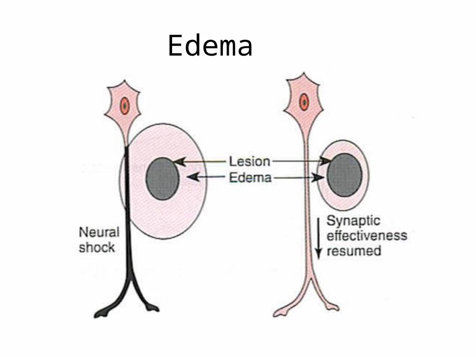
Metabolic Effect of Brain Injury: Excitotoxicity
• After brain injury, neurons deprived of oxygen die. These neurons also release excessive glutamate from their axon terminals, which causes surrounding neurons to overexcite and triggers a cascade of cell death, i.e. excitotoxicity.
• Damage after brain injury is not only limited to direct neuronal death, but also the indirect death from excitotoxicity.
Secondary injury after traumatic brain injury
Park E et al. CMAJ 2008;178:1163-1170

ischemia
Glutamate release
↑Ca++ influx
↑Intracellular H2O
Cell swelling
CELL DEATH
↑Glycolysis
Lactic acid
Protein enzyme
Oxygen free radicals

INTRACELLULAR RESPONSES TO INJURY AND RECOVERY OF FUNCTION
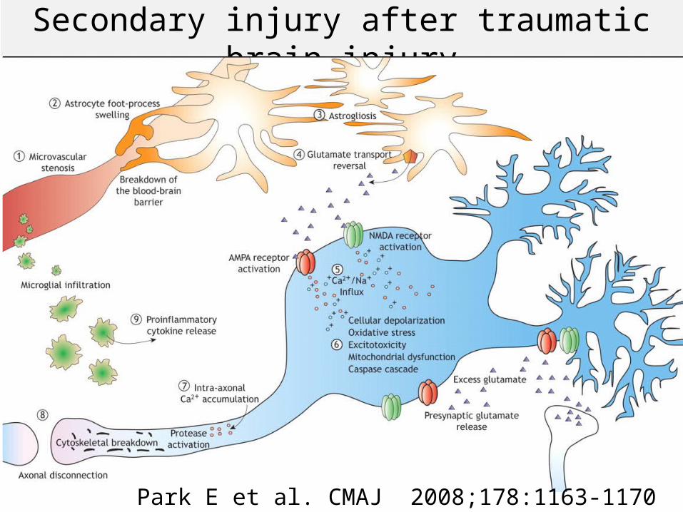
PNS and CNS Recovery: Collateral Sprouting
• Axon of remaining neuron forms a collateral sprout to reinnervate denervated target
With injury, younger rats develop more collateral sprouts than older rats

Collateral sprouting

PNS and CNS Recovery: Neural Regeneration
• Presynaptic axon and its target cell (postsynpatic ) are damaged
• Injured axon sprouts to new targets
• Regenerate the axon at 1mm/month

Regenerative sprouting in CNS is not functional and does not occur
• Neural regeneration occurs most frequently in PNS because Schwann cells produce nerve growth factor, which help recovery.
• Astrocytes and microglia form glial scars, which physically block axonal regeneration
• Oligodendrocytes produce Nogo (neurite outgrowth inhibitor), which inhibits axonal regeneration
• Axons don’t naturally regenerate very large distances

Synkinesis: aberrant regenerative sprouting in PNS
• Axon sprouting can cause problems when an inappropriate targets is innervated.
• After injury, motor axons innervate different muscle than they previously did, causing unwanted abnormal movements when the neurons fire.–e.g. Bell’s Palsy (CN VII): patients wink
when they intend to purse lips.• Typically lasts no more than a few months

Woman with a history of Bell palsy 18 years earlier and with synkinesis
(A–C) before treatment
(D–F) After PT and Chemo-denervation

• Recovery from early transient events, such as edema and diaschisis, neural shock
CNS Recovery: Recovery of Synaptic Effectiveness
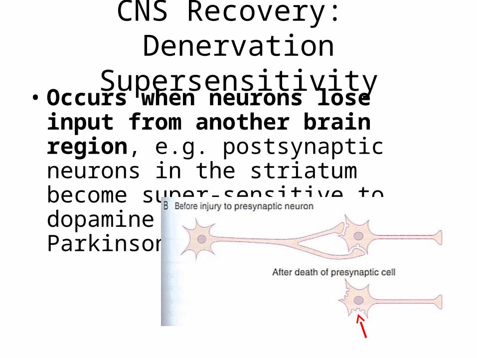
CNS Recovery: Denervation Supersensitivity
• Occurs when neurons lose input from another brain region, e.g. postsynaptic neurons in the striatum become super-sensitive to dopamine in patient with Parkinson

CNS Recovery: Synaptic Hypereffectiveness
• Occurs when only some branches of presynaptic axons are damaged
• Remaining axons receive all neurotransmitters that would normally be distributed among all branches
• Larger amount of neurotransmitters released to post- synaptic receptors

CNS Recovery: Unmasking of Silent Synapses
• In normal CNS, many neurons are not used due to competition of neural pathways
• Unused neurons become active

Functional Reorganization (remapping) of Cerebral Cortex
Training can expand cortical representation areas (i.e. cortical map)
A1 = pre-trainingA2 = post-trainingCortical area 3bIn adult monkey, sensory training for 3 months on task requiring repeated use of tips of distal phalanges of digits 2, 3, and sometimes 4

Functional Reorganization (remapping) of Cerebral Cortex
• Only patients with phantom limb pain (PLP) showed an expansion of areas representing lip into areas previously representing the hand on fMRI
Lotze, et al., 2001
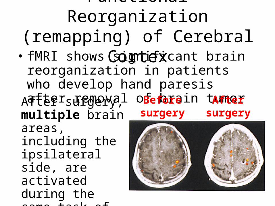
Functional Reorganization (remapping) of Cerebral Cortex
• fMRI shows significant brain reorganization in patients who develop hand paresis after removal of brain tumor
Before surgery
After surgery
After surgery, multiple brain areas, including the ipsilateral side, are activated during the same task of finger and thumb movement.

Structural Changes in Gray Matter and White Matter after Reduced Sensory
and Motor Input
48 hours later 16 days laterLanger, 2012
• Limb immobilization caused reduced nerve fiber density and cortical thickness in the brain

STRATEGIES AND PRINCIPLES TO ENHANCE NEURAL PLASTICITY

Effect of Training on CNS• Type of training
–Skill learning associated with cortical reorganization
–Strength training is not associated with cortical reorganization
• Early Intensive Training–Early, high-dose, constrain-induced
movement therapy (CIMT) results in the worse outcome compared to moderate intensity CIMT or conventional therapy

Effect of Training on CNS• Non-invasive cortical stimulation
–Stimulation applied during or shortly before skill training enhances motor learning. In contrast, stimulation after skill training interferes with the skill acquisition
–e.g. transcranial direct current stimulation (ionto- phoresor), rTMS
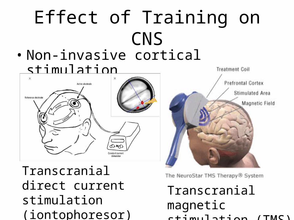
Effect of Training on CNS• Non-invasive cortical stimulation
Transcranial direct current stimulation (iontophoresor)
Transcranial magnetic stimulation (TMS)

Effect of Training on CNS
• Somatosensory stimulation –Using sensory-level electrical stimulation
combined with training–e.g. TENS to hand muscles increase the
size of cortical hand map–e.g. Cortical plasticity also occurs with
functional electrical stimulation applied to lower extremities: ↑descending input from corticospinal tract to activate TA

Effect of Training on CNS
• Somatosensory stimulation
Transcutaneous electrical nerve stimulation (TENS)
Functional electrical stimulation (FES)

Effect of Training on CNS• Constraint-Induced Movement Therapy
(CIMT) –Restrain unaffected limb and work
other limb intensely (e.g. put intact arm in a sling and use the
affected arm)–Affected limb must actively
engages in exercise, functional activities to benefit from CIMT

Principles of Experience-Dependent Plasticity
1. Use it or lose it2. Use it and
improve it3. Specificity4. Repetition matters5. Intensity matters
6. Time matters7. Salience matters
(training experience)
8. Age matters9. Transference10.Interference:

PLASTICITY AND LEARNING

Shift from short term to long term learning is reflected in a move along the continuum of neural modifiability.
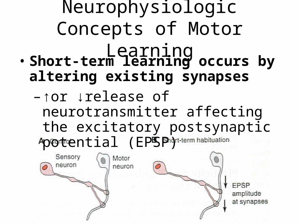
Neurophysiologic Concepts of Motor Learning
• Short-term learning occurs by altering existing synapses–↑or ↓release of neurotransmitter
affecting the excitatory postsynaptic potential (EPSP)

Neurophysiologic Concepts of Motor Learning
• Long-term learning occurs by the reduction or formation of new synapses or structural changes on neurons, e.g.–Habituation: decrease in synapses (C)–Sensitization: increase in synapses (D)

Procedural Learning: Role of Cerebellum
• Purkinje cells are output cells• Climbing fibers signal error, critical for
correcting ongoing movements• Mossy fibers bring sensory feedback
about ongoing movements, critical for controls movements
• When climbing fiber increases its activity, mossy fiber signals to Purkinje cells is reduced, which change the synaptic strength for the circuit


Procedural Learning: Role of Cerebellum
Monkeys move arm(1) Against an expected load (already learned)(2) Against an unexpectedly increased load(3) Against same load as in (2) after some practice
Simple spikes from mossy fibersComplex spikes from climbing fibers

Declarative Forms of Learning: Long-Term Potentiation
• LTP requires simultaneous firing of both presynaptic and postsynaptic cells
• Postsynaptic neuron must depolarize when the Glutamate binds to the NMDA receptor in order to open the ion channel

LTP conversion of silent synapses to active synapses
Lundy-Ekman Fig. 4-1
New dendritic spines formed
AMPA receptors inserted into membrane
Change in pre-synaptic cell to produce new synapse

Complex Form of Motor Learning
• Sensory cortex of cats is absolutely necessary to learn a new skill, how to supinate the forearm to retrieve food.
• Once learned, ablation of the sensory cortex will not affect the movement

Acquisition of Skills: Shift to Automaticity
• Automaticity during skill acquisition is associated with a reduction of brain activation in several regions
• Older adults or individuals with neurological diseases may activate more brain areas or increase the activity levels in order to perform the skills at the same level as health individuals