Embed Size (px)
Citation preview

RESEARCH ARTICLE
PTH1R Mutants Found in Patients with
Primary Failure of Tooth Eruption Disrupt G-
Protein Signaling
Hariharan Subramanian1,2☯, Frank Doring3☯, Sina Kollert3, Natalia Rukoyatkina1,4,
Julia Sturm3, Stepan Gambaryan4,5, Angelika Stellzig-Eisenhauer6, Philipp Meyer-
Marcotty6,7, Martin Eigenthaler1,6‡*, Erhard Wischmeyer3‡
1 Institute of Clinical Biochemistry and Pathobiochemistry, University of Wuerzburg, Wuerzburg, Germany,
2 Institute of Experimental Cardiovascular Research, University Medical Centre Hamburg-Eppendorf,
Hamburg, Germany, 3 Institute of Physiology, AG Molecular Electrophysiology, University of Wuerzburg,
Wuerzburg, Germany, 4 Sechenov Institute of Evolutionary Physiology and Biochemistry, Russian Academy
of Sciences, St. Petersburg, Russia, 5 Department of Cytology and Histology, St. Petersburg State
University, St. Petersburg Russia, 6 Department of Orthodontics, University Clinic of Wuerzburg, Wuerzburg,
Germany, 7 Department of Orthodontics, University Clinic of Goettingen, Goettingen, Germany
☯ These authors contributed equally to this work.
‡ These authors are joint senior authors on this work.
Abstract
Aim
Primary failure of tooth eruption (PFE) is causally linked to heterozygous mutations of
the parathyroid hormone receptor (PTH1R) gene. The mutants described so far lead to
exchange of amino acids or truncation of the protein that may result in structural changes of
the expressed PTH1R. However, functional effects of these mutations have not been inves-
tigated yet.
Materials and Methods
In HEK293 cells, PTH1R wild type was co-transfected with selected PTH1R mutants identi-
fied in patients with PFE. The effects on activation of PTH-regulated intracellular signaling
pathways were analyzed by ELISA and Western immunoblotting. Differential effects of wild
type and mutated PTH1R on TRESK ion channel regulation were analyzed by electrophysi-
ological recordings in Xenopus laevis oocytes.
Results
In HEK293 cells, activation of PTH1R wild type increases cAMP and in response activates
cAMP-stimulated protein kinase as detected by phosphorylation of the vasodilator stimu-
lated phosphoprotein (VASP). In contrast, the PTH1R mutants are functionally inactive and
mutant PTH1R/Gly452Glu has a dominant negative effect on the signaling of PTH1R wild
type. Confocal imaging revealed that wild type PTH1R is expressed on the cell surface,
whereas PTH1R/Gly452Glu mutant is mostly retained inside the cell. Furthermore, in
PLOS ONE | DOI:10.1371/journal.pone.0167033 November 29, 2016 1 / 16
a11111
OPENACCESS
Citation: Subramanian H, Doring F, Kollert S,
Rukoyatkina N, Sturm J, Gambaryan S, et al.
(2016) PTH1R Mutants Found in Patients with
Primary Failure of Tooth Eruption Disrupt G-Protein
Signaling. PLoS ONE 11(11): e0167033.
doi:10.1371/journal.pone.0167033
Editor: Peter A. Friedman, University of Pittsburgh
School of Medicine, UNITED STATES
Received: April 2, 2016
Accepted: November 8, 2016
Published: November 29, 2016
Copyright: © 2016 Subramanian et al. This is an
open access article distributed under the terms of
the Creative Commons Attribution License, which
permits unrestricted use, distribution, and
reproduction in any medium, provided the original
author and source are credited.
Data Availability Statement: All relevant data are
within the paper and its supporting information
files.
Funding: This work was supported by the
Interdisziplinares Zentrum fur Klinische Forschung
des Universitatsklinikum Wurzburg, grant Z-4/113.
Publication was supported by the Open Access
Publication Fund of the University of Wuerzburg.
The funders had no role in study design, data
collection and analysis, decision to publish, or
preparation of the manuscript.

contrast to wild type PTH1R which substantially augmented K+ currents of TRESK chan-
nels, coupling of mutated PTH1R to TRESK channels was completely abolished.
Conclusions
PTH1R mutations affect intracellular PTH-regulated signaling in vitro. In patients with pri-
mary failure of tooth eruption defective signaling of PTH1R mutations is suggested to occur
in dento-alveolar cells and thus may lead to impaired tooth movement.
Introduction
Parathyroid hormone (PTH), PTH-related peptide (PTHrP) and parathyroid hormone recep-
tor type 1 (PTH1R) play a key role in regulation of bone remodeling and plasma calcium levels
as reviewed by Taylor et al. [1]. Various autosomal dominant or recessive mutations in the
PTH1R gene have been identified to be associated with different diseases. The most severe
clinical disease is a complete loss of PTH1R function resulting in lethal Blomstrand chondro-
dysplasia characterized by advanced endochondral bone maturation and premature ossifica-
tion of all skeletal elements [2].
In contrast to this homozygous disease, we and others have described the incidence of het-
erozygous PTH1R mutations which may inactivate PTH1R function in Primary Failure of
Tooth Eruption (PFE) (MIM #125350). PFE is a rare autosomal, non-syndromic disorder with
incomplete eruption of mainly posterior teeth and growth deficiency of the alveolar process in
the affected region [3, 4]. The developing teeth remain below the occlusion level resulting in a
severe lateral open bite situation. Furthermore, movement of the teeth by orthodontic treat-
ment results in fusion of the dental cement with the surrounding bone making further tooth
movement impossible [5].
The genetic defects underlying PFE were initially identified by genetic screening in affected
families [3]. While the first reported 3 mutations (c.463G>T, c.543+1G>A, c.1050-3C>G) in
patients with PFE were predicted to generate loss-of-function proteins [3], the spectrum of
PTH1R mutations by now has been expanded by several investigators showing the occurrence
of more than 40 potentially pathogenic mutations [4, 6–8]. Furthermore, occurrence of spo-
radic cases of PTH1R mutations causing PFE have been identified by exome resequencing [9].
The PTH1R mutations identified so far are heterogenous and may result in proteolytic degra-
dation of the PTH1R precursor protein, truncation of the PTH1R protein or single amino acid
exchanges within the complex architecture of the receptor. However, until now no functional
data on the molecular and cellular effects of the PFE related mutations in eukaryotic cells exist.
The PTH1R gene encodes a secretin-like class II G protein-coupled receptor precursor that
is processed into a mature receptor protein of a 593 amino acids. The PTH1R receptor belongs
to the group of seven-helical-transmembrane receptors and mainly consists of a large extracel-
lular loop with a PTH binding site, seven transmembrane, helically shaped segments and an
intracellular signaling domain. PTH1R is highly expressed in osteoblasts and renal tubular
cells and regulates calcium homeostasis and bone formation. In addition to PTH agonist,
PTH1R is activated by PTHrP [10,11], a paracrine hormone that also regulates bone develop-
ment and epithelial-mesenchymal interactions in developing teeth.
Signal transduction of PTH1R is mediated by the intracellular domain which couples to
Gαs-proteins and thus increases the activity of adenylate cyclase (AC). This signaling pathway
is well characterized in osteoblasts [12], leading to increase of intracellular cAMP and
Disrupted Signaling by PTH1R Mutant
PLOS ONE | DOI:10.1371/journal.pone.0167033 November 29, 2016 2 / 16
Competing Interests: The authors have declared
that no competing interests exist.
accordingly to cAMP-dependent activation of enzymes such as protein kinase A (PKA). PKA
subsequently phosphorylates various signaling proteins. One major target protein is the vaso-
dilator-stimulated phosphoprotein (VASP) regulating cell adhesion and motility [13]. Alterna-
tively, the PTH1R is able to activate the Gq11/phospholipase C/ calcium/ PKC pathway [14].
One target of this pathway is the tandem-pore (K2P) potassium channel TRESK [15,16] which
is activated in a calcium-dependent manner.
In our present study, we used a cell model that allows transfection of PTH1R and its
mutants to study their effect on intracellular, PTH-activated signaling pathways. For initial
experiments, we selected two known mutations from patients with PFE, the PTH1R/
Trp339stop mutation resulting in a truncated protein and the PTH1R/Gly452Glu mutation
with single amino acid exchange and may affect the structure of the receptor [3, 10]. Both
mutants were functionally inactive and also in heterozygous circumstances strongly interfered
with the activation of physiological PTH1R receptor signaling pathways.
Materials and Methods
Ethics statement
The study was approved by the Ethic Committee of the University Clinic of Wuerzburg, vote
103/04 and 192/11.
Materials
Forskolin was purchased from Sigma Aldrich (Munich, Germany). PTH was obtained from
Tocris (Wiesbaden-Nordenstadt, Germany). Antibodies against Phospho-VASPSer157, Actin,
and GAPDH were from Cell Signaling (Frankfurt/Main, Germany) and were used in a 1:1000
dilution. Antibody against PTH1R was from Thermo Scientific (Bonn, Germany) and was
used in a 1:1000 dilution.
PTH1R constructs
Full-length human PTH1R cDNA was a gift from Dr. Lohse, Institute of Pharmacology of the
University of Wuerzburg. The mutants (c.G1016A (p.Trp339stop) and c.G1355A (p.Gly452-
Glu) were generated by QuikChange site-directed mutagenesis Kit (Stratagene, La Jolla, USA)
following the manufacturer’s protocol. To detect PTH1R in membrane fractions of oocytes
myc-tag was fused to the C-terminus of all constructs, respecting the premature stop codon of
the PTH1R/Trp339stop mutant. For biochemical assays in HEK293 cells cDNAs were sub-
cloned into pcDNA3 vector. The oocyte expression vector pSGEM was used to express recep-
tors and K+ channels for electrophysiological recordings and Western blots in Xenopus laevisoocytes. Cloning of mouse K2P channel TRESK has been previously described [15,16]. For
confocal imaging PTH1R wild type and PTH1R/Gly452Glu mutant were subcloned into
pEYFP-N1 vector to express PTH1R with c-terminal YFP.
Extraction and immunoblotting of oocyte membranes
Xenopus laevis oocytes injected with in vitro transcripts of myc-tagged PTH1R wild type,
PTH1R/Gly452Glu PTH1R/Trp339stop or H2O were incubated for 48 h in ND 96 solution (96
mM NaCl, 2 mM KCl, 1 mM MgCl2, 1 mM CaCl2, 5 mM HEPES, pH 7.4) and subsequently
homogenized by repeated pipetting in solubilization buffer (10mM HEPES, pH 7.9, 1mM
MgCl2, 83mM NaCl, 0,5mM PMSF, protease inhibitor cocktail complete [Roche]). Plasma
membranes from homogenates of 25 oocytes were isolated by sequential centrifugations at 2x
1000g (10 min) and 10000g (20 min) at 4˚C. Precipitates of the final centrifugation step were
Disrupted Signaling by PTH1R Mutant
PLOS ONE | DOI:10.1371/journal.pone.0167033 November 29, 2016 3 / 16
solubilized in modified Laemmli loading buffer (126 mM Tris/HCl pH 6,8, 300 mM DTT, 6%
SDS, 10% Glycerol 0,2% bromophenol blue). Equal amounts of precipitated membrane frac-
tions were subjected to PAGE and analyzed by Western immunoblots probed with monoclo-
nal mouse anti myc-tag antibody (1:500; clone 9B11; CST, Danvers, MA). For detection HRP-
conjugated goat anti mouse immunoglobulins (1:10,000; Jackson ImmunoResearch Laborato-
ries, West Grove, PA) were applied and after washing developed with self-prepared chemilu-
minescence reagent (0.1 M Tris pH 8.6, 1.25 mM Luminol, 0.6 mM p-Cumaric acid, 0.01%
H2O2).
Transfection and experiments on HEK293 cells
HEK293 cells were cultured in Dulbecco’s Modified Eagle’s Medium (DMEM), supplemented
with 10% fetal bovine serum. Cells were transiently transfected with PTH1R wild type and/or
mutant plasmids using METAFECTENE PRO (Biontex, Martinsried/Planegg, Germany) fol-
lowing the manufacturer’s protocol. 24 h after transfection cells were washed with PBS and left
in serum-free media for 2 h followed by stimulation with PTH or forskolin for 10 min. After
stimulation, cells were lysed for Western blot analysis.
Western blot analysis
For Western blot analysis cells were washed with PBS and lysed in 2x SDS gel loading buffer
(500 μl / confluent well of 6 well plate). Cell lysates were separated by SDS-PAGE, transferred
to nitrocellulose membranes followed by incubation with appropriate primary antibodies
overnight at 4˚C. For visualization of the signal, goat anti-rabbit or anti-mouse IgG conjugated
with horseradish peroxidase were used as secondary antibodies (dilution 1:10000 each), fol-
lowed by ECL detection. Blots were scanned using SilverFast software and analyzed densito-
metrically by NIH Image J software for uncalibrated optical density.
cAMP measurement
cAMP level in HEK293 cells was evaluated using a cAMP EIA Kit (Cayman Chemical, Ham-
burg, Germany) following the manufacturer’s instructions.
Confocal imaging
PTH1R wild type and PTH1R/Gly452Glu mutant fused with YFP were expressed in HEK293
cells. 24 hours post-transfection, cells were transferred to imaging medium (144 mM NaCl, 5
mM KCl, 1 mM CaCl2, 1 mM MgCl2, 10 mM HEPES, pH = 7.3) and incubated with CellMask™Deep red plasma membrane stain (Life Technologies GmbH Darmstadt, Germany) according
to manufacturer’s instruction. Then cells were mounted onto 63X objective in Zeiss LSM800
microscope and the images were captured with Zeiss Axiocam.
Electrophysiology
For heterologous gene expression in Xenopus laevis oocytes, capped run-off poly(A+) cRNA
transcripts from linearized cDNA of wild type and mutated PTH-receptors and channels were
synthesized and injected into defolliculated oocytes. Cells were incubated at 19˚C in ND96
solution (96 mM NaCl, 2 mM KCl, 1 mM MgCl2, 1 mM CaCl2, 5 mM HEPES, pH 7.4) supple-
mented with 100 μg/ml gentamicin and 2.5 mM sodium pyruvate. 48–72 hours after injection
two electrode voltage-clamp measurements were performed with a TURBO TEC-10 C ampli-
fier (npi, Tamm, Germany). Stimulation and data acquisition were controlled by Pulse soft-
ware (HEKA, Germany). Oocytes were placed in a small volume perfusion chamber with a
Disrupted Signaling by PTH1R Mutant
PLOS ONE | DOI:10.1371/journal.pone.0167033 November 29, 2016 4 / 16
constant flow of ND96 with 0.1% BSA or ND 96 with 0.1% BSA supplemented with different
concentrations of PTH.
Data analysis
All experiments were performed at least in triplicate and data shown are means ± S.D. Differ-
ences between groups were analyzed by one-way ANOVA or two-way ANOVA, respectively.
p<0.05 was considered statistically significant, p<0.01 as statistically highly significant. Origi-
nal data of ANOVA analysis are shown in supporting information files S1 and S2 Tables.
Results
PTH increases intracellular cAMP and induces cAMP/PKA-triggered
VASP phosphorylation in HEK293 cells transfected with PTH1R
In order to elucidate the efficacy of our expression system we transiently transfected HEK
293 cells with pcDNA3 or pcDNA3/PTH1R vector as described in methods. Controls and
stimulation with 10 nM or 100 nM PTH was performed for up to 10 min as indicated in Fig
1. After stimulation with 10 nM PTH, HEK 293 cells transfected with the PTH1R/pcDNA3
vector responded with a 4.7 ± 0.4 and 5.8 ± 0.6 -fold cAMP increase at 5 and 10 min, respec-
tively (Fig 1, upper panel). Increase of the PTH concentration to 100 nM did not result in a
further increase in cAMP levels as shown by a 4.9 ± 0.8 and 4.3 ± 0.6 fold cAMP increase at 5
and 10 min, respectively (p<0.01, number of independent experiments (n), n = 3). Cells
transfected with the empty pcDNA3 vector and stimulated by 10 nM or 100 nM PTH
showed no increase in cAMP compared to unstimulated control. To demonstrate intactness
of intracellular signaling pathways in the transfected cells, stimulation with 5μM Forskolin,
a strong direct activator for cAMP-regulated signaling pathways was performed. Forskolin
induced a 7.7 ± 0.6 or 8.3 ± 0.9 fold increase in cAMP in cells transfected with PTH1R or
empty vector, respectively (p<0.01, n = 3). Increase in cAMP resulted in corresponding
phosphorylation of the intracellular signaling protein VASP at the PKA-preferred phosphor-
ylation site Serine-157 (Fig 1, lower panel). The degree of VASP phosphorylation strongly
correlated with the increase of cAMP. Again, pcDNA3 transfected cells showed no increase
in VASP phosphorylation above baseline, whereas the PTH-independent cAMP-elevating
substance forskolin induced maximal VASP phosphorylation in both PTH1R and control
transfected cells.
PTH1R/Trp339stop and PTH1R/Gly452Glu mutants are functionally
inactive and do not activate intracellular cAMP-regulated pathways
In a second set of experiments we tested the effect of two different mutations of the PTH1R
protein. HEK293 cells were transfected with empty vector pcDNA3, pcDNA3:PTH1R wild
type, pcDNA3:PTH1R/Trp339stop mutant or pcDNA3:PTH1R/Gly452Glu mutant plasmids
as described in methods. 24 h after transfection cells were stimulated with 10 nM PTH or
10 μM forskolin (as a PTH-independent positive control of VASP phosphorylation). In cells
transfected with the empty pcDNA3 vector, stimulation with PTH did not induce VASP
phosphorylation, whereas stimulation with forskolin demonstrated the intactness of the intra-
cellular cAMP signaling pathway. Transfection of HEK293 with pcDNA3:PTH1R wild type
construct and stimulation with PTH increased intracellular cAMP and induced subsequent
phosphorylation of PKA-induced VASP phosphorylation at Serine 157 as shown in Fig 2A
(data for cAMP in supporting information file S3 Table).
Disrupted Signaling by PTH1R Mutant
PLOS ONE | DOI:10.1371/journal.pone.0167033 November 29, 2016 5 / 16

In contrast, cells transfected with pcDNA3:PTH1R/Trp339stop, which codes for a trun-
cated PTH1R protein showed no increase in cAMP and no phosphorylation of VASP after
stimulation with 10 nM PTH. The second mutant PTH1R/Gly452Glu, is supposed to alter the
structure of the PTH1R receptor by the exchange of glycine against glutamic acid at position
452. Again, cells transfected with the PTH1R/Gly452Glu mutant showed no increase in intra-
cellular cAMP after PTH stimulation and no subsequent VASP phosphorylation (Fig 2A, data
for cAMP in supporting information file S3 Table). Furthermore, increase of PTH concentra-
tion to 100 nM did not induce cAMP increase or VASP phosphorylation in PTH1R mutant
transfected cells (data not shown). Incubation of the cells with forskolin, a PTH1R-indepen-
dent elevator of cAMP, as a positive control resulted in strong cAMP/PKA-induced phosphor-
ylation of VASP in both wild type as well as mutant transfected cells (Fig 2A). As revealed by
confocal imaging, we identified co-localization of wild type PTH1R with the membrane dye
Fig 1. Activation of PTH1R by PTH in PTH1R transfected HEK293 cells increases intracellular cAMP and induces PKA-
triggered VASP phosphorylation. The upper panel depicts cAMP increase in HEK293 cells transfected with PTH1R (pcDNA3/
PTH1R transfected) or with the vector only (pcDNA3 transfected). Cells were stimulated for up to 10 min with 10 or 100 nM PTH as
indicated. For PTH-independent positive control cells were stimulated by 5 μM forskolin (as depicted). Data of thee independent
experiments are presented as mean ± SEM, n = 3, p< 0.05 compared to the pcDNA3 control. Phosphorylation of VASP and loading
control were documented by Western immunoblots with specific antibodies as indicated in the lower panel.
doi:10.1371/journal.pone.0167033.g001
Disrupted Signaling by PTH1R Mutant
PLOS ONE | DOI:10.1371/journal.pone.0167033 November 29, 2016 6 / 16

Fig 2. Stimulation and localization of PTH1R wild type or PTH1R mutants. (A) Extracts of stimulated (as indicated)
HEK293 cells transfected with pcDNA3, pcDNA3:PTH1R wild type (WT), pcDNA3:PTH1R/Trp339stop or pcDNA3:PTH1R/
Gly452Glu mutant were analyzed for VASP phosphorylation at the cAMP/PKA-preferred site Serine 157 (upper panel).
Blotting against actin was used as loading control and used for quantification of VASP phosphorylation (lower panel).
Representative blots of three independent experiments are shown. (B) Representative images from confocal imaging of YFP-
tagged wild type (WT-YFP) and PTH1R/Gly452Glu (452Glu-YFP) in transfected HEK293 cells. CellMask deep red membrane
dye was used for membrane staining. Scale bar 10 μM.
doi:10.1371/journal.pone.0167033.g002
Disrupted Signaling by PTH1R Mutant
PLOS ONE | DOI:10.1371/journal.pone.0167033 November 29, 2016 7 / 16
deep red (CellMask), whereas the PTH1R/Gly452Glu mutant was found to be intracellular and
did not colocalize with the membrane-staining dye (Fig 2B).
Dominant negative effect of PTH1R/Gly452Glu mutant
To identify whether mutant PTH1R/Gly452Glu has any dominant negative effect on wild type
PTH1R activity, increasing amounts of PTH1R/Gly452Glu plasmid were cotransfected with a
fixed amount of wild type PTH1R plasmid. After stimulation of cells with PTH, VASP phos-
phorylation was monitored. Western blot of VASP phosphorylation demonstrate that 5 or 10
times higher amounts of PTH1R/Gly452Glu plasmid transfected together with 100 ng wild
type vector significantly reduced VASP phosphorylation in comparison to the effect of wild
type PTH1R alone. This result documents a dominant negative effect of PTH1R/Gly452Glu
mutant on the wild type receptor (Fig 3).
Localization of PTH receptor variants
To explore proper expression and targeting of wild type and mutated PTH1R we analysed
myc-tagged proteins when expressed in Xenopus oocytes. Crude membrane fractions of
oocytes, injected with identical amounts of cRNA of each receptor construct (Fig 4) were iso-
lated and subjected to Western immunoblots. Specific signals, absent in H2O injected negative
controls, were found in samples of PTH1R wild type, which indicates proper expression of the
protein in oocytes. Also in preparations of mutant PTH1R/Gly452Glu specific protein bands
of the receptor were detected. Prominent bands were detected at apparent molecular weight of
>80 kDa and 175 kDa representing glycosylated monomeric and dimeric receptors. Although
proteins in both samples display a different and inhomogeneous band pattern probably result-
ing from incomplete denaturation of sticky proteins, the specific signals document expression
of PTH1R wild type and PTH1R/Gly452Glu mutant in membrane fractions in this expression
system. However samples of PTH1R/Trp339stop do not show any signal with the antibody
and thus document that the truncated protein is not located in membranes or not even
expressed in the oocyte. As truncation at Trp339 disrupts the fourth transmembrane segment
and eliminates the final three ones negative influence on the machinery of protein synthesis
are very likely.
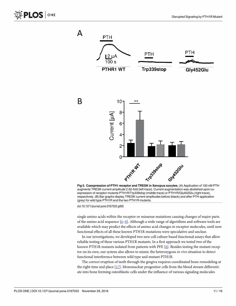
Coupling of PTH receptor 1 to TRESK K2P channels
In order to establish a physiological assay for PTH receptor activity we co-expressed the PTH1R
with the tandem-pore potassium channel TRESK in Xenopus oocytes. TRESK currents are acti-
vated by Gαq-coupled receptors via a phospholipase C-dependent pathway in a Ca2+-dependent
manner. Activation of PTH1R with 100 nM PTH augmented TRESK currents by 2.62 ±0.43
fold (Fig 5, upper left; p<0.01; number of independent experiments (n), n = 17). In contrast,
upon co-expression of the truncated receptor PTH1R/Trp339stop with TRESK channels, cur-
rent amplitude was unchanged (1.14±0.05 fold; n = 13). The same was true for the second
PTH1R mutant tested. Coupling of the mutant PTH1R/Gly452Glu to TRESK again failed to
augment TRESK currents significantly (1.08±0.09 fold; n = 14). To elucidate whether the effect
is specific for the exchange of Gly by Glu at position 452 we tested a conservative amino acid
exchange. Remarkably substitution of glycine 452 in the wild type PTH1R to alanine did not
significantly affect receptor function. In this case TRESK currents were significantly augmented
by 2.23±0.45 fold (p<0.05, n = 18) and thus were comparable to the effect of wild type PTH
receptors (data not shown). In summary, mutations leading to a truncated PTH receptor pro-
tein as well as a specific amino acid exchange at position 452, probably altering receptor struc-
ture, lead to loss of function of the PTH1R.
Disrupted Signaling by PTH1R Mutant
PLOS ONE | DOI:10.1371/journal.pone.0167033 November 29, 2016 8 / 16

Fig 3. PTH induced VASP phosphorylation in HEK293 cells transfected with various ratio of PTH1R
wild type and PTH1R/Gly453Glu mutant plasmids. (A) HEK293 cells were transfected with wild type and
PTH1R/Gly452Glu plasmids in a ratio 1:5 (100 ng WT and 500 ng mutant) or 1:10 (100 ng WT and 1μg
mutant) as indicated. As controls, pcDNA transfected cells and cells co-transfected with WT (100 ng) and
pcDNA3 (1 μg) were used. 48 hours after transfection, cells were stimulated as indicated and lysed for
Western immunoblots against P-VASP and GAPDH (loading control). Shown are representative blots of 3
independent experiments. (B) Bar graphs show quantification of phospho-VASP calculated with ImageJ
software (n = 3). **p<0.05.
doi:10.1371/journal.pone.0167033.g003
Disrupted Signaling by PTH1R Mutant
PLOS ONE | DOI:10.1371/journal.pone.0167033 November 29, 2016 9 / 16

Discussion
Primary failure of tooth eruption has been closely linked to heterozygous mutations in the
gene encoding for PTH1 receptor [3]. We and others have identified until now more than 40
mutations causing deletions of the PTH1R, distinct nucleotide changes leading to exchange of
Fig 4. Membrane localization of PTH1R wild type and mutants. Membrane fractions of Xenopus laevis
oocytes injected with cRNA of myc-tagged wild type (WT) and mutated PTH1R or H2O were analyzed by
Western immunoblotting. As revealed by myc-tag antibody specific signals (as indicated on the right) were
detected in preparations of PTH1R wild type and PTH1R/Gly452Gly whereas these protein bands were
absent in samples of PTH1R/Trp339stop and H2O (negative control). Loading of equal amounts of protein is
monitored by unspecific double bands (asterisk) in all samples. Quality and amount of cRNA injected into
oocytes for heterologous expression of receptors is documented by RNA gel (lower panel).
doi:10.1371/journal.pone.0167033.g004
Disrupted Signaling by PTH1R Mutant
PLOS ONE | DOI:10.1371/journal.pone.0167033 November 29, 2016 10 / 16
single amino acids within the receptor or missense mutations causing changes of major parts
of the amino acid sequence [6–8]. Although a wide range of algorithms and software tools are
available which may predict the effects of amino acid changes in receptor molecules, until now
functional effects of all these known PTH1R mutations were speculative and unclear.
In our investigations, we developed two new cell culture based functional assays that allow
reliable testing of these various PTH1R mutants. In a first approach we tested two of the
known PTH1R mutants isolated from patients with PFE [8]. Besides testing the mutant recep-
tor on its own, our system also allows to mimic the heterozygous in vivo situation to detect
functional interference between wild type and mutant PTH1R.
The correct eruption of teeth through the gingiva requires coordinated bone remodeling at
the right time and place [17]. Mononuclear progenitor cells from the blood stream differenti-
ate into bone forming osteoblastic cells under the influence of various signaling molecules
Fig 5. Coexpression of PTH1 receptor and TRESK in Xenopus oocytes. (A) Application of 100 nM PTH
augments TRESK current amplitude 2.62-fold (left trace). Current augmentation was abolished upon co-
expression of receptor mutants PTH1R/Trp339stop (middle trace) or PTH1R/Gly452Glu (right trace),
respectively. (B) Bar graphs display TRESK current amplitudes before (black) and after PTH application
(grey) for wild type PTH1R and the two PTH1R mutants.
doi:10.1371/journal.pone.0167033.g005
Disrupted Signaling by PTH1R Mutant
PLOS ONE | DOI:10.1371/journal.pone.0167033 November 29, 2016 11 / 16
including PTH, which plays a crucial regulatory role in this process [18]. Animal models using
transgenic mice have elucidated the importance of intact PTH signaling for tooth eruption
[17]. Missing links in PTH/PTH1R signaling pathways always resulted in failure of tooth erup-
tion and major changes in bone development [19]. PTH1R mediates the effects of PTH in
osteoblasts by stimulation of adenylate cyclase (AC) and by increase of cytosolic calcium con-
centration [20, 21]. Activation of AC increases intracellular cAMP in osteoblasts, subsequently
activating cAMP-dependent protein kinases (PKAs) that phosphorylate and activate several
cytoskeletal regulator proteins [21]. One major target of PKAs is the regulator protein VASP
(vasodilator stimulated phosphoprotein) that coordinates both cell adhesion and cell migra-
tion in many cell types [22].
In our experimental model system we used HEK293 cells which supply all necessary signal-
ing molecules of this PTH/cAMP/VASP signaling pathway except the PTH1R. Stimulation of
HEK293 with PTH therefore did not result in any detectable increase in cAMP or VASP phos-
phorylation, whereas PTH-independent stimulation of AC via forskolin resulted in strong
increase in cAMP and VASP phosphorylation. In this system, the transfection of human wild
type PTH1R and stimulation of the cells with PTH showed good response with regard to intra-
cellular cAMP signaling. This provides a new model system for testing the functional effects of
the known PTH1R mutants derived from patients with PFE [3, 6, 7, 8].
PTH1R belongs to the group of seven-helical-transmembrane receptors. Binding of ligands
to such receptors changes the configuration of the protein and mediates signaling effects inside
the cell via binding of G-proteins to the intracellular domains of the receptors. Changes in the
amino acid sequence of G-protein coupled receptors may have unexpected effects in functional
testing [23]. Mutant receptors may not interfere with signaling of the wild type protein, how-
ever, dominant negative effects with complete suppression of receptor signaling as well as
reduction of intracellular receptor signaling to a variable degree have been described in a vari-
ety of cases [23]. Even single amino acid mutations may result in loss as well as in gain of
receptor function.
PTH1R is highly expressed on osteoblasts and renal tubular cells and regulates calcium
homeostasis and bone formation. Besides PTH, PTH1R is further activated by PTHrP [10], a
paracrine hormone that also regulates bone development and epithelial-mesenchymal interac-
tions in developing teeth. A complete loss of PTH1R function results in lethal Blomstrand
chondrodysplasia (BOCD, OMIM #215045), characterized by advanced endochondral bone
maturation and premature ossification of all skeletal elements [2]. In EIKEN disease (MIM
#600002), multiple epiphyseal dysplasia and retarded ossification are found [24]. Jansen chon-
drodysplasia (MIM #156400) originates from a mutation in PTH1R that results in permanent
activation of the subsequent signaling pathways resulting in a dwarfism phenotype, micro-
gnathia, disorganized metaphyseal areas of the bones, hypercalcemia and hypophosphatemia
[25,26]. A further disease associated with PTH1R gene disorder is Ollier disease (OMIM
#166000), an enchondromatosis with an asymmetric dwarfism and epiphyseal fusion abnor-
mality [27].
Since a complete loss of PTH1R function is not compatible with live as seen in Bloomstrand
osteochondropathia [28], this possibility of complete destruction of PTH signaling or strong
dominant negative effect can be excluded for the mutants found in PFE patients. A single
amino acid mutation in the intracellular tail of PTH1R has been described to interfere with
cAMP signaling and cytosolic calcium transient [29]. Various autosomal dominant or reces-
sive mutations in the PTH1R gene have been identified to be associated with clinical symp-
toms, however, functional data of these mutants are missing.
The clinical symptoms in PFE are discrete and limited to mainly posterior teeth and growth
deficiency of the alveolar process in the affected region [3, 4]. Recently, an association with
Disrupted Signaling by PTH1R Mutant
PLOS ONE | DOI:10.1371/journal.pone.0167033 November 29, 2016 12 / 16

osteoarthritis was postulated [7]. In a first approach we tested the truncation mutant of
PTH1R (PTH1R/Trp339stop) and a single amino acid exchange mutant (PTH1R/Gly452Glu),
both were found in patients with PFE [8]. When transfected in HEK293 cells, the PTH1R/
Gly452Glu mutant on its own was functionally inactive probably because the mutant protein
was mostly retained within the cell and did not localize to the cell surface preventing the inter-
action with PTH ligand. On the other hand, our experiments show that PTH1R/Gly452Glu
has a dominant negative effect on wild type PTH1R and thus the mutation of one allele not
only reduces the amount of functional receptors but also negatively affects proper signaling of
the PTH1R from the healthy allele. Consequently our data suggest that patients with heterozy-
gous PTH1R mutations suffer from impaired receptor signaling due to gene dosage as well as
dominant negative effects. This might explain the distinct clinical implications of heterozygous
PTH1R mutations in patients with PFE. Besides the failure of tooth eruption in certain teeth
and certain developmental time points, no further clinical signs of missing PTH signaling have
been identified so far. Signaling of PTH regulates the two major intracellular responses,
increase in cAMP and cytosolic calcium mainly independent of each other in the various cell
systems of the organism. Therefore, the presence of PTH wild type receptor above a critical
threshold level may be sufficient to allow normal cell development and function in most cell
types despite the presence of a PTH1R mutant. However, in situations that may require maxi-
mal level of PTH signaling, the reduced PTH1R level in heterozygous PTH1R wild type/
mutant cells may not achieve this critical functional degree of PTH signaling. This is consistent
with our data suggesting that during tooth eruption, which is controlled by basal bone forma-
tion of dental osteoblasts, PTH-regulated signaling may drop below a critical threshold.
In a second approach we analyzed Gq-coupled Ca2+ signaling of PTH receptors by
electrophysiological recordings from Xenopus laevis oocytes. Prior, crude membrane fractions
of cRNA-injected oocytes were subjected to Western immunoblots and demonstrated that
RTH1R wild type and PTH1R/Gly452Glu mutant were properly expressed in the recombinant
system. However, the truncation mutant PTH1R/Trp339stop was not detected. This failure of
expression likely resulted from degradation of misfolded protein being selected by protein
quality control [30].
For the electrophysiological assay K2P channel TRESK was chosen to analyze the function
of Gq-coupled seven-helix receptor signaling quantitatively [16]. Co-expression of the receptor
together with TRESK in Xenopus oocytes allows to calculate the functional effect of different
PTH1R mutations present in patients with PFE. In accordance with the results of biochemical
experiments in HEK293 cells (Fig 2A) our electrophysiological recordings from Xenopusoocytes demonstrate that PTH1R/Trp339 and PTH1R/Gly452Glu are loss of function muta-
tions. As evident from immunocytochemistry in HEK cells the point mutant was not targeted
to the membrane whereas Western blots with oocytes show that the truncation mutant is not
properly expressed.
In conclusion, our newly developed testing systems allow to obtain functional data on all
described PTH1R mutants thereby providing a useful tool for the correct classification of the
known genetic PTH1R variants found in patients with PFE. Further investigations are cer-
tainly necessary to reveal exact molecular mechanisms of interference of the PTH1R mutants
with the wild type PTH1R signaling to elucidate the pathogenesis of PFE and to develop new
therapeutic strategies for this disease.
Supporting Information
S1 Table. ANOVA statistics 1.
(PZFX)
Disrupted Signaling by PTH1R Mutant
PLOS ONE | DOI:10.1371/journal.pone.0167033 November 29, 2016 13 / 16
S2 Table. ANOVA statistics 2.
(PZFX)
S3 Table. additional cAMP measurement data.
(XLSX)
Author Contributions
Conceptualization: ME HS ASE FD EW PMM.
Data curation: EW FD ME.
Formal analysis: EW FD ME.
Funding acquisition: ME EW ASE.
Investigation: HS SK SG NR JS FD.
Methodology: HS SG ME NR SK JS.
Project administration: ME FD EW SG.
Resources: ME ASE PMM EW FD.
Software: HS FD SG.
Supervision: ME EW.
Validation: ME HS FD EW.
Visualization: ME FD EW HS.
Writing – original draft: ME FD EW HS.
Writing – review & editing: ME FD EW HS.
References1. Taylor CW, Tovey SC. From parathyroid hormone to cytosolic Ca2+ signals. Biochem Soc Trans 2012;
40: 147–152. doi: 10.1042/BST20110615 PMID: 22260681
2. Jobert AS, Zhang P, Couvineau A, Bonaventura J, Roume J, LeMerrer M, Silve C. Absence of func-
tional receptors for parathyroid hormone and parathyroid hormone-related peptide in Blomstrand chon-
drodysplasia. J Clin Invest 1998; 102: 34–40. doi: 10.1172/JCI2918 PMID: 9649554
3. Decker E, Stellzig-Eisenhauer A, Fiebig BS, Rau C, Kress W, Saar K et al. PTH1R loss-of-function
mutations in familial, nonsyndromic primary failure of tooth eruption. Am J Hum Genet 2008; 83: 781–
786. doi: 10.1016/j.ajhg.2008.11.006 PMID: 19061984
4. Stellzig-Eisenhauer A, Decker E, Meyer-Marcotty P, Rau C, Fiebig BS, Kress W, et al. Primary failure of
eruption (PFE). Clinical and molecular genetic analysis. Orthod Fr 2013; 84: 241–250. doi: 10.1051/
orthodfr/2013055 PMID: 23993365
5. Pilz P, Meyer-Marcotty P, Eigenthaler M, Roth H, Weber BH, Stellzig-Eisenhauer A. Differential diagno-
sis of primary failure of eruption (PFE) with and without evidence of pathogenic mutations in the PTH1R
gene. J Orofac Orthop 2014; 75: 226–239. doi: 10.1007/s00056-014-0215-y PMID: 24825834
6. Frazier-Bowers SA, Simmons D, Koehler K, Zhou J Genetic analysis of familial non-syndromic primary
failure of eruption. Orthod Craniofac Res 2009; 12:74–81. doi: 10.1111/j.1601-6343.2009.01440.x
PMID: 19419450
7. Frazier-Bowers SA, Hendricks HM, Wright JT, Lee J, Long K, Dibble CF, Bencharit S. Novel mutations
in PTH1R associated with primary failure of eruption and osteoarthritis. J Dent Res 2014; 93: 134–139.
doi: 10.1177/0022034513513588 PMID: 24300310
8. Roth H, Fritsche LG, Meier C, Pilz P, Eigenthaler M, Meyer-Marcotty P, Stellzig-Eisenhauer A, Proff P,
Kanno CM, Weber BH. Expanding the spectrum of PTH1R mutations in patients with primary failure of
Disrupted Signaling by PTH1R Mutant
PLOS ONE | DOI:10.1371/journal.pone.0167033 November 29, 2016 14 / 16
tooth eruptions. Clin Oral Investig 2014; 18: 377–384. doi: 10.1007/s00784-013-1014-3 PMID:
23771181
9. Yamaguchi T, Hosomichi K, Narita A, Shirota T, Tomoyasu Y, Maki K, Inoue I. Exome resequencing
combined with linkage analysis identifies novel PTH1R variants in primary failure of tooth eruption in
Japanese. J Bone Miner Res 2011; 26: 1655–61. doi: 10.1002/jbmr.385 PMID: 21404329
10. Datta NS, Abou-Samra AB. PTH and PTHrP signaling in osteoblasts. Cell Signal 2009; 21:1245–1254.
doi: 10.1016/j.cellsig.2009.02.012 PMID: 19249350
11. Gensure RC, Gardella TJ, Juppner H. Parathyroid hormone and parathyroid hormone-related peptide,
and their receptors. Biochem Biophys Res Commun 2005; 32: 666–678.
12. Wang B, Ardura JA, Romero G, Yang Y, Hall RA, Friedman PA. Na/H exchanger regulatory factors con-
trol parathyroid hormone receptor signaling by facilitating differential activation of G(alpha) protein sub-
units. J Biol Chem 2010; 27; 285: 26976–26986. doi: 10.1074/jbc.M110.147785 PMID: 20562104
13. Eigenthaler M, Lohmann SM, Walter U, Pilz RB (1999) Signal transduction by cAMP-dependent protein
kinases and their emerging roles in the regulation of cell adhesion and gene expression. Rev Physiol
Biochem Pharmacol 1999; 135: 173–209.
14. Abou-Samra AB, Juppner H, Force T, Freeman MW, Kong XF, Schipani E, et al. Expression cloning of
a common receptor for parathyroid hormone and parathyroid hormone-related peptide from rat osteo-
blast-like cells: a single receptor stimulates intracellular accumulation of both cAMP and inositol trispho-
sphates and increases intracellular free calcium. Proc Natl Acad Sci U S A 1992; 89: 2732–6. PMID:
1313566
15. Dobler T, Springauf A, Tovornik S, Weber M, Schmitt A, Sedlmeier R et al. TRESK two-pore-domain K+
channels constitute a significant component of background potassium currents in murine DRG neu-
rones. J Physiol (Lond) 2007; 585.3, 867–879.
16. Kollert S, Dombert B, Doring F, Wischmeyer E. Activation of TRESK channels by the inflammatory
mediator lysophosphtidic acid balances nociceptive signaling. Sci Rep 2015,
17. Wise GE, Frazier-Bowers S, D’Souza RN. Cellular, molecular, and genetic determinants of tooth erup-
tion. Crit Rev Oral Biol Med 2002; 13: 323–334. PMID: 12191959
18. Boyle WJ, Simonet WS, Lacey DL. Osteoclast differentiation and activation. Nature 2003; 423: 337–
342. doi: 10.1038/nature01658 PMID: 12748652
19. Philbrick WM, Dreyer BE, Nakchbandi IA, Karaplis AC. Parathyroid hormone-related protein is required
for tooth eruption. Proc Natl Acad Sci USA 1998; 29: 11846–51
20. Prideaux M, Dallas SL, Zhao N, Johnsrud ED, Veno PA, Guo D, Mishina Y, Harris SE, Bonewald LF
Parathyroid hormone induces bone cell motility and loss of mature osteocyte phenotype through L-cal-
cium channel dependent and independent mechanisms. PLos One 2015; 10:e0125731. doi: 10.1371/
journal.pone.0125731 PMID: 25942444
21. Swarthout JT, D’Alonzo RC, Selvamurugan N, Partridge NC. Parathyroid hormone-dependent signaling
pathways regulating genes in bone cells. Gene 2002; 282: 1–17. PMID: 11814673
22. Doppler H, Storz P. Regulation of VASP phosphorylation: consequences for cell migration. Cell Adh
Migr 2013; 7: 482–6. doi: 10.4161/cam.27351 PMID: 24401601
23. Vassart G, Costagliola S. G protein-coupled receptors: mutations and endocrine diseases. Nat Rev
Endocrinol. 2011; 7: 362–372. doi: 10.1038/nrendo.2011.20 PMID: 21301490
24. Duchatelet S, Ostergaard E, Cortes D, Lemainque A, Julier C. Recessive mutations in PTH1R cause
contrasting skeletal dysplasias in Eiken and Blomstrand syndromes. Hum Mol Genet 2005; 14: 1–5.
doi: 10.1093/hmg/ddi001 PMID: 15525660
25. Schipani E, Kruse K, Juppner H. A constitutively active mutant PTH-PTHrP receptor in Jansen-type
metaphyseal chondrodysplasia. Science 1995; 268: 98–100. PMID: 7701349
26. Shimomura-Kuroki J, Farooq M, Sekimoto T, Amizuka N, Shimomura Y. Characterization of a PTH1R
missense mutation responsible for Jansen type metaphyseal chondrodysplasia. Odontology 2016 May
9. [Epub ahead of print].
27. Couvineau A, Wouters V, Bertrand G, Rouyer C, Gerard B, Boon LM, Grandchamp B, Vikkula M, Silve
C. PTH1R Mutations associated with Ollier disease result in receptor loss of function. Hum. Mol. Genet.
2008; 15: 2766–75.
28. Hoogendam J, Farih-Sips H, Wynaendts LC, Lowik CW, Wit JM, Karperien M. Novel mutations in the
parathyroid (PTH)/PTH-related peptide receptor type 1 causing Blomstrand osteochondrodysplasia
types I and II. J Clin Endocrinol Metab. 2007; 92: 1088–95. doi: 10.1210/jc.2006-0300 PMID:
17164305
29. Patterson EK, Hodsman AB, Hendy GN, Canaff L, Bringhurst FR, Fraher LJ. Functional analysis of a
type 1 parathyroid hormone receptor intracellular tail mutant [KRK(484–6)AAA]: effects on second
Disrupted Signaling by PTH1R Mutant
PLOS ONE | DOI:10.1371/journal.pone.0167033 November 29, 2016 15 / 16

messenger generation and cellular targeting. Bone 2010; 46: 1180–7. doi: 10.1016/j.bone.2009.12.005
PMID: 20006743
30. Chen B, Retzlaff M, Roos T, Frydman J. Cellular strategies of protein quality control. Cold Spring Harb
Perspect Biol 2011; 3: a004374. doi: 10.1101/cshperspect.a004374 PMID: 21746797
Disrupted Signaling by PTH1R Mutant
PLOS ONE | DOI:10.1371/journal.pone.0167033 November 29, 2016 16 / 16