Embed Size (px)
Citation preview
Hindawi Publishing CorporationDermatology Research and PracticeVolume 2012, Article ID 403908, 11 pagesdoi:10.1155/2012/403908
Review Article
Psychological Stress and the Cutaneous Immune Response:Roles of the HPA Axis and the Sympathetic Nervous System inAtopic Dermatitis and Psoriasis
Jessica M. F. Hall,1 desAnges Cruser,2 Alan Podawiltz,3 Diana I. Mummert,3
Harlan Jones,1 and Mark E. Mummert3, 4
1 Department of Molecular Biology and Immunology, University of North Texas Health Science Center, Fort Worth, TX 76107, USA2 Department of Medical Education, University of North Texas Health Science Center, Fort Worth, TX 76107, USA3 Department of Psychiatry and Behavioral Health, University of North Texas Health Science Center, Fort Worth, TX 76107, USA4 Department of Dermatology, University of Texas Southwestern Medical Center, Dallas, TX 75390, USA
Correspondence should be addressed to Mark E. Mummert, [email protected]
Received 11 May 2012; Revised 30 July 2012; Accepted 1 August 2012
Academic Editor: D. J. Tobin
Copyright © 2012 Jessica M. F. Hall et al. This is an open access article distributed under the Creative Commons AttributionLicense, which permits unrestricted use, distribution, and reproduction in any medium, provided the original work is properlycited.
Psychological stress, an evolutionary adaptation to the fight-or-flight response, triggers a number of physiological responses thatcan be deleterious under some circumstances. Stress signals activate the hypothalamus-pituitary-adrenal (HPA) axis and thesympathetic nervous system. Elements derived from those systems (e.g., cortisol, catecholamines and neuropeptides) can impactthe immune system and possible disease states. Skin provides a first line of defense against many environmental insults. A numberof investigations have indicated that the skin is especially sensitive to psychological stress, and experimental evidence shows thatthe cutaneous innate and adaptive immune systems are affected by stressors. For example, psychological stress has been shownto reduce recovery time of the stratum corneum barrier after its removal (innate immunity) and alters antigen presentation byepidermal Langerhans cells (adaptive immunity). Moreover, psychological stress may trigger or exacerbate immune mediateddermatological disorders. Understanding how the activity of the psyche-nervous -immune system axis impinges on skin diseasesmay facilitate coordinated treatment strategies between dermatologists and psychiatrists. Herein, we will review the roles of theHPA axis and the sympathetic nervous system on the cutaneous immune response. We will selectively highlight how the interplaybetween psychological stress and the immune system affects atopic dermatitis and psoriasis.
1. Introduction
Psychological stress can trigger the activation of numerousphysiological responses, including the endocrine, nervous,and immune systems [1–7]. Nearly 100 years ago, Cannonhypothesized that the release of substances (adrenalin,epinephrine, etc.) by the adrenal medulla during “painand the major emotions” (fear, rage, and asphyxia) was anevolutionary adaptation for survival [8]. For example, anencounter with a predator induces an acute psychologicalstress which in turn activates the release of substances fromthe adrenal medulla. Substances released by the adrenalmedulla induce profound physiological changes (increased
circulation to the lungs, heart and limbs; increased cardiacvigor and increased sugar content in the blood; cessationof the activities of the alimentary canal) that endow theintended prey to flee or to fight. However, the connotationof emotional distress as an adaptation for survival hasdramatically changed for most modern humans. Today, forexample, there may be psychological stress due to divorce orunemployment, with the peripheral physiological responsesassociated with stress being unwanted.
The concept that psychological stress impacts the healthof an individual has long been postulated. Accumulatingexperimental evidence is beginning to delineate how stresscan induce or exasperate disease processes. A comprehensive
2 Dermatology Research and Practice
understanding of the mechanisms whereby psychologicalstress contributes to disease processes may deepen ourunderstanding of the mind-body connection and may pro-vide novel approaches to patient treatment.
The skin constitutes the largest bodily organ and is bom-barded daily with environmental insults including infectiousand toxic agents, allergens, ultraviolet light, and mechanicaldamage. Therefore, the skin is equipped with innate andadaptive properties to respond to the myriad of environmen-tal factors encountered. In addition to environmental factors,skin also appears especially responsive to psychologicalstressors. Indeed, a number of psychodermatologic disordersassociated with stress have been reported, including (1)psoriasis, (2) atopic dermatitis, (3) pruritus, (4) alopeciaareata, (5) lichen planus, and (6) rosacea [9]. A plausibleinterprofessional arena between dermatology and psychiatryis elucidated by studies on outpatients in dermatology clinicsshowing psychiatric morbidity [10, 11]. In fact, cooccurringpsychiatric disorders in patients with skin disorders showa prevalence of around 30% [12]. The purpose of thispaper is to review the impact of psychological stress onthe cutaneous immune response and highlight the potentialrole of psychological stress in two skin diseases commonlyencountered in the clinic: atopic dermatitis and psoriasis.
2. Skin and the Neuroendocrine System
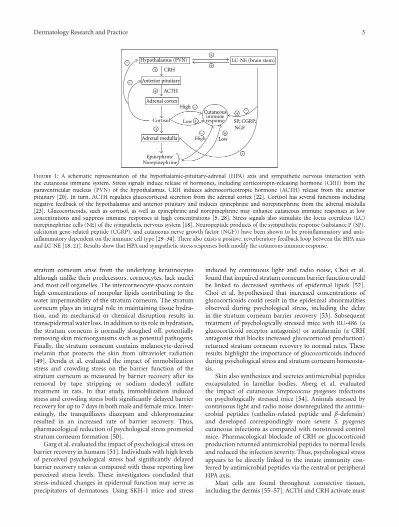
The central hypothalamic-pituitary-adrenal (HPA) axisis activated following stress signals such as 5-hydrox-ytryptamine [13, 14], acetylcholine [15], and inflammatorycytokines [16, 17]. Stress signals also activate the locuscoeruleus (LC) of the brain stem eliciting a sympatheticnervous system response. There exists a positive, rever-beratory feedback loop between these two major systems[18]. When the HPA axis is activated, stress hormones arereleased including corticotropin-releasing hormone (CRH)and arginine vasopressin [19] from the hypothalamus, whichinduces adrenocorticotropic hormone (ACTH) release fromthe anterior pituitary [20]. CRH also activates the LC-noradrenergic pathways resulting in norepinephrine secre-tion by the peripheral sympathetic nervous system andnorepinephrine and epinephrine secretion from the adrenalmedulla [21]. ACTH regulates secretion of glucocorticoidsincluding cortisol from the adrenal gland [22]. Cortisol neg-atively regulates CRH production in a feedback loop mech-anism [23]. Norepinephrine is a major neurotransmitterreleased by sympathetic fibers to innervated tissues, includ-ing the skin [24–26]. Activation of the sympathetic nervoussystem also leads to increased production of other factorsincluding catecholamines [27]. A highly schematic overviewof the central HPA axis and locus coeruleus/norepinephrine(LC-NE) sympathetic response to stress signals including thedownstream effects on the cutaneous immune response isshown in Figure 1.
Investigations have shown that human skin expressesCRH as well as CRH receptors (CRH-R). The CRH-R1αisoform is the predominant CRH receptor in skin andis expressed in all major cell populations of epidermis,
dermis, and subcutis. By contrast, CRH-R2 is expressedpredominately in hair follicles, sebaceous and eccrine glands,muscle and blood vessels [35]. CRH protein is also presentin murine skin although CRH mRNA has not been detected[35]. However, both mRNA and protein products for CRH-R1 and 2 have been detected in murine skin [36]. In additionto CRH, human skin also expresses urocortin I [37] andurocortin II mRNA [35]. CRH-R1 binds to urocortin I, butnot to urocortin II; while CRH-R2 binds to urocortin II, butnot urocortin I [38, 39] leading us to belief that the skin has adepth of responsiveness and interaction to the environmentthat is little understood. Finally, skin produces the precursorprotein, proopiomelanocortin protein (POMC) and POMCderived peptides that give rise to ACTH and other polypep-tide products [40, 41].
Ito et al. have shown that human hair follicles cansynthesize cortisol and that cortisol synthesis is regulated byendogenous feedback controls [42]. Thus, the skin appar-ently has a peripheral equivalent of the HPA axis that is fullyfunctional. The peripheral skin HPA axis may coordinateor fine tune peripheral stress responses with the centralHPA axis. In addition to expressing components of the HPAaxis, skin also produces a number of other neuroendocrinesignals including prolactin [43–45], melatonin [46], andcatecholamines [47, 48].
In addition to the HPA axis, the skin is highly inner-vated with sensory nerves that produce neurotrophins andneuropeptides. Sensory nerves derive from the dorsal rootganglion in the skin and C-fibers form the cutaneous sensorynervous system. Psychological stress leads to increasedconcentrations of cutaneous nerve growth factor (NGF)[29]. NGF has a number of biological activities including (1)axon sprouting of peptidergic and sympathetic neurons, (2)promoting cross-talk between neural cells, glia, and immunecells, and (3) facilitating monocyte/macrophage migrationthrough vascular endothelium [30]. NGF upregulates SP+nerve fibers in the dermis of stressed mice. Calcitoningene-related peptide (CGRP), a potent vasodilator, is alsoupregulated in response to NGF [29]. SP and CGRP havedifferent distributions within the skin with SP nerve fibersdetected in the dermis and subcutis and CGRP nerve fibersare in the epidermis around the distal hair follicle and thearrector pili muscle [31].
3. Impact of Psychological Stress on Innateand Adaptive Immunity in the Skin
The innate immune response consists of elements thatcontribute to the immediate and generic defense of the skin;immunological memory does not develop. By contrast, theadaptive immune response requires time for the develop-ment of a specific defense and can create immunologicalmemory. Psychological stress has been shown to impact bothinnate and adaptive immune responses.
3.1. Innate Immune Responses to Stress. The stratum cor-neum is terminally differentiated epidermis that forms theouter most layer of the skin. The corneocytes forming the
Dermatology Research and Practice 3
+ −
Epinephrine
Adrenal medulla
Cortisol
Low
Low
High
HighCutaneousimmune
Norepinephrine
response SP; CGRP;NGF
Adrenal cortex
Hypothalamus (PVN) LC-NE (brain stem)
ACTH
CRH
Anterior pituitary
+
+
+
+
+
+
+
+
−
−
−
−
Figure 1: A schematic representation of the hypothalamic-pituitary-adrenal (HPA) axis and sympathetic nervous interaction withthe cutaneous immune system. Stress signals induce release of hormones, including corticotropin-releasing hormone (CRH) from theparaventricular nucleus (PVN) of the hypothalamus. CRH induces adrenocorticotropic hormone (ACTH) release from the anteriorpituitary [20]. In turn, ACTH regulates glucocorticoid secretion from the adrenal cortex [22]. Cortisol has several functions includingnegative feedback of the hypothalamus and anterior pituitary and induces epinephrine and norepinephrine from the adrenal medulla[23]. Glucocorticoids, such as cortisol, as well as epinephrine and norepinephrine may enhance cutaneous immune responses at lowconcentrations and suppress immune responses at high concentrations [5, 28]. Stress signals also stimulate the locus coeruleus (LC)norepinephrine cells (NE) of the sympathetic nervous system [18]. Neuropeptide products of the sympathetic response (substance P (SP),calcitonin gene-related peptide (CGRP), and cutaneous nerve growth factor (NGF)) have been shown to be proinflammatory and anti-inflammatory dependent on the immune cell type [29–34]. There also exists a positive, reverberatory feedback loop between the HPA axisand LC-NE [18, 21]. Results show that HPA and sympathetic stress responses both modify the cutaneous immune response.
stratum corneum arise from the underlying keratinocytesalthough unlike their predecessors, corneocytes, lack nucleiand most cell organelles. The intercorneocyte spaces containhigh concentrations of nonpolar lipids contributing to thewater impermeability of the stratum corneum. The stratumcorneum plays an integral role in maintaining tissue hydra-tion, and its mechanical or chemical disruption results intransepidermal water loss. In addition to its role in hydration,the stratum corneum is normally sloughed off, potentiallyremoving skin microorganisms such as potential pathogens.Finally, the stratum corneum contains melanocyte-derivedmelanin that protects the skin from ultraviolet radiation[49]. Denda et al. evaluated the impact of immobilizationstress and crowding stress on the barrier function of thestratum corneum as measured by barrier recovery after itsremoval by tape stripping or sodium dodecyl sulfatetreatment in rats. In that study, immobilization inducedstress and crowding stress both significantly delayed barrierrecovery for up to 7 days in both male and female mice. Inter-estingly, the tranquillizers diazepam and chlorpromazineresulted in an increased rate of barrier recovery. Thus,pharmacological reduction of psychological stress promotedstratum corneum formation [50].
Garg et al. evaluated the impact of psychological stress onbarrier recovery in humans [51]. Individuals with high levelsof perceived psychological stress had significantly delayedbarrier recovery rates as compared with those reporting lowperceived stress levels. These investigators concluded thatstress-induced changes in epidermal function may serve asprecipitators of dermatoses. Using SKH-1 mice and stress
induced by continuous light and radio noise, Choi et al.found that impaired stratum corneum barrier function couldbe linked to decreased synthesis of epidermal lipids [52].Choi et al. hypothesized that increased concentrations ofglucocorticoids could result in the epidermal abnormalitiesobserved during psychological stress, including the delayin the stratum corneum barrier recovery [53]. Subsequenttreatment of psychologically stressed mice with RU-486 (aglucocorticoid receptor antagonist) or antalarmin (a CRHantagonist that blocks increased glucocorticoid production)returned stratum corneum recovery to normal rates. Theseresults highlight the importance of glucocorticoids inducedduring psychological stress and stratum corneum homeosta-sis.
Skin also synthesizes and secretes antimicrobial peptidesencapsulated in lamellar bodies. Aberg et al. evaluatedthe impact of cutaneous Streptococcus pyogenes infectionson psychologically stressed mice [54]. Animals stressed bycontinuous light and radio noise downregulated the antimi-crobial peptides (cathelin-related peptide and β-defensin)and developed correspondingly more severe S. pyogenescutaneous infections as compared with nonstressed controlmice. Pharmacological blockade of CRH or glucocorticoidproduction returned antimicrobial peptides to normal levelsand reduced the infection severity. Thus, psychological stressappears to be directly linked to the innate immunity con-ferred by antimicrobial peptides via the central or peripheralHPA axis.
Mast cells are found throughout connective tissues,including the dermis [55–57]. ACTH and CRH activate mast
4 Dermatology Research and Practice
cells, and human mast cells express CRH receptors [58].Recent work by Asadi et al. has shown that SP can inducethe expression of functional CRH receptor-1 in human mastcells [32]. Acute psychological stress is linked with mastcell activation and the release of IL-6. The finding thatserum levels of IL-6 are abrogated in mast cell-deficient micefollowing restraint stress as compared with their wildtypecounterparts underscores the importance of mast cells inthe production of systemic IL-6 [59]. Importantly, IL-6 cancross the blood/brain barrier [60] and activate the HPAaxis [61]. IL-6 can also induce immune reactions includinglymphocyte activation [62, 63] and increased antibodyproduction via CD4+ T-cell help [64]. Systemic effects of IL-6 include induction of fever [65] and acute phase proteinproduction [66, 67].
Mast cells also play a role in neurogenic inflammation.Singh et al. reported that restraint-induced stress resultedin significantly enhanced degranulation of mast cells inmice as compared with their nonstressed counterparts.Pretreatment of mice before stress with CRH antiserum, theneurotensin receptor antagonist SR48692 and capsaicin todeplete sensory neurons were all found to inhibit mast celldegranulation. These results suggested a role for neurogenicinflammation in psychological stress that is in additionto the HPA axis [68]. In fact, a number of investigatorshave shown that psychological stress activates the mastcell/nerve fiber interface leading to neurogenic inflammation[69, 70]. Shimoda et al. reported that administration of anantipsychotic drug (chlorpromazine) and anxiolytic reagents(tandospirone and CRA1000) significantly reduced degran-ulation of dermal mast cells in mice stressed by electricfoot shock [71]. These results may suggest that antipsychoticand anxiolytic agents may be effective treatments for stress-aggravated inflammatory skin diseases by inhibition of mast-cell degranulation [71].
3.2. Adaptive Immune Responses to Stress. The adaptive im-mune response requires the interaction of antigen-presentingcells (i.e., dendritic cell) with antigen-specific lymphocytes(i.e., T cells). Activation of lymphocytes requires theircomplex interplay with antigen presenting cells and co-stimulatory molecules on the surfaces of both cell types aswell as the production of cytokines.
Dhabhar and Mcewen investigated the impact of acutestress on contact hypersensitivity (CHS) reactions in ratsusing stress induced by a 2-hour confinement in a plex-iglass box [4]. Briefly, animals were sensitized using 2,4-dinitrofluorobenzene (DNFB), stressed on day 5 follow-ing sensitization and challenged on the pinna of the earon day 6 with ear swelling used as the read-out afterthe DNFB challenge. Acute stress markedly increased theear swelling response in stressed rats as compared withthe control animals. Elimination of glucocorticoid andepinephrine by adrenalectomy eliminated the stress-inducedenhancement, underscoring the importance of these hor-mones for immunomodulation. Moreover, administration ofcorticosterone or epinephrine at low doses enhanced stress-induced ear swelling suggesting that these hormones play
a role in immunoenhancement. On the other hand, highdoses of corticosterone or epinephrine had the oppositeeffect, that is, ear swelling was reduced. Therefore, theoutcome of corticosterone and epinephrine depends on theirconcentrations. Using a different contact sensitizing reagent(trinitrochlorobenzene) and isolation stress, Nakano alsofound that stress enhanced the cutaneous immune responseas evaluated by ear swelling [72]. However, stress alonedid not enhance the ear swelling response of mice treatedwith the contact irritant, sodium dodecyl sulfate. Theseresults suggested that elements of the adaptive immuneresponse were required for acute stress-induced immuneenhancement, as irritants do not develop immunologicalmemory.
In contrast to the results described above, Flint et al.showed that restraint stress prior to DNFB sensitizationresulted in suppression of the immune response [73]. Thus,experimental studies have provided seemingly contradictoryresults. It is, therefore, tempting to speculate that the natureof the sensitizing agent, the dose of the contact sensitizingagent and the timing of the stressor are all variables that areimportant for the ensuing immune response.
Different strains of mice may have different skin sensi-tivities to psychological stressors. Flint et al. reported thatC57BL/6 mice had blunted ear swelling responses to restraintstress as compared with BALB/c mice [74]. Importantly, earswelling responses in stressed C57BL/6 strain could not beenhanced even after exogenous corticosterone. The natureof the stressor may also impact the magnitude of the CHSresponse in animals. Bowers et al. compared CHS responsesin mice acutely or chronically stressed by restraint, forcedswim, isolation, handling, and low temperature [7]. Restraintstress and forced swim stress resulted in the most dramaticincrease in the CHS response as assessed by the ear swellingassay. Taken all together, experimental studies have shown acorrelation between acute stress and contact hypersensitivityresponses in rodents. However, further investigations arerequired to delineate conditions under which acute stresssuppresses immune responses and under which conditionsacute stress enhances immune responses. Findings that theoutcomes of acute psychological stress are related to mousestrain also suggest that genetic background may impact theinterplay between stress and skin inflammation.
The impact of chronic stress on skin immune responseshas also been investigated. Chronic stress has been reportedto lead to immunosuppression in a number of systems,including skin graft rejection [75]. In a model of chronicrestraint-induced stress, the CHS response was markedlysuppressed [3]. By contrast, other studies using chronicrestraint-induced stress resulted in enhanced CHS responses[7]. The thyroid axis may also modulate immune responsesduring chronic stress [76].
Among the factors that may account for psychologicalstress-induced changes in the adaptive immune responseare changes in the numbers, proportions, and distributionsof immune cells. Previous studies found that psychologicalstress markedly decreased the percentages of leukocytesin the blood [1, 2]. Interestingly, administration of cor-ticosterone to adrenalectomized mice closely mirrored the
Dermatology Research and Practice 5
decrease in blood leukocytes observed in stressed animals[2].
In addition to differences in cell numbers and distri-butions, psychological stress may modulate the activities ofimmunological cells. For example, stress has been shownto impair lymphocyte function [77]. Stress has also beenshown to decrease the density of Langerhans cells in theepidermis in both mice and humans [78, 79]. Langerhanscells are epidermal members of the dendritic cell familyof antigen-presenting cells. Conventionally, Langerhans cellshave been considered pivotal for the generation of adap-tive immunity although current studies suggest that theirimmunological activities may be considerably more complex(reviewed in [80]). A number of stress related moleculeshave been shown to impact Langerhans cells and dendriticcells. Hoetzenecker et al. have shown that corticosteroidsinduce the apoptosis of Langerhans cells and impair theirexpression of costimulatory molecules [81]. Studies in vitrohave shown that epinephrine inhibits antigen presentation inepidermal cell preparations as well as in purified Langerhanscells [82]. Glucocorticoids inhibit dendritic cell productionof IL-12 [83, 84]; and IL-12 suppression may skew theTH1/TH2 balance toward TH2 and thus impact the natureof the immune response [85]. Importantly, blockade of β2-AR with the antagonist ICI188, 551 impaired the migrationof Langerhans cells to the lymph nodes and blunted thesubsequent CHS response when mice were sensitized withthe fluorescein isothiocyanate contact sensitizing reagent[82]. In contrast to impaired dendritic cell activities, otherstress-related molecules appear to enhance dendritic cellfunctions. For example, Yanagawa et al. showed that nora-drenaline enhanced phosphatidylinositol 3-kinase-inducedantigen endocytosis by dendritic cells in vitro [86].
Neuropeptides can also impact the biological activitiesof antigen-presenting cells. Hosoi et al. showed that CGRPimpinged on Langerhans cells in the epidermis and CGRPwas directly detected on the surfaces of some Langerhanscells. Moreover, CGRP was found to inhibit the ability ofLangerhans cells to present antigen in vitro [33]. Recently,Ding et al. showed that treatment of Langerhans cellswith CGRP decreased antigen presentation to a TH1 T-cell clone but increased antigen presentation to a TH2 T-cell clone. Those researchers suggested that exposure ofLangerhans cells to nerve-derived CGRP may polarize theimmune response to a TH2 type of immunity [87]. Otherneuropeptides may also modulate the ability of Langerhanscells to effectively present antigen. Staniek et al. foundthat SP can bind to human Langerhans cells and impairT-cell proliferative responses in the mixed epidermal-celllymphocyte reaction. Based on those results the investigatorsconcluded that SP can impair antigen presentation [34].
In summary, psychological stressors and stress-relatedmolecules (e.g., epinephrine, glucocorticoids, and nora-drenaline) have been shown to impact various cell behaviors,costimulatory molecule expression and cytokine profiles ofimmune cells in skin adaptive immune responses, includingdendritic cells and lymphocyte immune cell subsets.
4. Psychological Stressand Human Skin Diseases
A number of skin diseases may be preceded or exacerbated bypsychological stress. In the following section, we review whatis known about the impact of psychological stress on atopicdermatitis and psoriasis. Our focus is on these two skindiseases because they are relatively common skin disorders.
4.1. Atopic Dermatitis. Atopic dermatitis is a chronic inflam-matory skin disorder characterized by eczematous lesionsand pruritus. It is a common disorder affecting 6% ofthe population in the USA [88]. Atopic dermatitis maybe the result of genetic predisposition and environmentalconditions, and no single etiologic agent is known. Analysesof sequential patch-testing skin biopsies have suggested thatatopic dermatitis has a biphasic TH1/TH2 T-cell response.Acute inflammation is primarily TH2 with a shift towardTH1 chronification [89, 90]. Psychological stress is known toaggravate atopic dermatitis and a psychological profile thatincludes anxiety, depression, and excitability has been linkedto this disease [91]. Traumatic events, including naturaldisasters, may increase psychological stress in the populationat large, exasperating the incidence of atopic dermatitissymptoms. For example, after the Great Hanshin earthquakein January 1995, subjective distress was found to be the rootcause for the enhanced symptoms of atopic dermatitis in thepopulations of the affected geographic areas [92].
Buske-Kirschbaum et al. analyzed leukocyte subsets,serum IgE levels, and cytokine concentrations in atopicdermatitis patients and nonatopic controls stressed in frontof an audience using the Trier Social Stress Test (TSST)[93]. Both groups showed significant elevations in thenumbers of serum lymphocytes, monocytes, neutrophils,and basophils with no differences between the two groups.However, eosinophil numbers were significantly higher inatopic dermatitis patients as compared with nonatopiccontrols. Similarly, IgE levels were significantly greater in theatopic dermatitis patients than their nonatopic counterparts.In both groups, TSST resulted in increased concentrationsof IFNγ and a reduction in IL-4 concentrations with nosignificant differences between the two groups. These studiesshowed that immunological similarities and differences existbetween atopic dermatitis patients and nonatopic individualssubjected to psychological stressors.
Interestingly, patients with atopic dermatitis have beenshown to have reduced production of cortisol and ACTH dueto experimental TSST stressors as compared with nonatopiccontrols. By contrast, catecholamine levels were significantlyhigher in atopic patients as compared with nonatopic con-trols. Thus, atopic dermatitis patients have blunted HPA axisreactivity as assessed by cortisol and ACTH measurements,but an overactive sympathetic adrenomedullary system assuggested by the high concentrations of catecholamine [94].Both the HPA axis and the SAM system suppress TH1 activitypotentially via IL-12, thus skewing the TH1/TH2 balancetoward TH2. Thus, flares in atopic dermatitis followingpsychological stress may reflect TH2 skewing to acute diseasesymptoms.
6 Dermatology Research and Practice
Concentrations of NGF and SP are elevated in in thesera of atopic dermatitis patients. Moreover, NGF and SPconcentrations have been positively correlated with diseaseseverity [95, 96]. Recently, Lonne-Rahm et al. comparedskin biopsies from patients with atopic dermatitis andchronically stressed atopic dermatitis patients [97]. Cortisolconcentrations were used to define which patients werepsychologically stressed. In both groups, the CD3+ cellinfiltrates expressed the 5-hydroxytryptamine 2A receptorand the serotonin transporter protein. Furthermore, thenumbers of mast cells were significantly greater in theskin lesions as compared with uninvolved skin. Likewise,nerve fibers were found in the epidermis and papillarydermis of involved skin as compared to uninvolved skin.In contrast, the number of SP and CGRP positive nervefibers was not significantly different between involved andnoninvolved skin. Nonetheless, chronic stress was correlatedwith greater numbers of 5-hydroxytryptamine 2A receptorpositive cells in the papillary dermis of involved skin. Theseresults showed that atopic dermatitis results in differencesin skin innervation and modulation of the serotonin systemthat also occurs in atopic dermatitis patients during chronicstress.
In summary, atopic dermatitis is a dermatological disor-der that is characterized initially as an acute TH2-mediateddisease that becomes TH1 polarized with chronicity. Atopicdermatitis seems to worsen in patients that are psycho-logically stressed, and adult atopic dermatitis patients havea constellation of psychological conditions that may placethem at risk for this dermatitis. Finally, it has been reportedthat atopic dermatitis patients have a blunted HPA responseand an overactive sympathetic adrenomedullary system thatmay exacerbate disease.
4.2. Psoriasis. Approximately 2% of the population in theUSA is diagnosed with psoriasis [98]. Most newly diagnosedpsoriasis patients are under the age of 30. Psoriatic arthritis,which is potentially debilitating, develops in 10–40% ofpsoriatics [99, 100].
Psoriasis is a multifactorial disease shaped by geneticsand environmental factors that include psychological stress[101]. Currently, it is believed that T cells play a significantrole in disease pathogenesis, particularly T cells expressingIL-17. Kryczek et al. have proposed that activated TH1 cellsare recruited into the skin and secrete IFNγ. In turn, theIFNγ induces local antigen-presenting cells to secrete IL-1and IL-23 that promote the expansion and survival of IL-17 expressing CD4+ and CD8+ T cells. Trafficking of IL-17expressing CD8+ T cells into the epidermis then promotesepidermal hyperplasia [102]. The T-cell-activating antigen(s)remain unknown.
Psychological stressors have been reported to precedethe onset of psoriasis in 44% of patients and to initiaterecurrent skin flares in 88% of psoriatics [103, 104]. Buske-Kirschbaum et al. have reported psoriatics exposed to theTSST stressor had greater numbers of CD4+ T cells andmonocytes in their blood as compared with a nonpsoriaticcontrol group. On the other hand, numbers of CD3+/CD25+
T cells were decreased in psoriatics as compared withnonpsoriatic controls. Psychological stress increased thenumbers of CD3+, CD8+, CD16+/CD56+ (i.e., NK cells), andCD3+/HLA-DR leukocytes in the blood, although the differ-ences between psoriatics and nonpsoriatics were insignificant[105]. Schmid-Ott et al. evaluated circulating levels of Tcells and NK cells in psoriatics and nonpsoriatic followingexperimental psychological stress [106]. In contrast to resultsobtained by Buske-Kirschbaum et al., levels of CD3+ T cellsincreased significantly only in the blood of psoriatics follow-ing psychological stress. Importantly, increased T-cell countswere due to increased numbers of CD8+ and CD3+CLA+
T cells (CLA, cutaneous lymphocyte-associated antigens).Similarly, CLA+ NK cells were increased significantly onlyin the circulation of psoriatics following psychological stress.Importantly, the CLA molecule is required for the traffickingof the cells to skin.
Interestingly, psoriasis patients who reported stress-exacerbated flares were found to have decreased levels ofcortisol and epinephrine [107]. Thus, similar to atopicdermatitis patients, it would appear that the response of theHPA axis is blunted in psoriatics sensitive to psychologicalstressors. By contrast, Karanikas et al. recently reported thatHPA axis reactivity was not correlated with psychopatholog-ical and immune parameters in psoriatics [108].
Psychological stress may also enhance neurogenic inflam-mation in psoriatics. Harvima et al. evaluated involvedand uninvolved skin from stressed and nonstressed patientsthrough immunohistochemistry [109]. CGRP and vasoactiveintestinal peptide nerve fibers were detected in the papillarydermis of the skin in stressed patients, whereas these nervefibers were only weakly detected in nonstressed individ-uals. Moreover, concentrations of neuropeptide degradingenzymes (i.e., chymase) were decreased in stressed patientsas compared with the nonstressed psoriatic controls.
In summary, psoriasis is a multifactorial disease witha strong T-cell component. Psychological stress has beenshown to trigger disease and exacerbate skin flares in somepatients. Experimental psychological stressors have beenshown to increase circulating levels of T cells, includingT cells expressing the requisite proteins for skin homing(e.g., CLA). Second, the response of the HPA axis may beblunted in psoriasis patients with stress sensitivity. However,discordant results suggest that more studies are needed todetermine the role of the HPA axis in psoriasis. Finally,psychological stress may enhance neurogenic inflammationin psoriatics.
5. Patient Treatment at the Intersection ofDermatology and Psychiatry
Psychotropic medications (antipsychotics, lithium, antide-pressants, and anticonvulsants) can lead to skin rash andskin allergy as well as severe skin reactions (Stevens-Johnsonsyndrome for treatment with anticonvulsants). On theother hand, adverse psychiatric effects to dermatologicalmedications and treatments can include depression (e.g.,isotretinoin and IFNα treatment) and psychosis (dapsone
Dermatology Research and Practice 7
treatment). Locala has recently reviewed skin diseases causedor exacerbated by psychotropic medications as well as psy-chiatric adverse effects of dermatologic medications [110].
The apparent psychophysiologic responses of manydermatoses may suggest that treatment programs structuredat the dermatology/psychiatry interface may be useful forpatient treatment, including programs that incorporate(1) psychotherapy, (2) biofeedback, (3) hypnosis, and (4)cognitive behavioral methods [111–113].
Hypnosis is just one example of psychiatric treat-ment augmenting dermatological treatments for dermatoses.Shenefelt performed a MEDLINE search that covered theyears 1966–1998 using search terms related to hypnosisand skin disease [111]. Results from MEDLINE showedthat a wide range of dermatological disorders could beimproved using hypnosis as an alternative or complementarytherapy for skin disease treatment, including (1) atopicdermatitis, (2) psoriasis, (3) alopecia areata, (4) rosacea, (5)vitiligo, (6) hyperhidrosis, and (7) ichthyosis vulgaris [111].Other psychiatric treatments may also benefit dermatologypatients. For example, one study showed that patientswho chose to participate in a cognitive behavioral therapyprogram reported reduced frequencies and numbers ofpsoriasis symptoms as long as 6 months after the programended [114].
6. Summary and Conclusions
The response to psychological stress is hypothesized to be anevolutionary adaptation for the fight-or-flight response. Incontrast, for contemporary humans, activation of the HPAaxis as a result of psychological stress can result in a numberof undesirable physiological responses including the exac-erbation of skin diseases. It has been shown that elementsof the HPA axis as well as the sympathetic nervous systemcan modulate the innate and adaptive cutaneous immuneresponses, and a number of experiments have suggested thatpsychological stress can impact disease development andprogression.
Recent studies have shown that skin has its own HPAaxis that may “fine tune” the response of the central HPAaxis. The skin is especially sensitive to psychological stressors.Indeed, cooccurring psychiatric disorders are prevalent inpatients with skin disorders. Both the innate and adaptivecutaneous immune responses are impacted by psychologicalstress as demonstrated in a number of experimental studiesin both laboratory rodents and humans. Mouse models ofcontact hypersensitivity strongly suggest that the nature ofthe sensitizing agent, the dose of the contact sensitizingagent, and the timing of the stressor are all variables thatare important for the ensuing immune response. Modulationof the cutaneous immune system by psychological stressmost likely affects the course of skin diseases, includingatopic dermatitis and psoriasis. Future investigations thatexplore the interconnections between psychological stressand the cutaneous innate and adaptive immune responseswill enhance our understanding of skin immunology andimmunological mediated skin diseases, provide unique
insight into the mind and body connection, and may leadto new treatment programs that will improve patient care.
Acknowledgment
This work was supported by NIH Grant RO1 AR48840.
References
[1] F. S. Dhabhar, A. H. Miller, M. Stein, B. S. McEwen, andR. L. Spencer, “Diurnal and acute stress-induced changes indistribution of peripheral blood leukocyte subpopulations,”Brain, Behavior, and Immunity, vol. 8, no. 1, pp. 66–79, 1994.
[2] F. S. Dhabhar, A. H. Miller, B. S. McEwen, and R. L. Spencer,“Effects of stress on immune cell distribution: dynamics andhormonal mechanisms,” Journal of Immunology, vol. 154, no.10, pp. 5511–5527, 1995.
[3] F. S. Dhabhar and B. S. McEwen, “Acute stress enhances whilechronic stress suppresses cell-mediated immunity in vivo: apotential role for leukocyte trafficking,” Brain, Behavior, andImmunity, vol. 11, no. 4, pp. 286–306, 1997.
[4] F. S. Dhabhar and B. S. Mcewen, “Enhancing versus suppres-sive effects of stress hormones on skin immune function,”Proceedings of the National Academy of Sciences of the UnitedStates of America, vol. 96, no. 3, pp. 1059–1064, 1999.
[5] F. S. Dhabhar, “Acute stress enhances while chronic stresssuppresses skin immunity: the role of stress hormones andleukocyte trafficking,” Annals of the New York Academy ofSciences, vol. 917, pp. 876–893, 2000.
[6] M. Altemus, B. Rao, F. S. Dhabhar, W. Ding, and R. D.Granstein, “Stress-induced changes in skin barrier functionin healthy women,” Journal of Investigative Dermatology, vol.117, no. 2, pp. 309–317, 2001.
[7] S. L. Bowers, S. D. Bilbo, F. S. Dhabhar, and R. J. Nelson,“Stressor-specific alterations in corticosterone and immuneresponses in mice,” Brain, Behavior, and Immunity, vol. 22,no. 1, pp. 105–113, 2008.
[8] W. B. Cannon, “The emergency function of the adrenalmedulla in pain and the major emotions,” American Journalof Physiology, vol. 33, no. 2, pp. 356–372, 1914.
[9] M. Jafferany, “Psychodermatology: a guide to understandingcommon psychocutaneous disorders,” Primary Care Com-panion to the Journal of Clinical Psychiatry, vol. 9, no. 3, pp.203–213, 2007.
[10] S. Aktan, E. Ozmen, and B. Sanli, “Psychiatric disorders inpatients attending a dermatology outpatient clinic,” Derma-tology, vol. 197, no. 3, pp. 230–234, 1998.
[11] A. Picardi, D. Abeni, C. F. Melchi, P. Puddu, and P. Pasquini,“Psychiatric morbidity in dermatological outpatients: anissue to be recognized,” British Journal of Dermatology, vol.143, no. 5, pp. 983–991, 2000.
[12] P. D. Shenefelt, “Psychodermatological disorders: recognitionand treatment,” International Journal of Dermatology, vol. 50,no. 11, pp. 1309–1322, 2011.
[13] S. Tsagarakis, P. Navara, L. H. Rees, M. Besser, and A. Gross-man, “Morphine directly modulates the release of stimulatedcorticotrophin-releasing factor-41 from rat hypothalamus invitro,” Endocrinology, vol. 124, no. 5, pp. 2330–2335, 1989.
[14] A. E. Calogero, R. Bernardini, A. N. Margioris et al., “Effectsof serotonergic agonists and antagonists on corticotropin-releasing hormone secretion by explanted rat hypothalami,”Peptides, vol. 10, no. 1, pp. 189–200, 1989.
8 Dermatology Research and Practice
[15] R. Wei, T. M. Phillips, and E. M. Sternberg, “Specific up-regulation of CRH or AVP secretion by acetylcholine orlipopolysaccharide in inflammatory susceptible Lewis ratfetal hypothalamic cells,” Journal of Neuroimmunology, vol.131, no. 1-2, pp. 31–40, 2002.
[16] J. Raber, O. Sorg, T. F. W. Horn et al., “Inflammatorycytokines: putative regulators of neuronal and neuro-endocrine function,” Brain Research Reviews, vol. 26, no. 2-3, pp. 320–326, 1998.
[17] A. Kariagina, D. Romanenko, S. G. Ren, and V. Chesnokova,“Hypothalamic-pituitary cytokine network,” Endocrinology,vol. 145, no. 1, pp. 104–112, 2004.
[18] I. J. Elenkov, R. L. Wilder, G. P. Chrousos, and E. S. Vizi,“The sympathetic nerve—an integrative interface betweentwo supersystems: the brain and the immune system,”Pharmacological Reviews, vol. 52, no. 4, pp. 595–638, 2000.
[19] R. Bernardini, A. Chiarenza, T. C. Kamilaris et al., “In vivoand in vitro effects of arginine-vasopressin receptor antago-nists on the hypothalamic-pituitary-adrenal axis in the rat,”Neuroendocrinology, vol. 60, no. 5, pp. 503–508, 1994.
[20] M. H. Whitnall, “Regulation of the hypothalamic corti-cotropin-releasing hormone neurosecretory system,” Progressin Neurobiology, vol. 40, no. 5, pp. 573–629, 1993.
[21] F. Tausk, I. Elenkov, and J. Moynihan, “Psychoneuroim-munology,” Dermatologic Therapy, vol. 21, no. 1, pp. 22–31,2008.
[22] C. Tsigos and G. P. Chrousos, “Hypothalamic-pituitary-adrenal axis, neuroendocrine factors and stress,” Journal ofPsychosomatic Research, vol. 53, no. 4, pp. 865–871, 2002.
[23] Y. Miyazaki, H. Yokozeki, S. Awad et al., “Glucocorticoidsaugment the chemically induced production and geneexpression of interleukin-1α through NF-κB and AP-1activation in murine epidermal cells,” Journal of InvestigativeDermatology, vol. 115, no. 4, pp. 746–752, 2000.
[24] B. G. Zimmerman and L. Whitmore, “Transmitter release inskin and muscle blood vessels during sympathetic stimula-tion,” The American Journal of Physiology, vol. 212, no. 5, pp.1043–1054, 1967.
[25] P. E. Pergola, D. L. Kellogg Jr., J. M. Johnson, W. A. Kosiba,and D. E. Solomon, “Role of sympathetic nerves in thevascular effects of local temperature in human forearm skin,”American Journal of Physiology, vol. 265, no. 3, pp. H785–H792, 1993.
[26] D. L. Kellogg Jr., “In vivo mechanisms of cutaneous vasodi-lation and vasoconstriction in humans during thermoregu-latory challenges,” Journal of Applied Physiology, vol. 100, no.5, pp. 1709–1718, 2006.
[27] R. Kvetnansky, E. L. Sabban, and M. Palkovits, “Cate-cholaminergic systems in stress: structural and moleculargenetic approaches,” Physiological Reviews, vol. 89, no. 2, pp.535–606, 2009.
[28] S. E. Sephton, F. S. Dhabhar, A. S. Keuroghlian et al., “Depres-sion, cortisol, and suppressed cell-mediated immunity inmetastatic breast cancer,” Brain, Behavior, and Immunity, vol.23, no. 8, pp. 1148–1155, 2009.
[29] R. A. Joachim, A. Kuhlmei, Q. T. Dinh et al., “Neuronalplasticity of the “brain-skin connection”: stress-triggered up-regulation of neuropeptides in dorsal root ganglia and skinvia nerve growth factor-dependent pathways,” Journal ofMolecular Medicine, vol. 85, no. 12, pp. 1369–1378, 2007.
[30] R. Levi-Montalcini, S. D. Skaper, R. Dal Toso, L. Petrelli,and A. Leon, “Nerve growth factor: from neurotrophin toneurokine,” Trends in Neurosciences, vol. 19, no. 11, pp. 514–520, 1996.
[31] E. M. J. Peters, V. A. Botchkarev, N. V. Botchkareva, D. J.Tobin, and R. Paus, “Hair-cycle-associated remodeling ofthe peptidergic innervation of murine skin, and hair growthmodulation by neuropeptides,” Journal of Investigative Der-matology, vol. 116, no. 2, pp. 236–245, 2001.
[32] S. Asadi, K. D. Alysandratos, A. Angelidou, A. Miniati, N.Sismanopoulos, and M. Vasiadi, “Substance P (SP) inducesexpression of functional corticotropin-releasing hormonereceptor-1 (CRHR-1) in human mast cells,” Journal ofInvestigative Dermatology, vol. 132, no. 2, pp. 324–329, 2012.
[33] J. Hosoi, G. F. Murphy, C. L. Egan et al., “Regulation ofLangerhans cell function by nerves containing calcitoningene-related peptide,” Nature, vol. 363, no. 6425, pp. 159–163, 1993.
[34] V. Staniek, L. Misery, J. Peguet-Navarro et al., “Bindingand in vitro modulation of human epidermal Langerhanscell functions by substance P,” Archives of DermatologicalResearch, vol. 289, no. 5, pp. 285–291, 1997.
[35] A. Slominski, A. Pisarchik, D. J. Tobin, J. E. Mazurkiewicz,and J. Wortsman, “Differential expression of a cutaneouscorticotropin-releasing hormone system,” Endocrinology, vol.145, no. 2, pp. 941–950, 2004.
[36] A. Slominski, J. Wortsman, A. Pisarchik et al., “Cutaneousexpression of corticotropin-releasing hormone (CRH), uro-cortin, and CRH receptors,” FASEB Journal, vol. 15, no. 10,pp. 1678–1693, 2001.
[37] A. Slominski, B. Roloff, J. Curry, M. Dahiya, A. Szczesniewski,and J. Wortsman, “The skin produces urocortin,” Journal ofClinical Endocrinology and Metabolism, vol. 85, no. 2, pp.815–823, 2000.
[38] S. Y. Hsu and A. J. W. Hsueh, “Human stresscopin andstresscopin-related peptide are selective ligands for the type 2corticotropin-releasing hormone receptor,” Nature Medicine,vol. 7, no. 5, pp. 605–611, 2001.
[39] D. K. Grammatopoulos and G. P. Chrousos, “Functionalcharacteristics of CRH receptors and potential clinical appli-cations of CRH-receptor antagonists,” Trends in Endocrinol-ogy and Metabolism, vol. 13, no. 10, pp. 436–444, 2002.
[40] A. Slominski, A. Szczesniewski, and J. Wortsman, “Liquidchromatography-mass spectrometry detection of corti-cotropin-releasing hormone and proopiomelanocortin-derived peptides in human skin,” Journal of ClinicalEndocrinology and Metabolism, vol. 85, no. 10, pp. 3582–3588, 2000.
[41] M. Kono, H. Nagata, S. Umemura, S. Kawana, and R.Y. Osamura, “In situ expression of corticotropin-releasinghormone (CRH) and proopiomelanocortin (POMC) genesin human skin,” The FASEB Journal, vol. 15, no. 12, pp. 2297–2299, 2001.
[42] N. Ito, T. Ito, A. Kromminga et al., “Human hair follicles dis-play a functional equivalent of the hypothalamic-pituitary-adrenal axis and synthesize cortisol,” FASEB Journal, vol. 19,no. 10, pp. 1332–1334, 2005.
[43] K. Foitzik, K. Krause, A. J. Nixon et al., “Prolactin and itsreceptor are expressed in murine hair follicle epithelium,show hair cycle-dependent expression, and induce catagen,”American Journal of Pathology, vol. 162, no. 5, pp. 1611–1621,2003.
[44] E. A. Langan, K. Foitzik-Lau, V. Goffin, Y. Ramot, and R.Paus, “Prolactin: an emerging force along the cutaneous-endocrine axis,” Trends in Endocrinology and Metabolism, vol.21, no. 9, pp. 569–577, 2010.
Dermatology Research and Practice 9
[45] Y. Ramot, T. Bıro, S. Tiede et al., “Prolactin—a novel neu-roendocrine regulator of human keratin expression in situ,”FASEB Journal, vol. 24, no. 6, pp. 1768–1779, 2010.
[46] A. Slominski, D. J. Tobin, M. A. Zmijewski, J. Wortsman, andR. Paus, “Melatonin in the skin: synthesis, metabolism andfunctions,” Trends in Endocrinology and Metabolism, vol. 19,no. 1, pp. 17–24, 2008.
[47] E. Weihe, B. Schutz, W. Hartschuh, M. Anlauf, M. K. Schafer,and L. E. Eiden, “Coexpression of cholinergic and nora-drenergic phenotypes in human and nonhuman autonomicnervous system,” Journal of Comparative Neurology, vol. 492,no. 3, pp. 370–379, 2005.
[48] B. Schutz, J. von Engelhardt, M. Gordes et al., “Sweat glandinnervation is pioneered by sympathetic neurons expressinga cholinergic/noradrenergic co-phenotype in the mouse,”Neuroscience, vol. 156, no. 2, pp. 310–318, 2008.
[49] R. Marks, “The stratum corneum barrier: the final frontier,”Journal of Nutrition, vol. 134, no. 8, aupplement, pp. 2017S–2021S, 2004.
[50] M. Denda, T. Tsuchiya, J. Hosoi, and J. Koyama, “Im-mobilization-induced and crowded environment-inducedstress delay barrier recovery in murine skin,” British Journalof Dermatology, vol. 138, no. 5, pp. 780–785, 1998.
[51] A. Garg, M. M. Chren, L. P. Sands et al., “Psychologicalstress perturbs epidermal permeability barrier homeostasis:implications for the pathogenesis of stress-associated skindisorders,” Archives of Dermatology, vol. 137, no. 1, pp. 53–59, 2001.
[52] E. H. Choi, B. E. Brown, D. Crumrine et al., “Mechanismsby which psychologic stress alters cutaneous permeabilitybarrier homeostasis and stratum corneum integrity,” Journalof Investigative Dermatology, vol. 124, no. 3, pp. 587–595,2005.
[53] E. H. Choi, M. Demerjian, D. Crumrine et al., “Glucocor-ticoid blockade reverses psychological stress-induced abnor-malities in epidermal structure and function,” AmericanJournal of Physiology, vol. 291, no. 6, pp. R1657–R1662, 2006.
[54] K. M. Aberg, K. A. Radek, E. H. Choi et al., “Psycholog-ical stress downregulates epidermal antimicrobial peptideexpression and increases severity of cutaneous infections inmice,” Journal of Clinical Investigation, vol. 117, no. 11, pp.3339–3349, 2007.
[55] H. T. Graham, O. H. Lowry, N. Wahl, and M. K. Priebat,“Mast cells as sources of tissue histamine,” The Journal ofexperimental medicine, vol. 102, no. 3, pp. 307–318, 1955.
[56] T. Cowen, P. Trigg, and R. A. J. Eady, “Distribution ofmast cells in human dermis: development of a mappingtechnique,” British Journal of Dermatology, vol. 100, no. 6, pp.635–640, 1979.
[57] R. A. J. Eady, T. Cowen, and T. F. Marshall, “Mast cell pop-ulation density, blood vessel density and histamine contentin normal human skin,” British Journal of Dermatology, vol.100, no. 6, pp. 623–633, 1979.
[58] J. Cao, N. Papadopoulou, D. Kempuraj et al., “Humanmast cells express corticotropin-releasing hormone (CRH)receptors and CRH leads to selective secretion of vascularendothelial growth factor,” Journal of Immunology, vol. 174,no. 12, pp. 7665–7675, 2005.
[59] M. Huang, J. Berry, K. Kandere, M. Lytinas, K. Karalis, and T.C. Theoharides, “Mast cell deficient W/WV mice lack stress-induced increase in serum IL-6 levels, as well as in peripheralCRH and vascular permeability, a model of rheumatoidarthritis,” International Journal of Immunopathology andPharmacology, vol. 15, no. 3, pp. 249–254, 2002.
[60] W. A. Banks, A. J. Kastin, and E. G. Gutierrez, “Penetrationof interleukin-6 across the murine blood-brain barrier,”Neuroscience Letters, vol. 179, no. 1-2, pp. 53–56, 1994.
[61] G. Mastorakos, G. P. Chrousos, and J. S. Weber, “Recom-binant interleukin-6 activates the hypothalamic-pituitary-adrenal axis in humans,” Journal of Clinical Endocrinologyand Metabolism, vol. 77, no. 6, pp. 1690–1694, 1993.
[62] A. Kitani, M. Hara, T. Hirose et al., “Autostimulatory effectsof IL-6 on excessive B cell differentiation in patients withsystemic lupus erythematosus: analysis of IL-6 productionand IL-6R expression,” Clinical and Experimental Immunol-ogy, vol. 88, no. 1, pp. 75–83, 1992.
[63] K. Takeda, T. Kaisho, N. Yoshida, J. Takeda, T. Kishimoto, andS. Akira, “Stat3 activation is responsible for IL-6-dependentT cell proliferation through preventing apoptosis: generationand characterization of T cell- specific stat3-deficient mice,”Journal of Immunology, vol. 161, no. 9, pp. 4652–4660, 1998.
[64] O. Dienz, S. M. Eaton, J. P. Bond et al., “The induction ofantibody production by IL-6 is indirectly mediated by IL-21produced by CD4+ T cells,” Journal of Experimental Medicine,vol. 206, no. 1, pp. 69–78, 2009.
[65] K. Kagiwada, D. Chida, T. Sakatani et al., “Interleukin (IL)-6, but not IL-1, induction in the brain downstream ofcyclooxygenase-2 is essential for the induction of febrileresponse against peripheral IL-1α,” Endocrinology, vol. 145,no. 11, pp. 5044–5048, 2004.
[66] J. V. Castell, M. J. Gomez-Lechon, M. David et al.,“Interleukin-6 is the major regulator of acute phase proteinsynthesis in adult human hepatocytes,” FEBS Letters, vol. 242,no. 2, pp. 237–239, 1989.
[67] J. V. Castell, T. Andus, D. Kunz, and P. C. Heinrich,“Interleukin-6: the major regulator of acute-phase proteinsynthesis in man and rat,” Annals of the New York Academyof Sciences, vol. 557, pp. 87–101, 1989.
[68] L. K. Singh, X. Pang, N. Alexacos, R. Letourneau, and T.C. Theoharides, “Acute immobilization stress triggers skinmast cell degranulation via corticotropin releasing hormone,neurotensin, and substance P: a link to neurogenic skindisorders,” Brain, Behavior, and Immunity, vol. 13, no. 3, pp.225–239, 1999.
[69] P. C. Arck, B. Handjiski, E. Hagen, R. Joachim, B. F. Klapp,and R. Paus, “Indications for a “brain-hair follicle axis(BHA)”: inhibition of keratinocyte proliferation and up-regulation of keratinocyte apoptosis in telogen hair folliclesby stress and substance P,” The FASEB Journal, vol. 15, no. 13,pp. 2536–2538, 2001.
[70] S. Pavlovic, M. Daniltchenko, D. J. Tobin et al., “Furtherexploring the brain-skin connection: stress worsens dermati-tis via substance P-dependent neurogenic inflammation inmice,” Journal of Investigative Dermatology, vol. 128, no. 2,pp. 434–446, 2008.
[71] T. Shimoda, Z. Liang, H. Suzuki, and S. Kawana, “Inhibitoryeffects of antipsychotic and anxiolytic agents on stress-induced degranulation of mouse dermal mast cells: exper-imental dermatology,” Clinical and Experimental Dermatol-ogy, vol. 35, no. 5, pp. 531–536, 2010.
[72] Y. Nakano, “Effect of chronic topical exposure to low-dosenoxious chemicals and stress on skin sensitivity in mice,”Journal of Occupational Health, vol. 49, no. 6, pp. 431–442,2007.
[73] M. S. Flint, D. B. Miller, and S. S. Tinkle, “Restraint-inducedmodulation of allergic and irritant contact dermatitis in maleand female B6.129 Mice,” Brain, Behavior, and Immunity, vol.14, no. 4, pp. 256–269, 2000.

10 Dermatology Research and Practice
[74] M. S. Flint and S. S. Tinkle, “C57BL/6 mice are resistantto acute restraint modulation of cutaneous hypersensitivity,”Toxicological Sciences, vol. 62, no. 2, pp. 250–256, 2001.
[75] R. Wistar Jr. and W. H. Hildemann, “Effect of stress on skintransplantation immunity in mice,” Science, vol. 131, no.3394, pp. 159–160, 1960.
[76] G. A. Cremaschi, G. Gorelik, A. J. Klecha, A. E. Lysionek, andA. M. Genaro, “Chronic stress influences the immune systemthrough the thyroid axis,” Life Sciences, vol. 67, no. 26, pp.3171–3179, 2000.
[77] R. W. Bartrop, E. Luckhurst, and L. Lazarus, “Depressedlymphocyte function after bereavement,” Lancet, vol. 1, no.8016, pp. 834–836, 1977.
[78] J. Hosoi, T. Tsuchiya, M. Denda et al., “Modification ofLC phenotype and suppression of contact hypersensitivityresponse by stress,” Journal of Cutaneous Medicine andSurgery, vol. 3, no. 2, pp. 79–84, 1998.
[79] C. E. Kleyn, L. Schneider, R. Saraceno et al., “The effects ofacute social stress on epidermal Langerhans’ cell frequencyand expression of cutaneous neuropeptides,” Journal ofInvestigative Dermatology, vol. 128, no. 5, pp. 1273–1279,2008.
[80] B. E. Clausen and J. M. Kel, “Langerhans cells: criticalregulators of skin immunity,” Immunology and Cell Biology,vol. 88, no. 4, pp. 351–360, 2010.
[81] W. Hoetzenecker, J. G. Meingassner, R. Ecker, G. Stingl, A.Stuetz, and A. Elbe-Burger, “Corticosteroids but not pime-crolimus affect viability, maturation and immune functionof murine epidermal langerhans cells,” Journal of InvestigativeDermatology, vol. 122, no. 3, pp. 673–684, 2004.
[82] K. Seiffert, J. Hosoi, H. Torii et al., “Catecholamines inhibitthe antigen-presenting capability of epidermal langerhanscells,” Journal of Immunology, vol. 168, no. 12, pp. 6128–6135,2002.
[83] I. J. Elenkov, D. A. Papanicolaou, R. L. Wilder, and G.P. Chrousos, “Modulatory effects of glucocorticoids andcatecholamines on human interleukin-12 and interleukin-10 production: clinical implications,” Proceedings of theAssociation of American Physicians, vol. 108, no. 5, pp. 374–381, 1996.
[84] P. Panina-Bordignon, D. Mazzeo, P. Di Lucia et al., “β2-agonists prevent Th1 development by selective inhibition ofinterleukin 12,” Journal of Clinical Investigation, vol. 100, no.6, pp. 1513–1519, 1997.
[85] K. Seiffert and R. D. Granstein, “Neuroendocrine regulationof skin dendritic cells,” Annals of the New York Academy ofSciences, vol. 1088, pp. 195–206, 2006.
[86] Y. Yanagawa, M. Matsumoto, and H. Togashi, “Enhanceddendritic cell antigen uptake via α2 adrenoceptor-mediatedPI3K activation following brief exposure to noradrenaline,”Journal of Immunology, vol. 185, no. 10, pp. 5762–5768, 2010.
[87] W. Ding, L. L. Stohl, J. A. Wagner, and R. D. Granstein,“Calcitonin gene-related peptide biases langerhans cellstoward Th2-type immunity,” Journal of Immunology, vol.181, no. 9, pp. 6020–6026, 2008.
[88] J. M. Hanifin, M. L. Reed, L. A. Drake et al., “A population-based survey of eczema prevalence in the United States,”Dermatitis, vol. 18, no. 2, pp. 82–91, 2007.
[89] T. Thepen, E. G. Langeveld-Wildschut, I. C. Bihari et al.,“Biphasic response against aeroallergen in atopic dermatitisshowing a switch from an initial T(H2) response to a T(H1)response in situ: an immunocytochemical study,” Journal ofAllergy and Clinical Immunology, vol. 97, no. 3, pp. 828–837,1996.
[90] M. Grewe, C. A. F. M. Bruijnzeel-Koomen, E. Schopf et al.,“A role for Th1 and Th2 cells in the immunopathogenesis ofatopic dermatitis,” Immunology Today, vol. 19, no. 8, pp. 359–361, 1998.
[91] H. Hashizume and M. Takigawa, “Anxiety in allergy andatopic dermatitis,” Current Opinion in Allergy and ClinicalImmunology, vol. 6, no. 5, pp. 335–339, 2006.
[92] A. Kodama, T. Horikawa, T. Suzuki et al., “Effect of stresson atopic dermatitis: investigation in patients after theGreat Hanshin Earthquake,” Journal of Allergy and ClinicalImmunology, vol. 104, no. 1, pp. 173–176, 1999.
[93] A. Buske-Kirschbaum, A. Gierens, H. Hollig, and D. H.Hellhammer, “Stress-induced immunomodulation is alteredin patients with atopic dermatitis,” Journal of Neuroimmunol-ogy, vol. 129, no. 1-2, pp. 161–167, 2002.
[94] A. Buske-Kirschbaum, A. Geiben, H. Hollig, E. Mor-schhauser, and D. Hellhammer, “Altered responsiveness ofthe hypothalamus-pituitary-adrenal axis and the sympa-thetic adrenomedullary system to stress in patients withatopic dermatitis,” Journal of Clinical Endocrinology andMetabolism, vol. 87, no. 9, pp. 4245–4251, 2002.
[95] M. Toyoda, M. Nakamura, T. Makino, T. Hino, M. Kagoura,and M. Morohashi, “Nerve growth factor and substanceP are useful plasma markers of disease activity in atopicdermatitis,” British Journal of Dermatology, vol. 147, no. 1,pp. 71–79, 2002.
[96] I. J. Wang, W. S. Hsieh, Y. L. Guo et al., “Neuro-mediatorsas predictors of paediatric atopic dermatitis,” Clinical andExperimental Allergy, vol. 38, no. 8, pp. 1302–1308, 2008.
[97] S. B. Lonne-Rahm, H. Rickberg, H. El-Nour, P. Marin, E. C.Azmitia, and K. Nordlind, “Neuroimmune mechanisms inpatients with atopic dermatitis during chronic stress,” Journalof the European Academy of Dermatology and Venereology, vol.22, no. 1, pp. 11–18, 2008.
[98] H. M. Sander, L. F. Morris, C. M. Phillips, P. E. Harrison,and A. Menter, “The annual cost of psoriasis,” Journal of theAmerican Academy of Dermatology, vol. 28, no. 3, pp. 422–425, 1993.
[99] J. M. Gelfand, D. D. Gladman, P. J. Mease et al., “Epidemi-ology of psoriatic arthritis in the population of the UnitedStates,” Journal of the American Academy of Dermatology, vol.53, no. 4, pp. 573–577, 2005.
[100] J. M. Gelfand, R. Weinstein, S. B. Porter, A. L. Neimann, J.A. Berlin, and D. J. Margolis, “Prevalence and treatment ofpsoriasis in the United Kingdom: a population-based study,”Archives of Dermatology, vol. 141, no. 12, pp. 1537–1541,2005.
[101] J. T. Elder, A. T. Bruce, J. E. Gudjonsson et al., “Moleculardissection of psoriasis: integrating genetics and biology,”Journal of Investigative Dermatology, vol. 130, no. 5, pp. 1213–1226, 2010.
[102] I. Kryczek, A. T. Bruce, J. E. Gudjonsson et al., “Inductionof IL-17+ T cell trafficking and development by IFN-γ:mechanism and pathological relevance in psoriasis,” Journalof Immunology, vol. 181, no. 7, pp. 4733–4741, 2008.
[103] M. S. Al’Abadie, G. G. Kent, and D. J. Gawkrodger, “Therelationship between stress and the onset and exacerbationof psoriasis and other skin conditions,” British Journal ofDermatology, vol. 130, no. 2, pp. 199–203, 1994.
[104] C. E. M. Griffiths and H. L. Richards, “Psychological influ-ences in psoriasis,” Clinical and Experimental Dermatology,vol. 26, no. 4, pp. 338–342, 2001.
[105] A. Buske-Kirschbaum, S. Kern, M. Ebrecht, and D. H.Hellhammer, “Altered distribution of leukocyte subsets and
Dermatology Research and Practice 11
cytokine production in response to acute psychosocial stressin patients with psoriasis vulgaris,” Brain, Behavior, andImmunity, vol. 21, no. 1, pp. 92–99, 2007.
[106] G. Schmid-Ott, B. Jaeger, T. Boehm et al., “Immunologicaleffects of stress in psoriasis,” British Journal of Dermatology,vol. 160, no. 4, pp. 782–785, 2009.
[107] A. Buske-Kirschbaum, M. Ebrecht, S. Kern, and D. H.Hellhammer, “Endocrine stress responses in TH1-mediatedchronic inflammatory skin disease (psoriasis vulgaris)—do they parallel stress-induced endocrine changes in TH2-mediated inflammatory dermatoses (atopic dermatitis)?”Psychoneuroendocrinology, vol. 31, no. 4, pp. 439–446, 2006.
[108] E. Karanikas, F. Harsoulis, I. Giouzepas, I. Griveas, andF. Chrisomallis, “Neuroendocrine stimulatory tests ofhypothalamus-pituitary-adrenal axis in psoriasis and cor-relative implications with psychopathological and immuneparameters,” Journal of Dermatology, vol. 36, no. 1, pp. 35–44, 2009.
[109] I. T. Harvima, H. Viinamaki, A. Naukkarinen et al., “Associa-tion of cutaneous mast cells and sensory nerves with psychicstress in psoriasis,” Psychotherapy and Psychosomatics, vol. 60,no. 3-4, pp. 168–176, 1993.
[110] J. A. Locala, “Current concepts in psychodermatology,”Current Psychiatry Reports, vol. 11, no. 3, pp. 211–218, 2009.
[111] P. D. Shenefelt, “Hypnosis in dermatology,” Archives ofDermatology, vol. 136, no. 3, pp. 393–399, 2000.
[112] P. D. Shenefelt, “Biofeedback, cognitive-behavioral methods,and hypnosis in dermatology: is it all in your mind?”Dermatologic Therapy, vol. 16, no. 2, pp. 114–122, 2003.
[113] M. M. Heller, E. S. Lee, and J. Y. Koo, “Stress as an influencingfactor in psoriasis,” Skin Therapy Letter, vol. 16, no. 5, pp. 1–4, 2011.
[114] D. G. Fortune, H. L. Richards, C. E. M. Griffiths, and C.J. Main, “Targeting cognitive-behaviour therapy to patient’simplicit model of psoriasis: results from a patient preferencecontrolled trial,” British Journal of Clinical Psychology, vol. 43,no. 1, pp. 65–82, 2004.

Submit your manuscripts athttp://www.hindawi.com
Stem CellsInternational
Hindawi Publishing Corporationhttp://www.hindawi.com Volume 2014
Hindawi Publishing Corporationhttp://www.hindawi.com Volume 2014
MEDIATORSINFLAMMATION
of
Hindawi Publishing Corporationhttp://www.hindawi.com Volume 2014
Behavioural Neurology
EndocrinologyInternational Journal of
Hindawi Publishing Corporationhttp://www.hindawi.com Volume 2014
Hindawi Publishing Corporationhttp://www.hindawi.com Volume 2014
Disease Markers
Hindawi Publishing Corporationhttp://www.hindawi.com Volume 2014
BioMed Research International
OncologyJournal of
Hindawi Publishing Corporationhttp://www.hindawi.com Volume 2014
Hindawi Publishing Corporationhttp://www.hindawi.com Volume 2014
Oxidative Medicine and Cellular Longevity
Hindawi Publishing Corporationhttp://www.hindawi.com Volume 2014
PPAR Research
The Scientific World JournalHindawi Publishing Corporation http://www.hindawi.com Volume 2014
Immunology ResearchHindawi Publishing Corporationhttp://www.hindawi.com Volume 2014
Journal of
ObesityJournal of
Hindawi Publishing Corporationhttp://www.hindawi.com Volume 2014
Hindawi Publishing Corporationhttp://www.hindawi.com Volume 2014
Computational and Mathematical Methods in Medicine
OphthalmologyJournal of
Hindawi Publishing Corporationhttp://www.hindawi.com Volume 2014
Diabetes ResearchJournal of
Hindawi Publishing Corporationhttp://www.hindawi.com Volume 2014
Hindawi Publishing Corporationhttp://www.hindawi.com Volume 2014
Research and TreatmentAIDS
Hindawi Publishing Corporationhttp://www.hindawi.com Volume 2014
Gastroenterology Research and Practice
Hindawi Publishing Corporationhttp://www.hindawi.com Volume 2014
Parkinson’s Disease
Evidence-Based Complementary and Alternative Medicine
Volume 2014Hindawi Publishing Corporationhttp://www.hindawi.com