Embed Size (px)
Citation preview
Psoriasis and Skin Cancer
Edward Pritchard
Long Cases
• You could get these!
• Last year’s finals! - Patient with recurrent SCC, with no symptoms. History focussing on skin exposure and social. Was asked about risk factors, macroscopic and microscopic appearance of different types of skin cancer and different treatments
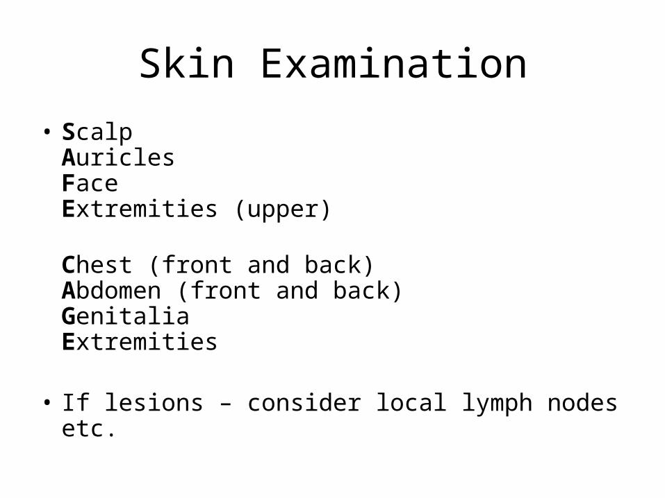
Skin Examination
• ScalpAuriclesFaceExtremities (upper)
Chest (front and back)Abdomen (front and back)GenitaliaExtremities
• If lesions – consider local lymph nodes etc.

Psoriasis
Definition
• Relapsing and remitting chronic skin condition characterised by scaly plaques
• Or inflammation of the dermis, with epidermal hyperproliferation
Epidemiology
• ~2% of the population• Peak incidence in early 20s and 50s
Precipitated byinfection, drugs (antimalarials, B-blockers, lithium), sunlight, stress, scars, burns
Pathophysiology
Immune mediated leads to increased speed of skin turnover (28 days to 4), causes thickening of the epidermis.

Symptoms and Signs
Typically well demarcated red, scaly, symmetrical, non itchy plaques
• 5 main presentations
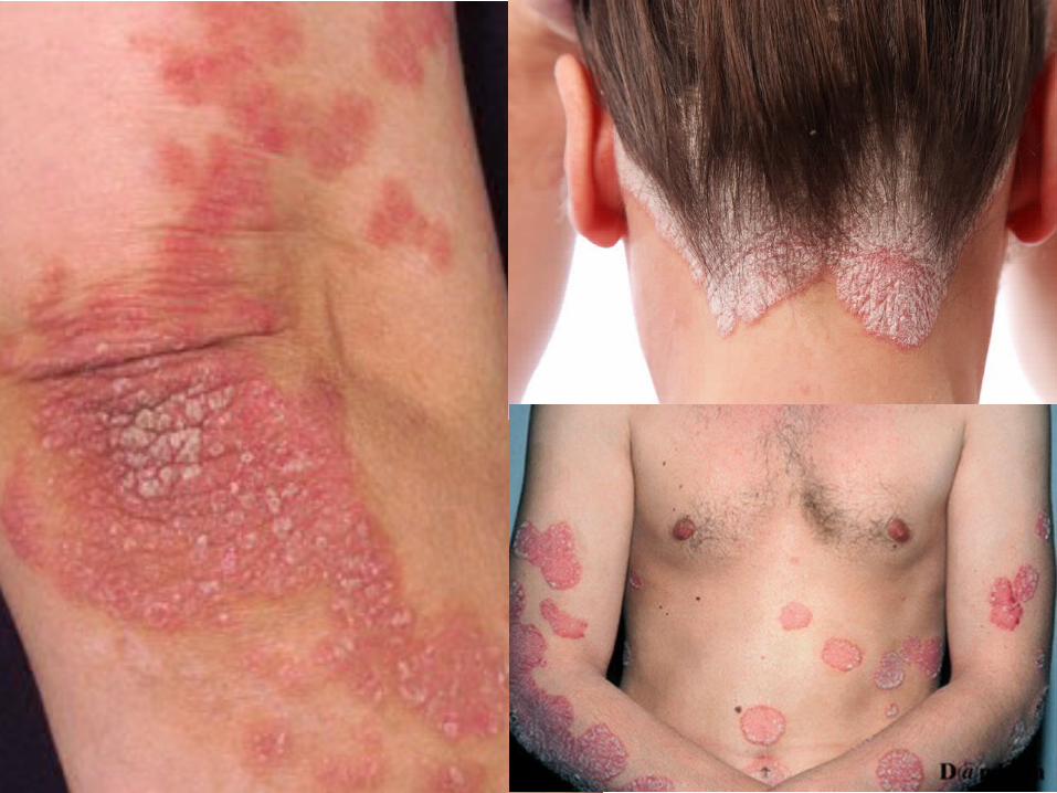
Plaque – typically on extensor surfaces and scalp
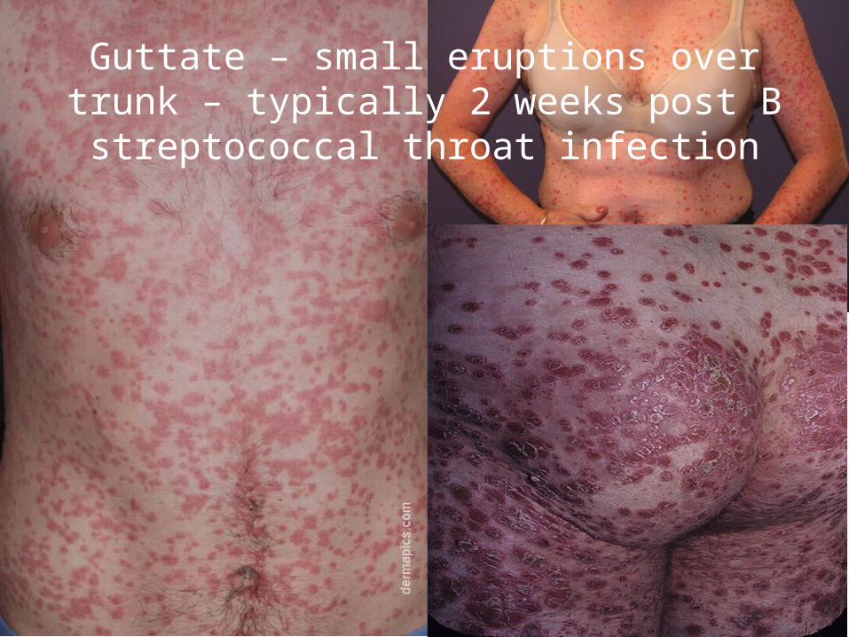
Guttate – small eruptions over trunk – typically 2 weeks post B streptococcal throat
infection
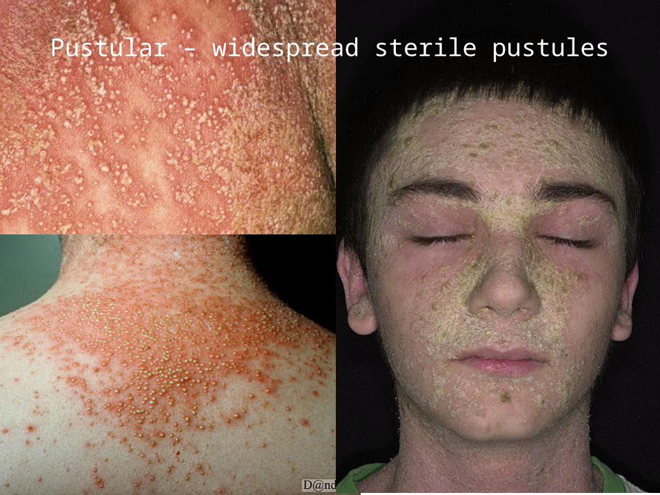
Pustular – widespread sterile pustules
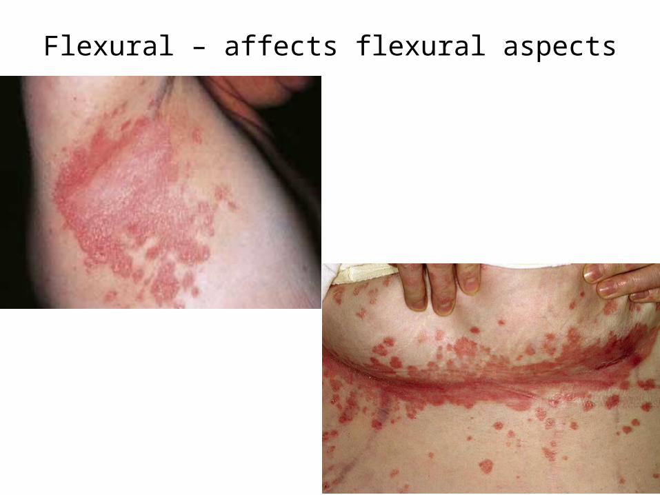
Flexural – affects flexural aspects
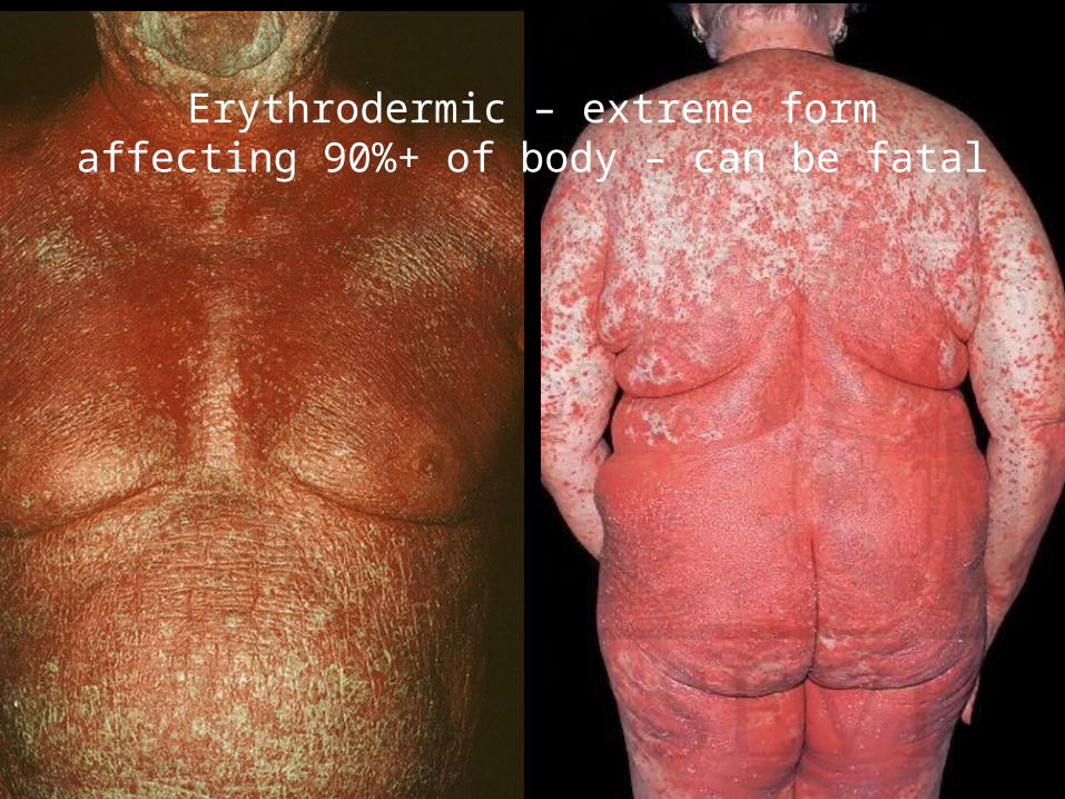
Erythrodermic – extreme form affecting 90%+ of body – can be fatal
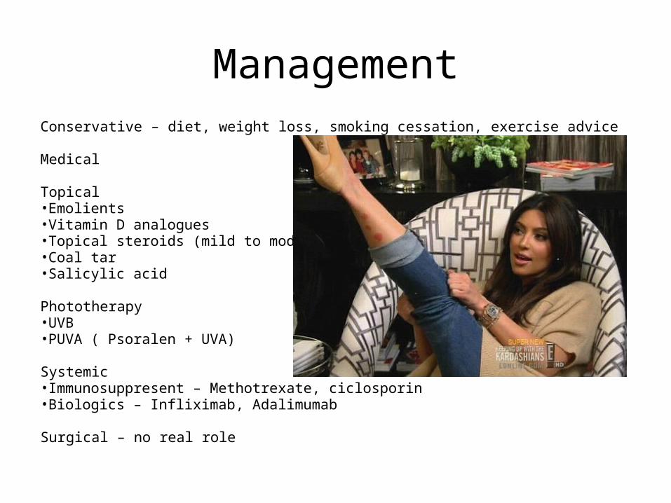
ManagementConservative – diet, weight loss, smoking cessation, exercise advice
Medical
Topical•Emolients•Vitamin D analogues•Topical steroids (mild to moderate)•Coal tar•Salicylic acid
Phototherapy•UVB•PUVA ( Psoralen + UVA)
Systemic•Immunosuppresent – Methotrexate, ciclosporin•Biologics – Infliximab, Adalimumab
Surgical – no real role

Skin Cancer
Aetiology/Risk Factors• Squamous cell – UV light exposure (sunbathing), fair
skin, radiation exposure, carcinogens, metastasise quickly
• Basal Cell – UV light exposure, radiation exposure, arsenic exposure, “never” metastasise – local tissue destruction
• Malignant melanoma – UV light exposure, metastasise rapidly
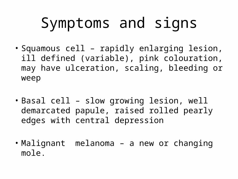
Symptoms and signs
• Squamous cell – rapidly enlarging lesion, ill defined (variable), pink colouration, may have ulceration, scaling, bleeding or weep
• Basal cell – slow growing lesion, well demarcated papule, raised rolled pearly edges with central depression
• Malignant melanoma – a new or changing mole.
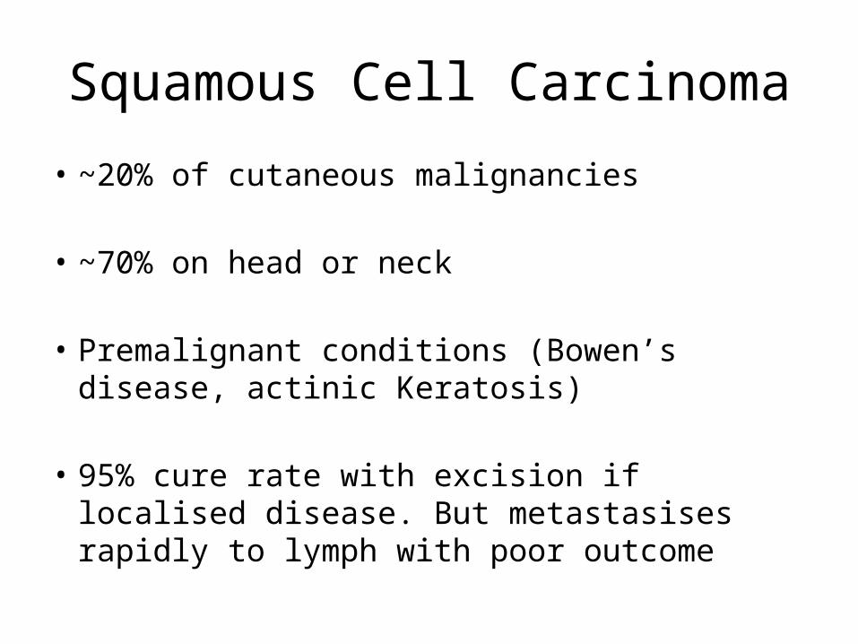
Squamous Cell Carcinoma
• ~20% of cutaneous malignancies
• ~70% on head or neck
• Premalignant conditions (Bowen’s disease, actinic Keratosis)
• 95% cure rate with excision if localised disease. But metastasises rapidly to lymph with poor outcome
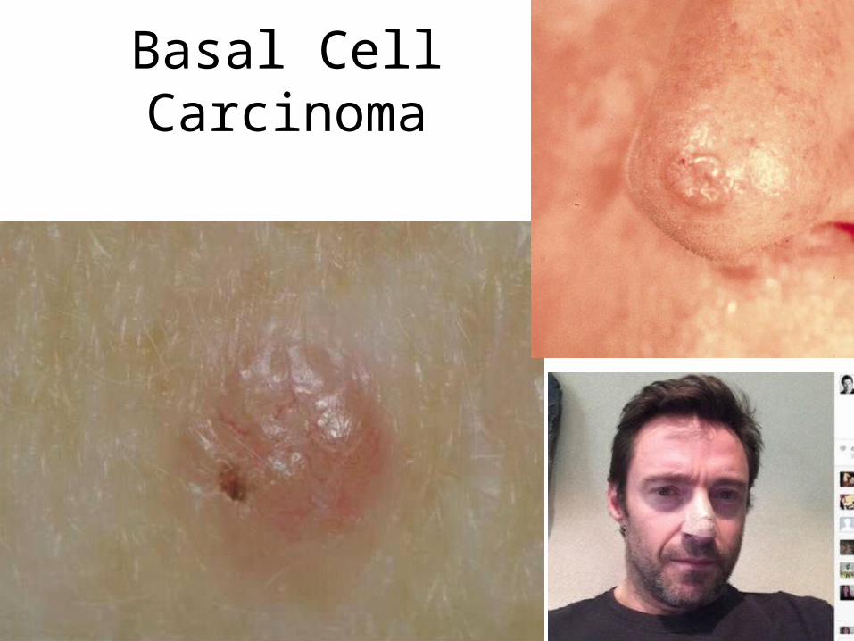
Basal Cell Carcinoma
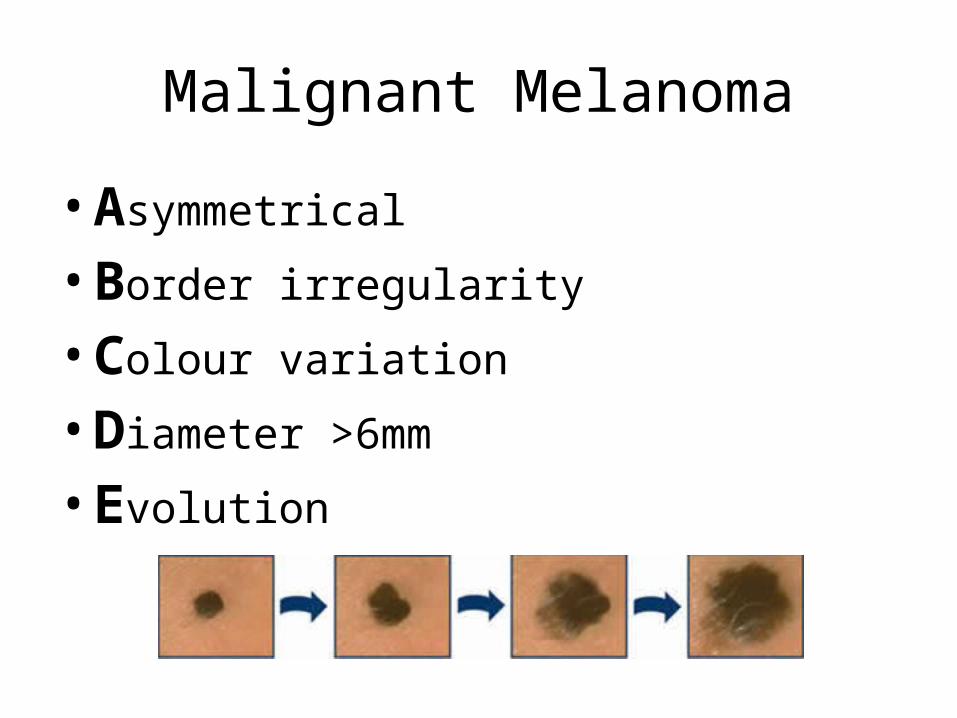
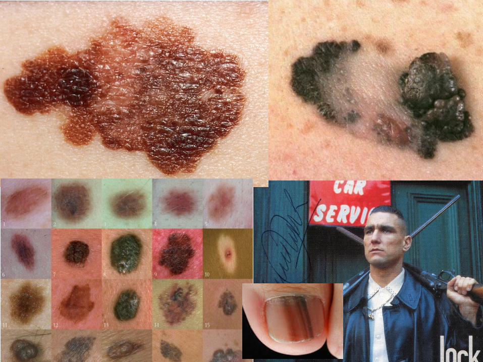
Malignant Melanoma
• Asymmetrical
• Border irregularity
• Colour variation
• Diameter >6mm
• Evolution
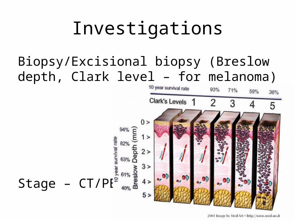
Investigations
Biopsy/Excisional biopsy (Breslow depth, Clark level – for melanoma)
Stage – CT/PET
Tx + PrognosisManagement• Conservative – reduce risk factors, smoking cessation• Medical – if for chemotherapy• Surgical – excision biopsy +/- lymph node resections
Prognosis• Basal cell – very good, fatality rare• Squamous cell – poor• Malignant melanoma – poorer (often metastasised at presentation)

Questions
• Thanks
![Psoriasis in Children and Adolescents: Diagnosis ......Guttate psoriasis is the second most common type of psoriasis in children [21, 29]. Griffiths and Barker defined guttate psoriasis](https://img.pdfslide.us/doc/110x75/5f501dea60f5a266c60b268c/psoriasis-in-children-and-adolescents-diagnosis-guttate-psoriasis-is-the.jpg)