Embed Size (px)
Citation preview
PSEUDOMONAS SYRINGAE PV. TOMATO (OKABE, 1933) YOUNG, DYE & WILKIE, 1978. A NEW BACTERIAL DISEASE OF TOMATO IN PORTUGAL.
Maria Helena M.C.F.C. Oliveira Joana M.C.P. Santa Marta Instituto Superior de Agronomia Tapada da Ajuda 1300 Lisboa
Abstract A new bacterial disease of tomato in greenhouse has been detected
for the first time in Portugal, in December 1982 and January 1983, by the authors. The observed symptomatology and the morphological, biochemical
physiological and pathogenicity tests demonstrated that the isolated bacteria is similar to Pseudomonas syvingae pv. tomato (Okabe, 1933) Young, Dye & Wilkie, 1978 (= P. tomato), the causal agent of bacterial speck of tomato.
Epidemiology and control measures are also inclused in this study.
1. Introduction An unusual disease, severely attacking three tomato cultivars
(Creon, Vemone and Adonia) in greenhouse was observed in December 1982 - January 1983, near Cartaxo. Symptoms of infection appeared on leaves, petioles, stems, flowers
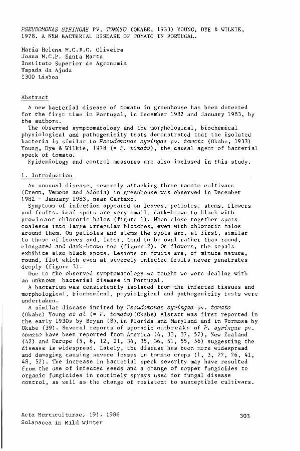
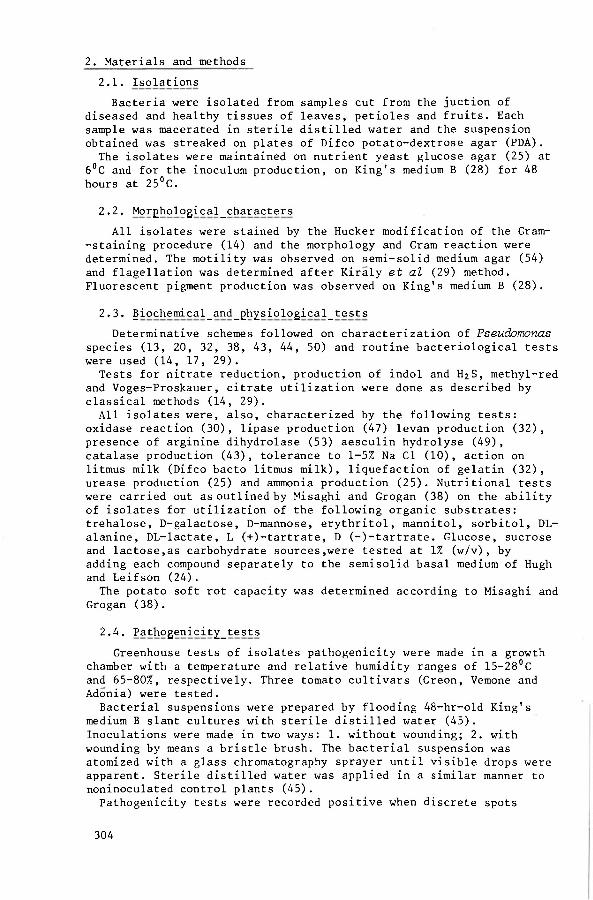
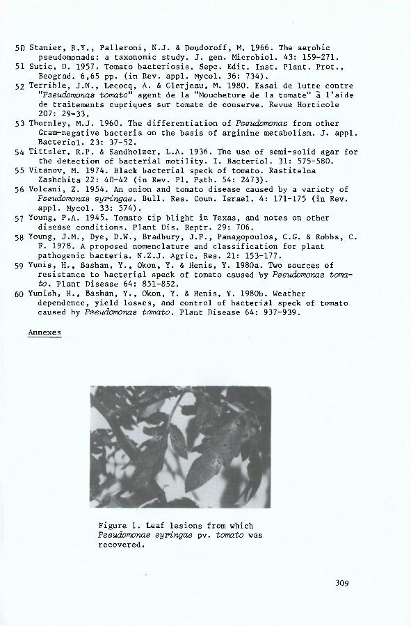
and fruits. Leaf spots are very small, dark-brown to black with prominant chlorotic halos (figure 1). When close together spots coalesce into large irregular blotches, even with chlorotic halos around them. On petioles and stems the spots are, at first, similar to those of leaves and, later, tend to be oval rather than round, elongated and dark-brown too (figure 2). On flowers, the sepals exhibite also black spots. Lesions on fruits are, of minute nature, round, flat which even at severely infected fruits never penetrates deeply (figure 3). Due to the observed symptomatology we tought we were dealing with
an unknown bacterial disease in Portugal. A bacterium was consistently isolated from the infected tissues and
morphological, biochemical, physiological and pathogenicity tests were undertaken. A similar disease incited by Pseudomonas syringae pv. tomato
(Okabe) Young et al (= P. tomato) (Okabe) Alstatt was first reported in the early 1930s by Bryan (8), in Florida and Maryland and in Formosa by Okabe (39). Several reports of sporadic outbreaks of P. syringae pv. tomato have been reported from America (4, 33, 37, 57), New Zealand (42) and Europe (5, 6, 12, 21, 34, 35, 36, 51, 55, 56) suggesting the disease is widespread. Lately, the disease has been more widespread and damaging causing severe losses in tomato crops (1, 3, 22, 26, 41, 48, 52). The increase in bacterial speck severity may have resulted from the use of infected seeds and a change of copper fungicides to organic fungicides in routinely sprays used for fungal disease control, as well as the change of resistent to susceptible cultivars.
Acta Horticulturae, 191, 1986 Solanacea in Mild Winter
303
2. Materials and methods 2.1. Isolations Bacteria were isolated from samples cut from the juction of
diseased and healthy tissues of leaves, petioles and fruits. Each sample was macerated in sterile distilled water and the suspension obtained was streaked on plates of Difco potato-dextrose agar (PDA). The isolates were maintained on nutrient yeast glucose agar (25) at
6°C and for the inoculum production, on King's medium B (28) for 48 hours at 25°C.
2.2. Morghological_characters All isolates were stained by the Hucker modification of the Gram-
-staining procedure (14) and the morphology and Gram reaction were determined. The motility was observed on semi-solid medium agar (54) and flagellation was determined after Kiraly et al (29) method. Fluorescent pigment production was observed on King's medium B (28).
2.3. §iochemical_and_ghYsiological_tests Determinative schemes followed on characterization of Pseudomonas
species (13, 20, 32, 38, 43, 44, 50) and routine bacteriological tests were used (14, 17, 29). Tests for nitrate reduction, production of indol and H2S, methyl-red
and Voges-Proskauer, citrate utilization were done as described by classical methods (14, 29). All isolates were, also, characterized by the following tests:
oxidase reaction (30), lipase production (47) levan production (32), presence of arginine dihydrolase (53) aesculin hydrolyse (49), catalase production (43), tolerance to 1-5% Na CI (10), action on litmus milk (Difco bacto litmus milk), liquefaction of gelatin (32), urease production (25) and ammonia production (25). Nutritional tests were carried out as outlined by Misaghi and Grogan (38) on the ability of isolates for utilization of the following organic substrates: trehalose, D-galactose, D-mannose, erythritol, mannitol, sorbitol, DL-alanine, DL-lactate, L (+)-tartrate, D (-)-tartrate. Glucose, sucrose and lactose,as carbohydrate sources,were tested at 1% (w/v), by adding each compound separately to the semisolid basal medium of Hugh and Leifson (24). The potato soft rot capacity was determined according to Misaghi and
Grogan (38).
2.4. PathogenicitY_tests Greenhouse tests of isolates pathogenicity were made in a growth
chamber with a temperature and relative humidity ranges of 15-28°C and 65-80%, respectively. Three tomato cultivars (Creon, Vemone and Adonia) were tested. Bacterial suspensions were prepared by flooding 48-hr-old King's
medium B slant cultures with sterile distilled water (45). Inoculations were made in two ways: 1. without wounding; 2. with wounding by means a bristle brush. The bacterial suspension was atomized with a glass chromatography sprayer until visible drops were apparent. Sterile distilled water was applied in a similar manner to noninoculated control plants (45) . Pathogenicity tests were recorded positive when discrete spots
304
surrounded by yellow halos were evident. Symptoms, generally developed in 4-6 days, but the final
observations were made 10 days after (41). Koch's postulates were completed by making isolations from infected
plants.
3. Results 3.1. Mor£hological_and_cultur§l_characteristics The isolated bacteria were gram-negative, rod-shaped, motile by
lophotrichous flagella, aerobics. A green, fluorescent pigment was produced on King's medium B.
3.2. Biochemical_and_£h^siological_reactions Bacteria were Na CI (1-5%) tolerant, catalase positive, active on
litmus milk (reduction and peptonization was observed), positive for levan and ammonia production, gelatin and aesculin hydrolysis. They were negative for oxidase, arginine dihydrolase, starch hydrolysis, nitrate reduction, lipase, urease, H2S, acetylmethylcarbinol production and methyl red reaction. Growth occurred with glucose, sucrose, lactose, D-mannose, D-galactose, mannitol, sorbitol, DL--alanine and citrate, but not with trehalose, erythritol, DL-lactate, L (+)-tartrate and D (-)-tartrate. All the isolates tested gave a negative potato soft rot reaction.
3.3. P§thogenicit^_tests All the isolates produced typical bacterial speck symptoms, on
Creon, Vemone and Adonia tomato plants, by the end of 4 days on leaves and 6 days on stems and petioles, after inoculation, regardless of the method of inoculation used.
These symptoms were similar to those observed under natural conditions in Cartaxo. Control plants were free of symptoms. Reisolation from test plant lesions yielded pure cultures of P. syrin-gae pv. tomato.
4. Discussion It was concluded from the evidence presented that the isolated
bacterium is indistinguishable from those previously referred as P. ringae pv. tomato (Okabe, 1933) Young, Dye and Wilkie, 1978, synonimous with P. tomato (Okabe) Alstatt (7, 9, 18, 19, 58). The isolates were characterized first as P. syringae types on the
basis of pigment production, negative oxidase, and arginine dihydrolase (9) and, later, as P. syringae pv. tomato on the basis of isolates capacity to use erythritol, DL-lactate and pathogenicity tests, as it's suggested by others (25, 38). Because the disease has been increasing in our country we thought it
was important to refer some approach about epidemiology and control. P. syringae pv. tomato survives on soil in association with tomato
debris and on roots of several weeds even in soils without history of tomato culture (40, 45, 46). The pathogen can, also, survives for long time on the surface of tomato seeds (2, 3, 27, 40, 45) in a state of reduced metabolism, suggesting the hypobiosis theory sensu Leben (31). Furthermore, P. syringae pv. tomato has been isolated from
apparently symptomless tomato leaves and foliage of weed hosts
305
constituting a resident population, that can maintain itself under adverse environmental conditions. When temperature and moisture are favourable this resident population can, then, multiply and infect (45, 46). Temperature of 23-25°C, relative humidity of 85-95% and a water film
on surface leaves are the most propitious to the infection (1, 2, 48, 6 0 ) . Concerning control measures of bacterial speck of tomato, prevention
is the key of success. It's advisable to use healthy seeds and to destruct the weeds.
Rotation crops and the use of resistent cultivars, as Rehovot - 13 (1) and Marmande (59) are, also, important. Contrarily, susceptible cultivars like VF-198 (1), Vemone, Creon and Adonia are unsuitable. Some authors pointed the elimination of seedborn inoculum by heat
treatment of seeds, at 52°C for 1 hour (16) or at 55°C for 30 minutes (40). Moreover if bacterial speck begins to develop in tomato crops, it
will be important to eliminate the infected plants and to spray the remaining ones as soon as disease is detected (15). Chemicals like copper, copper + maneb, streptomycin + mancozeb may
by effective until best control measures are improved (11, 22, 23, 48, 52, 60). Susceptibility of currently tomato cultivars and chemical control
are under investigation.
Aknowledgments The authors thank professor J. Pinto-Ganhao, Director of "Laborato-
rio de Patologia Vegetal Verissimo de Almeida" for helpfull discussions and support.
References 1 Bashan, Y. & Okon, Y. 1981. Inhibition of seed germination and
development of tomato plants in soil infested with Pseudomonas toma-to. Ann. appl. Biol. 98: 413-417.
2 Bashan, Y., Okon, Y. & Henis, Y. 1978. Infection studies of Pseudomo-nas tomato, causal agent of bacterial speck of tomato. Phytoparasitica 6: 135-143.
3 Bashan, Y., Okon, Y., Henis, Y. 1982. Long-term survival of Pseudomo-nas syringae pv. tomato and Xanthomonas campestr-Ls pv. vesioatoria in tomato and pepper seeds. Phytopathology 72: 1143-1144.
4 Basu, P.K. 1966. Conditions for symptomatological differentiation of bacterial canker, spot and speck on tomato seedlings. Can. J. PI. Sci. 46: 525-530 (in. Rev. appl. Mycol. 46: 439).
5 Berniac, M. 1971. Une bacteriose de la tomate nouvelle pour la France. C. r. hebd. Seanc. Acad. Agric. Fr. 57: 1495-1499.
6 Bosshard-Heer, E. & Vogelsanger, J. 1977. Ability of Pseudomonas toma to (Okabe) Alatatt to survive in different soils . Phytopath. Z. 90: 193-202 (cit. Rev. PI. Path. 57: 3108).
7 Breed, R.S., Murray, E.G.D. & Smith N.R. (eds.). 1957. Bergey's Manual of Determinative Bacteriology 7? ed., Williams & Wilkins, Baltimore.
8 Bryan, M.K. 1933. Bacterial speck of tomatoes. Phytopathology 23: 897--904.
9 Buchanan, R.E. & Gibbons, N.E. (eds.). 1974. Bergey's Manual of Determinative Bacteriology. 8? ed., Williams & Wilkins, Baltimore.
306
10 Burkholder, W.H. & Starr, M.P. 1948. The generic and specific characters of phytopathogenic species of Pseudomonas and Xanthomo-nas. Phytopathology 38: 494-502.
11 Chambers, S.C. & Merriman, P.R. 1975. Perennation and control of Pseu-domonas tomato in Victoria. Austr. Journ. of Agric. Res. 26: 657-663 (in Rev. PI. Path. 55: 1472).
12 Ciccarone, A. & Mezzetti, A. 1950. "La picchiettatura batterica del Po modoro" in Italia. Notiz. Malatt. Pianti 10: 33.
13 Clara, F.M. 1934. A comparative studie of the green-fluorescent bacterial plant pathogens. Cornell. Univ. Agr. Exp. Sta. Mem. 159.
14 Committe on Bacteriological Technic, Society of American Bacteriologists (eds.). 1957. Manual of Microbiological Methods. MacGraw-Hill, New York.
15 Conlin, K.C. & McCarter, S.M. 1980. Effectiveness of bactericides and bactericide-fungicide combinations in inhibiting Pseudomonas tomato and controlling bacterial speck. (Abstr.). Phytopathology 70: 566.
16 Devash, Y., Okon, Y. & Henis, Y. 1980. Survival of Pseudomonas tomato in soil and seeds. Phytopath. Z. 99: 175-185 (cit. Bashan & Okon, 1981) .
17 Dowson, W.J. 1957. Plant disease due to bacteria. Cambridge Univ. Press. (2? ed.) 232 pp.
18 Dye, D.W. 1974. The problem of nomenclature of the plant pathogenic pseudomonads. Rev. PI. Path. 53: 953-962.
19 Dye, D.W., Bradbury, J.F., Goto, M., Hayward, A.C., Lelliott, R.A. & Schroth, M.N. 1980. International standards for naming pathovars of phytopathogenic bacteria and a list of pathovar names pathotype strains. Rev. PI. Path. 59: 153-168.
20 Garrett, C.M.E., Panagopoulos, C.G. & Crosse, J.E. 1966. Comparison of plant pathogenic pseudomonads from fruit trees. J. Appl. Bact. 29: 342-356.
21 Golenia, A. & Pajewska, M. 1977. Investigations on Pseudomonas tomato (Okabe) Alstatt, the pathogen of bacterial spot of tomato (Solanum lyoopersicum). Roczniki Nauk Rolniczych 6: 97-112 (in Rev. PI. Path. 58: 1964).
22 Goode, M.J. & Sasser, M. 1980. Prevention - the key to controlling bacterial spot and bacterial speck of tomato Plant Disease 64: 831-834.
23 Hodosy, S. & Kiss, H.F. 1976. Results and consequences of fungicide trials in field tomato growing. Zoldsegtermesztesi Kutato Intezet Bulletinje. 10: 5-48 (in Rev. PI. Path. 55: 4852).
24 Hugh, R. & Leifson, E. 1953. The taxonomic significance of fermentative versus oxidative metabolism of carbohydrates by various gram negative bacteria J. Bact. 66: 24-26.
25 Jones, J.B., McCarter, S.M. & Gitaitis, R.D. 1981. Association of Pseudomonas syv-ingae pv. syringae with a leaf spot disease of tomato transplants in Southern Georgia. Phytopathology 71: 1281-1285.
26 Jones, J.B., McCarter, S.M. & Smitley, D.R. 1981. A vacuum infiltration inoculation technique for detecting Pseudomonas tomato in soil and plant tissue. Phytopathology 71: 1187-1190.
27 Kim, S.M. 1979. Dissemination of seed-born Pseudomonas tomato by transplant. (Abstr.) Phytopathology 69: 535.
28 King, E.O., Ward, M.K. & Raney, D.E. 1954. Two simple media for the demonstration of pyocyanin and fluorescin. J. Lab. Clin. Med. 44: 301-307.
307
29 Kirâly, Z., Klement, Z., Solymosy, F. & Voros, J. 1970. Methods in plant pathology. Akadémiai Kiado, Budapest, Hungary. 509 pp.
30 Kovacs, N. 1956. Identification of Pseudomonas pyooyanea by the oxidase reaction. Nature 178: 703.
31 Leben, C. 1974. Survival of plant pathogenic bacteria. Ohio Agric. Res. Dev. Cent., Spec. Circ. 100.21 pp.
32 Lelliott, R.A., Billing, E. & Hayward, A.C. 1966. A determinative scheme for the fluorescent plant pathogenic Pseudomonads. J. appl. Bact. 29: 470-489.
33 Livingston, J.E. 1942. Plant diseases occuring in Nebraska prior to July 18, 1942. Plant Dis. Reptr. 26: 368-370.
34 Majorana, G. 1961. La "machiettatura batterica" da Pseudomonas tomato (Okabe) Breed et al su Pomodoro, in Sicilia. Riv. Pat. Veg., Pavia, Ser. 3, 1: 377-386 (in Rev. appl. Mycol. 41: 255).
35 Marineescu, G. 1978. Recunoasterea si combaterea bacteriozelos de la tomate.Productia Vegetala. Horticultura 27: 13-17 (in Rev. PI. Path. 58: 4028).
36 Marras, F. 1963. Le malettie del Pomodoro in Sardegna. Riv. Ortoflorofruttic. ital. 47: 587-593 (in Rev. appl. Mycol. 44: 840).
37 McColloch, L.O.P., Foster, M.H. & Lutz, J.M. 1946. Observations on Mississipi tomato diseases during 1945. Plant Dis. Reptr. 30: 81-87.
38 Misaghi, I. & Grogan, R.G. 1969. Nutritional and biochemical of plant--pathogenic and saprophytic fluorescent pseudomonads. Phytopathology 59: 1436-1450.
39 Okabe, N. 1933. Bacterial diseases of plant occuring in Formosa. II Bacterial leaf spot of tomato. J. Soc. Trop. Agric. Taiwan. 5: 26--36 (cit. Smitley & McCarter, 1982).
40 Paulin, J.P. 1979. Maladie des taches noires ou moucheture. Les mala-dies des plantes. IIIes journées françaises d'études et d'informa-tion. Paris, France. 506 pg.
41 Pohronezny, K., Volin, R.B., Stall, R.E. 1979. An outbreak of bacterial speck on fresh-market tomatoes in South Florida. Plant. Dis. Reptr. 63: 13-17.
42 Reid, W.D. 1948. Tomato speck of tomato. N.Z.J. Sci. Tech. A. 30: 5-- 8 .
43 Rhodes, M.E. 1959. The characterization of Pseudomonads fluoresaens. J. gen. Microbiol. 21: 221-263.
44 Sands, D.C., Schroth, M.N. & Hildebrand, D.C. 1970. Taxonomy of the phytopathogenic pseudomonads. J. Bact. 101: 9-23.
45 Schneider, R.W. & Grogan, R.G. 1977. Bacterial speck of tomato: sources of inoculum and establishment of a resident population. Phytopathology 67: 388-394.
46 Schneider, R.W. & Grogan, R.G. 1977. Tomato leaf trichomes, a habitat for resident populations of Pseudomonas tomato. Phytopathology 67: 898-902.
47 Sierra, A.G. 1957. A simple method for the detection of lipolytic activity of micro-organisms and some observations on the influence of the contact between cells and fatty substracts. Antoni van Leeuwenholk. J. Microb. Serol. 23: 15-22.
48 Smitley, D.R. & McCarter, S.M. 1982. Spread of Pseudomonas syringae pv. tomato and role of epiphytic populations and environmental conditions in disease development. Plant Disease 66: 713-717.
49 -Sneath, P.H.A. 1956. Cultural and biochemical characteristics of the genus Chvomobaotevium. J. gen. Microbiol. 15: 70-98.
308
5D Stanier, R.Y., Palleroni, N.J. & Doudoroff, M. 1966. The aerobic pseudomonads: a taxonomic study. J. gen. Microbiol. 43: 159-271.
51 Sutic, D. 1957. Tomato bacteriosis. Sepc. Edit. Inst. Plant. Prot., Beograd. 6,65 pp. (in Rev. appl. Mycol. 36: 734).
52 Terrible, J.N., Lecocq, A. & Clerjeau, M. 1980. Essai de lutte contre "Pseudomonas tomato" agent de la "Moucheture de la tomate" ä l'aide de traitements cupriques sur tomate de conserve. Revue Horticole 207: 29-33.
53 Thornley, M.J. 1960. The differentiation of Pseudomonas from other Gram-negative bacteria on the basis of arginine metabolism. J. appl. Bacteriol. 23: 37-52.
54 Tittsler, R.P. & Sandholzer, L.A. 1936. The use of semi-solid agar for the detection of bacterial motility. I. Bacteriol. 31: 575-580.
55 Vitanov, M. 1974. Black bacterial speck of tomato. Rastitelna Zashchita 22: 40-42 (in Rev. PI. Path. 54: 2473).
56 Volcani, Z. 1954. An onion and tomato disease caused by a variety of Pseudomonas syringae. Bull. Res. Coun. Israel. 4: 171-175 (in Rev. appl. Mycol. 33: 574).
57 Young, P.A. 1945. Tomato tip blight in Texas, and notes on other disease conditions. Plant Dis. Reptr. 29: 706.
58 Young, J.M., Dye, D.W., Bradbury, J.F., Panagopoulos, C.G. & Robbs, C. F. 1978. A proposed nomenclature and classification for plant pathogenic bacteria. N.Z.J. Agric. Res. 21: 153-177.
59 Yunis, H., Bashan, Y., Okon, Y. & Henis, Y. 1980a. Two sources of resistance to bacterial speck of tomato caused by Pseudomonas toma-to. Plant Disease 64: 851-852.
60 Yunish, H., Bashan, Y., Okon, Y. & Henis, Y. 1980b. Weather dependence, yield losses, and control of bacterial speck of tomato caused by Pseudomonas tomato. Plant Disease 64: 937-939.
Annexes
Figure 1. Leaf lesions from which Pseudomonas syringae pv. tomato was recovered.
309
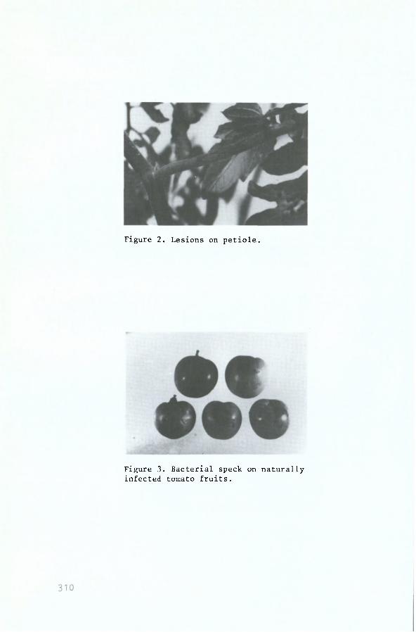
Figure 2. Lesions on petiole.
Figure 3. Bacterial speck on naturally infected tomato fruits.